Associate Dean of Admissions and Student Affairs Professor of Oral Biology School of Dentistry
Creighton University
Omaha, NE
This page intentionally left blank
Netter’s Advanced Head and Neck Flash Cards evolved from an expressed need for health professionals to learn and review the salient aspects of head and neck anatomy. Many of today’s courses in anatomy are shortened versions of their predecessors. Thus, the onus has been put on the student, as a lifelong learner, to fill in the areas that are often only briefly covered in lectures or problem-based learning (PBL) sessions. The anatomy of the head and neck is one of the most challenging areas of human anatomy to study and master. These flash cards help fill in that void.
Head and neck anatomy is composed of many structures that may seem minuscule to the naked eye but essential to the understanding of the whole. These flash cards are designed for users at all stages in their careers: the freshman student to the practicing clinician who desires a quick review. While Netter’s Advanced Head and Neck Flash Cards uses some of the images found in the 6th edition of Netter’s Atlas of Human Anatomy, this set is written at a more advanced level and is complemented with many more detailed images. A series of clinical correlates is incorporated into the appropriate sections of the set. An expanded section on imaging is included to aid the reader in his or her study.
The flash cards have been designed to test as well as to teach the reader. On the front of each card is an image with a series of labels to identify. On the reverse side are the answers to the labels and text or a table reviewing structure, function, and clinical relevance that is necessary for a sound foundation of head and neck anatomy.
Netter’s Advanced Head and Neck Flash Cards are a quick resource for studying the complex anatomy of the head and neck, which is the foundation for understanding so much of how the human body works in health and disease.
Neil S. Norton, PhD
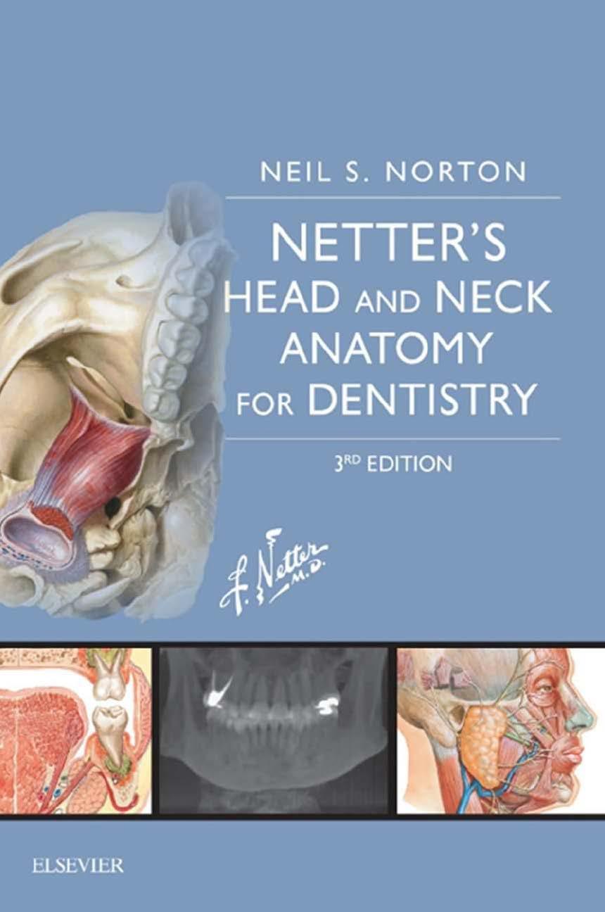
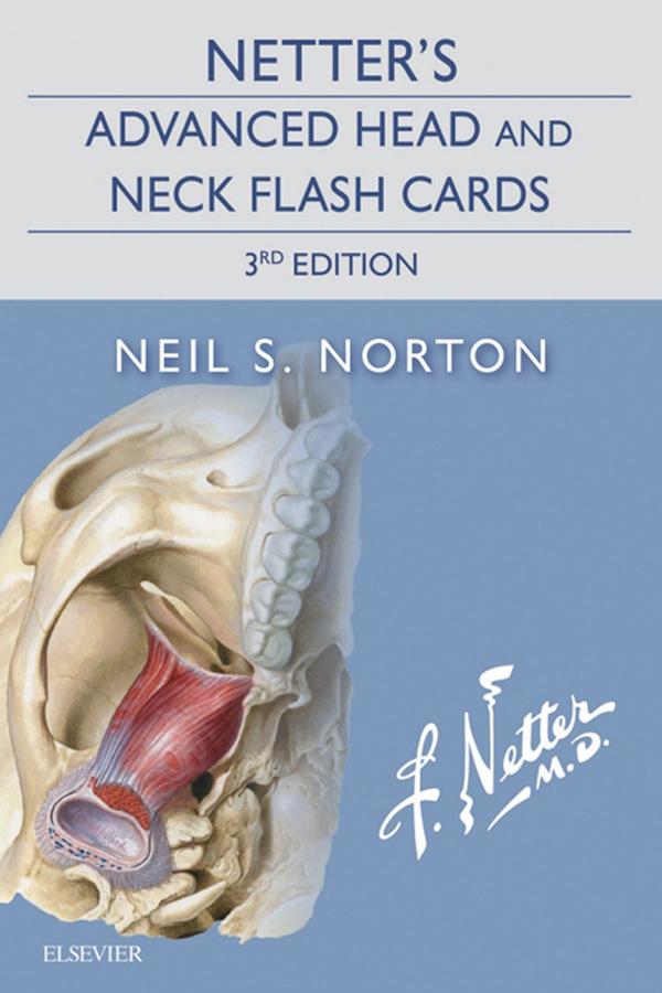
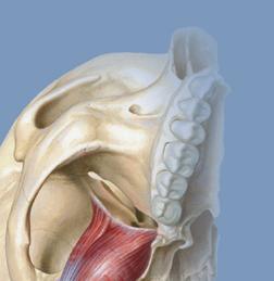
Netter’s Advanced Head and Neck Flash Cards
1600 John F. Kennedy Blvd. Ste 1800 Philadelphia, PA 19103-2899
NETTER’S ADVANCED HEAD AND NECK FLASH CARDS 3rd Edition
All rights reserved. No part of this publication may be reproduced or transmitted in any form or by any means, electronic or mechanical, including photocopying, recording, or any information storage and retrieval system, without permission in writing from the publisher. Permissions may be sought directly from Elsevier’s Rights Department: phone: (+1) 215 239 3804 (US) or (+44) 1865 843830 (UK); fax: (+44) 1865 853333; e-mail: healthpermissions@ elsevier.com. You may also complete your request on-line via the Elsevier website at http://www.elsevier.com/permissions
Notice
Knowledge and best practice in this field are constantly changing. As new research and experience broaden our knowledge, changes in practice, treatment and drug therapy may become necessary or appropriate. Readers are advised to check the most current information provided (i) on procedures featured or (ii) by the manufacturer of each product to be administered, to verify the recommended dose or formula, the method and duration of administration, and contraindications. It is the responsibility of the practitioner, relying on his or her own experience and knowledge of the patient, to make diagnoses, to determine dosages and the best treatment for each individual patient, and to take all appropriate safety precautions. To the fullest extent of the law, neither the Publisher nor the Author assumes any liability for any injury and/or damage to persons or property arising out of or related to any use of the material contained in this book.
The Publisher
ISBN: 978-0-323-44279-4
Executive Content Strategist: Elyse O’Grady
Content Strategist: Marybeth Thiel
Publishing Services Manager: Patricia Tannian
Project Manager: Stephanie Turza
Design Direction: Julia Dummitt
Illustration Buyer: Karen Giacomucci
Marketing Manager: Melissa Darling
Printed in China
Table of Contents
Section 1. Development
Section 2. Osteology
Section 3. Neuroanatomy
Section 4. Neck
Section 5. Scalp and Face
Section 6. Fossae of the Deep Face
Section 7. Nose, Nasal Cavity, and Paranasal Sinuses
Section 8. Oral Cavity
Section 9. Ear
Section 10. Orbit
Section 11. Imaging
Netter’s Advanced Head and Neck Flash Cards
This page intentionally left blank
1-1 to 1-11
Table of Contents
1-1 Embryological Development
1-2 Pharyngeal Arches
1-3 Cartilage Derivatives of Pharyngeal Arches
1-4 Pharyngeal Pouches
1-5 Development of the Cranium
1-6 Development of the Face
1-7 Development of the Palate
1-8 Development of the Tongue
1-9 Development of the Thyroid
1-10 Clinical Correlate
1-11 Clinical Correlate
Netter’s Advanced Head and Neck Flash Cards
This page intentionally left blank
Vertebrate Body Plan after 4 Weeks
Embryological Development
1. Ectoderm
2. Neural plate
3. Paraxial mesoderm
4. Intermediate mesoderm
5. Lateral plate mesoderm
6. Notochord
7. Neural crest (Future)
8. Endoderm
9. Neural plate forms neural tube
The developing embryo arises from 3 major germ layers:
• Ectoderm
• Endoderm
• Mesoderm
Mesoderm differentiates into:
• Paraxial mesoderm
• Intermediate mesoderm
• Lateral plate mesoderm
Ectoderm gives rise to 3 layers:
• Neuroectoderm
• Neural crest
• Epidermis
The head and neck are formed by:
• Paraxial mesoderm
• Lateral plate mesoderm
• Neural crest
• Ectodermal placodes
Most of the head and neck is formed from the pharyngeal region of the embryo.
See Figure 1-2
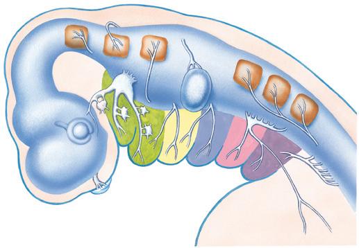
Pharyngeal Arches
1. Maxillary part of arch 1
2. Mandibular part of arch 1
3. Arch 2
4. Arch 3
5. Arch 4
6. Arch 6
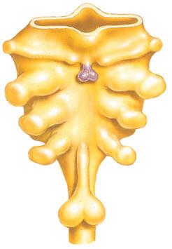
Pharyngeal arches start forming in the 4th week of development and develop as blocks separated by pharyngeal clefts (which are formed by ectoderm). Initially, 6 pharyngeal arches develop, but the 5th arch regresses. Compartments called pharyngeal pouches arise from the endoderm and extend toward the pharyngeal clefts (separated by pharyngeal membranes). Pharyngeal arches help form 4 of the 5 swellings (embryonic primordia) of the face:
• 2 mandibular processes (pharyngeal arch)
• 2 maxillary processes (pharyngeal arch)
• 1 frontonasal prominence
Composed of:
• External surface—ectoderm
• Internal surface—endoderm
• Central part—lateral plate mesoderm, paraxial mesoderm, and neural crest
Skeletal components and associated connective tissue develop from the neural crest cells whereas the muscular structures develop collectively from the mesoderm. Each arch is innervated by a cranial nerve that migrates with the muscles.
See Figure 1-3
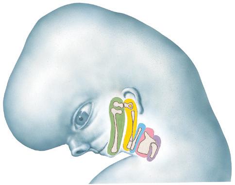
Embryo at 7 to 8 weeks
Cartilage primordia
Sphenomandibular ligament 4. Upper half of hyoid 5. Lower half of hyoid 6. Thyroid cartilage 7. Cricoid cartilage 8. Greater cornu of hyoid 9. Stylohyoid ligament 10. Styloid process 11. Stapes
CARTILAGE STRUCTURES FROM MESODERM CONNECTIVE TISSUE STRUCTURES FROM NEURAL CREST NERVE
CARTILAGE STRUCTURES FROM NEURAL CREST
Trigeminal
Sphenomandibular ligament
Anterior ligament of the malleus (both from Meckel’s cartilage which degenerates in adulthood)
Stylohyoid ligament
Connective tissue of tonsil
Connective tissue of thymus and inferior parathyroid Glossopharyngeal
Incus
Malleus
Malleus Incus (both from Meckel’s cartilage which degenerates in adulthood)
1 Develops into Maxillary process Mandibular process
Lesser cornu of hyoid Body of hyoid (Sup.) Styloid process Stapes
Facial 3 Greater cornu of hyoid Inferior part of hyoid body
Vagus (via superior laryngeal)
Connective tissue of superior parathyroid and thyroid
Epiglottis Thyroid (from lateral plate mesoderm)
Vagus (via recurrent laryngeal)
Arytenoid Cricoid Cuneiform Corniculate
Pharyngeal
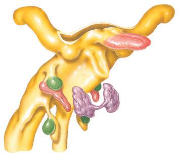
Pharyngeal Pouches
1st pouch
2nd pouch
3rd pouch
4th pouch
Inferior parathyroid
Thymus
Superior parathyroid
Figure 1-6
Epithelium of pharyngotympanic (auditory) tube and tympanic cavity
Tonsillar sinus (fossa)
Epithelium of palatine tonsil
Inferior parathyroid gland (from dorsal part)
Thymus (from ventral part)
Superior parathyroid gland (from dorsal part)
Ultimobranchial body (from ventral part)
Tubotympanic recess
Opposite 1st pharyngeal cleft, separated by 1st pharyngeal membrane
Primordial palatine tonsils
Opposite 2nd pharyngeal cleft, separated by 2nd pharyngeal membrane
Divides into dorsal and ventral part
Dorsal part migrates inferiorly toward thorax
Opposite 3rd pharyngeal cleft, separated by 3rd pharyngeal membrane
Divides into dorsal and ventral part
Ventral part is invaded by neural crest to form parafollicular (or C) cells
Opposite 4th pharyngeal cleft, separated by 4th pharyngeal membrane
1
2
3
4
Chondrocranium at 9 weeks
Membrane bones at 9 weeks
Development of the Cranium
1. Pharyngeal arch mesenchyme for viscerocranium
2. Head mesenchyme for neurocranium
3. Intramembranous ossification
4. Cartilage from pharyngeal arches for viscerocranium
5. Cartilage from somite sclerotomes and neural crest for neurocranium
6. Endochondral ossification
The cranium is formed from:
• Lateral plate mesoderm (neck region)
• Paraxial mesoderm
• Neural crest
Cranium development is divided into 2 parts:
• Viscerocranium—forms the bones of the face (from the pharyngeal arches) forms completely from neural crest
• Neurocranium—forms the bones of the cranial base and cranial vault and the function is to protect and surround the brain and organs of special sense (olfaction, vision, auditory, and equilibrium)
It can be divided into:
• Membranous neurocranium (forms from neural crest and paraxial mesoderm)
• Cartilaginous neurocranium (forms from neural crest and paraxial mesoderm)
Bony skull is formed by either of 2 mechanisms:
• Intramembranous ossification
• Endochondral ossification See Figure
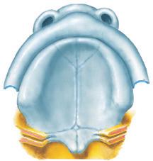
Ventral view at 5 to 6 weeks
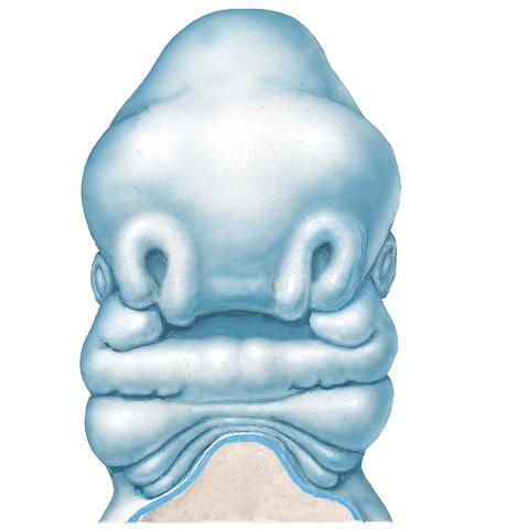
Development of the Face
1. Frontonasal prominence
2. Medial nasal prominence
3. Lateral nasal prominence
4. Maxillary prominence of arch 1
5. Mandibular prominence of arch 1
The face is formed mainly from neural crest, which makes 3 swellings (prominences) that surround the stomodeum:
• Frontonasal prominence
• Maxillary prominence (from the 1st pharyngeal arch)
• Mandibular prominence (from the 1st pharyngeal arch)
Lateral to the frontonasal prominence, 2 additional areas of ectoderm form the 2 nasal placodes that invaginate in the center to form nasal pits, creating ridges of tissue on either side of the pits:
• Lateral nasal prominence
• Medial nasal prominence
Fusion of the medial nasal prominences at the midline results in formation of the intermaxillary segment.
See Figure 1-10
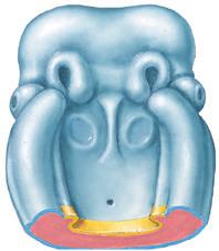
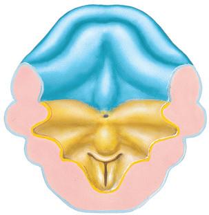
Roof of stomodeum (inferior view; 6 to 7 weeks)
Development of the Palate
Palate formation (inferior view; 7 to 8 weeks)
1
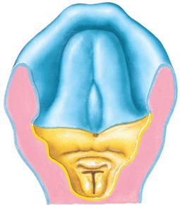
Roof of oral cavity (inferior view; 8 to 10 weeks)
2
1 2 3
Development of the Palate
1. Primary palate
2. Secondary palate (Lateral palatine process)
3. Maxillary prominence of arch 1
Formed by the:
• Primary palate (intermaxillary segment)
• Secondary palate (protrusions from the maxillary prominences)
The intermaxillary segment is the initial portion of the palate in development and contains the central and lateral incisors. Swellings of the maxillary prominence form shelves (lateral palatine processes) that project medially and are separated by the tongue. When the tongue no longer occupies the space between the palatal shelves, these lateral palatine processes fuse together to form the secondary palate. The primary and secondary palatal tissues all meet at the incisive foramen. Primary and secondary palates and the nasal septum fuse to form the definitive palate.
See Figure 1-13
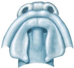
Development of the Tongue
Floor of oral cavity and pharynx (superior view; 5 to 6 weeks)
Floor of oral cavity and pharynx (superior view; 6 to 7 weeks)