Table of Contents
Cover image
Title page
Copyright
Consulting Editors
Preface
Dedication
Preface to the First Edition
About the Editors
Acknowledgments
1. Introduction
Electronic Bonus Plates
General Anatomy
Systematic Anatomy
Electronic Bonus Plates
2. Head and Neck
Electronic Bonus Plates
Surface Anatomy
Bones and Joints
Neck
Nose
Mouth
Pharynx
Larynx and Endocrine Glands
Eye
Ear
Brain and Meninges
Cranial and Cervical Nerves
Cerebral Vasculature
Regional Imaging
Structures with High Clinical Significance
Cranial Nerves
Branches of Cervical Plexus
Muscles
Electronic Bonus Plates
3. Back
Electronic Bonus Plates
Surface Anatomy
Vertebral Column
Spinal Cord
Muscles and Nerves
Cross-Sectional Anatomy
Structures with High Clinical Significance
Muscles
Electronic Bonus Plates
4. Thorax
Electronic Bonus Plates
Surface Anatomy
Thoracic Skeleton
Mammary Glands
Thoracic Wall and Diaphragm
Lungs, Trachea, and Bronchi
Heart
Mediastinum
Cross-Sectional Anatomy
Structures with High Clinical Significance
Muscles
Electronic Bonus Plates
5. Abdomen
Electronic Bonus Plates
Surface Anatomy
Abdominal Wall
Peritoneal Cavity
Stomach and Intestines
Liver, Gallbladder, Pancreas, and Spleen
Visceral Vasculature
Visceral Nerves and Plexuses
Kidneys and Suprarenal Glands
Lymphatics
Regional Imaging
Cross-Sectional Anatomy
Structures with High Clinical Significance
Muscles
Electronic Bonus Plates
6. Pelvis
Electronic Bonus Plates
Surface Anatomy
Bony Pelvis
Pelvic Diaphragm and Viscera
Urinary Bladder
Female Internal Genitalia
Female Perineum and External Genitalia
Male Perineum and External Genitalia
Homologies of Male and Female Genitalia
Male Internal Genitalia
Rectum and Anal Canal
Vasculature
Nerves of Perineum and Pelvic Viscera
Cross-Sectional Anatomy
Structures with High Clinical Significance
Muscles
Electronic Bonus Plates
7. Upper Limb
Electronic Bonus Plates
Surface Anatomy
Shoulder and Axilla
Arm
Elbow and Forearm
Wrist and Hand
Nerves and Vasculature
Regional Imaging
Structures with High Clinical Significance
Nerves of Brachial Plexus
Muscles
Electronic Bonus Plates
8. Lower Limb
Electronic Bonus Plates
Surface Anatomy
Hip, Buttock, and Thigh
Knee
Leg
Ankle and Foot
Nerves
Regional Imaging
Structures with High Clinical Significance
Nerves of Lumbosacral Plexus
Muscles
Electronic Bonus Plates
References
Index
e-Appendix A. Plate Pearls
e-Appendix B. Study Guides
Copyright
ELSEVIER
1600 John F. Kennedy Blvd.Ste. 1600 Philadelphia, PA 19103-2899
ATLAS OF HUMAN ANATOMY: CLASSIC REGIONAL APPROACH,EIGHTH EDITION
Standard Edition:978-0-323-68042-4
Professional Edition:978-0-323-79373-5
International Edition: 978-0-323-79374-2
Copyright © 2023 by Elsevier Inc.
Previous editions copyrighted 2019, 2014, 2011, 2006, 2003, 1997, 1989
All rights reserved. No part of this publication may be reproduced or transmi�ed in any form or by any means, electronic or mechanical, including photocopying, recording, or any information storage and retrieval system, without permission in writing from the publisher. Details on how to seek permission, further information about the Publisher’s permissions policies and our arrangements with organizations such as the Copyright Clearance Center and the Copyright Licensing Agency can be found at our website: www.elsevier.com/permissions.
This book and the individual contributions contained in it are protected under copyright by the Publisher (other than as may be noted herein).
Permission to use Ne�er Art figures may be sought through the website Ne�erImages.com or by emailing Elsevier’s Licensing Department at H.Licensing@elsevier.com.
Notices
Knowledge and best practice in this field are constantly changing. As new research and experience broaden our understanding, changes in research methods, professional practices, or medical treatment may become necessary. Practitioners and researchers must always rely on their own experience and knowledge in evaluating and using any information, methods, compounds, or experiments described herein. In using such information or methods they should be mindful of their own safety and the safety of others, including parties for whom they have a professional responsibility. With respect to any drug or pharmaceutical products identified, readers are advised to check the most current information provided (i) on procedures featured or (ii) by the manufacturer of each product to be administered, to verify the recommended dose or formula, the method and duration of administration, and contraindications. It is the responsibility of practitioners, relying on their own experience and knowledge of their patients, to make diagnoses, to determine dosages and the best treatment for each individual patient, and to take all appropriate safety precautions. To the fullest extent of the law, neither the Publisher nor the authors, contributors, or editors, assume any liability for any injury and/or damage to persons or property as a ma�er of products liability, negligence or otherwise, or from any use or operation of any methods, products, instructions, or ideas contained in the material herein.
International Standard Book Number: 978-0-323-68042-4
Publisher: Elyse O’Grady
Senior Content Strategist: Marybeth Thiel
Publishing Services Manager: Catherine Jackson
Senior Project Manager/Specialist: Carrie Ste�
Book Design: Renee Duenow
Consulting Editors
Chief Contributing Illustrator and Art Lead Editor
Carlos A.G. Machado, MD
Terminology Content Lead Editors
Paul E. Neumann, MD, Professor, Department of Medical Neuroscience, Faculty of Medicine, Dalhousie University, Halifax, Nova Scotia, Canada
R. Shane Tubbs, MS, PA-C, PhD
Professor of Neurosurgery, Neurology, Surgery, and Structural and Cellular Biology, Director of Surgical Anatomy, Tulane University School of Medicine, Program Director of Anatomical Research, Clinical Neuroscience Research Center, Center for Clinical Neurosciences, Department of Neurosurgery, Tulane University School of Medicine, New Orleans, Louisiana
Department of Neurology, Tulane University School of Medicine, New Orleans, Louisiana
Department of Structural and Cellular Biology, Tulane University School of Medicine, New Orleans, Louisiana
Professor, Department of Neurosurgery, and Ochsner Neuroscience Institute, Ochsner Health System, New Orleans, Louisiana
Professor of Anatomy, Department of Anatomical Sciences, St. George’s University, Grenada
Honorary Professor, University of Queensland, Brisbane, Australia
Faculty, National Skull Base Center of California, Thousand Oakes, California
Electronic Content Lead Editors
Brion Benninger, MD, MBChB, MSc
Professor of Medical Innovation, Technology, & Research
Director, Ultrasound, Professor of Clinical Anatomy, Executive Director, Medical Anatomy Center, Department of Medical Anatomical Sciences, Faculty, COMP and COMP-Northwest, Faculty College of Dentistry
Western University of Health Sciences, Lebanon, Oregon and Pomona, California
Faculty, Sports Medicine, Orthopaedic & General Surgery
Residencies, Samaritan Health Services, Corvallis, Oregon Faculty, Surgery, Orthopedics & Rehabilitation, and Oral
Maxillofacial Surgery, Oregon Health & Science University, Portland, Oregon, Visiting Professor of Medical Innovation and Clinical Anatomy, School of Basic Medicine, Peking Union Medical College, Beijing, China, Professor of Medical Innovation and Clinical Anatomy Post Graduate Diploma Surgical Anatomy, Otago University, Dunedin, New Zealand
Todd M. Hoagland, PhD
Clinical Professor of Biomedical Sciences and Occupational Therapy
Marque�e University College of Health Sciences, Milwaukee, Wisconsin
Educational Content Lead Editors
Jennifer K. Brueckner-Collins, PhD, Distinguished Teaching Professor, Vice Chair for Educational Programs, Department of Anatomical Sciences and Neurobiology, University of Louisville School of Medicine, Louisville, Kentucky
Martha Johnson Gdowski, PhD, Associate Professor and Associate Chair of Medical Education, Department of Neuroscience, University of Rochester School of Medicine and Dentistry, Rochester, NY
Virginia T. Lyons, PhD, Associate Professor of Medical Education, Associate Dean for Preclinical Education, Geisel School
of Medicine at Dartmouth, Hanover, New Hampshire
Peter J. Ward, PhD, Professor, Department of Biomedical Sciences, West Virginia School of Osteopathic Medicine, Lewisburg, West Virginia
Emeritus Editor
John T. Hansen, PhD, Professor Emeritus of Neuroscience and former Schmi� Chair of Neurobiology and Anatomy and Associate Dean for Admissions University of Rochester Medical Center, Rochester, New York
Editors of Previous Editions
First Edition
Sharon Colacino, PhD
Second Edition
Arthur F. Dalley II PhD
Third Edition
Carlos A.G. Machado, MD
John T. Hansen, PhD
Fourth Edition
Carlos A.G. Machado, MD
John T. Hansen, PhD
Jennifer K. Brueckner, PhD
Stephen W. Carmichael, PhD, DSc
Thomas R. Gest, PhD
Noelle A. Granger, PhD
Anil H. Waljii, MD, PhD
Fifth Edition
Carlos A.G. Machado, MD
John T. Hansen, PhD
Brion Benninger, MD, MS
Jennifer K. Brueckner, PhD
Stephen W. Carmichael, PhD, DSc
Noelle A. Granger, PhD
R. Shane Tubbs, MS, PA-C, PhD
Sixth Edition
Carlos A.G. Machado, MD
John T. Hansen, PhD
Brion Benninger, MD, MS
Jennifer Brueckner-Collins, PhD
Todd M. Hoagland, PhD
R. Shane Tubbs, MS, PA-C, PhD
Seventh Edition
Carlos A.G. Machado, MD
John T. Hansen, PhD
Brion Benninger, MD, MS
Jennifer Brueckner-Collins, PhD
Todd M. Hoagland, PhD
R. Shane Tubbs, MS, PA-C, PhD
Other Contributing Illustrators
Rob Duckwall, MA (DragonFly Media Group)
Kristen Wienandt Marzejon, MS, MFA
Tiffany S. DaVanzo, MA, CMI
James A. Perkins, MS, MFA
International Advisory Board
Nihal Apaydin, MD, PhD
Professor of Anatomy, Faculty of Medicine, Department of Anatomy, Ankara University
Chief, Department of Multidisciplinary Neuroscience, Institute of Health Sciences, Ankara, Turkey
Hassan Amiralli, MD, MS, FUICC
Professor and Chair, Department of Anatomy, American University of Antigua College of Medicine, Antigua, West Indies
Former Professor of Surgery, Muhimbili University of Health Sciences, Daressalaam, Tanzania
Belinda R. Beck, BHMS(Ed), MS, PhD, Professor of Anatomy and Exercise Science, Director, Bone Densitometry Research Laboratory, Griffith University, Gold Coast Campus , Queensland, Australia
Jonathan Campbell, MD, FAAOS, Assistant Professor of Orthopaedic Surgery , Division of Sports Medicine, Medical College of Wisconsin, Milwaukee, Wisconsin
Francisco J. Caycedo, MD, FAAOS, St. Vincent’s Hospital, Birmingham, Alabama
Thazhumpal Chacko Mathew, MSc, PhD, FRCPath, Professor, Faculty of Allied Health Sciences, Health Sciences Centre, Kuwait University, Kuwait City, Kuwait
Eduardo Cotecchia Ribeiro, MS, PhD, Associate Professor of Descriptive and Topographic Anatomy, School of Medicine, Federal University of São Paulo, São Paulo, Brazil
William E. Cullinan, PhD, Professor and Dean, College for Health Sciences, Marque�e University, Milwaukee, Wisconsin
Elisabeth Eppler, MD, University Lecturer, Institute of Anatomy , University of Berne, Berne, Swi�erland
Christopher Kelly, MD, MS, North Carolina Heart and Vascular, Raleigh, North Carolina
Michelle D. Lazarus, PhD, Director, Centre for Human Anatomy Education, Monash Centre for Scholarship in Health Education (MCSHE) Curriculum Integration Network Lead, Monash Education Academy Fellow, Monash University, Clayton, Victoria, Australia
Robert G. Louis, MD, FAANS, Empower360 Endowed Chair for Skull Base and Minimally Invasive Neurosurgery, Chair, Division of Neurosurgery, Pickup Family Neurosciences Institute, Hoag Memorial Hospital, Newport Beach, California
Chao Ma, MD, Department of Human Anatomy, Histology & Embryology, Peking Union Medical College, Beijing, China
Diego Pineda Martínez, MD, PhD
Chief, Department of Innovation in Human Biological Material, Professor of Anatomy, Faculty of Medicine of the National Autonomous University of Mexico President, Mexican Society of Anatomy, Mexico City, Mexico
William J. Swar�, PhD, Emeritus Professor of Cell Biology and Anatomy, Louisiana State University Health Sciences Center, New Orleans, Louisiana
Kimberly S. Topp, PT, PhD, FAAA, Professor and Chair Emeritus, Department of Physical Therapy and Rehabilitation Science, School of Medicine, University of California San Francisco, San Francisco, California
Ivan Varga, PhD, Professor of Anatomy, Histology, and Embryology, Faculty of Medicine, Comenius University, Bratislava, Slovak Republic
Robert J. Ward, MD, CCD, DABR, Chief Executive Officer, Sullivan’s Island Imaging, LLC, Sullivan’s Island, South Carolina;, Professor of Radiology, Saint Georges University, Grenada, West Indies
Alexandra L. Webb,
BSc, MChiro,
PhD,
Associate
Professor, Deputy Director, Medical School, College of Health and Medicine, Australian National University, Canberra, ACT, Australia
Preface
The illustrations comprising the Ne�er Atlas of Human Anatomy were painted by physician-artists, Frank H. Ne�er, MD, and Carlos Machado, MD. Dr. Ne�er was a surgeon and Dr. Machado is a cardiologist. Their clinical insights and perspectives have informed their approaches to these works of art. The collective expertise of the anatomists, educators, and clinicians guiding the selection, arrangement, labeling, and creation of the illustrations ensures the accuracy, relevancy, and educational power of this outstanding collection.
You have a copy of the regionally organized 8th edition with English language terminology. This is the traditional organization and presentation that has been used since the first edition. Also available is a Latin terminology option that is also regionally organized, as well as an option with English terminology organized by body system. In all cases, the same beautiful and instructive Art Plates and Table information are included.
New to this Edition
New Art
More than 20 new illustrations have been added and over 30 art modifications have been made throughout this edition. Highlights include new views of the temporal and infratemporal fossa, pelvic fascia, nasal cavity and paranasal sinuses, plus multiple new perspectives of the heart, a cross-section of the foot, enhanced surface anatomy plates, and overviews of many body systems. In
these pages you will find the most robust illustrated coverage to date for modern clinical anatomy courses.
Terminology and Label Updates
This 8th edition incorporates terms of the Terminologia Anatomica (2nd edition), as published by the Federative International Programme on Anatomical Terminology in 2019 (h�ps://fipat.library.dal.ca/ta2) and adopted by the International Federation of Associations of Anatomy in 2020. A fully searchable database of the updated Terminologia Anatomica can be accessed at h�ps://ta2viewer.openanatomy.org. Common clinical eponyms and former terminologies are selectively included, parenthetically, for clarity. In addition, a strong effort has been made to reduce label text on the page while maximizing label information through the use of abbreviations (muscle/s = m/mm.; artery/ies = a./aa.; vein/s = v./vv.; and nerve/s = n./nn.) and focusing on the labels most relevant to the subject of each Plate.
Nerve Tables
The muscle tables and clinical tables of previous editions have been so positively received that new tables have been added to cover four major nerve groups: cranial nerves and the nerves of the cervical, brachial, and lumbosacral plexuses.
Future of the Netter Anatomy Atlas
As the Ne�er Atlas continues to evolve to meet the needs of students, educators, and clinicians, we welcome suggestions! Please use the following form to provide your feedback: h�ps://tinyurl.com/Ne�erAtlas8
Preface to the First Edition
I have often said that my career as a medical artist for almost 50 years has been a sort of “command performance” in the sense that it has grown in response to the desires and requests of the medical profession. Over these many years, I have produced almost 4,000 illustrations, mostly for The CIBA (now Ne�er) Collection of Medical Illustrations but also for Clinical Symposia. These pictures have been concerned with the varied subdivisions of medical knowledge such as gross anatomy, histology, embryology, physiology, pathology, diagnostic modalities, surgical and therapeutic techniques, and clinical manifestations of a multitude of diseases. As the years went by, however, there were more and more requests from physicians and students for me to produce an atlas purely of gross anatomy. Thus, this atlas has come about, not through any inspiration on my part but rather, like most of my previous works, as a fulfillment of the desires of the medical profession.
It involved going back over all the illustrations I had made over so many years, selecting those pertinent to gross anatomy, classifying them and organizing them by system and region, adapting them to page size and space, and arranging them in logical sequence. Anatomy of course does not change, but our understanding of anatomy and its clinical significance does change, as do anatomical terminology and nomenclature. This therefore required much updating of many of the older pictures and even revision of a number of them in order to make them more pertinent to today’s ever-expanding scope of medical and surgical practice. In addition, I found that there were gaps in the portrayal of medical knowledge as pictorialized in the illustrations I had previously
done, and this necessitated my making a number of new pictures that are included in this volume.
In creating an atlas such as this, it is important to achieve a happy medium between complexity and simplification. If the pictures are too complex, they may be difficult and confusing to read; if oversimplified, they may not be adequately definitive or may even be misleading. I have therefore striven for a middle course of realism without the clu�er of confusing minutiae. I hope that the students and members of the medical and allied professions will find the illustrations readily understandable, yet instructive and useful.
At one point, the publisher and I thought it might be nice to include a foreword by a truly outstanding and renowned anatomist, but there are so many in that category that we could not make a choice. We did think of men like Vesalius, Leonardo da Vinci, William Hunter, and Henry Gray, who of course are unfortunately unavailable, but I do wonder what their comments might have been about this atlas.
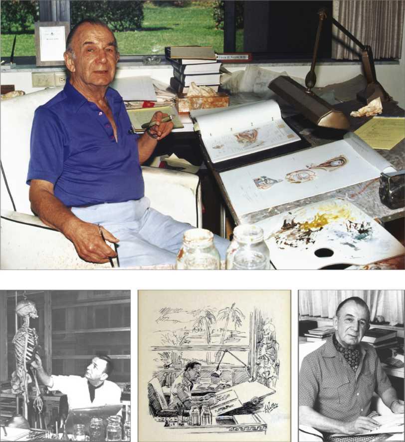
Frank H. Ne�er, MD (1906–1991)
Frank H. Netter, MD
Frank H. Ne�er was born in New York City in 1906. He studied art at the Art Students League and the National Academy of Design before entering medical school at New York University, where he received his Doctor of Medicine degree in 1931. During his student years, Dr. Ne�er’s notebook sketches a�racted the a�ention of the medical faculty and other physicians, allowing him to augment his income by illustrating articles and textbooks. He continued illustrating as a sideline after establishing a surgical practice in 1933, but he ultimately opted to give up his practice in favor of a full-time commitment to art. After service in the United States Army during World War II, Dr. Ne�er began his long collaboration
with the CIBA Pharmaceutical Company (now Novartis Pharmaceuticals). This 45-year partnership resulted in the production of the extraordinary collection of medical art so familiar to physicians and other medical professionals worldwide.
Icon Learning Systems acquired the Ne�er Collection in July 2000 and continued to update Dr. Ne�er’s original paintings and to add newly commissioned paintings by artists trained in the style of Dr. Ne�er. In 2005, Elsevier Inc. purchased the Ne�er Collection and all publications from Icon Learning Systems. There are now over 50 publications featuring the art of Dr. Ne�er available through Elsevier Inc.
Dr. Ne�er’s works are among the finest examples of the use of illustration in the teaching of medical concepts. The 13-book Ne�er Collection of Medical Illustrations, which includes the greater part of the more than 20,000 paintings created by Dr. Ne�er, became and remains one of the most famous medical works ever published. The Ne�er Atlas of Human Anatomy, first published in 1989, presents the anatomic paintings from the Ne�er Collection. Now translated into 16 languages, it is the anatomy atlas of choice among medical and health professions students the world over.
The Ne�er illustrations are appreciated not only for their aesthetic qualities, but, more importantly, for their intellectual content. As Dr. Ne�er wrote in 1949 “clarification of a subject is the aim and goal of illustration. No ma�er how beautifully painted, how delicately and subtly rendered a subject may be, it is of li�le value as a medical illustration if it does not serve to make clear some medical point.” Dr. Ne�er’s planning, conception, point of view, and approach are what inform his paintings and what make them so intellectually valuable.
Frank H. Ne�er, MD, physician and artist, died in 1991.