Orthopaedic Surgeon, Duke Orthopaedics, Durham, North Carolina
Keith Bengtson, MD
Clinical Director, Hand Rehabilitation, Physical Medicine and Rehabilitation, Mayo Clinic, Rochester, Minnesota
Anthony Beutler, MD
Sports Medicine Fellowship Director and Associate Professor, Family Medicine, Uniformed Services University, Bethesda, Maryland
Amrut Borade, MD
Clinical Research Fellow, Division Of Shoulder Surgery, The Johns Hopkins School Of Medicine, Baltimore, Maryland
Clinical Research Fellow, Department of Orthopaedic Trauma, Geisinger Health System, Danville, Pennsylvania
Jay E. Bowen, DO
Assistant Professor, Department of Physical Medicine and Rehabilitation, Rutgers—New Jersey Medical School, Newark, New Jersey
J. W. Thomas Byrd, MD
Clinical Professor, Department of Orthopaedic Surgery and Rehabilitation, School of Medicine, Vanderbilt University, Nashville, Tennesee
Larry H. Chou, MD
Medical Director, Sports and Spine Rehabilitation Division, Premier Orthopaedic and Sports Medicine Associates, LTD, Havertown, Deleware
Clinical Assistant Professor, Department of Physical Medicine and Rehabilitation, University of Pennsylvania School of Medicine, Philadelphia, Pennsylvania
Matthew J. Fanous, MD
Resident Physician, Physical Medicine and Rehabilitation, The Ohio State University Wexner Medical Center, Columbus, Ohio
Heather R. Galgon, DO
Attending Physician, Swedish Spine, Sports and Musculoskeletal Medicine, Swedish Medical Group, Providence Healthcare System, Issaquah, Washington
Mederic M. Hall, MD
Assistant Professor, Department of Orthopaedics and Rehabilitation, University of Iowa, Iowa City, Iowa
Contributors
Lisa Huynh, MD
Clinical Assistant Professor, Division of Physical Medicine and Rehabilitation, Department of Orthopaedics, Stanford University, Redwood City, California
David J. Kennedy, MD
Clinical Assistant Professor, Division of Physical Medicine and Rehabilitation, Department of Orthopaedics, Stanford University, Redwood City, California
John Kincaid, MD
Nerve Conduction Studies and Needle EMG, Department of Neurology, Indiana University, Indianapolis, Indiana
Brian Krabak, MD, MBA, FACSM
Clinical Professor, Department of Rehabilitation, Orthopedics and Sports Medicine, University of Washington and Seattle Children’s Sports Medicine, Seattle, Washington
Gerard A. Malanga, MD
New Jersey Regenerative Institute, Cedar Knolls, New Jersey
Clinical Professor, Department of Physical Medicine and Rehabilitation, Rutgers School of Medicine, New Jersey Medical School, Newark, New Jersey
Kenneth Mautner, MD
Associate Professor, Department of Physical Medicine and Rehabilitation, Emory University, Atlanta, Georgia
Edward G. McFarland, MD
The Wayne Lewis Professor of Shoulder Surgery Director, Division of Shoulder Surgery, The Department of Orthopaedic Surgery, The Johns Hopkins University, Baltimore, Maryland
Matthew S. Mendez-Zfass, MD
Physician, Department of Orthopedic Surgery, Lenox Hill Hospital, New York, New York
Kenneth D. Montgomery, MD
Head Team Physician and Chairman, Medical Department, New York Jets Football Team Orthopedic Surgeon, Tri-County Orthopedics, Cedar Knolls, New Jersey
Francis G. O’Connor, COL, MC, USA, MD, PhD
Professor and Chair, Military and Emergency Medicine
Associate Director, Consortium for Health and Military Performance (CHAMP), Uniformed Services University of the Health Sciences, “America’s Medical School”, Bethesda, Maryland
Tutankhamen Pappoe, MD
Medical Director, Pain Management, Phoenix Neurological and Pain Institute, Phoenix, Arizona
Joel M. Press, MD
Medical Director, Spine and Sports Rehabilitation Centers, Rehabilitation Institute of Chicago Professor, Physical Medicine and Rehabilitation, Feinberg School of Medicine, Chicago, Illinois
Lt Col Ross A. Schumer, MD
Department of Orthopaedic Surgery and Rehabilitation, San Antonio Military Medical Center, San Antonio, Texas
Jeffrey A. Strakowski, MD
Clinical Associate Professor, Department of Physical Medicine and Rehabilitation, The Ohio State University Associate Director of Medical Education, Physical Medicine and Rehabilitation, Riverside Methodist Hospital
Director of Musculoskeletal Research, The McConnell Spine Sport and Joint Center, Columbus, Ohio
Walter Sussman, DO
Primary Care Sports Medicine Fellow, Department of Sporrts Medicine, Physical Medicine and Rehabilitation, Emory University, Atlanta, Georgia
Andrew Willis, MD
Faculty, Orthopedic Sports Medicine Fellowship, Department of Orthopedic Surgery, Lenox Hill Hospital, New York, New York
Attending Orthopedic Surgeon, Department of Orthopedic Surgery, Morristown Medical Center, Morristown, New Jersey
David N. Woznica, MS, MD
Clinical Instructor, Department of Orthopaedics and Rehabilitation, Yale University School of Medicine, New Haven, Connecticuit
Video Table of Contents
Video 2-1
Sensory Exam
Video 2-2 Manual Muscle Testing
Video 2-3 Reflex Exam
Video 2-4 Hoffman’s Sign
Video 3-1 Spurling Test
Video 3-2 Shoulder Abduction Test
Video 3-3 Neck Distraction Test
Video 3-4 Lhermitte Sign
Video 3-5 Adson Test
Video 3-6 Roos Test
Video 4-1 Empty Can Test
Video 4-2 Drop Arm Test
Video 4-3 Patte Test
Video 4-4 Lift-off Test
Video 4-5 Bear Hug Test
Video 4-6 Lateral Scapular Slide Test
Video 4-7 Scapular Assistance Test
Video 4-8 Yergason’s Test
Video 4-9 Speed’s Test
Video 4-10 Neer Sign Test
Video 4-11 Hawkins Test
Video 4-12 Yocum’s Test
Video 4-13 Fulcrum Test
Video 4-14 Apprehension Test
Video 4-15 Relocation Test
Video 4-16 Sulcus Sign Test
Video 4-17 Active Compression Test
Video 4-18 Biceps Load Test
Video 4-19 Biceps Load Test II
Video 4-20 New Pain Provocation Test
Video 4-21 Crank Test
Video 4-22 Anterior Slide Test
Video 4-23 Compression-Rotation Test
Video 4-24 Scarf Test
Video 5-1 Tinel’s Cubital Tunnel
Video 5-2 Cozen Test
Video 5-3 Resisted Middle-Finger Extension Test
Video 5-4 Resisted Wrist Extension Test
Video 5-5 Biceps Hook Test
Video 5-6 Biceps Squeeze Test
Video 5-7 Varus Stress Test
Video 5-8 Pivot Shift Test For Posterolateral Rotatory Instability
Video 5-9 Posterolateral Drawer Test
Video 5-10 Valgus Test
Video 5-11 Milking Maneuver
Video 6-1 Allen Test
Video 6-2 Tinel Sign at Wrist
Video 6-3 Phalen’s Test
Video 6-4 Reverse Phalen’s Test
Video 6-5 Carpal Compression Test
Video 6-6 Finkelstein’s Test
Video 6-7 Thumb Basilar Joint Grind Test
Video 6-8 Watson Test
Video 6-9 Lunotriquetral Test
Video 6-10 Shear Test
Video 6-11 Ulnocarpal Stress Test
Video 6-12 Ulnar Collateral Test
Video 7-1 Inspection of the Lumbar Spine
Video 7-2 Lower Extremity Manual Motor Testing
Video 7-3 Lower Extremity Sensory Examination
Video 7-4 Lower Extremity Reflex Examination
Video 7-5 Straight-Leg Raise Test
Video 7-6 Crossed Straight-Leg Raise Test
Video 7-7 Bowstring Sign
Video 7-8 Slump Test
Video 7-9 Ankle Dorsiflexion Test
Video 7-10 Femoral Nerve Stretch Test
Video 7-11 Crossed Femoral Nerve Test
Video 7-12 Waddell Signs
Video 7-13 Hoover Test
Video 7-14 Standing Flexion Test
Video 7-15 Seated Flexion Test
Video 7-16 Gillet Test
Video 7-17 Compression Test
Video 7-18 Gapping Test
Video 7-19 Patrick Test
Video 7-20 Gaenslen Test
Video 7-21 Thigh Thrust Test
Video 7-22 Active Straight-Leg Raise and Active Assisted Straight-Leg Raise
Video 8-1 Thomas Test
Video 8-2 Ely Test
Video 8-3 Rectus Femoris Contracture Test
Video 8-4 Ober Test
Video 8-5 Piriformis Test
Video 8-6 Popliteal Angle Test
Video 8-7 Stinchfield Test
Video 8-8 Log Roll Test
Video 8-9 Quadrant Test (Scour Test)
Video 8-10 Axial Hip Distraction Test
Video 8-11 Resisted Sit-Up Test
Video 8-12 Resisted Hip Adduction Test
Video 8-13 Leg-Length Discrepancy Test
Video 9-1 Anterior Drawer Test
Video 9-2 Lachman Test
Video 9-3 Pivot Shift Test
Video 9-4 Posterior Sag Sign
Video 9-5 Posterior Drawer Test
Video 9-6 Quadriceps Active Test
Video 9-7 Valgus Stress Test
Video 9-8 Varus Stress Test
Video 9-9 Patellofemoral Grinding Test
Video 9-10 Patellar Compression Test (Clarke Sign)
Video 9-11 Patellar Apprehension Test
Video 9-12 Joint Line Tenderness
Video 9-13 McMurray Test
Video 9-14 Bounce-Home Test
Video 9-15 Thessaly Test
Video 10-1 Anterior Drawer Test
Video 10-2 Talar Tilt Test
Video 10-3 External Rotation or Kleiger Test
Video 10-4 Syndesmotic Ligament Palpation Test
Video 10-5 Syndesmosis Squeeze Test
Video 10-6 Thompson Test
An Evidence-Based Approach to the Musculoskeletal Physical Examination
Gerard A. Malanga, MD | Kenneth Mautner, MD
Since the publication of the first edition of this textbook, much has changed in the world, including the passing of our dear colleague and coeditor of this textbook, Dr. Scott Nadler. This is not only a great personal loss but also a loss to the many residents and fellows who will never benefit from his teaching skills; still others who will be deprived of his masterful speaking skills; and the medical community in general, which has lost a great mind and researcher.
We are pleased that over the past decade, there appears to be an increased interest in orthopedic physical examination with a great deal of peer-reviewed literature published in orthopedic, rehabilitation, physical therapy, and many other journals and textbooks. New tests have been developed and studied for various orthopedic conditions.1,2 In addition, a greater emphasis has been placed on education in the performance of the orthopedic physical examination in medical schools and in the related specialty areas of physical medicine rehabilitation; rheumatology; and obviously, orthopedic surgery.
Unfortunately, as noted in the introduction of the first edition of this textbook, there continues to be overemphasis on and increased use of imaging studies such as magnetic resonance imaging (MRI), and recently, musculoskeletal ultrasonography. While these technologies offer incredible resolution of musculoskeletal pathology, the medical literature has clearly demonstrated a significant number of falsepositive findings with their use. Moreover, the escalation in the use of MRI and other imaging studies is believed to be due to their indiscriminate use without appropriate indications. It is therefore clear that the appropriate use of imaging studies should require a thorough history and physical examination beforehand.
Overreliance on imaging and other diagnostic studies can additionally result in improper diagnosis and unnecessary treatment. Multiple studies have demonstrated the incidence of MRI abnormalities in normal subjects. This has been reported in the spine,3 shoulder,4 knee,5 and other areas. LaPrade and colleagues5 noted that 24% of normal individuals have findings consistent with grade II meniscal tears on MRI, and they recommended that clinicians match clinical signs and symptoms with MRI findings before surgical intervention. O’Shea and coworkers6 noted that the correct diagnosis was made in 83% of patients using the history and physical examination alone in the diagnosis of knee injuries. This, along with the significant findings on MRI in asymptomatic individuals, brings into question the need for MRI or ultrasonography as part of a standard
INTRODUCTION
screen for musculoskeletal injury. It also highlights the importance of a properly performed clinical examination.
Musculoskeletal complaints represent some of the most common reasons for patient encounters by primary care physicians.7 This trend is likely to increase secondary to societal changes in health and fitness, which has led our aging population to be more physically active than in years past. A poor history or physical examination can lead to inappropriate diagnostic testing and will influence patient outcome because treatment may be directed toward abnormal findings on imaging study rather than being based on the patient’s complaint and physical exam findings.8 The history can quickly produce a more discrete differential diagnosis and includes the mechanism of injury; the quality, location, and referral of pain; and the associated functional deficits. The fact that the physical examination is not performed in isolation but rather in conjunction with the history needs to be stressed. The information gained from the history helps focus the physical examination, especially if the physician has a good understanding of the underlying anatomy and biomechanics.
The physical examination of the musculoskeletal system is often limited by a lack of research into the sensitivity and specificity for the disease processes that these tests are used to assess. Sackett and Rennie9 noted that studies have been limited in regard to the physical exam, though the capability of reporting sensitivity, specificity, and predictive power would be similar to commonly studied laboratory tests. Reliability or reproducibility in regard to the translation of skills between the same or different clinicians at various time points during the course of disease is also poorly defined. Finally, there is no true gold standard by which to assess the validity of the different maneuvers because even reported surgical pathology may be a normal anatomic or age-related change in a variety of disease processes. This does not imply that physical examination maneuvers should be abandoned but rather that they need to be more completely understood and, ultimately, refined. Unfortunately, these issues are not understood by clinicians in various specialties and disciplines, leading to a promulgation of myths rather than facts.
Another issue arises when clinicians fail to recognize the importance of using more than one examination maneuver to make their diagnosis. Andersson and Deyo10 identified improved sensitivity, specificity, and positive predictive value with the utilization of combinations of tests rather than tests in isolation. This has also been shown in the physical examination of the shoulder4,7 and other joints. Although this
Reliability and Validity of Physical Examinations
Heather R. Galgon, DO | Larr y H. Chou, MD
INTRODUCTION
In 1880, John Venn, a priest and lecturer in Moral Science at Caius College, Cambridge University, England, introduced and popularized Venn diagrams (Fig. 1.1).1 Each circle represents a distinct domain that interacts with and is overlapped by other domains. The areas of overlap are more significant than the circles themselves, for within the overlapping areas “truth” can be found. While these diagrams were originally designed as models for mathematics and logic, they can also be used in the philosophy and practice of modern clinical medicine.
The “truth” in medicine represents the underlying diagnosis giving rise to a patient’s symptoms and signs. In this version of the Venn diagram, the large circle represents a patient’s relevant clinical history. It is the largest of the circles and where the majority of useful information can be found. Partially overlapping the patient’s history is a smaller circle representing the physical examination. The exam substantiates findings from the clinical history, but the nonoverlapping area represents its ability to identify issues not uncovered in the patient’s history. Last, the smallest circle represents additional clinical testing—whether laboratory, imaging, or electrophysiologic—that can further refine and confirm the true diagnosis denoted by the black dot. The true diagnosis lies within the clinical history, is supported by the physical examination, and is corroborated with other clinical studies.
In modern medicine, the bulk of clinical practice is predicated on the research question and the quality of support by which the question is answered. When critically reviewing the literature, it is paramount to understand these concepts. Indeed, an appreciation of the scientific method is necessary to fully understand the merits and pitfalls of the medical literature such that a conclusion can be properly applied to a specific clinical scenario. This chapter discusses the concepts of validity and reliability, how they give rise to sensitivity and specificity for diagnostic tests, and how the reported statistics of the various diagnostic tests should be interpreted in the clinical setting.
In contrast to observational cohort, case-control, and cross-sectional studies, the evaluation of diagnostic tests is different. Most observational studies attempt to show an association between the test result (a predictor variable) and the disease. In contrast, diagnostic studies attempt to discriminate between the diseased and the nondiseased. It is insufficient to merely identify an association between the test result and the disease.2 The concepts of specificity and
sensitivity as well as positive and negative predictive value are discussed here.
VALIDITY
Validity represents the truth, whether it is deduced, inferred, or supported by data. There are two types of validity: internal and external.3 Internal validity is the degree to which the results and conclusions from a research study correctly represent the data that were measured in the study. That is, the truth in the study can be correctly explained by the results of the study. However, it is important to recognize that this may not correctly answer the clinical question at hand. While a conclusion can be properly reached based on the available study findings, if the question asked or methods used are incorrect, then meaningful interpretation of the results is suspect. Once internal validity issues are satisfied, then the greater issue is that of external validity.
External validity is the degree to which the internal validity conclusions can be generalized to situations outside of the study. This is the sine qua non of meaningful clinical research. That is, can the conclusion of a study that has correctly interpreted its results be used outside of that specific research setting? The variables designed in a study must correctly represent the phenomena of interest. A research study that is so contrived or so artificially oversimplified to a degree that does not exist in the real world clinical setting is of guarded value.
Errors in study design and measurement tools greatly affect validity. How well a measurement represents a phenomenon is influenced by two major sources of error: sampling error and measurement error. In order for a study to be generalizable, the study population needs to parallel the target population. That is, the inclusion criteria for entrance into the study must represent the clinical characteristics and demographics of the population for which the study is intended. The sample size needs to be sufficiently large to avoid bias and increase power (see the following section). It is important to recognize that reporting errors can also occur, though these should be, and often are, identified in the peer review process.
Likewise, measurement errors need to be avoided so that valid conclusions can be drawn from the results. This brings up the concept of the accuracy and precision of a measurement (Fig. 1.2). Accuracy is the degree to which the study measurement reflects the actual measurement. In other words, accuracy represents the validity of the study, whether
3. Hulley SB, Newman TB, Cummings SR. Getting started: the anatomy and physiology of research. In: Hulley SB, Cummings SR, eds. Designing Clinical Research. Baltimore: Williams & Wilkins; 1988:1-11.
4. Hulley SB, Cummings SR. Planning the measurements: precision and accuracy. In: Hulley SB, Cummings SR, eds. Designing Clinical Research. Baltimore: Williams & Wilkins; 1988:31-41.
5. Rabin A, Irrgang JJ, Fitzgerald GK, et al. The intertester reliability of the Scapular Assistance Test. J Orthop Sports Phys Ther. 2006; 36:653-660.
6. Hulley SB, Gove S, Browner WS, et al. Choosing the study subjects: specification and sampling. In: Hulley SB, Cummings SR, eds.
Designing Clinical Research. Baltimore: Williams & Wilkins; 1988: 18-30.
7. Andersson GB, Deyo RA. History and physical examination in patients with herniated lumbar discs. Spine. 1996;21(suppl 24): 10S-18S.
8. Katz JW, Fingeroth RJ. The diagnostic accuracy of ruptures of the anterior cruciate ligament comparing the Lachman test, the anterior drawer sign, and the pivot shift test in acute and chronic knee injuries. Am J Sports Med. 1986;14:88-91.
testing is performed by asking the patient to identify numbers that are written on the palm. Temperature sense can be tested with a cold tuning fork or with test tubes containing warm and cold water. When cold sensation is being tested, the body part must be normally warm.12
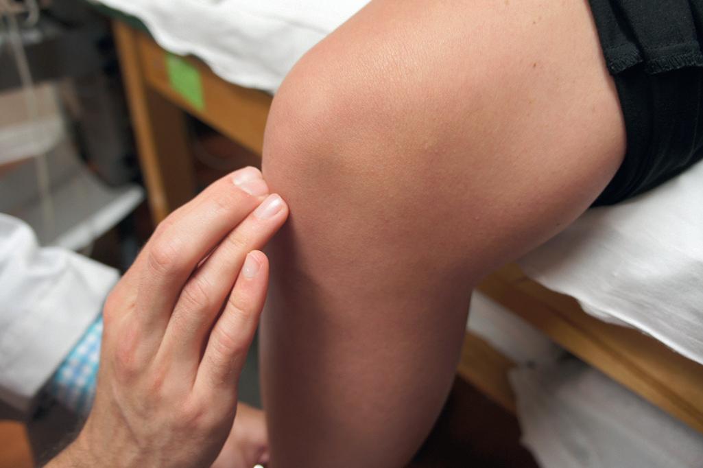
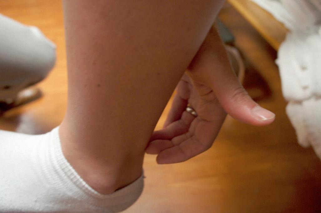
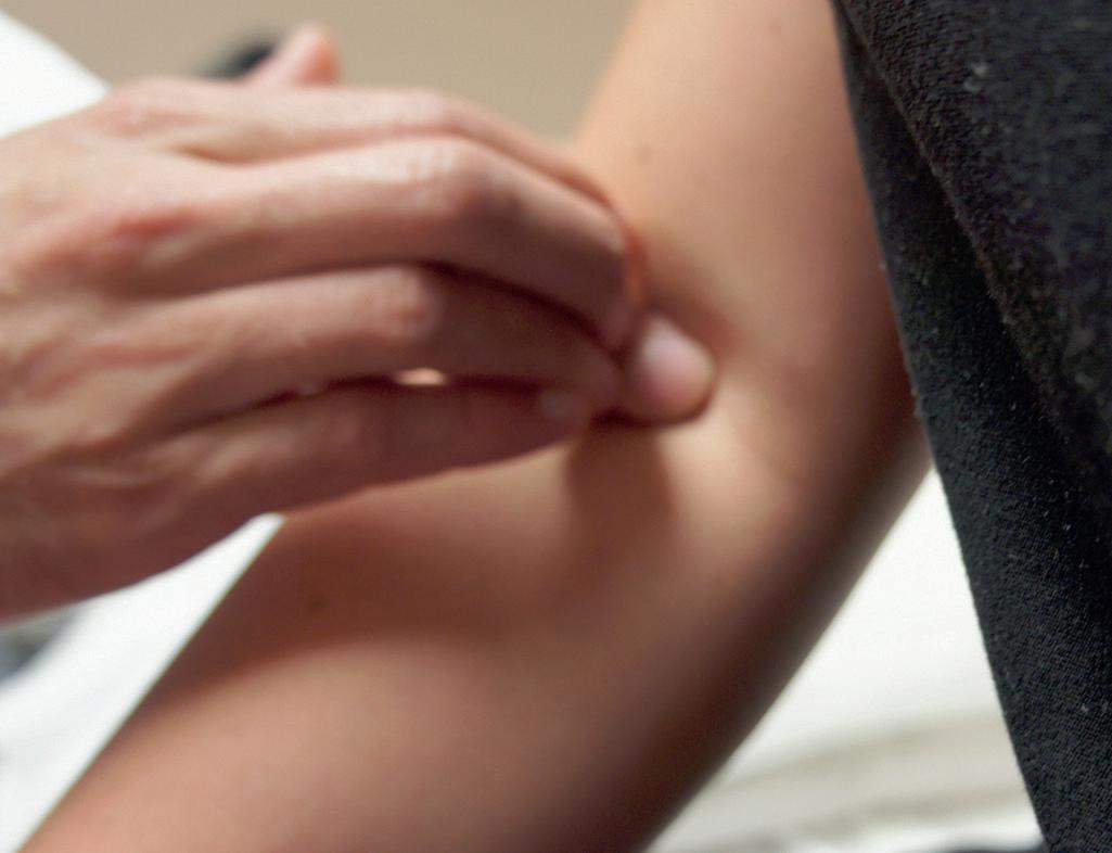
Joint position sense is tested by moving the terminal phalanx of a patient’s finger or toe up or down a few degrees. If the patient cannot identify these tiny movements with eyes closed, similar testing should be performed on the larger joints such as the metacarpal phalangeal joint or wrist. The body part being tested should be grasped on the sides rather than the dorsal or ventral aspect to prevent the patient from using pressure cues to detect movement.
To test vibration sense, the examiner places a finger under the patient’s distal interphalangeal joint and presses a lightly tapped 128-cycle tuning fork on top of the joint. The patient detects the vibration and then notes its extinction about the same time as the examiner, who feels it through the patient’s digit.13 The age and size of the patient should be considered when assessing abnormalities of vibration sense.14 Devices designed to improve quantitative measurement of vibration sensation can also be used.15,16
PROVOCATIVE MANEUVERS
Provocative maneuvers for eliciting sensory symptoms are notoriously nonspecific for reliably distinguishing true neurologic deficit, but they can sometimes provide clinical clues to the source of pain complaints. An example of this is a patient with chronic pain such as fibromyalgia, whose complaints of paresthesias are magnified by muscle palpation. Tender or “trigger” points in muscle can lead to reporting sensations described as paresthesias but are not related to an identifiable neurologic deficit.
Other maneuvers can potentially lead to dynamic nerve compression and provide clinical clues that contribute to
localization of the source of a pain generator, but they are not specific for either neurologic deficit or nerve entrapment. Examples include the Spurling test in cervical radiculopathy, Adson test in thoracic outlet entrapment, and Phalen sign in median nerve entrapment at the carpal tunnel. Techniques of this nature can potentially produce neurologic or neurologic-like symptoms, but they cannot be expected to alter the sensory examination.
Tapping over a suspected focal peripheral neuropathy to reproduce neuritic symptoms and assess for sensitivity is a frequently cited technique and has been termed the Tinel sign. Tinel originally described this technique as a method to determine the location of recovery of regenerating axons after trauma. In his description, the presence of the sign at the location that was being tapped was indicative that the nerve had regenerated to that position.17,18 Localized sensitivity can develop over an area of peripheral nerve injury; however, percussion over a normal peripheral nerve in a superficial location will also induce pain and paresthesias. The use of this technique should not be considered reliable confirmation of a focal entrapment neuropathy.
MOTOR EXAMINATION
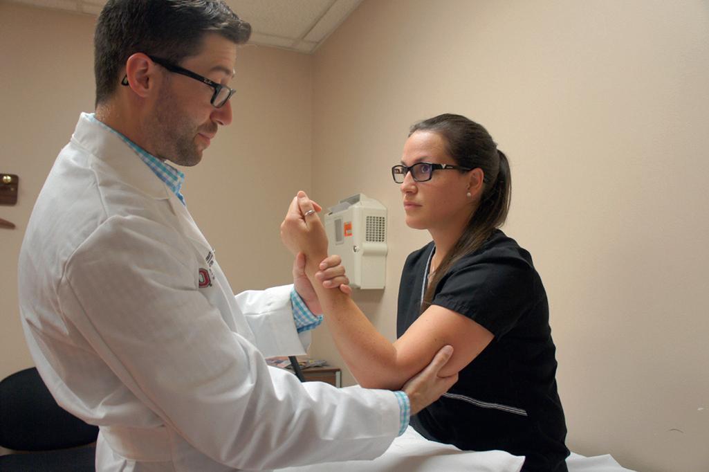
The role of motor testing is to assess the patient’s strength. Reports of weakness are not always due to a true motor deficit. Some complain of weakness when they are actually referring to fatigue, malaise, or incoordination. Strength testing is not the same as power, which refers to the rate of performing work. Manual muscle testing is the most commonly used technique for testing strength. With manual muscle testing, the strength of specific muscle groups is tested against resistance, and one side of the body is compared with the other. It is performed by providing a counterforce on a specific point on the limb against the patient’s best effort19 (Fig. 2.5; Video 2-2).
There are a variety of different classification systems for grading manual muscle testing. Most are based on the Medical Research Council 0–5 scale20 (Table 2.1).
There have been significant modifications of this format by many authors. Most of these scales give ordinal data. This
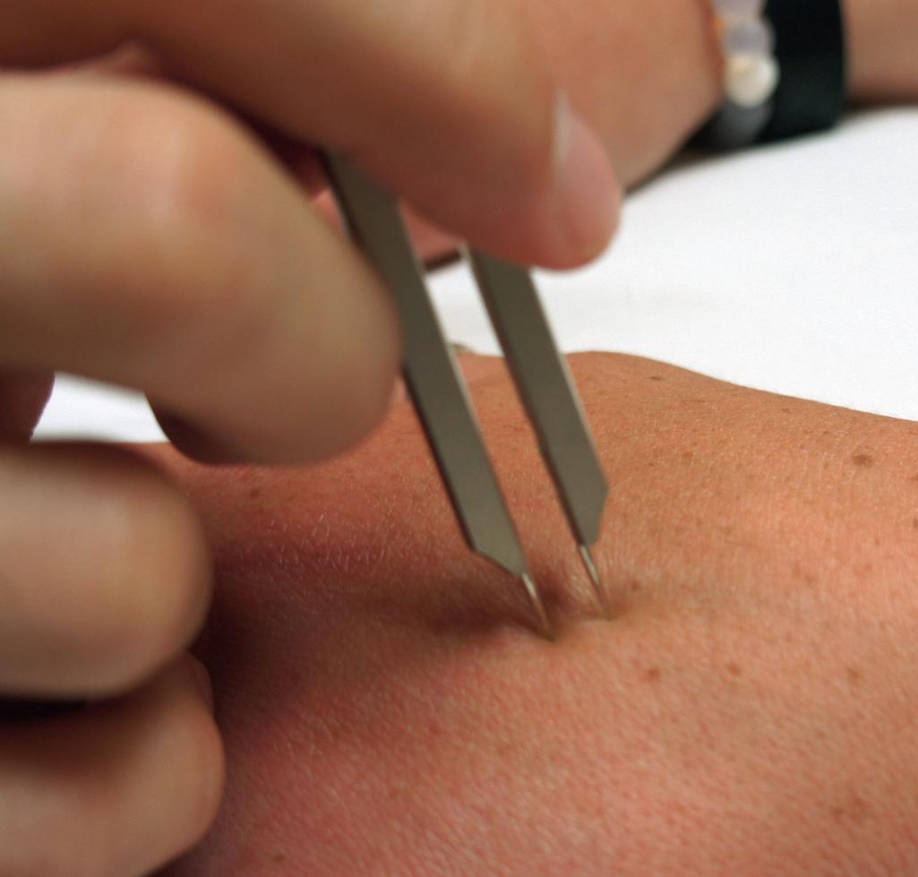
Figure 2.4 Demonstration of the use of calipers to assess two-point discrimination.
Figure 2.5 Demonstration of the technique of manual muscle testing.
tions, diseases, and drugs.60-62 The findings should always be used in clinical context. The diagnostic specificity of clearly abnormal reflexes is relatively good, but the presence of normal reflexes does not exclude a neurologic deficit.63
PATHOLOGICAL REFLEXES
CLONUS
The most frequently used grading system for muscle stretch reflexes is that of the National Institute of Neurological Disorders and Stroke (NINDS) scale.54 This is a 5-point scale that ranges from 0 to 5. Zero represents an absent reflex and is virtually always considered abnormal.55,56 Other values are defined as follows: A score of 1+ represents a diminished reflex and might be normal or abnormal; 2+ represents normal reflexes; 3+ represents a brisk reflex and might be normal or abnormal; 4+ represents a brisk reflex with the presence of clonus, a series of involuntary muscular contractions and relaxations; and 4+ is always considered abnormal.
Consideration should be given to the age of the patient when interpreting reflexes.57 Individuals without neurologic deficit will experience a decline in reflex response with aging.58,59 Multiple other factors can influence muscle stretch reflexes, including endocrine changes, medica-
Clonus is a series of involuntary rhythmic muscle contractions elicited by a rapid passive stretching of a muscle. It is due to a lesion in descending motor neurons and is often associated with increased muscle stretch reflexes and spasticity.64,65 Clonus is most commonly seen at the ankle but can be found at the ankle, knee, wrist, jaw, or elbow.66 Clonus can be seen in virtually any muscle group and generally repeats with a frequency of 5 to 8 Hz.67
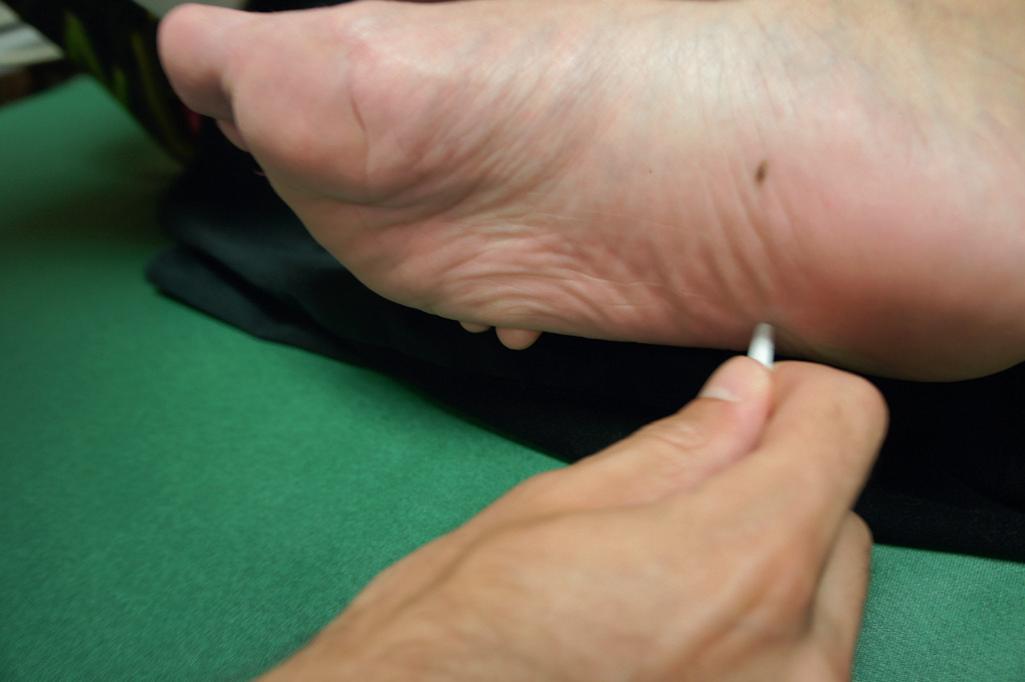
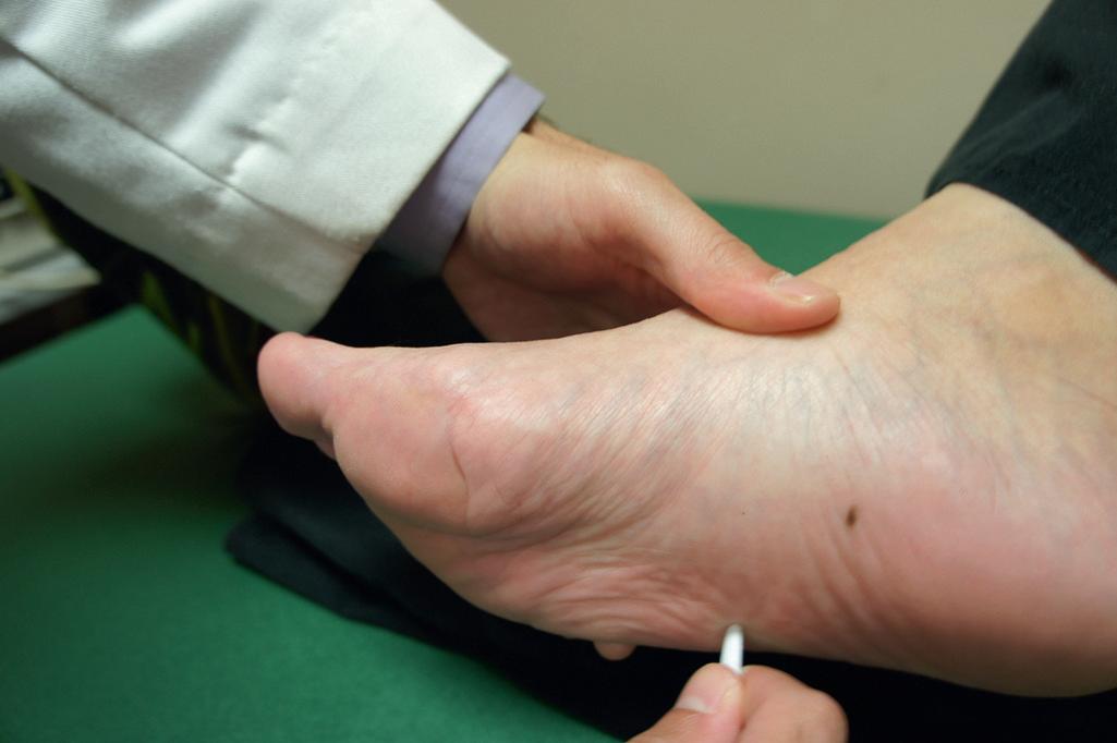
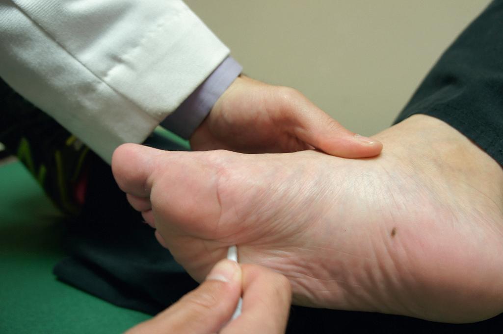
PLANTAR REFLEX
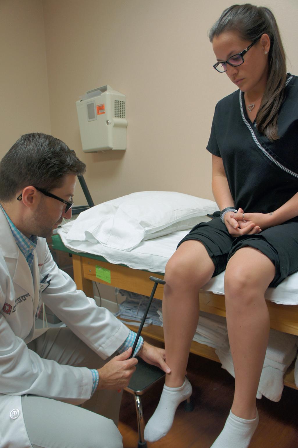
The plantar reflex is a reflex elicited when the sole of the foot is stroked, often with a blunt instrument (Fig. 2.10). In normal adults, the reflex causes flexion of the hallux (Fig. 2.11). An abnormal response, also known as the Babinski sign, named after Joseph Babinski,68,69 is an extension of the hallux (Fig. 2.12). An abnormal plantar reflex can represent dysfunction of the pyramidal tract. It can sometimes be the (C7–C8), quadriceps (ie, patellar) (L3–L4), semimembranosus (L5, S1), and gastrocnemius (aka Achilles or ankle) (S1, S2).53 Side-to-side comparisons should always be used with these reflexes to assess for variations.
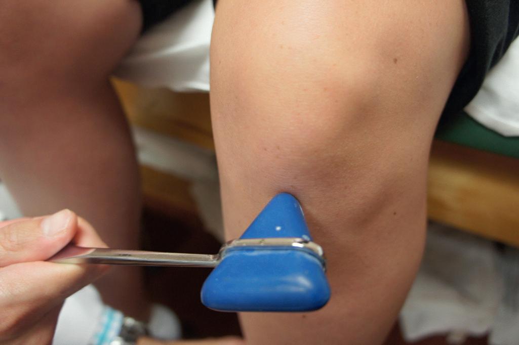
Figure 2.8 Demonstration of the use of different reflex hammers for eliciting muscle stretch reflexes. A, Babinski reflex hammer. B, Taylor hammer.
Figure 2.7 Demonstration of the technique for eliciting the muscle stretch reflex. In this case, the quadriceps reflex is elicited by briskly striking the patellar tendon, with the patient’s knee in a relaxed and flexed position.
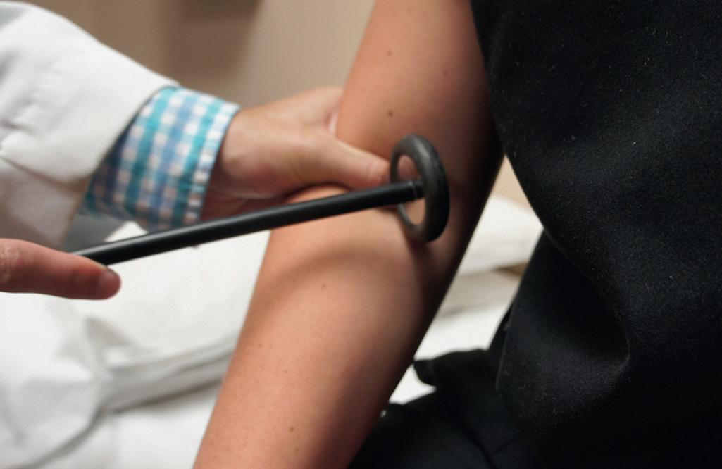
2.9 Demonstration of the manual technique for eliciting muscle stretch reflexes without a hammer. The fingers are tapped briskly on the tendon. A, Patellar. B, Achilles. C, Biceps Brachii.
first and only indication of a CNS lesion. The initial response of the toe should be carefully observed when the reflex appears abnormal. Normal withdrawal with extension of the toes can occur by the patient after the reflex is complete.
HOFFMAN’S REFLEX
Hoffman’s reflex (aka finger flexor reflex) is performed by tapping or flicking the terminal phalanx of the long or ring finger. The Hoffman’s sign is a positive test that is
Figure 2.10 Demonstration of the method for assessing the plantar reflex. The lateral side of the foot is stroked from the heel along a curve to the area of the metatarsal pads. The progression of movement is shown in A, B and C
demonstrated by reflexive flexion of the terminal phalanx of the thumb with this maneuver (Fig. 2.13, Video 2-4).
Unlike the Babinski sign, the Hoffman’s sign can be seen in normal adults that are naturally somewhat hyperreflexic. The finding more likely represents upper motor neuron pathology when it is an acute onset or unilateral. Hoffman’s sign should be interpreted within the context of other neurologic signs and is not considered a good screening test in asymptomatic individuals.70,71 The mechanism is
Figure
A
B C
A
B C
66. Uysal H, Boyraz I, Yağcıoğlu S, et al. Ankle clonus and its relationship with the medium-latency reflex response of the soleus by peroneal nerve stimulation. J Electromyogr Kinesiol. 2011;21: 438-444.
67. Boyraz I, Uysal H, Koc B, et al. Clonus: definition, mechanism, treatment. Med Glas (Zenica). 2015;12:19-26.
68. Bassetti C. Babinski and Babinski sign. Spine. 1995;20:25912594.
69. Furukawa T. [Joseph Babinski’s contribution to neurological symptomatology]. [Article in Japanese] Brain Nerve. 2014;66:12791286.
70. Grijalva RA, Hsu FP, Wycliffe ND, et al. Hoffmann sign: clinical correlation of neurological imaging findings in the cervical spine and brain. Spine. 2015;40:475-479.
71. Tejus MN, Singh V, Ramesh A, et al. An evaluation of the finger flexion, Hoffman’s and plantar reflexes as markers of cervical spinal cord compression—A comparative clinical study. Clin Neurol Neurosurg. 2015;134:12-16.
72. Hoffmann G, Kamper DG, Kahn JH, et al. Modulation of stretch reflexes of the finger flexors by sensory feedback from the proximal upper limb poststroke. J Neurophysiol. 2009;102:14201429.
Physical Examination of the Cervical Spine
Lisa Huynh, MD | David J. Kennedy, MD
INTRODUCTION
The annual prevalence of neck pain is estimated to range between 30% and 50%,1 and nearly half of all individuals will experience neck pain in their lifetime.2 History and physical examination can provide important clues in determining the etiology of symptoms. Many specialized provocative tests have been described for physical examination of the neck and cervical spine. These tests are routinely performed by clinicians with varying experience and skill. This may lead to error in both the technique and the interpretation of findings.
Several key principles exist in examination of the cervical spine: (1) The exam should be systematic to avoid missing key steps. (2) Generally, exam maneuvers should be done in a stepwise manner so that less painful movements are performed first and most painful movements are completed last; this ensures the least amount of pain carryover, which may confound exam findings. (3) Of crucial concern for any examination of the cervical spine is the ability to differentiate pathologies that merely cause pain from those that adversely affect sensitive neural tissues associated with the cervical spinal cord and its nerve roots.
This chapter provides a comprehensive overview of the physical examination of the cervical spine. For each test, the original description, currently performed technique, reliability, validity, and clinical significance are discussed, based on a comprehensive search of the existing literature. The goal is not necessarily to learn every examination maneuver performed for neck pain but rather to understand the limitations, reliability, and scientifically proven validity of some of the commonly used tests.
INSPECTION
Inspection should begin by noting the position of the head in relation to the line of gravity, which passes through the external auditory meatus; odontoid process; the cervical, thoracic, thoracolumbar, and lumbosacral spine; and the sacral promontory. One should carefully assess not only the upper cervical region but also the relative curvature of the thoracolumbar and lumbosacral spines because the relative positioning of the cervical spine may be influenced by the curvature below. The forward-head position can also be the direct cause of the loss of cervical motion. Caillet3
reported a 25% to 50% loss of head rotation with a forwardly protruded head and a significant increase in the gravity-induced weight of the head brought on by this postural abnormality. The forward-head posture thus increases the work requirements of the capital and cervical musculature. Additional features associated with damage to the neural structures may be noted by observation/inspection, including limb muscle atrophy, clumsiness, and balance problems on gait.
PALPATION
Palpation is a common component of cervical spine evaluation. It should be systematic and focus on palpation of the midline spinous processes, the paraspinal musculature, and the underlying zygapophyseal joints (z-joints), as well as the associated cervical spinal musculature. Studies have addressed the accuracy of palpation in identifying the structure or level of the spine and interexaminer reliability. In a study of 69 patients, experienced anesthesiologists were asked to identify the C7 spinous process by palpation, which was then compared with fluoroscopy.4 The C7 level was chosen because it was believed to be the most prominent and easiest level to identify. The physicians in this study were only able to correctly identify the C7 spinous process 47.9% of the time when compared with fluoroscopy. This was increased to 77.1% with the addition of neck flexionextension. Additional concerns with the reliability of the cervical spine palpatory examination have been highlighted in a systematic comprehensive review of the literature, which showed overall poor interexaminer reliability for all palpation.5 Reviewers also found that the level of clinical experience did not improve the reliability in that experienced clinicians fared no better than students in terms of palpatory reliability. Segmental range of motion as assessed by palpation was also found to have very low reliability. Additionally, palpation for soft tissue had very low reliability in all regions tested. Reviews of the literature on manual palpation of trigger points have shown poor reproducibility; however, the majority of studies had poor methodological quality.6
Studies on the validity of palpation in the cervical spine are lacking. In 1995, Sandmark and Nisell7 found in a study of 75 patients with self-reported pain that palpation over the facet joints was the most appropriate screening to corroborate self-reported neck dysfunction. This was based on a