OVERVIEW of MUSCLE STRENGTH ASSESSMENT
While attempts to quantify human muscle strength have been occurring for hundreds of years,35 agreement on the definition of muscle strength is not evident in the literature. Some authors use the term muscle strength to refer to the ability of muscle to develop tension106 or torque.25 Other authors provide a more restrictive definition that limits strength to force generated over a single, unlimited episode against an immovable resistance,69 whereas still others advocate the use of muscle power as a substitute for muscle strength.103 In recognition of the fact that human muscle must be capable of force generation in a static position and through a range of motion, the term muscle strength will be defined herein according to Knuttgen and Kraemer67 as “the maximal force a muscle or muscle group can generate at a specified or determined velocity.”
Tests of muscle strength have been, and continue to be, performed for many and varied reasons. Strength may be tested in relation to athletic performance, a practice common at least since the time of the ancient Greeks with the original Olympic Games. In a health care setting, strength concerns are more often related to the diagnosis and treatment of disease and restoration of function. Muscle strength testing is an essential component of the examination of all patients, particularly those with musculoskeletal or neuromuscular pathology. Numerous such pathologies leave the patient with less than optimal muscle strength and control, and clues to diagnosing such disorders are often found during examination of muscle strength. For example, muscle testing is used in patients with spinal cord injury to help determine the level of the lesion and the degree of damage to the cord. Patterns of muscle strength loss may help differentiate between two possible diagnoses (e.g., C6 radiculopathy vs. median nerve lesion). Strength testing may be used to determine a patient’s ability to perform activities of daily living or to assess an injured athlete’s ability to return to competition.
FACTORS AFFECTING MUSCLE
STRENGTH: RELATIONSHIP OF MUSCLE
STRENGTH WITH AGE AND SEX
Muscle strength does not remain constant over an individual’s life span but demonstrates a pattern of gradual
increase during childhood and into young adulthood followed by a gradual decline through the remainder of the person’s life. Many researchers have examined muscle strength development in children. Several of these investigations report a strong relationship between the steady increases in muscle strength and increases in muscle size experienced during growth and development.* Among the most predictive factors of muscle strength in both sexes are muscle crosssectional area,34,61,62 height,34,94,104,112,114 weight,94,104,112,114,119 and age.† Muscle strength in boys is greater than that of girls from as early as age 9 or 10 years114 and appears to be due, at least until puberty, to body mass and height, both of which are larger in males than in females as a group.34,100 However, longitudinal studies indicate that at about the time of peak height velocity, the rate of increase in muscle strength in males becomes disproportionate to body mass and height increases, whereas the rate of muscle strength increase in females remains proportionate.94,104 This disproportionately high increase in male strength has been attributed to plasma testosterone levels, which rise rapidly around the time of peak height velocity.104
Increases in muscle strength appear to continue until sometime between age 20 and 30.4,5,12,27,89 Strength then shows slight declines until the fifth or sixth decade, when rates of decline in muscle strength increase.‡ Declines in muscle strength do not appear to be consistent between muscle groups§ or between men and women.60,89,98 Loss of isometric muscle strength from ages 20 to 80 has been reported to range from 32% to 52% in women27,59,89,99,136 and from 34% to 60% in men,27,89 with declines in muscle strength in women starting later than those in men.27,75,118
Although the reasons for declines in strength associated with aging have not been fully elucidated, possible mechanisms are varied. Total muscle mass declines with aging, with reductions estimated to be somewhere between 20% and 40% between the third and ninth
*References 34, 61, 62, 94, 104, 119
†References 6, 62, 88, 94, 102, 114
‡References 5, 26, 27, 57, 89, 118, 127
§References 5, 9, 26, 64, 76, 105, 115, 118.
decades,* although such declines may be ameliorated with physical activity.10,66,86,126 Motor neurons in the spinal cord also are lost during the aging process, particularly after age 60, which may account for some of the declines in muscle force.121 Additionally, the number and size of muscle fibers are apparently reduced with aging, contributing to a decline in the contractile capabilities of the muscle.†
APPROACHES TO MUSCLE STRENGTH TESTING
Using the definition of muscle strength as “the maximal force a muscle or muscle group can generate at a specified or determined velocity,” one can measure muscle strength in a variety of ways, depending on the velocity of motion and type of resistance used in the test. Although a variety of methods of examining muscle strength exist, there are basically three different approaches to muscle strength testing that are described in the literature and used clinically: isotonic, isokinetic, and isometric testing.
Isotonic strength testing has traditionally been defined as the testing of strength using a constant external resistance.21 However, the term isotonic (Greek, isos: “equal” and tonos: “straining”) is a misnomer in this situation, because isotonic properly refers to constant muscle tension, a situation that very rarely occurs in muscle, rather than constant external resistance. Regardless of the misuse of the term, isotonic strength testing typically involves the use of free weights or resistance machines and may use testing techniques such as the one-repetition maximum (1-RM), which is considered by many to be the gold standard in muscle strength assessment.68 However, such testing may prove overtaxing to the subject, because several repetitions of weight lifting normally are necessary as the examiner attempts to discover the maximum weight the subject can lift or move.21 Additionally, such testing may be time consuming, lack the portability of other muscle strength testing methods, and cause injury and tests gross strength of muscle groups rather than strength of individual muscles.52
Isokinetic strength testing was developed in the 1960s and involves measurement of muscle strength by having the subject provide resistance through the range of motion at a constant velocity.91 Isokinetic dynamometers generate an isokinetic torque curve, and muscle strength is determined by measuring the highest point on the curve.33 Because peak torque is normally used to define muscle strength when using isokinetic dynamometry, the result is measurement of strength at only one point in the range of motion (although isokinetic
dynamometry is capable of providing strength data throughout the range of motion).7,131 Reliability of isokinetic testing is high, provided testing protocols are followed strictly.24,36 Like isotonic testing, isokinetic testing tests gross strength of muscle groups rather than strength of individual muscles. Isokinetic testing costs may be prohibitive, with the price of instruments estimated at $40,000.
Isometric (Greek, isos: “equal” and metron: “measure”) testing of muscle strength involves having the muscle generate force against an immovable resistance so that muscle length remains the same throughout the test. Thus, factors that can confound a muscle test, such as variability in muscle length and velocity of joint motion, are eliminated. The two most commonly used methods of isometric muscle testing, manual muscle testing (MMT) and handheld dynamometry (HHD), are highly portable and inexpensive, with MMT requiring no equipment other than the examiner’s hands. A disadvantage of isometric strength testing is that, because muscle length is held constant, isometric testing provides muscle strength data at only one point in the range of motion.
SELECTION OF THE APPROPRIATE TESTING TOOL
No single method of examining muscle strength is appropriate in every situation. Therefore, the examiner needs to have an arsenal of muscle testing tools available and the knowledge of the advantages and disadvantages of each method so that the optimal tool may be selected and the desired information may be obtained for each patient. In some cases, selection of the testing method may be limited by constraints such as the patient’s condition, equipment availability, or other factors, and the data obtained may be less than perfect. For example, because children younger than 3 to 4 years do not have the ability to cooperate with complex testing situations such as those used with MMT, HHD, or isokinetic testing,93 muscle strength testing is best accomplished by observing functional activities (see Chapter 6) in such patients. However, the data obtained are not as quantifiable as those available with methods such as HHD or isokinetic dynamometry. Compromises such as these are frequently necessary in assessing muscle strength, and the examiner must make the judgment as to the best testing method in each situation. Generally, the following guidelines will assist the examiner in choosing the best method of muscle strength assessment.
*References 4, 44, 64, 73, 121, 124, 136.
†References 44, 56, 70, 72, 73, 122
1. Select the tool that is most appropriate for the patient’s strength. Patients with significant weakness are best assessed using MMT or functional muscle testing.82,97 HHD and other instrumented forms of muscle testing typically are not sensitive enough to detect very low levels of muscle strength,
although there is some evidence that isokinetic dynamometry using the continuous passive motion mode may be effective in measuring strength in very weak muscles.120 Conversely, patients with muscle strength levels in the range of 4 or 5 using MMT would optimally be tested using HHD or isokinetic dynamometry, because MMT does not allow clearly quantifiable discrimination between gradations of muscle strength in these ranges37,109,110,123 (see Validity of Manual Muscle Testing).
2. Select the tool that is most appropriate for the patient’s age. As mentioned previously, methods of muscle strength assessment requiring the following of complex instructions or concentrated attention are not useful in patients under the age of 3 or 4 as a rule, although cognitive and attention skills of the individual patient may expand or contract that age range. Additionally, elderly patients may not be able to tolerate certain positions or may lack the motor control or balance to perform certain tasks required of some testing methods. Such limitations must be considered in selection of the testing method.
3. Select the tool that fits the testing environment. Sophisticated equipment may not be available or practical in all situations. For example, in the home environment, the examiner is unlikely to have access to an isokinetic dynamometer, whereas a handheld dynamometer may be readily available.
4. Select the testing method for which the tools are available. This rule goes almost without saying. Obviously, isokinetic dynamometry cannot be performed without access to an isokinetic dynamometer.
5. When more than one reliable testing method is available, select the method that provides the most quantifiable data. Many methods of muscle strength testing contain a large subjective component. Objective, quantifiable data are preferred when making assessments about a patient’s strength or inferences about changes in patient status. Use of quantifiable methods of strength testing results in more reliable normative data on which to base patient strength assessment.
Methods of assessing strength may seem almost as numerous as the reasons for testing. Most of this text is devoted to a description of noninstrumented and minimally instrumented methods of assessing muscle strength. Such testing methods are portable, economical, easily learned (with practice), and applicable to a wide range of patients. Strength testing methods that use more cumbersome or sophisticated instrumentation, although described briefly elsewhere in this chapter, are beyond the scope of this text. Readers are referred to sources cited at the end of this chapter for more in-depth discussions of these types of muscle testing.54,68 Due to the complexity of testing techniques and the time required to perform comprehensive
muscle strength testing, screening examinations for muscle strength often are used to reveal areas of weakness requiring further investigation. Manual muscle screening and some of the functional muscle tests described in Chapters 5 and 6 of this text are examples of screening examinations for muscle strength. In many instances, assessment of muscle strength occurs as part of an overall assessment of nervous system function. For this reason, techniques for assessment of other nervous system functions, such as peripheral sensation, reflex testing, and cranial nerve integrity, are included in Chapters 8 and 9. The remainder of this chapter, as well as Chapters 2 through 4, is devoted to a discussion of MMT, and Chapter 7 is devoted to handheld dynamometry.
NONINSTRUMENTED MUSCLE STRENGTH TESTING: MANUAL MUSCLE TESTING
Credit for the first published description of MMT in the United States is generally attributed to Wilhelmine Wright,135 an assistant to orthopedic surgeon Robert W. Lovett, M.D. Lovett first used manual and then spring balance muscle testing in his treatment of patients with poliomyelitis in the early twentieth century.77,79,87 Wright132 published and apparently was instrumental in developing the MMT techniques used by Lovett. Initially, the system of grading muscle strength used by Lovett was based on three possible classifications: normal, partially paralyzed, or wholly paralyzed. Later, this grading system was modified to include the grades of normal, good, fair, poor, trace, and totally paralyzed.78
Since the time of Lovett, several individuals have contributed to and modified his muscle testing method. In a 1927 article in the American Journal of Surgery, 80 LeRoy Lowman, M.D., recommended a grading scale for muscle testing that included the use of numeric grades (0 to 9) and plus/minus designations for the grades of fair through normal. In the 1930s, the Kendalls (Henry O. and Florence P.) introduced the concept of percentages in grading manual muscle tests and published their work on muscle testing in a text that is now in its fourth edition and is widely used in the United States and other parts of the world.63 In the 1940s, a second major manual of muscle testing was published, this authored by Daniels et al.31 These authors included positions for testing the muscle both against gravity and with gravity “eliminated” and used a grading scale from zero to normal, much like the one used by Lovett. What has come to be commonly known as the Daniels and Worthingham method of muscle testing has been published in a somewhat modified form by Hislop and Montgomery.51
Several individuals have examined the reliability and validity of MMT (see later) and have made strides
toward improving the standardization of muscle testing techniques.* Concerns over the lack of quantifiable data available from MMT have led to the development of instrumented forms of muscle testing such as the handheld dynamometer and the isokinetic dynamometer.7,33,95 These instruments are gaining wider acceptance clinically and are quite useful in certain situations (particularly when the patient’s strength is in the good to normal range). However, MMT remains the method of choice for assessing the strength of patients whose muscle test grades fall below fair and is the most convenient and inexpensive method of strength assessment currently available. MMT remains the preferred method of strength assessment when determining motor scores following spinal cord injury according to International Standards for Neurological Classification of Spinal Cord Injury (ISCSCI) and American Spinal Injury Association (ASIA) standards.19,41,108,117,134
MANUAL MUSCLE TESTING VERSUS MANUAL MUSCLE SCREENING
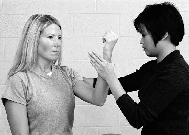
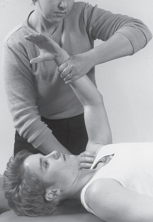
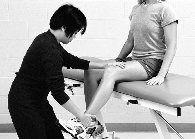
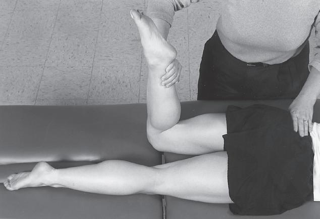
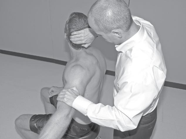
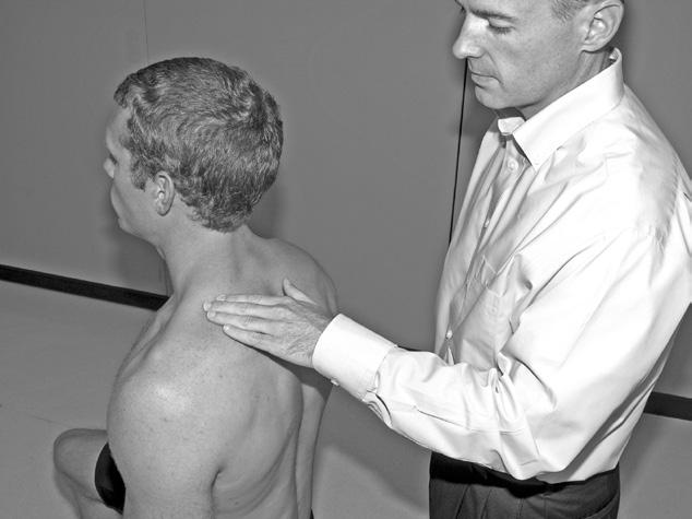
Performing a manual muscle test involves extensive time, effort, and attention to detail to ensure that the results obtained are as accurate as possible. To avoid biasing the results in a manual muscle test, the patient’s positioning, along with the examiner’s technique, must be standardized and adhered to. Due to the extensive time and effort needed to perform a comprehensive manual muscle test, manual muscle screening frequently is used to provide a quick overview of the patient’s muscle strength. The information thus obtained can help the examiner identify potential areas of strength deficit that then can be investigated further with more standardized methods of strength assessment. During manual muscle screening, muscle strength is assessed by placing the patient in positions of convenience, rather than in specific positions in which the muscles are working against, or outside the influence of, gravity. The strength of muscle groups then are tested through manual resistance supplied by the examiner, allowing rapid, although crude, assessment of muscle group strength. Screening of muscle strength is part of the upper and lower quarter screening examination and is useful as a survey of the overall strength of a patient prior to functional activity or training in activities of daily living.81 Figures 1-1 to 1-4 demonstrate differences in the testing techniques used in manual muscle screening, versus MMT, of the triceps brachii (elbow extensor) and biceps femoris (knee flexor) muscles. Manual muscle screening is a valuable tool for the rapid survey of gross strength but does not provide data that are accurate or quantifiable enough for diagnosis of neuromuscular disease or for evaluation of
*References 11, 14, 15, 20, 23, 30, 39, 40, 42, 43, 46, 49, 50, 53, 55, 65, 74, 83-85, 92, 97, 107, 108, 111, 113, 125, 128, 129.
that muscle is acting against gravity.
patient progress in terms of strength gains. In addition, because MMT does not have high sensitivity, its use as a screening tool may result in missed strength deficits unless those deficits are significant.18
Resisted movement testing, used to differentiate between contractile and noncontractile sources of musculoskeletal pain, also uses a form of MMT.29,81 During resisted movement testing, the patient’s joint is placed in a mid-range position, and isometric testing of muscle
Fig. 1-1
Manual muscle screening of triceps brachii muscle. Note lack of specific patient positioning in regard to gravity.
Fig. 1-2
Manual muscle testing of triceps brachii muscle. Note positioning of patient so
1-3 Manual muscle screening of biceps femoris muscle. Note lack of specific patient positioning in regard to gravity.
Fig. 1-4 Manual muscle testing of biceps femoris muscle. Note positioning of patient so that muscle is acting against gravity.
strength is performed to uncover any muscle weakness or pain that might be present with resisted motion. The presence of weakness with or without pain during isometric contractions performed in such neutral joint positions points to a muscular contribution to the musculoskeletal pathology. However, due to the lack of strict adherence to gravity-resisted versus gravityeliminated positioning during resisted movement testing, the information obtained about muscle strength cannot be graded or quantified any further than application of the terms “strong” or “weak.”
RELIABILITY OF MANUAL MUSCLE TESTING
A limited number of studies investigating the reliability of MMT have been performed. One of the earliest investigations occurred during the 1952 gammaglobulin field trials for the treatment of poliomyelitis.74 Lilienfeld
et al.74 reported on the interrater reliability of a standardized protocol for MMT by comparing experienced examiners both with novice examiners and with other experienced examiners. Although only descriptive statistics were reported, the authors found complete agreement (assigning of the same MMT grade) between examiners in 60% to 66% of the tests and agreement within plus or minus one full grade in 91% to 95% of the manual muscle tests given. Similar results were reported by Blair14 in a later study. However, in the studies of Lilienfeld et al.74 and Blair,14 the MMT scores were modified by applying a weighting factor for muscle bulk, which makes comparison with later studies difficult.
Somewhat lower levels of reliability were reported by Iddings et al.53 who examined both interrater and intrarater reliabilities of MMT. These authors reported levels of complete interrater agreement of 41% to 51% and agreement within plus or minus one full grade of 87% to 93%. Intrarater agreement was somewhat higher, with complete agreement occurring in 54% to 65% of the tests, whereas agreement within plus or minus one full grade was reported in 96% to 98% of the tests. The examiners in the study by Iddings et al.,53 unlike those in the studies of Lilienfeld et al.74 and Blair,14 did not use a standard MMT protocol. Each examiner was allowed to use the MMT technique of his or her choice. No weighting for muscle bulk was included in this or any subsequent report on the reliability of MMT.
In a study designed to improve the standardization of MMT for use with patients with chronic renal disease, Silver et al.113 examined the interrater reliability of a standardized protocol of MMT based on the techniques described by Daniels et al.31 Like the previous authors, Silver et al.113 reported their results only in terms of descriptive statistics and found complete agreement among examiners in 67% of the muscle tests performed, with agreement within a one-half grade occurring in 97% of the muscles tested. The apparently stronger reliabilities reported in this study (97% agreement within a one-half grade versus 95% within one full grade reported by earlier groups) may have been due to the strong emphasis placed by Silver et al.113 on standardization of the technique of muscle testing and grading used.
At least three groups have used inferential statistics to analyze the reliability of MMT in the normal population. Wadsworth et al.129 measured the intrarater reliability of MMT using five muscle groups: shoulder abductors, elbow extensors, wrist extensors, hip flexors, and knee flexors. Muscle testing protocols were standardized, and the examiner performing the testing had 8 years of clinical experience. Test-retest reliability coefficients were 0.63 for knee flexors, 0.74 for hip flexors, and 0.98 for shoulder abductors. Because grades on both test and retest for wrist and elbow extensors were the same, correlation coefficients for these muscle groups could not be calculated.
Fig.
In an article published in the same year as the study by Wadsworth et al.,129 Frese et al.43 reported the interrater reliability of manual muscle tests of the gluteus medius and middle trapezius muscles. Interrater reliability coefficients ranged from 0.04 to 0.66, including muscle test grades below 3 (fair). Although one would expect somewhat lower interrater reliability than intrarater reliability, the reliabilities reported in the Frese et al.43 study are quite poor. Differences in design between the studies by Wadsworth et al.129 and Frese et al.43 may account for much of the variation seen in the reported reliabilities. Examiners in the Frese et al.43 study did not use standard techniques for muscle testing but were allowed to test each muscle using the technique with which the examiner was most comfortable. The muscle testing techniques of both Kendall et al.63 and Daniels and Worthingham32 were used, but the two techniques use different methods of positioning the patient and resisting the muscle, particularly for the middle trapezius. Additionally, the examiners participating in the Frese et al.43 study had an average of 2.3 years of experience compared with the 8 years of experience possessed by the examiner in the other study by Wadsworth et al.129 No mention was made in the Frese et al.43 study of the establishment of intrarater reliability before the investigation of interrater reliability.
A study that examined both intrarater and interrater reliabilities of MMT in the healthy population was published by Brandsma et al.20 in 1995. These researchers focused on reliability of MMT for the intrinsic muscles of the hand. Comparison was made of MMT scores obtained by one experienced examiner in a test-retest situation (intrarater reliability) and by two experienced examiners on the same group of patients (interrater reliability). Both intrarater and interrater reliabilities were calculated using Cohen’s weighted kappa and ranged from 0.71 to 0.96 for intrarater and from 0.72 to 0.93 for interrater, results similar to those reported by Wadsworth et al.129 and appreciably higher than those reported by Frese et al.43
Reliability of MMT in individuals with musculoskeletal and neuromuscular pathology has also been examined. In a study by Florence et al.,42 physical therapists, all possessing 16 to 20 years of experience, performed manual muscle tests on 18 different muscle groups in males with Duchenne’s muscular dystrophy. A standardized method of muscle testing was used, and a modified Medical Research Council (MRC) scale was used in grading the muscles. Intrarater reliability was calculated using Cohen’s kappa, and results for individual muscle groups ranged from 0.65 to 0.93. The intrarater reliability of MRC grades 0 to 5 ranged from 0.80 to 0.99, with the highest reliabilities found in grades below 3 and the lowest reliabilities in grades 3+ (0.80), 4 (0.83), and 5 (0.83).
In a later study, another group of investigators examined the interrater reliability of performing MMT on a
group of 12 children with varying types of muscular dystrophy.39 Muscle testing was confined to those groups previously shown to be tested reliably,42 namely shoulder abductors, elbow and hip flexors, knee extensors, and ankle dorsiflexors. All examiners (n = 12) underwent 3 days of training by an experienced tester, and a modified MCR scale was used to grade the muscle tests.39 Examiners were divided into three groups of four testers for assessment of interrater reliability. Initially, reliability between the three groups ranged from 0.62 to 0.76, thus an additional training session was scheduled. Interrater reliability (intraclass correlation coefficients [ICCs]) averaged 0.90 subsequent to the additional training. Intrarater reliability was reported for one of the three groups of examiners and averaged 0.95 (ICC) over the 2 years of study.
Paternostro-Sluga and colleagues97 used a modification of the MRC scale, as well as the original scale, when examining the reliability of MMT in a group of 31 patients with lesions involving the radial nerve or the C7 nerve root fibers. Strength testing of the wrist and finger extensors was performed by five “specialists in physical medicine and rehabilitation” using a strictly standardized protocol. Both intrarater and interrater reliability was calculated using weighted kappa values, with kappa values between 0.61 and 0.80 considered substantial agreement and values above 0.80 considered “almost perfect agreement.” Intrarater reliability was reported as almost perfect agreement with all kappa values above 0.80. Interrater reliability was somewhat lower with kappa values ranging from 0.77 to 0.81.
Other groups of investigators have examined the reliability of MMT in patient groups with pathologies such as juvenile idiopathic inflammatory myopathy (JIIM),50,55 Charcot-Marie-Tooth disease,128 and a variety of illnesses resulting in a stay in the intensive care unit (ICU).40 All studies examined interrater reliability of MMT. Reliability was reported as excellent in ICU survivors, “almost perfect” in 95% of muscles tested in patients with Charcot-Marie-Tooth disease, and anywhere from excellent to quite variable in children with JIIM.50,55
Some studies that have examined reliability of MMT in individuals with pathology have used a collapsed method of scoring muscle strength. One group of researchers used the MRC sumscore, which was defined by the authors as the “summation of the strength of six muscle groups tested on both sides according to the MRC scale.”65 The six muscle groups included in the sumscore were the shoulder abductors, elbow flexors, wrist extensors, hip flexors, knee extensors, and ankle dorsiflexors. Manual muscle tests of these six muscle groups were performed bilaterally on a group of patients with Guillain-Barré syndrome, and then the MRC scores (0 to 5) for all muscle groups (six per side) were summed to achieve the MRC sumscore.
Calculation of ICCs for interrater reliability for both more and less experienced examiners resulted in ICCs of 0.98 and 0.96, respectively, for the two groups of examiners.
Collapsed scores of muscle strength are frequently used in studies examining muscle strength in patients following spinal cord injury. Several studies have examined the reliability of the motor examination component of the ISNCSCI, which uses MMT to examine the strength of 10 upper extremity muscle groups (5 on each side of the body) and 10 lower extremity muscle groups (5 on each side), grading each group on a 0-to-5 scale.2 A total motor score is then calculated by summing the scores for all 20 muscle groups. Upper extremity muscle scores (UEMS) and lower extremity muscle scores (LEMS) may be calculated by summing the scores for just the upper or lower extremity muscles, respectively. Both intrarater and interrater reliability for total motor scores and for UEMS and LEMS have been reported to be excellent among trained examiners.84,92,108
Most of the studies just cited indicate good intrarater reliability for MMT. In instances where lower intrarater reliabilities are reported, the examiners failed to use a standardized protocol for MMT.53 A similar observation can be made regarding the conflicting data reported for interrater reliability. Those studies reporting lower interrater reliabilities used a research protocol wherein each examiner was allowed to use his or her own method of MMT.43,53 Higher interrater reliability was achieved when a uniform method of MMT was used by all examiners.* Clinicians should be aware that unless standardized methods of MMT are used, the reliability of MMT will suffer.
VALIDITY OF MANUAL MUSCLE TESTING
Validity has been defined as “the degree to which an instrument measures what it is purported to measure.”28 One method of assessing validity is to perform correlational studies in which two different tests are administered to the same group of patients.90 Several researchers have compared MMT with muscle testing using HHD, strain gauge, or isokinetic dynamometer to determine whether a correlation exists between these forms of muscle testing.† Beasley11 was the first to examine the correlation between MMT and HHD. He compared the two methods of muscle testing on the knee extensor muscles of a group of normal and postpolio children aged 9 to 12 years. The results of Beasley’s study,11 which included only children with knee extensor muscles of grade fair or higher, found poor correlations between MMT and HHD scores particularly in muscles
with grades of normal or good. The children in Beasley’s study with as much as a 50% reduction in knee extensor strength were assigned an MMT grade of normal, and the same MMT grade was assigned to muscles with as much as a 20% to 25% difference in strength as measured by HHD.11
Studies performed subsequent to Beasley’s study11 have demonstrated similar problems with correlation between MMT and HHD, MMT and strain gauge, and MMT and isokinetic dynamometry above an MMT grade of good (4).* However, for muscles with an MMT grade below 4, high correlations have been demonstrated between MMT and HHD15,17,49,96,111 and between MMT and strain gauge.1 Schwartz et al.111 also demonstrated that specific MMT grades (from poor-plus to good) correspond to discrete ranges of myometry scores, indicating that “although MMT is a subjective measurement of strength, there are associated underlying objective parameters as represented by myometry data.”
The high correlations exhibited between MMT and HHD for muscles with an MMT grade below 4 demonstrate that both methods of testing measure strength, a good indication of the validity of MMT. That MMT has predictive validity has been reported in a number of studies. MMT scores at 72 hours after spinal cord injury have been shown to be good predictors of functional muscle recovery at the zone of injury22 and of functional status of patients with quadriplegia at rehabilitation discharge.71 ASIA motor upper extremity subscale scores have been reported to explain as much as 72% of the upper cord score of the Functional Independence Measure (FIM), and ASIA lower extremity subscale scores may explain up to 66% of FIM lower cord scores.83 Motor scores also have been demonstrated to be superior to neurologic level in predicting feeding score on the Quadriplegia Index of Function in patients with SCI.85 In another study involving patients with quadriplegia, MMT scores of six shoulder muscles were shown to have a very strong correlation (Spearman’s rank correlation coefficient of 0.95) with the FIM motor score and the FIM transfer score.46 Strong correlations between MMT scores and FIM scores also have been reported in patients with Duchenne muscular dystrophy.125
Discriminative validity of MMT has been demonstrated in placebo-controlled trials involving patients with idiopathic inflammatory myopathy. In two separate trials, increases in strength were detected by MMT in patients who received pharmacotherapeutic agents but not in those who received placebo medication.23,30 In summary, MMT is a valid method of measuring muscle strength as substantiated by several groups of researchers. MMT is designed to measure the entire range of muscle strength, from 0 (no evidence of muscle
*References 14, 20, 39, 65, 74, 113.
†References 1, 3, 11, 15, 17, 38, 49, 111
*References 1, 3, 15, 17, 38, 96, 111
contraction) through 5 (movement through the complete range of motion against gravity and maximum resistance). Other forms of muscle strength testing, such as HHD and isokinetic dynamometry, are limited in their ability to accurately assess the lower grades of muscle strength. However, when MMT scores exceed grade 4, the MMT loses much of its ability to discriminate between gradations of strength, whereas HHD and isokinetic dynamometry retain their sensitivities in these ranges. In instances in which documentation of strength levels is critical above an MMT grade of 4, the examiner might be well advised to use an alternative form of measuring muscle strength such as HHD or isokinetic dynamometry.37
CONSIDERATIONS IN THE PERFORMANCE OF THE MANUAL MUSCLE TEST
MMT is designed to measure muscle strength, which has been defined as the ability of the muscle to develop tension against resistance.69,106 The amount of tension generated by a muscle in a given situation will vary according to a number of factors, including number and firing rate of motor units activated, length of the muscle at the time of the contraction, muscle cross-sectional area, fiber type composition of the muscle, point of application of resistance, stabilization techniques used, and motivational state of the subject.* Although the anatomic and physiologic factors influencing muscle strength cannot be controlled in a testing situation, many factors such as patient positioning, stabilization, point of force application, and use of motivation can and should be controlled. These factors should be standardized for each muscle test to maintain consistency and to optimize the validity and reliability of MMT.
Instructing and Motivating the Patient
Patients should be provided with explicit instructions before performing any examination technique, including strength testing. Studies have indicated that standardization of instructions may improve the reliability of the results of strength testing.25 Such testing techniques require the full cooperation and best effort of the patient, factors that are likely to be enhanced as the patient’s understanding of the purpose of the test increases.
Before beginning the procedure, describe to the patient exactly what will be taking place and why the measurement must be performed. If any testing apparatus is to be used during the procedure, show the patient the tool, and explain, in layperson’s terms, its purpose and how it will be used. Instruct the patient in
the position he or she is to assume, again using layperson’s terms and avoiding terms such as supine or prone. Explain to the patient the necessity of exerting maximum effort during the examiner’s resistance of each movement for the results of the testing to be meaningful. Detailed explanations of every step of the procedure should not be provided initially, as this will only confuse the patient. A brief, general explanation is best at this point, and further explanations may be given once the procedure is in progress, remembering to use layperson’s terms with all explanations provided. An example of initial patient instructions is as follows:
Ms. Bates, I need to measure how much strength you have in the muscles that straighten your elbow. This information will tell me how much your strength is improving and will give me an idea of what changes we need to make in your plan of care. At a certain point during the test, I will push against your arm and will ask you to try to keep me from bending your elbow. When I do that, I want you to try as hard as you can to keep me from bending your elbow. I will need you to lie on this table on your back so that I can test your muscles.
Patient Positioning
Proper positioning of the patient during muscle testing is essential in ensuring that the appropriate muscle is being tested and in preventing substitution by other muscles. The choice of a preferred patient position for each MMT is based on several criteria. For a position to be considered optimal, all criteria should be met. Although this is not an exhaustive list, the major criteria in selecting a preferred patient position for measurement of muscle strength are as follows:
1. The distal joint segment should be placed in the desired position in relation to the pull of gravity. Gravity plays an integral part in MMT, and the patient’s ability or inability to move the designated part against gravity determines the position in which the patient should be placed for the muscle test. Patients who are able to move the appropriate joint actively through the complete range of motion against gravity are placed in the so-called gravityresisted position for testing. That is, the patient is positioned so that the distal segment of the joint must move against gravity to complete the range of motion. An individual who is unable to move the distal segment of the tested joint through the complete range of motion against gravity is placed in the so-called gravity-eliminated position to test the muscle (see Clinical Comment: Gravity-Eliminated Testing Position).
*References 8, 13, 47, 58, 116, 130, 133.
2. The patient should be positioned so that the proximal segment of the joint is most easily and optimally stabilized. Stabilization of the proximal joint segment is of critical importance during MMT.
Stabilization may be provided via several avenues, including use of a firm testing surface, patient positioning, muscle activity by the patient, and manual holds by the examiner.130 The origin of the muscle being tested must be firmly fixed so that maximal contraction against the insertion can occur, although stabilization over the muscle belly being tested should be avoided. Failure to provide such fixation may cause underestimation of strength by the examiner due to the patient’s inability to produce optimal force with the muscle or may result in substitutions by other muscles in an effort to compensate for an inability of the agonist to generate sufficient force.48,106,116,130 The examiner should be consistent in the use and technique of stabilization each time a muscle test is performed.
3. The patient should be positioned so that the motion to be performed is not restricted in any way. Motion should not be blocked by external objects, such as the examining table, or by internal forces, such as muscle tightness in the antagonistic muscle group.
4. The joint should be positioned at the beginning of the range of motion for the movement to be performed. Such positioning allows observation of the movement through the complete range of motion, an observation that is necessary to accurate grading of muscle strength.
5. The patient should be positioned so that the examiner’s body mechanics are optimized during stabilization and application of resistance. The examiner should not endure undue stress in providing stabilization of the proximal joint segment or while resisting the distal segment (gravity-resisted test only). Aids to patient positioning, such as the use of a highlow examining table, a stool for the examiner, or other devices, may be needed to facilitate optimal body mechanics for the examiner.
6. The patient must be able to assume the position. In some cases, this criterion cannot be met (see Case 2-1, Chapter 2), and an alternative position must be used. In any instance in which an alternative
position is used, the examiner should design the position so that it adheres to the previous five criteria as closely as possible.
FURTHER
EXPLORATION: PATIENT POSITIONING—GRAVITY-RESISTED VERSUS “GRAVITY-ELIMINATED”
The following activities are designed to help the student evaluate and design preferred and alternative patient positions for muscle strength testing.
1. Without looking at the specific manual muscle tests described in this text, design a testing position that would allow each of the muscles listed in item 2 to work in the following scenarios:
a. Against gravity
b. In a “gravity-eliminated” position
c. With gravity (assisted by the pull of gravity)
2. The following muscles are to be tested:
a. Biceps brachii
b. Pectoralis major
c. Semimembranosus
d. Peroneus longus
3. Apply the criteria for positioning (listed in the previous section) to each of the positions you devised. How well does each position meet the criteria listed? Make modifications to your devised position as needed, so that it adheres more closely to the positioning criteria.
FURTHER EXPLORATION: PALPATING MUSCLE CONTRACTION
The following activities are designed to assist the student in detection of muscle contraction via manual palpation.
1. Ask a partner to sit in a comfortable chair without arms.
2. Position yourself in front of and to one side of your partner, with one hand on the superior aspect of your partner’s shoulder and the other hand (fingertips) on the anterior aspect of your partner’s arm (see Fig. 2-90).
Box 1-1 CLINICAL COMMENT: GRAVITY-ELIMINATED TESTING POSITION
The term gravity eliminated has been commonly used with reference to MMT for many years. However, the term is rather misleading in that gravity is not eliminated, but the effects of gravity are lessened in this position. The examiner positions the patient so that movement of the distal joint segment is neither directly resisted nor directly assisted by gravity.
3. Ask your partner to strongly contract the elbow flexors (partner’s elbow will flex) while you palpate the muscle contraction.
4. Now, ask your partner to contract the elbow flexors weakly, producing little or no movement of the elbow.
5. Have your partner repeatedly perform weak contractions of the elbow flexors while you palpate the entire length of the biceps brachii muscle. Determine where along the muscle the weak contraction is best detected.
6. Repeat the exercise for the knee extensors (see Fig. 4-67), palpating the quadriceps muscles over the anterior aspect of the thigh.
Box 1-2
CLINICAL COMMENT: MUSCLE PALPATION
An examiner should always confirm a contraction of the muscle being tested by palpating the muscle during the patient’s active movement. Palpation is critical for very weak muscles, especially if little to no active movement is produced (grades below 2). Additionally, palpation of the muscle during active movement by the patient assists the examiner in ruling out the possibility of substitution by other muscles.
FURTHER EXPLORATION: STABILIZATION
The following activities are designed to help the student appreciate the effects of proper versus improper positioning on the muscle strength test.
1. Perform a gravity-resisted test of the muscle groups listed next, first including, and then excluding, the stabilization described. Refer to the page numbers provided for a description of each test.
a. Elbow flexors, pp. 95-102
b. Shoulder horizontal adductors, pp. 73-82
c. Hip lateral rotators, pp. 275-278
d. Knee flexors, pp. 283-286
2. As each muscle group is tested, observe the results of the test with and without stabilization. Answer the following questions.
a. What differences do you observe in each situation?
b. Is the patient able to generate a more forceful contraction with the proximal joint segment stabilized or unstabilized?
c. Do substitutions by other muscle groups occur more often when the proximal joint segment is stabilized or unstabilized?
3. Would stabilization of the proximal joint segment be more, or less, important in a patient with muscle weakness? Why?
APPLICATION OF RESISTANCE
Testing muscles with MMT grades above 3 requires the application of manual resistance by the examiner. In this text, the “break test” is used to apply resistance. During a break test, the examiner applies resistance against the patient’s body part, increasing the resistance applied until the patient’s muscular contraction is overcome by the examiner (the patient “breaks”) or until maximum resistance has been applied and held for 4 to 5 seconds.16,25,116 Both “make” and “break” tests are used in MMT, and both have been found to be reliable, although more force appears to be produced by
muscles tested under the “break” than under the “make” method.16
When applying resistance, the examiner should generally apply the force perpendicular to the distal end of the distal segment of the joint being tested. With the exception of a few cases, resistance should not be applied any further distally than the distal end of the bone on which the muscle being tested inserts (i.e., resistance for the deltoid should be applied over the distal end of the humerus). Although this technique may result in a shorter lever arm for the application of resistance, the possible confounding of the results that can occur when one applies resistance over weak distal joints is avoided.
For most muscle tests in this text, resistance is applied at the end of the gravity-resisted range of motion. In many cases, this is not the strongest point in the range of motion for the muscle being tested.13,133 However, application of force at the end of the gravity-resisted range of motion provides a consistent point for the application of resistance and may help prevent the examiner from overestimating the patient’s strength, a documented tendency in MMT grades above 4.11 As maximum force generated by the muscle changes significantly with muscle length and joint angle,8,47,116 the examiner must exercise extreme care to apply resistance at the same point in the range of motion each time the muscle is tested. To do otherwise would result in an unreliable muscle test.
FURTHER EXPLORATION: LEVER ARM LENGTH
The following activities are designed to help the student understand the effects of lever arm length on the perceived strength of the muscle contraction.
1. Perform a gravity-resisted test of the muscle groups listed next, using the two different points of resistance described. Refer to the page numbers provided for a description of the patient positioning and stabilization for each test.
a. Shoulder abductors, pp. 61-68
i. Resistance just proximal to elbow
ii. Resistance at wrist
b. Scapular adductors (middle trapezius), pp. 23-29
i. Resistance on scapula
ii. Resistance at wrist
2. As each muscle group is tested, observe the results of the test for each resistance point utilized. Answer the following questions.
a. Does the patient seem stronger or weaker when using the more proximal resistance point? The more distal point?
b. Would changing the position of the intervening joint(s) make a difference in the results when the distal resistance point is used (e.g., flexing the elbow)?
3. Could pathology involving the intervening joint affect the results of the muscle test when the distal resistance point is used? If so, how?
Box 1-3 CLINICAL COMMENT: USE OF RESISTANCE DURING MMT
If a patient is able to move through the complete range of motion when placed in a gravity-resisted position, one knows at that point that the muscle test grade is at minimum a 3. Manual resistance is then applied by the examiner and the muscle’s strength is assessed according to the scale in Table 1-1
Resistance is not applied to a muscle that is unable to move through the complete range of motion against gravity.
GRADING SCALE
As discussed, a variety of methods have been used in grading the MMT. The scale used in this text is a modification of the MRC scale in that either numbers or word/letters can be used (Table 1-1). Plus/minus designations are included at some points, but extreme caution is advised when assigning plus/minus grades, especially when the testing is being performed by a novice examiner. The accurate use of plus/minus designations requires considerable skill and experience in the art of MMT.
Definitions for each grade are provided in Table 1-1 with the corresponding number/word designation. One should be aware that grades above 3 tend to be much more subjective than grades 3 or below because of the inability to precisely define the amount of force that constitutes “maximum,” “moderate,” or “minimal” resistance. Additionally, evidence indicates that there are serious questions regarding the ability to differentiate strength differences in the grades above 3 or to identify losses in muscle strength of up to 50%.* Given the subjectivity of muscle grades above 3, one should be particularly careful regarding the method of application of manual resistance. To maintain consistency in testing, each individual examiner must not deviate from the amount of resistance that the examiner defines as maximal, moderate, and minimal. One might be tempted to assign a grade of 5 to the triceps muscle of an elderly female who “breaks” under moderate resistance, rationalizing that this patient’s strength is probably normal for someone of her age and gender. Such allowances and rationalizations result in poor reliability of MMT both in repeated tests by the same examiner (intrarater reliability) and between tests by
*References 11, 15, 49, 107, 111, 129
Table 1-1. MUSCLE TEST GRADES
NUMBER (AND LETTER) GRADE WORD GRADE DEFINITION
0
1 (Tr)
2− (P−)
2 (P)
2+ (P+)
3− (F−)
3 (F)
3+ (F+)
4 (G)
5 (N)
Zero No evidence of contraction by vision or palpation
Trace Slight contraction; no motion
Poor minus Movement through partial test range in gravityeliminated position
Poor Movement through complete test range in gravityeliminated position
Poor plus Movement through complete test range in gravityeliminated position and through up to one half of test range against gravity
Fair minus Movement through complete test range in gravityeliminated position and through more than one half of test range against gravity
Fair Movement through complete test range against gravity
Fair plus Movement through complete test range against gravity and able to hold against minimum resistance
Good Movement through complete test range against gravity and able to hold against moderate resistance
Normal Movement through complete test range against gravity and able to hold against maximum resistance
different examiners (interrater reliability). By maintaining a consistent definition of each level of resistance an examiner is able to ensure that reliability in testing is maximized.
The use of numerals as grades for muscle testing should not lead one to think that muscle test grades are interval measures. Michaels90 defines an interval scale as “one on which the categories are numerical units and the intervals between the units are assumed to be of equal size.” The intervals between muscle test grades are not equal, and therefore the MMT grading scale is more appropriately classified as an ordinal scale. One should not then consider that the difference in strength between the muscle test grades of 5 and 4 is equivalent to the difference in strength between the muscle test grades of 4 and 3. Such assumptions are invalid and cannot be made regarding ordinal data.
Table 1-2 provides a format and rationale for performing a manual muscle test. The use of a standardized procedure for all muscle tests will assist the examiner in achieving consistency, which is critical in establishing reliability of MMT.
Table 1-2. FORMAT AND RATIONALE FOR MANUAL MUSCLE TEST STEPS RATIONALE
1. Explain purpose of procedure to patient.
2. Place patient in gravity-resisted position.
3. Stabilize proximal joint segment.
4. Instruct patient in specific movement to be performed while passively moving distal segment through ROM.
5. Return distal segment to starting position.
6. Ask patient to perform movement while muscle is being palpated and stabilization maintained.
7. Apply resistance (only if patient can complete ROM against gravity) by moving palpating hand to appropriate position for resisting the muscle.
8. Reposition patient in gravity-eliminated position (if patient is unable to complete ROM against gravity), and repeat steps 3 to 6. Do not apply resistance
9. Assign appropriate muscle grade.
ROM, Range of motion.
Box 1-4
Patient needs to be fully engaged in process to achieve accurate strength measurement.
Assume patient can move against gravity unless patient is known to be unable to do so, then use gravity-eliminated position.
Proper stabilization of proximal muscle attachment allows tested muscle to produce optimal contraction and decreases likelihood of substitution by synergists.
Shows patient exact movement expected and allows assessment of patient’s available ROM.
Patient must return to starting position so full movement can be performed actively.
Muscle is palpated to confirm presence of active muscle contraction.
Resistance is applied only if patient is able to complete movement against gravity. Since palpation is no longer necessary, palpating hand should be used to apply resistance. OR
Patients unable to move against gravity should be retested in a position where the resistance of gravity on the movement is lessened. If patient is unable to complete movement against gravity, resistance is not appropriate.
Grade should be assigned based on scale in Table 1-1
CLINICAL COMMENT: RANGE OF MOTION
The criteria for assigning a strength grade during MMT are based in part on the patient’s ability to perform the movement in question through the complete range of motion. Complete range of motion refers to the total range of movement possible for the joint
REFERENCES
1. Aitkens S, Lord J, Bernauer E, et al. Relationship of manual muscle testing to objective strength measurements. Muscle Nerve. 1989;12: 173-177.
2. American Spinal Injury Association. International Standards for Neurological Classification of Spinal Cord Injury. Atlanta, GA: Author; 2000.
3. Andersen H, Jakobsen J. A comparative study of isokinetic dynamometry and manual muscle testing of ankle dorsal and plantar flexors and knee extensors and flexors. Eur Neurol. 1997;37: 239-242.
4. Asmussen E, Heeboll-Nielsen K. Isometric muscle strength in relation to age in men and women. Ergonomics. 1962;5:167-169.
5. Bäckman E, Johansson V, Hager B, et al. Isometric muscle strength and muscular endurance in normal persons aged between 17 and 70 years. Scand J Rehab Med. 1995;27:109-111.
6. Baldauf KL, Swenson DK, Medeiros JM, et al. Clinical assessment of trunk flexor muscle strength in healthy girls 3 to 7 years of age. Phys Ther. 1984;64:1203-1208.
7. Baltzopoulos V, Brodie DA. Isokinetic dynamometry: applications and limitations. Sports Med. 1989;8(2):101-116.
8. Bandy WD, Lovelace-Chandler V. Determinants of muscle strength. Phys Ther Pract. 1992;2:1-10.
9. Barber A. Upper cervical spine flexor muscles: age related performance in asymptomatic women. Austral J Physiother. 1994;40: 167-172.
being tested unless the patient has some structural impairment making complete movement at the joint in question impossible (i.e., loose body in the joint). In such a case, the patient’s available range of motion becomes the complete range of motion for the joint.
10. Bassey EJ, Bendall MJ, Pearson M. Muscle strength in the triceps surae and objectively measured customary walking activity in men and women over 65 years of age. Clin Sci. 1988;74:85-89.
11. Beasley WC. Influence of method on estimates of normal knee extensor force among normal and postpolio children. Phys Ther Rev 1956;36:21-41.
12. Bemben MG, Massey BH, Bemben DA, et al. Isometric muscle force production as a function of age in healthy 20- to 74-yr-old men. Med Sci Sports Exerc. 1991;23:1302-1310.
13. Bender JA, Kaplan HM. The multiple angle testing method for the evaluation of muscle strength. J Bone Joint Surg. 1963;45A:135-140.
14. Blair L. The role of the physical therapist in the evaluation studies of the poliomyelitis vaccine field trials. Phys Ther Rev. 1957; 37:437-447.
15. Bohannon RW. Manual muscle test scores and dynamometer test scores of knee extension strength. Arch Phys Med Rehabil. 1986; 67:390-392.
16. Bohannon RW. Make tests and break tests of elbow flexor muscle strength. Phys Ther. 1988;68:193-194.
17. Bohannon RW. Measuring knee extensor muscle strength. Am J Phys Med Rehabil. 2001;80:13-18.
18. Bohannon RW. Manual muscle testing: does it meet the standards of an adequate screening test? Clin Rehabil. 2005;19:662-667.
19. Bracken MB, Shepard MJ, Holford TR, et al. Administration of methylprednisolone for 24 and 48 hours or tirilazad mesylate for 48 hours in the treatment of acute spinal cord injury. JAMA. 1997;277:1597-1604.
20. Brandsma JW, Schreuders TAR, Birke JA, et al. Manual muscle strength testing: intraobserver and interobserver reliabilities for the intrinsic muscles of the hand. J Hand Ther. 1995;8:185-190.
21. Brown LE, Weir JP. ASEP procedures recommendation, I: assessment of muscular strength and power. J Exerc Phys. 2001;4:1-21.
22. Brown PJ, Marino RJ, Herbison GJ, et al. The 72-hour examination as a predictor of recovery in motor complete quadriplegia. Arch Phys Med Rehabil. 1991;72:546-548.
23. Bunch TW, Worthington JW, Combs JJ, et al. Azathioprine with prednisone for polymyositis. A controlled, clinical trial. Ann Intern Med. 1980;92:365-369.
24. Byl NN, Wells L, Grady D, et al. Consistency of repeated isokinetic testing: effect of different examiners, sites, and protocols. Isokinet Ex Sci. 1991;1:122-130.
25. Caldwell LS, Chaffin DB, Dukes-Dobos FN, et al. A proposed standard procedure for static muscle strength testing. Am Ind Hyg Assoc J. 1974;35:201-206.
26. Christ CB, et al. Maximal voluntary isometric force production characteristics of six muscle groups in women aged 25 to 74 years. Am J Hum Biol. 1992;4:537-545.
27. Clement FJ. Longitudinal and cross-sectional assessments of age changes in physical strength as related to sex, social class, and mental ability. J Gerontol. 1974;29:423-429.
28. Currier DP. Elements of Research in Physical Therapy. 2nd ed. Baltimore: Williams & Wilkins; 1984.
29. Cyriax J. Textbook of Orthopaedic Medicine. 8th ed. Philadelphia: Baillière Tindall; 1982.
30. Dalakas MC, Illa I, Dambrosia JM, et al. A controlled trial of highdose intravenous immune globulin intrafusions as treatment for dermatomyositis. N Engl J Med. 1993;329:1993-2000.
31. Daniels L, Williams M, Worthingham C. Muscle Testing: Techniques of Manual Examination. Philadelphia: WB Saunders; 1947.
32. Daniels L, Worthingham C. Muscle Testing: Techniques of Manual Examination. 5th ed. Philadelphia: WB Saunders; 1986.
33. Delito A. Isokinetic dynamometry. Muscle Nerve. 1990;S53–S57.
34. De Ste Croix MBA, Armstrong N, Welsman JR, et al. Longitudinal changes in isokinetic leg strength in 10-14-year-olds. Ann Hum Biol 2002;29:50-62.
35. Duvall EN, Houtz SJ, Hellebrandt FA. Reliability of a single effort muscle test. Arch Phys Med. 1947;April:213-218.
36. Dvir Z. Clinical applicability of isokinetics: a review. Clin Biomech 1991;6:133-144.
37. Dvir Z. Grade 4 in manual muscle testing: the problem with submaximal strength assessment. Clin Rehabil. 1997;11:36-41.
38. Ellenbecker TS. Muscular strength relationship between normal grade manual muscle testing and isokinetic measurement of the shoulder internal and external rotators. Isokinet Exerc Sci. 1996; 6:51-56.
39. Escolar DM, Henricson EK, Mayhew J, et al. Clinical evaluator reliability for qualitative and manual muscle testing measures of strength in children. Muscle Nerve. 2001;24:787-793.
40. Fan E, Ciesla ND, Truong AD, et al. Inter-rater reliability of manual muscle strength testing in ICU survivors and stimulated patients. Intensive Care Med. 2010;36:1038-1043.
41. Fawcett JW, Curt A, Steeves JD, et al. Guidelines for the conduct of clinical trials for spinal cord injury as developed by the ICCP panel: spontaneous recovery after spinal cord injury and statistical power needed for therapeutic clinical trials. Spinal Cord. 2007;45:190-205.
42. Florence JM, Pandya S, King WM, et al. Intrarater reliability of manual muscle test (Medical Research Council scale) grades in Duchenne’s muscular dystrophy. Phys Ther. 1992;72:115-126.
43. Frese E, Brown M, Norton BJ. Clinical reliability of manual muscle testing: middle trapezius and gluteus medius muscles. Phys Ther 1987;67:1072-1076.
44. Frontera WR, Hughes VA, Fielding RA, et al. Aging of skeletal muscle: a 12-yr longitudinal study. J Appl Physiol. 2000;88: 1321-1326.
45. Frontera WR, Hughes VA, Lutz KJ, et al. A cross-sectional study of muscle strength and mass in 45- to 78-yr-old men and women. J Appl Physiol. 1991;71:644-650.
46. Fujiwara T, Hara Y, Akaboshi K, et al. Relationship between shoulder muscle strength and functional independence measure (FIM) score among C6 tetraplegics. Spinal Cord. 1999;37:58-61.
47. Gordon AM, Huxley AF, Julian FJ. The variation in isometric tension with sarcomere length in vertebrate muscle fibres. J Physiol 1966;184:170-192.
48. Hart DL, Stobbe TJ, Till CW, et al. Effect of trunk stabilization on quadriceps femoris muscle torque. Phys Ther. 1984;64:1375-1380.
49. Herbison GJ, Issac Z, Cohen ME, et al. Strength post-spinal cord injury: myometer vs manual muscle test. Spinal Cord. 1996;34: 543-548.
50. Hicks J, Wesley R, Koziol D, et al. Validation of manual muscle testing in the assessment of juvenile dermatomyositis. Arthritis Rheum. 2000;43:S194.
51. Hislop HJ, Montgomery J. Daniels and Worthingham’s Muscle Testing 7th ed. Philadelphia: WB Saunders; 1995.
52. Hurley BF. Age, gender, and muscular strength. J Gerontol Ser A 1995;50A:41-44.
53. Iddings DM, Smith LK, Spencer WA. Muscle testing, II: reliability in clinical use. Phys Ther Rev. 1961;41:249-256.
54. Jacoby SM. Isokinetics in rehabilitation. In: Prentice WE, Voight MJ, eds. Techniques in Musculoskeletal Rehabilitation. New York: McGrawHill; 2001:153-166.
55. Jain M, Smith M, Cintas H, et al. Intra-rater and inter-rater reliability of the 10-point manual muscle test (MMT) of strength in children with juvenile idiopathic inflammatory myopathies (JIIM). Phys Occup Ther Pediatr. 2006;26:5-17.
56. Jakobsson F, Borg K, Edstrom L. Fibre-type composition, structure and cytoskeletal protein location of fibres in anterior tibial muscle. Acta Neuropathol. 1990;80:459-468.
57. Jan MH, Chai HM, Lin YF, et al. Effects of age and sex on the results of an ankle plantar-flexor manual muscle test. Phys Ther. 2005; 85:1078-1084.
58. Johnson BL, Nelson JK. Effect of different motivational techniques during training and in testing upon strength performance. Res Q. 1967;38:630-636.
59. Johnson T. Age-related differences in isometric and dynamic strength and endurance. Phys Ther. 1982;62:985-989.
60. Jordan A, Mehlsen J, Bulow M, et al. Maximal isometric strength of the cervical musculature in 100 healthy volunteers. Spine. 1999; 24:1343-1348.
61. Kanehisa H, Ilegawa S, Tsunoda N, et al. Strength and crosssectional areas of reciprocal muscle groups in the upper arm and thigh during adolescence. Int J Sports Med. 1995;16:54-60.
62. Kanehisa H, Yata H, Ikegawa S, et al. A cross-sectional study of the size and strength of the lower leg muscles during growth. Eur J Appl Physiol. 1995;72:150-156.
63. Kendall FP, McCreary EK, Provance PG. Muscles: Testing and Function. 4th ed. Baltimore: Williams & Wilkins; 1993.
64. Kent-Braun JA, Ng AV. Specific strength and voluntary muscle activity in young and elderly women and men. J Appl Physiol 1999;87:22-29.
65. Kleyweg RP, Van Der Meché FGA, Schmitz PIM. Interobserver agreement in the assessment of muscle strength and functional abilities in Guillain-Barré syndrome. Muscle Nerve. 1991;14:1103-1109.
66. Klitgaard H, Mantoni M, Schiaffino S, et al. Function, morphology and protein expression of ageing skeletal muscle: a cross-sectional study of elderly men with different training backgrounds. Acta Physiol Scand. 1990;140:119-139.
67. Knuttgen HG, Kraemer WJ. Terminology and measurement in exercise performance. J Appl Sport Sci Res. 1987;1:1-10.
68. Kraemer WJ, Fry AC. Strength Testing: Development and Evaluation of Methodology. Physiological Assessment of Human Fitness. Champaign, IL: Human Kinetics; 1995:115-138.
69. Kroemer KH. Human strength: terminology, measurement and interpretation of data. Hum Factors. 1972;12:515-522.
70. Larsson L, Sjodin B, Karlsson J. Histochemical and biochemical changes in human skeletal muscle with age in sedentary males, age 22-65 years. Acta Physiol Scand. 1978;103:31-39.
71. Lazar RB, Yarkony GM, Ortolano D, et al. Prediction of functional outcome by motor capability after spinal cord injury. Arch Phys Med Rehabil. 1989;70:819-822.
72. Lexell J, Taylor T. Variability in muscle fiber areas in whole human quadriceps muscle: effects of increasing age. J Anat. 1991;174: 239-249.
73. Lexell J, Taylor T, Sjostrom M. What is the cause of the ageing atrophy? Total number, size and proportion of different fiber types studied in whole vastus lateralis muscle from 15- to 83-year-old men. J Neurol Sci. 1988;84:275-294.
74. Lilienfeld AM, Jacobs M, Willis M. A study of the reproducibility of muscle testing and certain other aspects of muscle scoring. Phys Ther Rev. 1954;34:279-289.
75. Lindle RS, Metter EJ, Lynch NA, et al. Age and gender comparisons of muscle strength in 654 women and men aged 20-93 yr. J Appl Physiol. 1997;83:1581-1587.
76. Liu F, Carlson L, Watson K. Quantitative abductor pollicis brevis strength testing: reliability and normative values. J Hand Surg 2000;25A:752-759.
77. Lovett RW. The treatment of infantile paralysis: preliminary report. JAMA. 1915;64:2118.
78. Lovett RW The Treatment of Infantile Paralysis. Philadelphia: P Blakiston; 1916.
79. Lovett RW, Martin EG. Certain aspects of infantile paralysis with a description of a method of muscle testing. JAMA. 1916;66:729-733.
80. Lowman CL. A method of recording muscle tests. Am J Surg 1927;3:588-591.
81. Magee DJ. Orthopedic Physical Assessment. 4th ed. Philadelphia: WB Saunders; 2002.
82. Mahony K, Hunt A, Daley D, et al. Inter-tester reliability and precision of manual muscle testing and hand-held dynamometry in lower limb muscles of children with spina bifida. Phys Occup Ther Pediatr. 2009;29:44-59.
83. Marino RJ, Graves DE. Metric properties of the ASIA motor score: subscales improve correlation with functional activities. Arch Phys Med Rehabil. 2004;85:1804-1810.
84. Marino RJ, Jones L, Kirshblum S, et al. Reliability and repeatability of the motor and sensory examination of the International Standard for Neurological Classification of Spinal Cord Injury. J Spinal Cord Med. 2008;31:166-170.
85. Marino RJ, Rider-Foster D, Maissel G, et al. Superiority of motor level over single neurological level in categorizing tetraplegia. Paraplegia. 1995;33:510-513.
86. Marks R. The effect of ageing and strength training on skeletal muscle. Austral J Physiother. 1992;38:9-19.
87. Martin EG, Lovett RW. A method of testing muscular strength in infantile paralysis. JAMA. 1915;65:1512-1513.
88. Mathiowetz V, Wiemer DM, Federman SM. Grip and pinch strength: norms for 6- to 19-year-olds. Am J Occup Ther. 1986;40:705-711.
89. Metter EEJ, Conwit R, Tobin J, et al. Age-associated loss of power and strength in the upper extremities in women and men. J Gerontol 1997;52A:B267–B276.
90. Michaels E: Evaluation and research in physical therapy. Phys Ther 1982;62:828-834.
91. Moffroid M, Whipple R, Hofkosh J, et al. A study of isokinetic exercise. Phys Ther. 1969;49:735-747.
92. Mucahey MJ, Gaughan J, Betz RR. Agreement of repeated motor and sensory scores at individual myotomes and dermatomes in young persons with complete spinal cord injury. Spinal Cord 2009;47:56-61.
93. Mulcahey MJ, Gaughan J, Betz RR, et al. The international standards for neurological classification of spinal cord injury: reliability of data when applied to children and youths. Spinal Cord 2007;45:452-459.
94. Nevill AM, Holder RL, Baxter-Jones A, et al. Modeling development changes in strength and aerobic power in children. J Appl Physiol. 1998;84:963-970.
95. Newman LB. A new device for measuring muscle strength: the myometer. Arch Phys Med Rehabil. 1949;30:234-237.
96. Noreau L, Vachon J. Comparison of three methods to assess muscular strength in individuals with spinal cord injury. Spinal Cord 1998;36:716-723.
97. Paternostro-Sluga T, Grim-Steiger M, Posch M, et al. Reliability and validity of the Medical Research Council (MRC) scale and a modified scale for testing muscle strength in patients with radial palsy. J Rehabil Med. 2008;40:665-671.
98. Phillips BA, Lo SK, Mastaglia FL. Muscle force measured using “break” testing with a hand-held myometer in normal subjects aged 20 to 69 years. Arch Phys Med Rehabil. 2000;81:653-661.
99. Pousson M, Lepers R, Van Hoecke J. Changes in isokinetic torque and muscular activity of elbow flexor muscles with age. Exp Gerontol. 2001;36:1687-1698.
100. Ramos E, Frontera WR, Llopart A, et al. Muscle strength and hormonal levels in adolescence: gender related differences. Int Sports Med. 1998;19:526-531.
101. Rider LG, Giannini EH, Harris-Love M, et al. Defining clinical improvement in adult and juvenile myositis. J Rheumatol. 2003; 30:603-617.
102. Robertson A, Deitz J. A description of grip strength in preschool children. Am J Occup Ther. 1988;42:647-652.
103. Rothstein JM. Muscle biology: clinical considerations. Phys Ther 1982;62:1823-1830.
104. Round JM, Jones DA, Honour JW, et al. Hormonal factors in the development of differences in strength between boys and girls during adolescence: a longitudinal study. Ann Hum Biol. 1999; 26:49-62.
105. Salo PK, Ylinen JJ, Malkia EA, et al. Isometric strength of the cervical flexor, extensor, and rotator muscles in 220 healthy females aged 20 to 59 years. J Orthop Sports Phys Ther. 2006;36:495-502.
106. Sapega AA. Muscle performance evaluation in orthopaedic practice. J Bone Joint Surg. 1990;72A:1562-1574.
107. Sarantini AJ, Gleim GW, Melvin M, et al. The relationship between subjective and objective measurements of strength. J Orthop Sports Phys Ther. 1980;2:15-19.
108. Savic G, Bergstrom EM, Frankel HL, et al. Inter-rater reliability of motor and sensory examinations performed according to American Spinal Injury Association standards. Spinal Cord. 2007;45:444-451.
109. Schreuders TAR, Roebroeck ME, Jaquet JB, et al. Long-term outcome of muscle strength in ulnar and median nerve injury: comparing manual muscle strength testing, grip and pinch strength dynamometers and a new intrinsic muscle strength dynamometer. J Rehabil Med. 2004;36:273-278.
110. Schreuders TAR, Selles RW, Roebroeck M, et al. Strength measurements of the intrinsic hand muscles: a review of the development and evaluation of the Rotterdam intrinsic hand myometer. J Hand Ther. 2006;19:393-402.
111. Schwartz S, Cohen ME, Herbison GJ, et al. Relationship between two measures of upper extremity strength: manual muscle test compared to hand-held myometry. Arch Phys Med Rehabil. 1992; 73:1063-1068.
112. Sepic SB, Murray MP, Mollinger LA, et al. Strength and range of motion in the ankle in two age groups of men and women. Am J Phys Med. 1986;65:75-84.
113. Silver M, McElroy A, Morrow L, et al. Further standardization of manual muscle testing for clinical study: applied in chronic renal disease. Phys Ther. 1970;50:1456-1465.
114. Sinaki M, Limburg PJ, Wollan PC, et al. Correlation of trunk muscle strength with age in children 5 to 18 years old. Mayo Clin Proc 1996;71:1047-1054.
115. Sinaki M, Nwaogwugwu NC, Phillips BE, Mokri M. Effect of gender, age, and anthropmetry on axial and appendicular muscle strength. Am J Phys Med Rehabil. 2001;80:330-338.
116. Smidt GL, Rogers MW. Factors contributing to the regulation and clinical assessment of muscular strength. Phys Ther. 1982;62: 1283-1290.
117. Steeves JD, Lammertse D, Curt A, et al. Guidelines for the conduct of clinical trials for spinal cord injury (SCI) as developed by the ICCP panel: clinical trial outcome measures. Spinal Cord. 2007; 45:206-221.
118. Stoll T, Huber E, Seifert B, et al. Maximal isometric muscle strength: normative values and gender-specific relation to age. Clin Rheumatol. 2000;19:105-113.
119. Tabin GC, Gregg JR, Bonci T. Predictive leg strength values in immediately prepubescent and postpubescent athletes. Am J Sports Med. 1985;13:387-389.
120. Tiffreau V, Ledoux I, Eymard B, et al. Isokinetic muscle testing for weak patients suffering from neuromuscular disorders: a reliability study. Neuromuscul Disord. 2007;17:524-531.
121. Tomlinson BE, Irving D. The number of limb motor neurons in the human lumbosacral cord throughout life. J Neurol Sci. 1977;34: 213-219.
122. Tomonaga M. Histochemical and ultrastructural changes in senile human skeletal muscle. J Am Geriatr Soc. 1977;25:125-131.
123. Tyler TF, Nahow RC, Nicholas SJ, et al. Quantifying shoulder rotation weakness in patients with shoulder impingement. J Shoulder Elbow Surg. 2005;14:570-574.
124. Tzankoff SP, Norris AH. Effect of muscle mass decrease on agerelated BMR changes. J Appl Physiol. 1977;43:1001-1006.
125. Uchikawa K, Liu M, Hanayama K, et al. Functional status and muscle strength in people with Duchenne muscular dystrophy living in the community. J Rehabil Med. 2004;36:124-129.
126. Vandervoort AA. Effects of ageing on human neuromuscular function: implications for exercise. Can J Sport Sci. 1992;17:178-184.
127. Vandervoort AA, Hayes KC. Plantarflexor muscle function in young and elderly women. Eur J Appl Physiol. 1989;58:389-394.
128. Vinci P, Serrao M, Pierelli F, et al. Lower limb manual muscle testing in the early stages of Charcot-Marie-Tooth disease type 1a. Funct Neurol. 2006;21:159-163.
129. Wadsworth CT, Krishnan R, Sear M, et al. Intrarater reliability of manual muscle testing and hand-held dynametric muscle testing. Phys Ther. 1987;67:1342-1347.
130. Wakim KG, Gersten JW, Martin GM. Objective recording of muscle strength. Arch Phys Med Rehabil. 1950;31:90-99.
131. Watkins MR, Harris BA. Evaluation of isokinetic muscle performance. Clin Sports Med. 1983;2:37-53.
132. Williams M. Manual muscle testing, development and current use. Phys Ther Rev. 1956;36:797-805.
133. Williams M, Stutzman L. Strength variation through the range of joint motion. Phys Ther Rev. 1959;39:145-152.
134. Wirz M, van Hedel HJ, Rupp R, et al. Muscle force and gait performance: relationships after spinal cord injury. Arch Phys Med Rehabil 2006;87:1218-1222.
135. Wright WG. Muscle training in the treatment of infantile paralysis. Boston Med Surg J. 1912;167:567-574.
136. Young A, Stokes M, Crowe M. Size and strength of the quadriceps muscles of old and young women. Eur J Clin Invest. 1984;14: 282-287.
This page intentionally left blank