Portugal in a European context: essays on taxation and fiscal policies in late medieval and early modern Western Europe, 1100-1700 Rodrigo Da Costa Dominguez
Mosby’s Comprehensive Review of Practical Nursing for the NCLEX PNu00ae Exam E Book (MOSBY’S COMPREHENSIVE REVIEW OF PRACTICAL NURSING FOR NCLEX PN) (Ebook PDF)
No part of this publication may be reproduced or transmitted in any form or by any means, electronic or mechanical, including photocopying, recording, or any information storage and retrieval system, without permission in writing from the publisher. Details on how to seek permission, further information about the Publisher‘s permissions policies and our arrangements with organizations such as the Copyright Clearance Center and the Copyright Licensing Agency, can be found at our website: www.elsevier.com/permissions.
This book and the individual contributions contained in it are protected under copyright by the Publisher (other than as may be noted herein).
Notice
Practitioners and researchers must always rely on their own experience and knowledge in evaluating and using any information, methods, compounds or experiments described herein. Because of rapid advances in the medical sciences, in particular, independent verification of diagnoses and drug dosages should be made. To the fullest extent of the law, no responsibility is assumed by Elsevier, authors, editors or contributors for any injury and/or damage to persons or property as a matter of products liability, negligence or otherwise, or from any use or operation of any methods, products, instructions, or ideas contained in the material herein.
Previous editions copyrighted 2008, 1999, and 1993.
Library of Congress Control Number: 2020930388
Executive Content Strategist: Alexandra Mortimer
Content Development Manager: Rebecca Gruliow
Content Development Specialist: Anne Snyder
Publishing Services Manager: Julie Eddy
Senior Project Manager: Abigail Bradberry
Design Direction: Maggie Reid
Printed in Canada
Last digit is the print number: 9 8 7 6 5 4 3 2 1
Contributors
Martha Warren Bidez, PhD*
Professor
Department of Biomedical Engineering University of Alabama at Birmingham Birmingham, Alabama
Chapter 6 Clinical Biomechanics in Implant Dentistry
Diana Bronstein, DDS, MS
Associate Director of Predoctoral Periodontology
Nova Southeastern University Ft. Lauderdale, Florida
Chapter 41 Peri-Mucositis and Peri-Implantitis Diagnosis, Classification, Etiologies, and Therapies
Foreword
After 50 years of involvement in dental implant evaluation and research and 47 years of clinical implant practice, I feel greatly honored as well as having a substantial professional responsibility to provide the Foreword to Misch’s Contemporary Implant Dentistry authored by Dr. Randolph R. Resnik. Why? This book should, simply put, have an incalculable influence on dentistry for years to come.
Since 1972 I have also served continuously on the Executive Committee of the International Congress of Oral Implantologists (ICOI). Today, the ICOI is one of the largest implant societies in the world. For many years, Dr. Carl E. Misch and I were CoChairman of the ICOI. Since his death, I have acted as CEO. ICOI’s mission has always been to promote worldwide dental implant education, research and international fraternity.
Having known Dr. Randy Resnik for many years, I can assure you that he is a shining example of a multi-talented individual who has pursued these goals and has dedicated his life to oral implantology/implant dentistry and expanding the impact of the Contemporary Implant Dentistry texts.
Because of his extensive teaching and mentoring background, he appreciates like few others the “gestalt” of oral implantology/ implant dentistry. With the exponential growth of this field, fueled by exceptional professional acceptance and growing consumer awareness, Dr. Resnik has been able to thoughtfully identify the numerous sources of complications that can occur and propose many solutions. Further, he makes a strong case that dental implants are for the many, not just the privileged few. In this view several clinicians around the world are attempting to influence manufacturers to lower the price of implants or the required number of implants used in specific cases to increase their availability to patients and yet obtain satisfactory results.
Having spent many hours discussing the question with Dr. Resnik, I can assure you that he feels, as I do, that implants are the purview of generalists as well as specialists worldwide. What determines the elements of treatment that individual practitioners do should be determined by how well they train, by how much they are committed to lifelong education, and by how well they are influenced by mentors who are open, honest and caring, such as Dr. Resnik.
Several aspects of Misch’s Contemporary Implant Dentistry have to be emphasized so that casual reading is not encouraged. There are eight sections with 42 chapters, all of which have been updated. Further, approximately 20 chapters are brand new and present indepth multiple new topics. Dr. Resnik is very aware of how much and how fast the field of oral implantology/implant dentistry is changing. To this end, Dr. Resnik has asked multiple colleagues, researchers and specialists to contribute their knowledge.
Misch’s Contemporary Implant Dentistry, authored by Dr. Randolph R. Resnik, is a classic guide for the student and the young practitioner and a valuable reference for well-experienced clinicians.
With great personal and professional respect, Kenneth W. M. Judy, DDS, FAGD, FACD, MICD
CEO & Co-Chairman, ICOI
Clinical Professor, New York University College of Dentistry, New York, New York
Clinical Professor Department of Oral Implantology, Dental Medicine Section of Oral, Diagnostic and Rehabilitation Sciences, Division of Prosthodontics, Columbia University College of Dental Medicine, New York, New York
To my wife Diane, and children Christopher and Allison, for their patience and understanding along with enriching my life.
Carl E. Misch Dedication
The sign of a true genius is someone who has the innate ability to foresee what the future beholds. This is reflective of Dr. Carl E. Misch’s life. Over 30 years ago, he was responsible for pioneering the foundation and protocols that are universally utilized today in the mainstream field of dental implantology. He had the unbelievable foresight to develop these concepts, usually against much resistance, to unprecedented perfection. When Carl, like other gifted geniuses, leave this life, the accomplishments they achieved reveal the true impact they have made on our daily lives.
Carl will always be known as one of the true “fathers” in implant dentistry, as most techniques and procedures today are based on his original principles and classifications. He had more to do with the inception, evolution and current theories of today’s implant dentistry than any other practitioner in the field. He dedicated his life’s work to the field of implant dentistry and worked painlessly every day to achieve these accomplishments.
Carl had a singular focus toward the understanding that if properly utilized, dental implants would have significant positive impacts on the health of the population at large. His passion was centered on perfecting the clinical outcomes of implant patients and his vision allowed implant dentistry to become a reality. He
was a true innovator that has led to dental implants becoming the standard of care in dentistry even though he went against the odds and encountered much resistance.
Carl will be remembered as the consummate clinician, researcher, educator and father. He lived and taught what he believed, teaching right up to the end of his life. He was relentless and determined to further implant dentistry in the medical community. Not only did he continue teaching every one of us about dental implantology, he was also imparting further wisdom with his love for life. Carl was able to stimulate a renaissance in oral implantology that will continue to impact the field forever.
That is the beauty of life. Certain geniuses come along with great gifts. The best of these decide to dedicate their lives to sharing those gifts with others. That is a great description of Dr. Carl E. Misch, and I, as well as the rest of our profession, will never forget him. His legacy will live on in the clinicians he has educated, the teachers he has influenced, and the patients who will benefit from his tireless and profound work.
Carl, thank you for allowing me to continue your legacy. You are truly missed and you are in our thoughts every day. Rest in peace, my friend!
Preface
The use of dental implants in the field of dentistry has become a widely acceptable treatment modality to rehabilitate patients with edentulous sites. Dental implant clinicians and researchers continue to dedicate a significant amount of time and resources to the future development of the field. The global dental implant market continues to grow at an unprecedented rate, expected to exceed 7.0 billion by 2024. With an ever-increasing public awareness of the benefits of dental implantology, the popularity of dental implant rehabilitation will continue to increase for the future. A growing number of the population experience partial or complete edentulism, and the dental implant is now the preferred method of choice to replace a single, multiple, or completely edentulous sites. Therefore, it is imperative the dental implant clinician have a strong foundation of the accepted principles for treatment planning, radiographic evaluation, surgical procedures, prosthetic rehabilitation and postoperative care.
In the fourth edition of Contemporary Implant Dentistry, the underlying theme of past editions is clearly maintained with respect to the science-based concept of implant dentistry. This new edition is a comprehensive overview of all surgical aspects of implant dentistry, which include eight sections and 42 chapters. Each chapter in this book is specifically written to be related to all other chapters in the text with the concept of consistent and predictable care as the priority. The fourth edition has nearly tripled in size from the first edition written in the early 1990s. New chapters on treatment planning, implant surgery, pharmacology, medical evaluation, immediate placement and immediate loading, bone grafting techniques, Botox and dermal fillers, and the treatment of peri-implant disease have been added to this fourth edition.
The first part of the fourth edition Contemporary Implant Dentistry is related to the scientific basis for dental implants. It presents the rationale for the use of dental implants as inert replacements for missing teeth and why biomechanics play such a significant role in the treatment planning process. A comprehensive outline of the terminology is explained with clear and concise examples. Science based research is used as the basis for discussing implant design and biomaterials, along with the physiologic bone response to these materials.
The second part of this book discusses the biomechanical properties which relate to the dental implant process. The pioneering stress theorem concepts postulated by Dr. Carl Misch are the basis for these chapters as the various force factors which dental implants are exposed to are presented. The effects of these forces along with how different implant surfaces relate to the stresses are discussed in detail.
The third part of Contemporary Implant Dentistry provides information concerning the related basic sciences of oral implantology. The medical evaluation chapter details medical conditions and medications which have direct and indirect effects on the short
and long-term success of dental implants. The radiographic evaluation chapter allows the reader to have a comprehensive understanding of normal anatomy as well as anatomic and pathologic variants related to dental implantology. An updated pharmacology chapter encompasses all prophylactic and therapeutic medications related to pre- and postoperative care of dental implants. And lastly, applied anatomy of the head and neck is discussed with an overview on possible infectious episodes that may result from dental implant treatment.
The fourth part of Contemporary Implant Dentistry is based upon all aspects of the treatment planning process. The pioneering classifications from Dr. Carl Misch including available bone, prosthetic options, key implant positions and bone density are updated. A new chapter added to this section details the use of interactive cone beam computerized tomography (CBCT) in the treatment planning process. Valuable treatment planning concepts are discussed with a generic protocol for the use of CBCT.
The fifth part of Contemporary Implant Dentistry discusses generalized treatment planning concepts related to anatomical regions within the oral cavity. Single , multiple, and fully edentulous treatment planning principles are presented according to anatomic areas in the anterior and posterior maxilla and mandible. The edentulous treatment planning process for fixed versus removable prostheses are compared with respect to anatomic areas in the maxilla and mandible.
The sixth part of Contemporary Implant Dentistry is dedicated to the implant surgery process. A new chapter related to surgical techniques entails basic surgical principles and protocols, as well as the armamentarium required in the field of oral implantology. Various surgical protocols are discussed related to the specific anatomy in the maxilla and mandible. In addition, a full array of possible complications of implant surgery with respect to etiology, management, and prevention is presented. And lastly, new classifications and protocols related to immediate implant placement surgery along with immediate loading techniques are explained in science- and research-based techniques.
The seventh part of Contemporary Implant Dentistry discusses all aspects of soft and hard tissue rehabilitation. A detailed chapter explains guidelines and techniques for atraumatic extraction and socket grafting. A new chapter specifically discussing the available bone substitutes and membranes, with advantages and disadvantages based on science and the latest research is presented. In addition, updated and comprehensive bone grafting chapters on guided tissue regeneration, maxillary sinus augmentation, intraoral bone grafts, and extraoral techniques are included in this part. And lastly, a new chapter related to the use of Botox and dermal fillers is added to this section which includes the use for esthetic and functional aspects of oral implantology.
The last section of Contemporary Implant Dentistry is related to the postoperative care, specifically the treatment of
peri-implant disease with an emphasis on treatment protocols. The last chapter includes a detailed protocol and treatment techniques on the maintenance of dental implants.
In summary, Contemporary Implant Dentistry has been used over the years as a textbook for dental schools, dental residents, postgraduate programs, lab technicians, general dentists, and dental specialists. The translations into many languages has shown
the popularity and acceptance of this textbook in the field of oral implantology worldwide. The fourth edition of this textbook comprehensively updates the reader on all aspects of dental implantology with the goal of elevating the educational standards through a science-based approach.
Randolph R. Resnik, DMD, MDS
Acknowledgments
I would like to express my sincere gratitude for the many individuals who helped shape my career and provided the foundation for the writing of this book. First and foremost, I would never have had the ambition, aspiration, and discipline to write this book if not for the two mentors in my life, my late father, Dr. Rudolph Resnik, and the true pioneer in oral implantology, Dr. Carl E. Misch. My father was the perfect role model, educator, clinician, and a true pioneer in the field of fixed prosthetics. He was my hero and best friend, and the number one reason I am where I am today. His endless support and encouragement motivated me to give 100% to every endeavor that I ever pursued.
Secondly, Dr. Carl Misch was not only my mentor, but also a very close friend. His endless energy and ability to foresee the future of oral implantology and its impact on dentistry allowed me to be at the forefront of this challenging profession. His dedication and contributions to the field of oral implantology are unprecedented and will never be forgotten. The scientific basis for his classifications and principles will be an integral component in the field forever.
I would also like to acknowledge the thousands of doctors, whom over the past 30 years, have attended my various lectures, symposiums and especially the past graduates of the Misch International Implant Institute. It is through their inquisitiveness and ambition to learn that has empowered me to write the Fourth Edition of Contemporary Implant Dentistry. They have given me
the determination and desire to raise the standard of care in our profession and elevate implant dentistry to the next level.
I am sincerely thankful to all the additional chapter authors for sharing their expertise with the writing of this book. Their dedication to implant dentistry, and especially their friendship and personal support to me, is greatly appreciated: Dean Jon Suzuki, Steven Caldwell, Robert Resnik, Christopher Resnik, David Datillo, Joseph Cillo, Neil Park, Grant Bullis, Mauri Kerr, Amanda Sheehan, Kevin Suzuki, Diana Bronstein, Ralph Powers, Francine Misch- Dietsh, and Mohamed Sharowry.
A special note of thanks to the staff at Elsevier Publishing for their, encouragement, enthusiasm and guidance with the content of this book. In particular, Content Strategist, Alexandra Mortimer and Senior Content Development Specialist, Anne E. Snyder, for their dedication and endless hours of work in the development and creativity of this book. Without their help, this book would never have come to fruition.
At last but not least, I would like to thank my family, for their support and encouragement they gave me during this project, despite the sacrifice and burden it often imposed on them. My wife, Diane, who’s unwavering support always gives me the strength to succeed. So proud of both of my children, Christopher, currently in a residency program at the University of Pittsburgh and soon to be third generation Prosthodontist and my beautiful daughter, Allison, who is currently in medical school at Georgetown University.
Part I: Scientific Basis
1 Rationale for Dental Implants, 2
Randolph R. Resnik and Carl E. Misch
2 Terminology in Implant Dentistry, 20
Neil I. Park and Mayuri Kerr
3 Functional Basis for Dental Implant Design, 48 Grant Bullis
4 Bone Physiology, Metabolism, and Biomechanics, 69
W. Eugene Roberts
5 Biomaterials for Dental Implants, 108
Jack E. Lemons, Francine Misch-Dietsh, and Randolph R. Resnik
Part II: Biomechanical Properties of Dental Implants
6 Clinical Biomechanics in Implant Dentistry, 140
Martha Warren Bidez and Carl E. Misch
7 Stress Treatment Theorem for Implant Dentistry, 152
Carl E. Misch and Randolph R. Resnik
8 Treatment Planning: Force Factors Related to Patient Conditions, 174
Randolph R. Resnik and Carl E. Misch
9 Dental Implant Surfaces, 197
Neil I. Park and Mayuri Kerr
Part III: Fundamental Science
10 Medical Evaluation of the Dental Implant Patient, 210
Randolph R. Resnik and Robert J. Resnik
11 Radiographic Evaluation in Oral Implantology, 275
Randolph R. Resnik
12 Applied Anatomy for Dental Implants, 331
Mohamed Sharawy
13 Dental Implant Infections, 341
Joseph E. Cillo, Jr.
14 Pharmacology in Implant Dentistry, 359
Randolph R. Resnik
Part IV: Treatment Planning Principles
15 Interactive Computed Tomography and Dental Implant Treatment Planning, 384
Randolph R. Resnik
16 Available Bone and Dental Implant Treatment Plans, 415
Carl E. Misch and Randolph R. Resnik
17 Prosthetic Options in Implant Dentistry, 436
Randolph R. Resnik and Carl E. Misch
18 Bone Density: A Key Determinant for Treatment Planning, 450
Randolph R. Resnik and Carl E. Misch
19 Treatment Plans Related to Key Implant Positions and Implant Number, 467
Carl E. Misch and Randolph R. Resnik
Part V: Edentulous Site Treatment Planning
20 Treatment Plans for Partially and Completely Edentulous Arches in Implant Dentistry, 480
Carl E. Misch and Randolph R. Resnik
21 Preimplant Prosthodontic Factors Related to Surgical Treatment Planning, 495
Carl E. Misch, Randolph R. Resnik, and Francine Misch-Dietsh
22 Single and Multiple Tooth Replacement: Treatment Options, 531
Randolph R. Resnik and Neil I. Park
23 Treatment Planning for the Edentulous Posterior Maxilla, 553
Randolph R. Resnik and Carl E. Misch
24 The Edentulous Mandible: Fixed Versus Removable Prosthesis Treatment Planning, 567
Randolph R. Resnik and Carl E. Misch
25 The Edentulous Maxilla: Fixed versus Removable Treatment Planning, 589
Randolph R. Resnik and Carl E. Misch
Part VI: Implant Surgery
26 Basic Surgical Techniques and Armamentarium, 602
Christopher R. Resnik and Randolph R. Resnik
27 Implant Placement Surgical Protocol, 644
Randolph R. Resnik
28 Ideal Implant Positioning, 670
Randolph R. Resnik and Carl E. Misch
29 Maxillary Anterior Implant Placement, 706
Randolph R. Resnik and Carl E. Misch
30 Mandibular Anatomic Implications for Dental Implant Surgery, 737
4. Bone Physiology, Metabolism, and Biomechanics, 69
5. Biomaterials for Dental Implants, 108
1 Rationale for Dental Implants
RANDOLPH R. RESNIK AND CARL E. MISCH
The goal of modern dentistry is to restore the patient to normal contour, function, comfort, esthetics, speech, and health by removing a disease process from a tooth or replacing teeth with a prosthesis. What makes implant dentistry unique is the ability to achieve this goal, regardless of the atrophy, disease, or injury of the stomatognathic system.1 However, the more teeth a patient is missing, the more challenging this task becomes. As a result of continued research, diagnostic tools, treatment planning, implant designs, advanced materials, and techniques, predictable success is now a reality for the rehabilitation of many challenging clinical situations.
The impact of dental implants has surely affected the field of dentistry in the United States. The number of dental implants placed in the United States has increased more than 10-fold from 1983 to 2002, and another fivefold from 2000 to 2005. More than 1 million dental implants are inserted each year and the industry is expected to be a $10 billion industry in 2020.2,3 More than 90% of interfacing surgical specialty dentists currently provide dental implant treatment on a routine basis in their practices, 90% of prosthodontists restore implants routinely, and more than 80% of general dentists have used implants to support fixed and removable prostheses, compared with only 65% 15 years ago.4-7
Despite these figures demonstrating implants are incorporated into dentistry more than ever before, there is still a great deal of room for continued growth. Utilization of dental implants varies widely in different countries. For example, it is estimated that the number placed each year per 10,000 people is 230 for Israel (the greatest number); 180 for South Korea and Italy; 140 for Spain and Switzerland; 100 for Germany; 60 each for Brazil, the Netherlands, and the United States; 50 for Japan and France; 40 for Canada and Australia; and Taiwan and the United Kingdom, at 20 per year, use implants less often. The six countries with the greatest use of implants (five in Europe and South Korea) accounted for more than half the total market growth from 2002 to 2007. A long-term growth of 12% to 15% is expected in the future in most countries using implants at this time (Fig. 1.1).
The percentage of teeth replaced with an implant, rather than traditional fixed or removable prostheses, also dramatically varies by country. In countries such as Israel, Italy, and South Korea, 30% to 40% of teeth replaced incorporate a dental implant. In Spain, Switzerland, Germany, and Sweden, 20% to 26% of restorations to replace teeth are supported by an implant, whereas
in Brazil and Belgium approximately 13% to 16% of restorations use an implant. Surprisingly, the United States, Japan, France, and Canada use implants in 10% or fewer of the teeth replaced, however this number is increasing (Fig. 1.2).8
Increasing Demand for Dental Implants
The increased need and use of implant-related treatments result from the combined effect of several factors, including (1) patients living longer, (2) age-related tooth loss, (3) patients are more socially active and esthetic conscious, (4) a higher incidence of partial and complete edentulism, (5) conventional prosthesis complications, and (6) the inherent advantages of implant-supported restorations.
Patients Living Longer
According to the literature, age is directly related to every indicator of tooth loss9,10; therefore the aging population is an important factor to consider in implant dentistry. When Alexander the Great conquered the ancient world, he was only 17 years old. However, life expectancy at that time was only 22 years of age. From 1000 BCE to CE 1800, life span remained less than 30 years (Fig. 1.3). The latest statistics from the National Center for Health Statistics show that the average American life expectancy is approximately 78.6 years, with women (81.1 years) living approximately 5 years longer than men (76.1 years). The group older than age 65 is projected to increase from 12% in 2000 to more than 20% of the population before 2025 (Fig. 1.4).11
In addition, not only is the percentage of the population over 65 years increasing, but the overall population as a whole is increasing. The population in 2000 was 282 million and is projected to increase 49% to 420 million by 2050. Considering the effect of both a population increase and a greater percentage of that population being older than age 65, a dramatic overall increase in patient numbers can be expected. In 2003, 35 million people were older than age 65. This number is expected to increase 87% by 2025, resulting in almost 70 million people being older than age 65 9 ( Fig. 1.5 ). Because older people are more likely to be missing teeth, the need for implant dentistry will dramatically increase over the next several decades.
• Fig. 1.1 Implant used to replace teeth varies by country. Estimated implant use per 10,000 people per year is greatest in Israel, South Korea, and Italy. (From Misch CE. Rationale for dental implants. In: Misch CE, ed. Dental Implant Prosthetics. 2nd ed. St Louis: Mosby; 2015.)
• Fig. 1.2 Implant versus nonimplant tooth replacement (percentage) varies greatly by country. In the United States only 1 of every 10 teeth replaced incorporates an implant. (From Misch CE. Rationale for dental implants. In: Misch CE, ed. Dental Implant Prosthetics. 2nd ed. St Louis: Mosby; 2015.)
• Fig. 1.3 Average life expectancy remained approximately 20 to 30 years for several hundred years of human civilization. Since the late 18th century, there has been a gradual increase in life span. (Redrawn from Le Figaro Magazine, Paris, 2004.)
Life expectancy has increased significantly past the age of retirement. A 65 year old person can now expect to live more than 20 additional years, and an 80-year-old person can expect to live 9.5 more years10 (Fig. 1.6). Women represent two-thirds of the population older than age 65. It is not unusual for a 70-year-old patient to ask, “Is it worth it for me to spend a lot of money to repair my mouth at my age?” The response should be very positive because the patient’s life expectancy will extend for two more decades on average, and his or her current oral situation will normally become worse if not corrected.
Over 69% of Americans between 35 and 44 years have at least one missing tooth. According to the National Center for Health Statistics, 91% of the people in the United States aged 20 to 64 had dental caries in their permanent teeth. The National Health and Nutrition Examination survey estimated that approximately 42% of the children aged 2 to 11 years have tooth caries, and over 23% are left untreated. The National Institute of Dental and Craniofacial Research has determined that tooth loss in American adults begins between the ages of 35 and 45, and more than 24% of adults older than 74 years are completely edentulous.12
Age-Related Tooth Loss
The aging process directly affects the oral cavity with negative consequences. As the tooth enamel wears away, teeth become more vulnerable to disease processes and eventual tooth loss. Many medications directly affect the teeth, especially causing xerostomia. Xerostomia not only weakens the teeth, but also results in hard and soft tissue loss. Therefore, a direct correlation between the aging process and tooth loss exists.
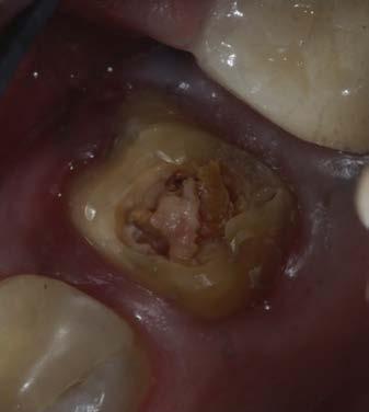
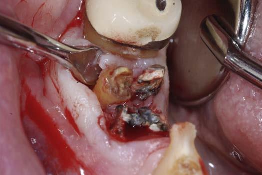
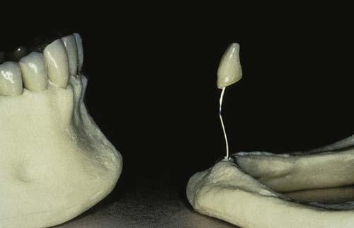
The posterior regions of the oral cavity are the most common areas for single-tooth loss13 (Fig. 1.7). The first molars are the first permanent teeth to erupt in the mouth and, unfortunately, are often the first teeth lost as a result of decay, failed endodontic therapy, or fracture (usually after endodontics).
The molar teeth are vitally important for maintenance of the arch form and proper occlusal schemes. In addition, the adult patient often has one or more crowns as a consequence of previous larger restorations required to repair the integrity of the tooth. Longevity reports of crowns have yielded very disparate results. The mean life span at failure has been reported as approximately 10.3 years. Other reports range from a 3% failure rate at 23 years to a 20% failure rate at 3 years. The primary cause of failure of the crown is caries followed by periodontal disease and endodontic therapy.14 The tooth is at risk for extraction as a result of these complications, which are the leading causes of single posterior tooth loss in the adult (Fig. 1.8, Fig. 1.9).15
Researchers have found a direct correlation of tooth loss in the elderly population exhibiting physical and mental decline. The data showed that subjects who had lost all their natural teeth performed approximately 10% worse in both memory and mobility (walking) than counterparts with natural teeth. Usually, tooth loss is less with patients of higher socioeconomic status. However, in this study, the link between total tooth loss and mobility (slower walking speed) remained significant when all variables were taken into consideration.
Patients More Socially Active and Esthetic Conscious
With patients living longer, their social pleasures, including dining and dating, are continuing into their elderly years. In the past, treatment of elderly patients emphasized nonsurgical approaches and palliative treatment. Today, the full scope of dental services for elderly patients is increasing in importance to both the public and the profession because of the increasing age of our society. Studies have shown that
• Fig. 1.5 Adult population older than the age of 60 years will increase by 87% from the year 2000 to the year 2025. (From Misch CE. Rationale for dental implants. In: Misch CE, ed. Dental Implant Prosthetics. 2nd ed. St Louis: Mosby; 2015.)
• Fig. 1.6 When a person reaches age 65 years, he or she may often feels an investment in health is less appropriate. A 65-year-old healthy woman will live 23 more years 50% of the time and 29 more years 25% of the time. Her present oral condition will become worse during this extended time frame if treatment is not rendered.
elderly patients that are more socially active will have a slower progression of health declines than elderly people who become less socially active. Engaging older people have been shown to be more motivated to maintain their health than their less-engaged peers. Therefore with patients living longer, patient education is vitally important as the demand for more comprehensive dental implant treatment will be most definitely increasing in the future to maintain social activity.
Higher Prevalence of Partial and Complete Edentulism
Partial Edentulism
Currently, the prevalence of partial edentulism in the general population has resulted in an increased need for dental implants.
or have minimal restorations.
• Fig. 1.8 Posterior molar tooth exhibiting caries and endodontic fracture, which are two of the most common complications leading to an unrestorable tooth.
• Fig. 1.9 Posterior missing tooth is a frequent occurrence in a general practice. The most common single tooth missing is the first molar. which results in many dental arch complications. (From Misch CE. Rationale for dental implants. In: Misch CE, ed. Dental Implant Prosthetics. 2nd ed. St Louis: Mosby; 2015.)
• Fig. 1.10 There are more than 44 million people in the United States missing at least one quadrant of posterior teeth (most often in the mandible). (From Misch CE. Rationale for dental implants. In: Misch CE, ed. Dental Implant Prosthetics. 2nd ed. St Louis: Mosby; 2015.)
Various studies have shown this pattern to be as high as 48% of the population. Many variables which have been associated this increase include gender, ethnicity, and chronic disease. In addition, adults exhibiting partial edentulism were 22.6% more likely to be from rural areas and 31.5% from depressed locations.16
As stated previously, the most common missing teeth are have been shown to be molars.17 Partial free-end edentulism is of particular concern because in these patients, teeth are often replaced with removable partial prostheses. Implant placement in the posterior regions is often challenging because of the location of the maxillary sinus and the mandibular canal. Mandibular free-end edentulism frequency is greater than its maxillary counterpart in all age groups. Unilateral free-end edentulism is more common than bilateral edentulism in both maxillary and mandibular arches in the younger age groups (ages 25–44). About 13.5 million persons in these younger age groups have free-end edentulism in either arch (Fig. 1.10).
In 45- to 54-year-old patients, 31.3% have mandibular freeend edentulism, and 13.6% have free-end edentulism in the maxillary arch. Approximately 9.9 million persons in the 45- to 54-year-old group have at least one free-end edentulous quadrant, and almost half of these have bilateral partial edentulism. The pattern of posterior edentulism evolves in the 55- to 64-yearold group, in which 35% of mandibular arches show free-end
• Fig. 1.7 (A and B) The most common tooth to be lost is the first molar. Approximately 80% of the time, the adjacent teeth are unrestored
edentulism compared with 18% of maxillary arches. As a result, approximately 11 million individuals in this age group are potential candidates for implants. An additional 10 million show partial free-end edentulism at age 65 or older. Additional US survey studies have documented approximately 44 million people to have at least one quadrant of posterior missing teeth. For example, if each of these arches requires three implants to support a fixed prosthesis, 132 million implants, added to the 192 million for edentulous patients, would be required.18-20
Total Edentulism
Although the percentage of patients with total edentulism is decreasing because of the baby-boomer population, the total number of patients exhibiting edentulism that will require treatment will increase in the future. In the past, full arch extractions were mainly indicated because of the combined pathologic processes of dental caries, periodontal disease, or as a method to reduce the costs associated with dental treatment. However, because of the high success rate of dental implants today, it is not uncommon for full-mouth extractions to be completed when teeth are questionable, especially in anticipation of future implant placement. Similar to other pathologic outcomes of disease, the occurrence of total loss of teeth is directly related to the age of the patient. The rate of edentulism increases approximately 4% per 10 years in early adult years and increases to more than 10% per decade after age 70.21
The average total edentulous rate worldwide is approximately 20% at age 60, although there is wide disparity between the countries with the highest and lowest rates. For example, in the 65- to 74-year age group, the total edentulous rate in Kenya and Nigeria was 0%, whereas the Netherlands and Iceland have a 65.4% and 71.5% rate, respectively. The edentulous Canadian rate was 47% at ages 65 to 69 and 58% from ages 70 to 98 (with Quebec at 67% for those older than age 65 compared with Ontario with a 41% rate). 22
In the United States the comparison of edentulism from 1957 to 2012 decreased from 19% to 5%. Income is often related to education and may also play a role in the rate of edentulism in the United States from 1988 to 1994, studies reported an edentulous rate of 22% for those with less than 8 years of education, 12% for those with 9 to 11 years of school, 8% for those with 12 years of school, and 5% for individuals with more than 12 years of education.
Studies show that edentulism in the United States is rarely seen in high-income individuals. The level of education is inversely proportional to edentulism. Geographically, edentulism was found to be highest in states that are bordered by the Appalachian Mountains and the Mississippi Delta. The lowest prevalence was found in California, Connecticut, Hawaii, and Minnesota. The prevalence in southern states is nearly twice that in western states (Fig. 1.11).23
In the National Institute of Dental Research national surveys, the occurrence of total edentulism (absence of teeth) of a single arch (35 times more frequent in the maxilla) was slight in the 30- to 34-year-old age group, but it increased at around age 45 to 11% and then remained constant after 55 years at approximately 15% of the adult population. A total of approximately 12 million individuals in the United States have edentulism in one arch, representing 7% of the adult population overall. With the passing of generations born in the mid-20th century, the rate of decline in edentulism is projected to slow, reaching approximately 2.6% by the year 2050. This continuing decline, however will be offset by population aging.The projected number of edentulous people in
• Fig. 1.11 Age-standardized edentulism prevalence among adults aged ≥25 years in the United States in 2010. (From Slade GD, Akinkugbe AA, Sanders, AE. Projections of U.S. edentulism prevalence following 5 decades of decline. J Dent Res. 2014;93(10):959–965.)
• Fig. 1.12 The US population completely edentulous rate ranges from 5% for 40 year olds to 44% for those older than age 75. As a result, 20 million people (10.5% of the population) in the United States have no teeth. An additional 12 million people (7% of the adult population) have no maxillary teeth opposing at least some mandibular teeth.
2050 will be approximately 8.6 million. This will be 30% lower than the 12.2 million edentulous people in 2010.23
The present younger population is benefiting from today’s advanced knowledge and restorative techniques. Edentulism has been noted in 5% of employed adults aged 40 to 44, gradually increasing to 26% at age 65, and almost 44% in seniors older than age 75 (Fig. 1.12).24 As expected, older persons are more likely to be missing all their teeth. Gender was not found to be associated with tooth retention or tooth loss once adjustments were made for age. The percentages of one- or two-arch edentulism translate into more than 30 million people, or about 17% of the entire US adult population. To put these numbers in perspective, 30 million people represent approximately the entire US African American population, or the entire population of Canada. Although the edentulism rate is decreasing every decade, the elderly population is rising so rapidly that the adult population in need of one or two complete dentures will actually increase from 33.6 million adults in 1991 to 37.9 million adults in 2020. The total number of edentulous arches is estimated at 56.5 million in 2000, 59.3 million in 2010, and 61 million in 2020. Complete edentulism, therefore, remains a significant concern, and affected patients often
• BOX 1.1
Consequences of Complete Edentulism
• Continued bone loss of the maxilla and mandible
• Negative soft tissue changes of the face and jaws
• Negative facial esthetic changes
• Decreased masticatory function
• Increased Health Issues
• Negative dietary effects
• Psychological Issues
• Patients Less Socially Active
From Misch CE. Rationale for dental implants. In: Misch CE, ed. Dental Implant Prosthetics. 2nd ed. St Louis: Mosby; 2015.
require dental implant treatment to solve several related problems. For example, to show the need for implant treatment with the edentulous group, if four implants were used to help support each complete edentulous arch in 2000, a total of 226 million implants would have been required. However, only approximately 1 million implants were inserted for all patient treatment (partially or completely edentulous) that year. Almost 70% of dentists spend less than 1% to 5% of their treatment time on edentulous patients, leaving a great unfulfilled need for implant dentistry.25
When the partially edentulous figures are added to the complete edentulous percentages, almost 30% of the adult US population are candidates for a complete or partial removable prosthesis. The need for additional retention, support and stability, and the desire to eliminate a removable prosthesis are common indications for dental implants. As a result, 74 million adults (90 million arches) are potential candidates for dental implants. Because a minimum of five appointments is required to implant and restore a patient, every US dentist would need approximately 20 appointments every month for 20 years to treat the present posterior partial and complete edentulous population with implant-supported prostheses. The population’s evolution to an increased average age, combined with the existing population of partially and completely edentulous patients, guarantees implant dentistry’s future for several generations of dentists.
In the elderly population, tooth loss is more common. The babyboomer population in the United States is the major purchaser of elective plastic surgery and antiaging procedures and medications. This generation is destined to be the most affluent older generation ever in the United States, and they will inherit the largest inflationadjusted transfer of wealth in history at approximately $10 trillion.26 This propensity for discretionary spending has fueled unprecedented growth in implant dentistry during the last decade, and it is expected to continue. The 65-year-plus population in the United States is expected to increase at annual rates of 1.5% to 3% from 2010 through 2035. The population of 65+ age group will increase from 12.4% of the population in 2000 to 20.6% in 2050.27,28
Anatomic Consequences Of Edentulism
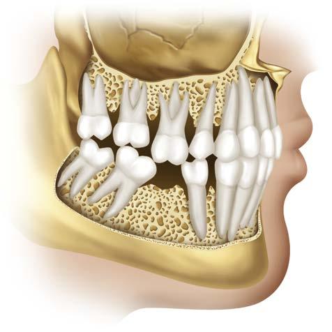
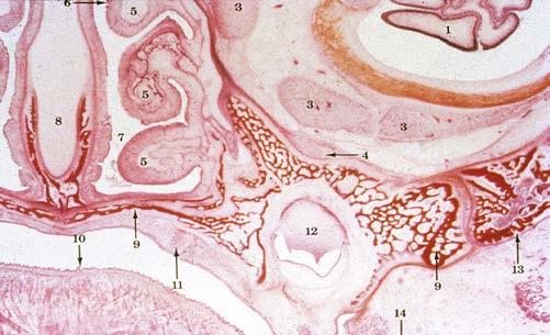
Hard Tissue Loss. Basal bone forms the dental skeletal structure, contains most of the muscle attachments, and begins to form in the fetus before teeth develop (Box 1.1). Alveolar bone first appears when the Hertwig root sheath of the tooth bud evolves (Fig. 1.13). The alveolar bone does not form in the absence of primary or secondary tooth development. The close relationship between the tooth and the alveolar process continues throughout life. Wolff’s law (1892) stated that bone remodels in relationship to the forces applied. Every time the function of bone is modified, a definite change occurs in the internal architecture and external configuration.29,30 In dentistry, the consequences of complete edentulous and remaining bone volume was noted by Misch in 1922, in which he described the skeletal structure of a 90-year-old woman without teeth for several decades.31
• Fig. 1.13 The alveolar bone forms as a result of the tooth root formation. When no tooth root is present, the alveolar process does not form (i.e., ectodermal dysplasia when partial or complete anodontia of both primary and secondary teeth occurs).
Continuous loss for 25 years
• Fig. 1.14 After the initial extraction of teeth, studies have shown the average first-year bone loss is more than 4 mm in height and 30% in crestal bone width. Although the rate of bone loss is slower after the first year, the bone loss is continuous throughout life.
Bone requires stimulation to maintain its form and density. Roberts and colleagues32 reported that a 4% strain to the skeletal system maintains bone and helps balance the resorption and formation phenomena. Teeth transmit compressive and tensile forces to the surrounding bone. These forces have been measured as a piezoelectric effect in the imperfect crystals of durapatite that compose the inorganic portion of bone. When a tooth is lost, the lack of stimulation to the residual bone causes a decrease in trabeculae and bone density in the area, with loss in external width, then height, of the bone volume.32 There is a 25% decrease in the width of bone during the first year after tooth loss and an overall 4-mm decrease in height during the first year after extractions for an immediate denture. In a pioneering longitudinal 25-year study, demonstrated continued bone loss during this time span; in comparing the bone loss of the maxilla to the mandible, a fourfold greater loss was observed in the mandible (Fig. 1.14).33 Although, initially the mandibular bone height is twice that of the maxilla, maxillary bone loss is very significant in the long-term edentulous patient. In fact, maxillary implant placement and bone graft procedures may be more challenging in comparison to the mandible.
Prostheses also contribute to bone loss. In general, a tooth is necessary for the development of alveolar bone, and stimulation of this bone is required to maintain its density and volume. A removable denture (complete or partial) does not stimulate and maintain bone; rather, it accelerates bone loss. The load from
mastication is transferred to the bone surface only and not the entire bone. As a result, blood supply is reduced and total bone volume loss occurs. This issue, which is of utmost importance, has been observed but not addressed until recently in traditional dentistry. Most often dentists overlook the insidious bone loss that will occur after tooth extraction. Therefore, it is imperative patients be educated about the anatomic changes and the potential consequences of continued bone loss. The bone loss accelerates when the patient wears a poorly fitting soft tissue–borne prosthesis. Patients do not understand that bone is being lost over time and at a greater rate beneath poorly fitting dentures (Fig. 1.15). Patients infrequently return for follow-up visits for evaluation of their edentulous condition; instead, they will return for a repair of the prosthesis. Hence the traditional method of tooth replacement (e.g. removable prosthesis) often affects bone loss in a manner not sufficiently considered by the doctor and the patient. Bone loss has been shown to increase with the use of a poorly fitting soft tissue–borne prosthesis. Patients should be informed of periodic evaluations to reline or fabricate a new prosthesis (Fig. 1.16).
Preventive dentistry has traditionally emphasized methods to decrease tooth loss. No predictable therapy had been accepted by the profession to avoid the bone changes resulting from tooth loss. Today, the profession must consider the loss of both teeth
and bone. The loss of teeth causes remodeling and resorption of the surrounding alveolar bone and eventually leads to atrophic edentulous ridges. The rate and amount of bone loss may be influenced by such things as gender, hormones, metabolism, parafunction, and ill-fitting dentures (Box 1.2). Yet almost 40% of denture wearers have been wearing an ill-fitting prosthesis for more than 10 years. Patients wearing dentures day and night place greater forces on the hard and soft tissues, which accelerates bone loss. Nonetheless, studies have shown that approximately 80% of dentures are worn both day and night.34 Atrophic edentulous ridges are associated with anatomic problems that often impair the predictable results of traditional dental therapy (Fig. 1.17; Box 1.3).
Loss of bone in the maxilla or mandible is not limited to alveolar bone; portions of the basal bone also may be resorbed, especially in the posterior aspect of the mandible in which severe resorption may result in catastrophic bone loss.35 The contents of the mandibular canal or mental foramen eventually become dehiscent and serve as part of the support area of the prosthesis. As a result, acute pain and transient to permanent nerve impairment of the areas supplied by the mandibular nerve are possible. The body of the mandible is also at increased risk of pathologic fracture, even under very low impact forces. The mandibular fracture causes the jaw to shift to one side and makes stabilization and an esthetic result most difficult to obtain during treatment of the fracture.
•
BOX 1.2 Factors Effecting Rate and Amount of Bone Loss
• Gender
• Medications
• Hormones
• Age
• Metabolism
• Bone Quality
• Parafunction (Increased Biting Force)
• Ill-fitting prosthesis
• Facial type (brachiocephalic versus dolichocephalic)
• Time period dentures are worn
• Past History of Dental Disease
Modified from Misch CE. Rationale for dental implants. In: Misch CE, ed. Dental Implant Prosthetics. 2nd ed. St Louis: Mosby; 2015.
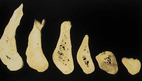
• Fig. 1.15 Atwood described six different stages of resorption in the anterior mandible. Stage 1 represents the tooth and surrounding alveolar process and basal bone. Stages II and III illustrate the initial residual ridge after tooth loss. Stages IV to VI primarily describe a continuous loss in length of anterior residual bone.
• Fig. 1.16 Loss of bone height in the mandible may be significant resulting in loss of function. This vertical bone loss has a large impact on restoring the patient back to dental health. The patient should understand that to restore the hard and soft tissue loss, more extensive treatment is usually indicated.
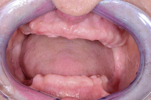
• Fig. 1.17 Maxillary and Mandibular edentulous arches depicting irregular bone resorption with varying degrees of quality soft tissue (i.e. attached tissue).
In the maxilla, extensive bone loss can also be problematic. In some cases, the complete anterior ridge and even the anterior nasal spine may be resorbed in the maxilla, causing pain and an increase in maxillary denture movement during function. Masticatory forces generated by short facial types (brachiocephalics) can be three to four times that of long facial types (dolichocephalics). Short facial–type patients are at increased risk for developing severe atrophy.
Many of these similar conditions exist in the partially edentulous patient wearing a removable soft tissue–borne prosthesis (e.g. removable partial denture) (Fig. 1.18). In addition, the natural abutment teeth, on which direct and indirect retainers are designed, experience significant lateral forces. Because these teeth
are often compromised by deficient periodontal support or large restorations, the resultant forces may be damaging. These forces may result in an increase in mobility of the removable prosthesis and greater soft tissue support. These conditions often will lead to accelerated the bone loss in the edentulous regions (see Box 1.3).
Soft Tissue Consequences. As bone loses width, then height, then width and height again, the attached gingiva gradually decreases. A very thin attached tissue usually lies over the advanced atrophic mandible or maxilla. The increased zones of nonkeratinized gingiva are prone to abrasions caused by the overlaying prosthesis. In addition, unfavorable high muscle attachments and hypermobile tissue often complicate the situation (Fig. 1.19).
• Continued loss of supporting bone width
• Prominent mylohyoid and internal oblique ridges with increased sore spots
• Progressive decrease in keratinized mucosa surface
• Prominent superior genial tubercles with sore spots and increased denture movement
• Muscle attachment near crest of ridge
• Elevation of prosthesis with contraction of mylohyoid and buccinator muscles serving as posterior support
• Forward movement of prosthesis from anatomic inclination (angulation of mandible with moderate to advanced bone loss)
• Thinning of mucosa, with sensitivity to abrasion
• Loss of basal bone
• Possible Nerve Impairment from dehiscent mandibular neurovascular canal
• More active role of tongue in mastication
• Effect of bone loss on esthetic appearance of lower third of face
• Increased risk of mandibular body fracture from advanced bone loss
• Loss of anterior ridge and nasal spine, causing increased denture movement and sore spots during function
As the bone resorbs from Division A to Division B, the resultant narrow residual ridge will often cause discomfort when pressure (from a prosthesis) is applied to the ridge. This often occurs in the posterior mandible, as atrophy may cause a prominent mylohyoid and internal oblique ridges covered by thin, movable, unattached mucosa. In severe atrophy cases the anterior residual alveolar process will continue to resorb, and the superior genial tubercles (which are approximately 20 mm below the crest of bone when teeth are present) eventually become the most superior aspect of the anterior mandibular ridge. This results in excessive movement of the prosthesis during function or speech. This condition is further compromised by the vertical movement of the distal aspect of the prosthesis during contraction of the mylohyoid and buccinator muscles and the anterior incline of the atrophic mandible compared with that of the maxilla.36
The thickness of the mucosa on the atrophic ridge is also related to the presence of systemic disease and the physiologic changes that accompany aging. Conditions such as hypertension, diabetes, anemia, and nutritional disorders have a deleterious effect on the vascular supply and soft tissue quality under removable prostheses. These disorders result in a decreased oxygen tension to the basal cells of the epithelium. Surface cell loss occurs at the same rate, but the cell formation at the basal layer is slowed. As a result, the
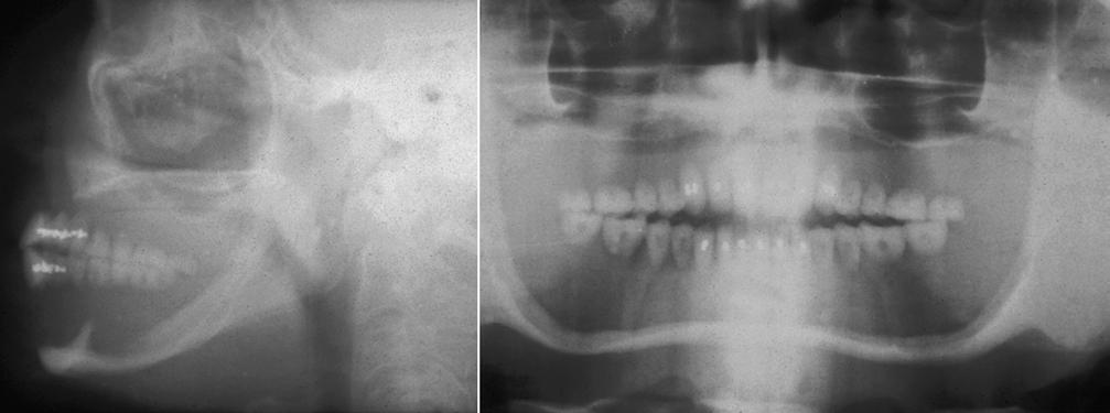
• Fig. 1.18 (A) Lateral cephalogram of a patient demonstrates the restored vertical dimension of occlusion with a denture. However, because of the advanced basal bone loss in the mandible, the superior genial tubercles (red arrow) are positioned above the residual anterior ridge. The body of the mandible is only a few millimeters thick, and the mandibular canal is completely dehiscent. In the maxillary anterior ridge, only the nasal spine remains (not the original alveolar ridge), and the posterior maxillary bone is very thin because of basal bone loss at the crest and the pneumatization of the maxillary sinus. (B) A denture may restore the vertical dimension of the face, but the bone loss of the jaws can continue until the basal bone becoems pathologically thin.
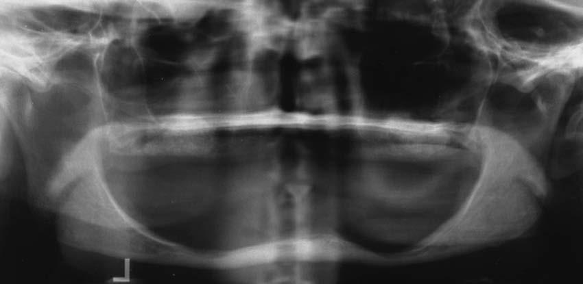
• Fig. 1.19 Resorption of an edentulous mandible may result in dehiscence of the mandibular canal and associated nerve impairment. In addition, a conventional removable prosthesis is often difficult to wear because of the associated discomfort from the exposed nerve. The soft tissue is often thin and is usually hypersensitive, especially if the patient is wearing a conventional removable prosthesis
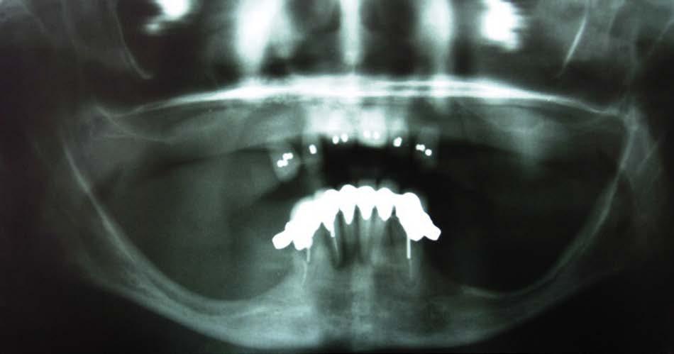
• Fig. 1.20 Panoramic radiograph exhibiting extensive mandibular posterior atrophy. Note that the anterior teeth have maintained the bone in the anterior mandible and has resulted in the degradation of the premaxilla (Combination Syndrome). Wearing of a mandibular class I removable partial denture has escalated the posterior bone loss.
thickness of the surface tissues gradually decreases. Therefore, soft tissue irritation usually results.
The tongue of the patient with edentulous ridges often enlarges to accommodate the increase in space formerly occupied by teeth. At the same time, the tongue is used to limit the movements of the removable prostheses and takes a more active role in the mastication process. As a result, the removable prosthesis decreases in stability. The decrease in neuromuscular control, often associated with aging, further compounds the problems of traditional removable prosthodontics. The ability to wear a denture successfully may be largely a learned, skilled task. The aged patient who recently became edentulous may lack the motor skills needed to adjust to the new conditions (Fig. 1.20; Box 1.4).
• BOX 1.4 Soft Tissue Consequences of Edentulism
• Attached, keratinized gingiva is lost as bone is lost
• Unattached mucosa for denture support causes increased soft spots
• Thickness of tissue decreases with age, and systemic disease causes more sore spots for dentures
• Tongue increases in size, which decreases denture stability
• Tongue has more active role in mastication, which decreases denture stability
• Decreased neuromuscular control of jaw in the elderly