Additive and Traditionally Manufactured Components: A Comparative Analysis of Mechanical Properties (Additive Manufacturing Materials and Technologies) 1st Edition
All rights reserved. No part of this publication may be reproduced, stored in a retrieval system, or transmitted, in any form or by any means, electronic, mechanical, photocopying, recording or otherwise, except as permitted by law. Advice on how to obtain permission to reuse material from this title is available at http://www.wiley.com/go/ permissions.
The right of Elizabeth Bahar Houshmand to be identified as the author of the editorial material in this work has been asserted in accordance with law.
Registered Offices
John Wiley & Sons, Inc., 111 River Street, Hoboken, NJ 07030, USA
John Wiley & Sons Ltd, The Atrium, Southern Gate, Chichester, West Sussex, PO19 8SQ, UK
Editorial Office
9600 Garsington Road, Oxford, OX4 2DQ, UK
For details of our global editorial offices, customer services, and more information about Wiley products visit us at www.wiley.com.
Wiley also publishes its books in a variety of electronic formats and by print‐on‐demand. Some content that appears in standard print versions of this book may not be available in other formats.
Limit of Liability/Disclaimer of Warranty
The contents of this work are intended to further general scientific research, understanding, and discussion only and are not intended and should not be relied upon as recommending or promoting scientific method, diagnosis, or treatment by physicians for any particular patient. In view of ongoing research, equipment modifications, changes in governmental regulations, and the constant flow of information relating to the use of medicines, equipment, and devices, the reader is urged to review and evaluate the information provided in the package insert or instructions for each medicine, equipment, or device for, among other things, any changes in the instructions or indication of usage and for added warnings and precautions. While the publisher and authors have used their best efforts in preparing this work, they make no representations or warranties with respect to the accuracy or completeness of the contents of this work and specifically disclaim all warranties, including without limitation any implied warranties of merchantability or fitness for a particular purpose. No warranty may be created or extended by sales representatives, written sales materials or promotional statements for this work. The fact that an organization, website, or product is referred to in this work as a citation and/or potential source of further information does not mean that the publisher and authors endorse the information or services the organization, website, or product may provide or recommendations it may make. This work is sold with the understanding that the publisher is not engaged in rendering professional services. The advice and strategies contained herein may not be suitable for your situation. You should consult with a specialist where appropriate. Further, readers should be aware that websites listed in this work may have changed or disappeared between when this work was written and when it is read. Neither the publisher nor authors shall be liable for any loss of profit or any other commercial damages, including but not limited to special, incidental, consequential, or other damages.
Library of Congress Cataloging‐in‐Publication Data
Name: Houshmand, Elizabeth Bahar, editor.
Title: Microneedling : global perspectives in aesthetic medicine / edited by Elizabeth Bahar Houshmand.
Other titles: Cosmetic and aesthetic dermatology series.
Description: Hoboken, NJ : John Wiley & Sons, Inc., 2021. | Series: Cosmetic and aesthetic dermatology series | Includes bibliographical references and index.
Identifiers: LCCN 2020055815 (print) | LCCN 2020055816 (ebook) | ISBN 9781119431923 (cloth) | ISBN 9781119431831 (adobe pdf) | ISBN 9781119431947 (epub)
Set in 10/12.5pt MinionPro by SPi Global, Pondicherry, India
List of Editors and Contributors, vi
Cosmetic and Aesthetic Procedures in Dermatology Series, viii
Preface, ix
1 Introduction to Microneedling, 1 Elizabeth Bahar Houshmand
2 A Short History of Skin Needling, 10 Desmond Fernandes
3 The Value of Medical Needling in Burn Scars, 22 Matthias Aust, Desmond Fernandes, and Richard Bender
4 Skin Care Used with Microneedling, 41 Chytra V. Anand and Parinitha Rao
5 Treatment of Hyperpigmentation with Microneedling, 52 Atchima Suwanchinda
6 Treatment of Acne and Acne Scars with Microneedling, 81 Stuti Khare Shukla and Michael H. Gold
7 Microneedling and Platelet-Rich Plasma (PRP), 98 Elizabeth Bahar Houshmand Index, 113
List of Editors and Contributors
Michael H. Gold, MD
Gold Skin Care Center
Tennessee Clinical Research Center
Nashville, TN, USA
Elizabeth Bahar Houshmand, MD, FAAD, FABIM Houshmand Dermatology and Wellness
Dallas, TX, USA
Atchima Suwanchinda, MD, MSc School of Anti-aging and Regenerative Medicine, Mae Fah Luang University
Ramathibodi University Hospital, Mahidol University Bangkok, Thailand
Chytra V. Anand, MD
Kosmoderma Clinics Bangalore, India
Desmond Fernandes, MB, BCh, FRCS (Edin) Department of Plastic and Reconstructive Surgery Faculty of Medicine University of Cape Town
Matthias Aust, MD Aust Aesthetik Landsberg am Lech, Germany
Parinitha Rao, MBBS, MD
Kosmoderma Clinics Bangalore, India
Richard Bender, MD
St. Vinzenz Hospital Cologne Plastische Chirurgie
Cologne, Germany
Stuti Khare Shukla, MD
Elements of Aesthetics Clinics
Dr. Stuti Khare’s Skin & Hair Clinics Mumbai, India
Cosmetic and Aesthetic Procedures in Dermatology Series
The scope and field of cosmetic surgery have changed and grown over the past decade. We have a myriad of new and exciting noninvasive or minimally invasive techniques and treatments that have helped propel this growth. With injectables, energy‐based devices, and skincare, we now have the tools at our disposal to reverse signs of aging and to successfully treat cosmetic concerns with minimal downtime and with outstanding clinical results.
Several years ago, we began discussing the idea of producing a series of cosmetic dermatology textbooks edited by masters in their fields and written by some of the best minds in our discipline. While it is not always easy to get these kinds of projects off the ground, we are pleased that with persistence and great guidance from the Wiley teams, we are now able to present an entire series on the various aspects of cosmetic dermatology.
Through this textbook series on cosmetic and aesthetic dermatology, clinicians will have the tools available to help treat patients with the most advanced techniques and with the most appropriate guidance that has been presented to date. Each volume in this series has been meticulously thought out; each is edited by one of the most significant thought leaders in our field, and each chapter will bring to life this incredible field we live in, and illuminate how we are able to transform lives and make our patients better.
We hope you enjoy this series of books as they are rolled out. It is our joy and pleasure to bring them to you.
Michael H. Gold, MD Series Editor
Preface
The skin is our canvas. As a board certified dermatologist, it is my mission to help all my patients achieve healthy, beautiful skin. Through education, teaching, and research this is possible. This textbook is a continuation of this mission, inspired by the drive to create the best possible outcomes for my patients, and the love of being a physician and educator.
Over the last several years, minimally invasive procedures have significantly increased in the worlds of dermatology, plastic surgery, and aesthetic medicine. This textbook is a global consortium of the leaders in these fields and experts on microneedling.
Microneedling is an outstanding option for patients of all skin types and ethnicities who desire measurable clinical results from treatments with little to no downtime. It is a relatively new option in dermatology and has been utilized for a broad range of applications, including skin rejuvenation, acne scarring, rhytides, surgical scars, dyschromia, melasma, enlarged pores, and transdermal drug delivery.
Microneedling is a safe, minimally invasive, and effective aesthetic treatment for several different dermatologic conditions. Given its expedient post‐treatment recovery, minimal side effect profile, and significant clinical results, microneedling is a valuable alternative to more invasive procedures. The chapters of this book discuss the various applications in detail.
I want to thank my family and mentors for their support always. I also want to give a special thank‐you to Dr. Michael Gold. He is an innovator in our field and I am happy to call him my dear friend, mentor, and colleague.
Elizabeth Bahar Houshmand Houshmand Dermatology and Wellness, Dallas, TX, USA
Introduction
Microneedling is a minimally invasive procedure that uses fine needles to puncture the epidermis. The microwounds created stimulate the release of growth factors and induce collagen production. The epidermis remains relatively intact during the procedure.
Microneedling initially was utilized as a collagen induction therapy for facial scars and skin rejuvenation, but it is now widely used for multiple indications, including transdermal delivery system for therapeutic drugs and in combination therapies. The indications for microneedling have grown as research and clinical applications have expanded widely in dermatology and dermatologic surgery.
In this textbook, the authors highlight the constantly evolving research and developments in microneedling techniques and instruments, along with microneedling’s applications in dermatology and aesthetic medicine. We are honored to provide a comprehensive and global perspective from key opinion leaders in dermatology and plastic surgery from around the world.
History
Microneedling, or percutaneous collagen induction therapy, was introduced in the 1990s for the treatment of scars, striae, and laxity [1]. The use of needles for nonablative skin treatment was first described by Orentreich and Orentreich in 1995
Microneedling: Global Perspectives in Aesthetic Medicine, First Edition. Edited by Elizabeth Bahar Houshmand.
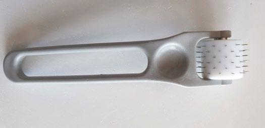
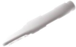
Figure 1.1 Original microneedling roller created by Dr. Desmond Fernandes in 2001. Fixed needle length of 3.0 mm multiuse roller; designed for reuse on a single patient for approximately six treatment sessions. The original rollers were not able to be autoclaved at that time. They were sterilized by soaking in instrument cleaning fluid. Source: Dr. Desmond Fernandes.
as subcision surgery, which is the release of depressed scars and wrinkles with a needle from their attachment to the underlying skin. This controlled trauma leads to the formation of connective tissue to fill the created gap.
In 1996, skin needling using a roller device was introduced by Fernandes at the International Society of Aesthetic Plastic Surgery (ISAPS) congress in Taipei [2]. In 1997, Camirand and Doucet introduced dry tattooing without pigment as needle dermabrasion and proposed it as a technique to improve the appearance of scars [3].
Fernandes, in 2001, developed the original percutaneous collage induction dermaroller with needles. His pilot roller device was a drum‐shaped tool, with a cylinder and 3 mm needles that reach the fibroblasts deep in the reticular layer (see Figure 1.1).
Zeitter et al. confirmed Fernandes’s findings and made a modified roller. They concluded that 1 mm needles show similar results to 3 mm needles, with the advantage of less downtime, swelling, and pain [3, 4].
Mechanism of action
The mechanism of action is thought to be a disruption of the epidermis and dermis. Micropunctures are created using microneedles, which produce a controlled skin injury without damaging the epidermis. The mechanical microinjury results in the classic wound‐healing cascade and stimulates cellular proliferation and migration through the stimulation of growth factors (see Figure 1.2).
These microinjuries lead to minimal superficial bleeding and set up a wound‐healing cascade with release of various growth factors, such as platelet‐derived growth factor (PDGF), transforming growth factor alpha and beta (TGFα and TGFβ), connective tissue activating protein, connective tissue growth factor, and fibroblast growth factor (FGF) [5]. The needles also break down the scar strands
and allow them to revascularize. Neovascularization and neocollagenesis are initiated by migration and proliferation of fibroblasts and laying down of an intercellular matrix [6, 7]. A fibronectin matrix forms five days after injury and determines the deposition of collagen, resulting in skin tightening persisting for five to seven years in the form of collagen III. The depth of neocollagenesis has been found to be 5–600 μm with a 1.5 mm length needle. Histological examination of the skin treated with four microneedling sessions one month apart shows up to 400% increase in collagen and elastin deposition at six months postoperatively, with a thickened stratum spinosum and normal rete ridges at one year postoperatively [8]. Collagen fiber bundles appear to have a normal lattice pattern rather than parallel bundles as in scar tissue [9].
The devices used create transient epidermal and dermal openings ranging in size from 25 to 3000 um in depth as a microinjury, with the goal of stimulating the inherent skin repair mechanisms. These microwounds or microinjuries initiate the release of growth factors, which trigger and stimulate collagen and elastin formation in the dermis. That leads to healthier skin with improved texture. The microwounds are microchannels and heal following the classic wound‐healing cascade: inflammation, proliferation, and remodeling. This cascade is brought on by the needles’ disruption of the stratum corneum; the endothelial lining and the subendothelial matrix recruits platelets and neutrophils to the site of injury. Needling exposes thrombin and collagen fragments, which attract and activate platelets. The platelets form a plug and initiate the clotting cascade, which involves local platelet aggregation, inflammation, and blood coagulation through increased levels of thrombin and fibrin.
The needles carry an electric potential that stimulates fibroblast proliferation [10]. The mechanical injury triggers the release of potassium and proteins that
Figure 1.2 The electric pen‐shaped device has adjustable settings to control the speed and depth of needle penetration. Source: skvalval/Shutterstock.
alter intercellular resting potential, drawing in fibroblasts and stimulating neocollagenesis and revascularization [6].
Research has shown up‐regulation of TGFβ3, a cytokine that prevents aberrant scarring; increased gene expression for collagen type I; and elevated levels of vascular endothelial growth factor, fibroblast growth factor, and epidermal growth factor [11–13]. Histological studies have shown huge variation in epidermal thickness. Randomized murine studies have reported statistically significant epidermal thickening from 140% up to 685% after microneedling plus topical vitamins A and C when compared to control [13, 14]. This is thought to be one of the reasons microneedling is effective for scar therapy and notable skin rejuvenation.
A human study of 480 patients treated with microneedling plus topical vitamins A and C reported thickening of the stratum spinosum lasting up to one year [8, 15].
Increased collagen types I, III, and VII and tropoelastin in human biopsies were found after six sessions of microneedling, ten with elevated levels of collagen type I and elastin persisting at six months. The number of melanocytes was unchanged postprocedurally.
These results support the safe use of this modality in patients with darker skin types [8, 15]. Having a safe and effective treatment modality for all skin types is advantageous in an aesthetic practice.
The devices
Modern microneedling devices consist of rollers, stamps, and pens. Needling devices have evolved over the past decade through a variety of advancements. Currently, there are multiple devices based on needle length, drum size, and automation. To date, there are five FDA‐approved pen devices. Physicians and providers need to consider important factors like needle length, needle material, and clinical indications in selecting which device to utilize [9].
Pens
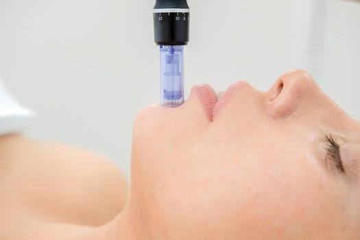
Most pens utilize sterile single‐use cartridges and variable needle length to be able to customize the treatment depending on the unique characteristics of the patient’s skin and the area being treated. They are automated and the physician has the ability to adjust the needle length for customized treatment options and the pressure and depth during treatment can be more uniform (see Figure 1.3) [16].
The pen itself is reusable, and most pens have a protective disposable sleeve. The needle tips are the disposable/consumable in these devices. Because of their size the tips are able to treat curved and small areas such as the nasal ala and the
Figure 1.3 Pen: Single‐use cartridge with adjustable frequency and needle length. Source: Sakurra/Shutterstock.
periocular and perioral areas. Most devices have a rechargeable battery that operates in two modes: high speed mode (700 cycles/minute) and low speed mode (412 cycles/minute) in a vibrating stamplike manner [17].
The devices contain multiple fine needles, ranging from 0.5 to 1.5 mm in length, that are rolled onto the skin. Needles between 1.5 and 3.0 mm are available but are preferred for the use of scars and damaged skin. The roller device is a drum‐shaped tool with a cylindrical head that is rolled back and forth to induce thousands of tiny pores in the stratum corneum and papillary dermis.
The length of needle selected for an individual patient depends upon the indication for microneedling and on the thickness of epidermis and dermis of the skin being treated. For treating acne and other scars, on average a needle length of 1.5–2 mm is utilized. When microneedling is used as a procedure to treat skin aging and wrinkles, a needle length of 0.5 mm or 1.0 mm is recommended [18]. The frequency interval for microneedling depends upon the indication for which the procedure is being done as well as the needle length of the dermaroller device used. Microneedling generally requires more than one session and a series of treatments is usually recommended.
Five basic types of medical dermarollers, which are registered with the FDA, have been described in the dermaroller series by Anastassakis and most dermarolling devices are adopted from these elementary types [19].
Stamps
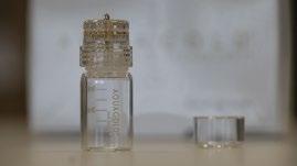
Stamps were popular in the late 1990s and have made a resurgence recently. The stamps currently offered have attached microchambers which have the ability to directly administer a form of mesotherapy using the stamping device. Stamps have different needle lengths (0.2–3 mm) and a diameter of 0.12 mm These are useful in the administration of treatment to scars and anatomically small surface areas such as the perioral, periocular, and nasal regions where greater control is beneficial, and may be used on isolated scars and wrinkles (see Figure 1.4) [3, 20–22].
1.4
Source: Aquavit Pharmaceuticals, Inc.
Rollers
Rollers have many fine‐gauged needles that are on a cylindrical surface that pierces the skin on an angle. The rollers are fixed; the parameters are uniform for each device that you use. Unlike pens, you can not mechanically adjust rollers. The quality of rollers is also critical. Patients are seeking at‐home rollers but the quality of the needles is paramount. Needles that are dull or loose may cause tears in the skin and foreign body reactions, including but not limited to granulomas.
The most important factor is needle length. A high ratio of tip length to diameter (13:1) is an important property of good needles [9]. The length of needle selected for an individual patient depends upon the indication for microneedling. For treating acne and other scars as a routine, a needle length of 1.5–2 mm is usually used. When microneedling is used as a procedure to treat aging skin and wrinkles, a needle length of 0.5 mm or 1.0 mm is usually recommended [19]. The needle length to use will also depend on the thickness of the epidermis and dermis of the skin for optimal results.
Given their design and mechanics, rollers are able to pierce the skin deeper when at a 90‐degree angle or perpendicular to the skin. Fernandes showed that with the use of rollers you have an intact epidermis with microchannels spaced out with about a four‐cell width distribution [4]. The provider’s technique with these devices is critical. Tearing of the epidermis may occur if performed incorrectly, with too much pressure, or at an increased speed. The needle rollers themselves are variable based on the materials used, needle length and diameter, and total number of needles. The quality of the rollers is also critical to evaluate.
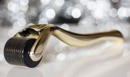
Needle length is generally 0.2–3.00 mm, with a diameter of > 0.25mm and the materials are variable: stainless steel, titanium, or silver and gold. Stainless steel is the most common type of needle, silver and gold offer antimicrobial properties and carry less of a risk of allergic reactions, and titanium needles usually stay sharper longer (see Figure 1.5).
Figure
Microneedling stamp: fixed needle length with customizable vial for needling infusion.
1.5 Current rollers with fixed needle length; some current models are autoclavable. Source: marcinm111/Shutterstock.
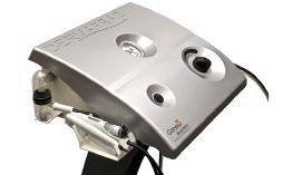
DermaFrac
DermaFrac treatment is a newer modification of microneedling combining microdermabrasion, microneedling, simultaneous deep tissue serum infusion, and light emitting diode (LED) therapy. DermaFrac treatments target aging and sun damaged skin, acne, enlarged pores, uneven skin tone, wrinkles, fine lines, hyperpigmentation, and superficial scars. It takes approximately 45 minutes to complete a full face treatment when all four modalities are used. This noninvasive, cost‐effective treatment carries the advantage of having no downtime, with individualized selection of serums for infusion (see Figure 1.6) [22].
Clinical considerations
Microneedling is not only used for rejuvenation of the skin. Its use in dermatology and aesthetic medicine has expanded to include the treatment of acne scars, alopecia, dyspigmentation, alopecia, striae, and for many other indications. It can be utilized alone or in combination with other treatment modalities, such as chemical
Figure
Figure 1.6 DermaFrac™: Microneedling device combining simultaneous customized infusion followed by LED light therapy. Source: Genesis Biosystems, Inc.
peels, platelet‐rich plasma, radiofrequency, subcision, punch elevation, and lasers. It is often used in conjunction with a topical formulation to enhance its penetration and action.
Microneedling is safely used for enhanced drug delivery to the deeper epidermis and dermis by bypassing the stratum corneum. This strategy has been utilized for burn patients and for rejuvenation, allowing cosmeceuticals to be delivered more deeply. Caution is necessary in deciding which topicals to use during delivery, as inflammation may occur and granulomas have been noted.
Conclusion
Microneedling is a popular treatment in dermatology and aesthetic medicine. Since the development of the first dermaroller over 20 years ago, a variety of new microneedling devices have been introduced. Accordingly, the applications of microneedling in dermatology and aesthetic medicine have expanded indications over the past several years.
Evidence‐based treatment of the skin for a variety of indications have been shown to be safe on all skin types. Microneedling is an effective modality of treatment, especially in patients with Fitzpatrick’s IV and V skin types because it overcomes the side effects of scarring and hyperpigmentation resulting from other procedures in which the epidermis is compromised. It certainly promises to be a valuable technique with its numerous applications and its ever‐expanding modifications.
References
1 Orentreich DS, Orentreich N. Subcutaneous incisionless (subcision) surgery for the correction of depressed scars and wrinkles. Dermatol Surg. 1995;21:543–549. [PubMed: 7773602]
2 Bahuguna A. Micro needling ‐ Facts and Fictions. Asian J Med Sci. 2013;4:1–4.
3 Camirand A, Doucet J. Needle dermabrasion. Aesthet Plast Surg. 1997;21:48–51. [PubMed: 9204168]
4 Fernandes D. Minimally invasive percutaneous collagen induction. Oral Maxillofac Surg Clin North Am. 2006;17:51–63. [PubMed: 18088764]
5 Falabella AF, Falanga V.Wound healing. The Biology of the Skin. Parethenon: New York; 2001. pp. 281–299.
6 Fabbrocini G, Fardella N, Monfrecola A, et al. Acne scarring treatment using skin needling. Clinical and Experimental Dermatology. 2009;34:874–879.
7 Majid I, Sheikh G, September PI. Microneedling and its applications in dermatology InPrime. 7. Vol. 4. London: Informa Healthcare; 2014. Sep 15, pp. 44–49.
8 Aust MC, Fernandes D, Kolokythas P, et al. Percutaneous collagen induction therapy. An alternative treatment for scars, wrinkles, and skin laxity. Plast Reconstr Surg. 2008;121:1421–1429.
9 Nair PA, Arora TH. Microneedling using dermaroller: A means of collagen induction therapy. GMJ. 2014;69:24–27.
10 Jaffe L. Control of development by steady ionic currents. Fed Proc. 1981;40:125–127.
11 Aust MC, Reimers K, Gohritz A, et al. Percutaneous collagen induction. Scarless skin rejuvenation: fact or fiction? Clin Exp Dermatol. 2010 Jun;35(4):437–439.
12 Murata H, Zhou L, Ochoa S, et al. TGF‐beta 3 stimulates and regulates collagen synthesis through TGF‐beta‐dependent and independent mechanisms. J Invest Dermatol. 1997;108:258–262.
13 Aust MC, Reimers K, Kaplan HM, et al. Percutaneous collagen induction regeneration in place of cicatrisation? J Plast Reconstr Aesthet Surg. 2011;64:97–107.
14 Zeitter S, Sikora Z, Jahn S, et al. Microneedling: Matching the results of medical needling and repetitive treatments to maximize potential for skin regeneration. Burns. 2014;40:966–973.
15 Aust MC, Knobloch K, Vogt PM. Percutaneous collagen induction as a novel therapeutic option for Striae distensae. Plast Reconstr Surg. 2010;126:4.
16 McCrudden MT, McAlister E, Courtenay AJ, et al. Microneedle applications in improving skin appearance. Exp Dermatol. 2015;24:561–566.
17 Arora S, Gupta BP. Automated microneedling device–A new tool in dermatologist’s kit–A review. J Pak Med Assoc. 2012;22:354–357.
18 Doddaballapur S. Microneedling with dermaroller. J Cutan Aesthet Surg. 2009;2:110–111. [PMCID: PMC2918341] [PubMed: 20808602]
19 Anastassakis K. The Dermaroller Series. [Last accessed June 22, 2016] http://www. mtoimportadora.com.br/site_novo/wp.content/uploads/2014/04/Dr.‐Anastassakis‐Kostas.pdf.
20 Bhardwaj D. Collagen induction therapy with dermaroller. Community Based Med J. 2013;1:35–37.
21 McCrudden MT, McAlister E, Courtenay AJ, et al. Microneedle applications in improving skin appearance. Exp Dermatol. 2015;24:561–566. [PubMed: 25865925]
22 Lewis W. Is microneedling really the next big thing? Wendy Lewis explores the buzz surrounding skin needling. Plast Surg Pract. 2014;7:24–28.
A Short History of Skin Needling
Desmond Fernandes Department of Plastic and Reconstructive Surgery, Faculty of Medicine, University of Cape Town
Introduction
I first started researching skin needling as we know it today in my medical practice in Cape Town on 20 volunteers starting in 1997. The treated cases had acne scarring, burn scars, and fine wrinkles. My first iteration of needling started in 1994 with deep dermal needling parallel to the skin surface of the upper lip skin to treat upper lip creases. This horizontal needling is not the same as subcision that Orentreich was doing for deep acne scars and wrinkles [1]. The two‐year experience of this method was presented at the Asian Society of Aesthetic Plastic Surgery in Taipei in 1996.
Vertically oriented needling through the skin arose from the work of Camirand and Doucet for treating linear scars (e.g. face‐lift scars) [2], which was discussed at the ISAPS meeting in São Paulo in 1997. I first started to use a tattoo artist’s instrument, just as Camirand had (equivalent to the common pen‐style needling done nowadays), to do needling of burn scars, wrinkles, and acne scars. That technique I found to be too laborious when doing the whole face and the needles could not penetrate as deeply as I felt they should. I also felt that the holes were too close to each other. That drove me to design a roller tool that I believed would be easier to use and give better, safer results.
I said the results of needling stemmed from the “inflammatory” cascade of growth factors and in particular TGF‐beta‐3 (TGFβ3), which had been described by Fergusson and his team as the regeneration factor [3]. I believed that a normal matrix was developed after needling to replace degenerated or scar collagen.
Microneedling: Global Perspectives in Aesthetic Medicine, First Edition. Edited by Elizabeth
My own personal histological studies had shown the increase of nonscar woven collagen and increased vertically oriented elastin. I also believed that vitamin A is so good for scars and regeneration of photoaged skin because of its stimulating growth factors [4–6].
Prof. Matthias Aust visited me in 2003, was intrigued by skin needling, and decided to research it at Hannover Medical School, Germany. His research subsequently revealed the platelet derived growth factors responsible for regeneration of the epidermis and matrix in rats in 2004 and was published in 2008 [7].
Today we know that skin needling is the safest way to treat scarred or aged skin and now we need to concentrate on the timing of needling and the chemicals we should use to promote better results. At this stage, vitamins A and C in the cosmetic form seems the best ingredients to use with needling, but research on the importance of selected peptides also offers great promise.
Because this chapter focuses on the history of skin needling, it necessarily includes a substantial autobiographical focus due to my role in bringing skin needling to the attention of the medical profession.
My clinical research in skin needing started in 1994 and at that time I was also trying to understand why cosmetic forms of vitamin A rejuvenated skin. I believed that growth factors had to be involved. However, I was also looking at the difficult problem of wrinkles/creases on the upper lip. I believed that these upper‐lip lines were like scars between the skin and the deeper layer. I thought that if I could repeatedly pass a needle through the fibrous tissue under the lip lines and perforate them sufficiently they would then allow these “contractures” between the skin and the muscle to expand and thereby flatten out the wrinkles on the upper lip. That process was, in fact, the first iteration of skin needling, which I call horizontal needling. I still use this technique in conjunction with vertical needling.
I simply passed a needle backwards and forwards to create “tunnels” that were as close to the surface as I could manage and would thereby cause fenestration of any fibrous bands that were pulling in the wrinkles of the upper lip down to deeper tissue.
I started initially with a gauge 15 needle that I pierced through the upper‐lip skin laterally and then pushed it medially backwards and forwards horizontally immediately under the skin. This is not the same as the subcision technique described by Orentreich, which completely transects scars or wrinkles away from the subcutaneous tissue [1]. I found that there were dense areas immediately under the wrinkles and I hoped the perforations into them would allow the skin to lift upwards. At that stage I thought we might make some scar tissue, but I did not think that we would cause tissue regeneration.
I did a presentation on my experience with skin needling with horizontal needling at the International Society of Plastic Surgery (ISAPS) meeting in 1996 in Taipei. I met Andre Camirand in São Paulo in 1997, when he mentioned that he could improve face‐lift scars by “needle abrasion.” He used a tattoo gun with a flat array of needles to abrade the scars that penetrated to the normal level used by
tattoo artists. I then read his paper in which he spoke about piercing the skin vertically with a tattoo device to treat face‐lift scars [2]. That made sense to me because until then I periodically struggled with hematomas that had formed under the skin, which meant that people were really uncomfortable because they had rather thick, bruised, lips that took time to settle. With vertical needling I had no problems like this.
I conducted the first research into skin needling as we know it today as part of my medical practice in Cape Town, working with 20 volunteers starting in 1997. My results convinced me to take skin needling into the realms of burn scars and photoaging wrinkles. The treated cases had acne scarring, burn scars, and fine wrinkles. Dr. Hilton Kaplan worked with me on virtually all of the early cases and we tried to explain why we were getting the results that we saw. I was impressed by the changes induced by skin needling and so were my patients, so it was easy to continue my research work and get more experience.
Since I was already looking at the question of why cosmetic versions of vitamin A rejuvenated skin, I came to the conclusion that both vitamin A and skin needling must be releasing growth factors. I was heavily influenced by the excellent research of Ferguson and his team, who were trying to prevent the formation of scars [3, 8]. His conclusion was that TGF‐beta 3 (TGFβ3) was the dominant promoter of regeneration. His studies showed that TGF‐beta 1 and 2 were the dominant growth factors in normal healing that causes scars. TGFβ3 makes only a transient appearance and disappears within 24 hours. In fetal wounds TGFβ3 dominates with Interleukin 10 (IL10) and hyaluronic acid, whereas TGFβ1 and TGFβ2 disappear rapidly. Ferguson felt that added TGFβ3 would change the paradigm of healing to scarless healing, i.e. regeneration. His results impressed me. In the case of skin needling the release of growth factors from platelets was much more intense than Vitamin A stimulating keratinocytes. However, at that stage we still had to wait for evidence that vitamin A stimulated growth factors, but eventually that was demonstrated [5]. Vitamin A is also one of the most powerful stimulants for hyaluronic acid production [9–11]. Since hyaluronic acid works synergistically with TGFβ3, this may be an important way that vitamin A helps improve scarring.
I first started to use a tattoo artist’s instrument, just as Camirand had (equivalent to the common pen‐style needling done nowadays) to do needling of burn scars, wrinkles, and acne scars. I recognized the risk of, but fortunately never experienced, excoriation of the epidermis because I had been professionally trained to do tattooing. I only wanted the needles to penetrate the blood vessels in the papillary dermis. That pen‐type technique I found to be too laborious when doing the whole face and the needles could not penetrate as deeply as I felt they should. I also felt that the holes were too close together. I concluded that the needles needed to be longer so that they would prick not only the vessels of the upper papillary dermis but also those in the reticular dermis. For safety and to reduce skin resistance to penetration I needed needles more widely spaced apart in a roller‐type device.
My aim was to get to the reticular dermis, which is about 1.0 mm below the surface of the skin on the face. That drove me in 1997 to design rollers with 1.0 to 3.0 mm length needles. I believe a roller tool is easier to use and gives better, safer results. I submitted the design for patent in 1998 but the idea could not be patented because Dr. Pistor (the “father” of mesotherapy) had designed and patented a very elegant stainless‐steel roller with about 12 long sharp needles in the 1950s. I also designed a “patter” or stamper device, but I mainly used the rolling device.
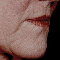
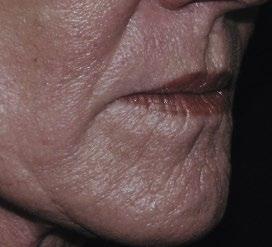
My very first case of skin needling was to correct upper‐lip creases. The patient was put onto vitamin A cosmetics and after three weeks I used regional anesthesia to numb her upper lip and I needled her upper lip intensively at 1.0 mm with the pen‐style device. I covered the needled area with skin‐colored micropore and asked her to apply the vitamin A cosmetics to the surface. She swelled considerably and after about five days I removed the micropore tapes and her lip looked intact but rather swollen. A month later I could see that the lip wrinkles looked better but not sufficiently so; therefore I advised a second needling done in the same way and after another month I felt that a third needling would give us a good result. By the end of the fourth month I was happy to sit back and wait for improvement. At that time, I realized that unlike laser treatments, where the results were obvious and best at about a month, with skin needling one needs to wait at least six months because there is progressive and well‐defined improvement month after month. After that the changes are more subtle. In my first case, I was able to follow up for five years and I can say that if you do intensive needling together with applying vitamin A cosmetic skin care, then the excellent result can be maintained for at least four years, as shown in Figure 2.1.
While I started using a pen‐type tattooing device, I felt that because the needle holes were too close together this produced a lot of inflammation. Once I started (a) (b)
Figure 2.1 The very first case of needling; long‐term result. (a) Before any treatment. (b) Four years after one needling per month for three sessions and continuous use of topical vitamin A and C. Source: Des Fernandes.
rolling I found that I got much less inflammation even after very intensive needling. After a few months I could see that the results were the same as those I got from using the pen‐type of device. I found out that percutaneous needling is ideal to get changes in the reticular dermis and stimulate the production of collagen and elastin fibers.
Dr. Hugo Nel of Medunsa University was fascinated by the concept and did histology on roller‐needled skin and reported to me personally that he could find no evidence of scar tissue and only found normal lattice‐style collagen. Normal healthy dermal collagen is in a web or latticework and is not cross‐linked, whereas scar collagen (cross‐linked collagen III) is more densely laid down and the fibers lie in parallel bundles and are cross‐linked. This was the first indication suggesting that skin needling caused regeneration instead of scarring. This was important evidence to support my concept that needling caused the release of platelet‐derived growth factors (especially TGF‐beta 3) and was responsible for regeneration as described by Ferguson.
My classical technique of skin needling using a roller
The roller is the safest and most effective way to needle the whole face, stretch marks, deep acne scars, or burn scars. I generally don’t use it for linear scars or fresh scars, where I prefer to use the pen‐style device. I also use the pen‐style device when I needle the upper eyelids and close to the lower eyelid’s eyelashes. I believe we should all become expert in using both techniques. The roller device is far easier to use and requires less training.
By rolling backwards and forwards in various directions, or by repeatedly rolling in one direction with some pressure, one can achieve an even distribution of holes. The skin should be kept taught and be needled as densely as possible to maximize the release of platelets and hence the secretion of growth factors. Usually, as the needle holes get too close to each other, the needle “slips” into an established hole and so it seems impossible to overtreat the skin and make the holes too close to each other. The needles penetrate right through the epidermis but do not excoriate or remove it, so the epidermis is only punctured and will rapidly heal. Histological studies show that the needle seems mainly to divide cells from each other rather than cutting through the cells, so many cells are spared injury. The epidermis and particularly the stratum corneum remain intact, except for these tiny holes, which are about four cells in diameter. The needles penetrate about 1.5 to 2 mm into the dermis. Naturally, the skin bleeds for a short time, but that soon stops.
Contrary to popular myth, the needle holes are simple, and the skin never gets slits or cuts as the needle exits the skin. I have examined this very carefully and
studied it with slow motion video. The skin is tented upwards as the needle lifts the skin but then because the roller is moving in the opposite direction the needle simply slides out of the skin. There is only one hole through which the blood may escape, and I believe more blood (and platelets) remain in the skin and hence could give a better final result. The more platelets, the richer the concentration of platelet‐derived growth factors. The skin develops multiple microbruises in the dermis that initiate the complex cascade of growth factors that results in collagen production. One advantage of the roller that can never be replicated by the mechanical pen‐type devices is that through one hole many arterioles are cut or pierced as the needle transits from entrance to exit. This is very different from when one uses a vertical stamper or pen device: each punctured blood vessel is under a hole through which blood may escape and potentially less blood remains in the dermis. The mechanical devices merely penetrate the blood vessels immediately in the vertical pathway of the needle.
After the bleeding stops, there is a serous ooze that must to be wiped from the surface of the skin. Wet gauze swabs soak up most of this serous ooze. As the skin swells, the holes are closed, the edges of the epidermis are approximated, and the ooze stops. Chemicals, however, may still penetrate the skin, so only safe molecules should be used topically. After this serous leak has stopped, the skin is washed and then covered with vitamins A and C [12]. There is no other special postneedling care, but I have found that exposing the skin to a low pH immediately after needling seems to facilitate even better results.
When I had sufficient histology to prove in my own clinical cases that we were regenerating skin, I understood that – for the first time in medical history – a medical intervention could cause regeneration instead of scar formation when treating scars or photodamaged skin. Till then all other treatments, such as CO2 laser or heavy peeling, had focused on scarring skin to tighten and smoothen it. In fact, skin needling is also known as percutaneous collagen induction (PCI)_ therapy and the automatic production particularly of TGF‐beta 3 opened up a new paradigm in treating scars.
I collected enough evidence to present a paper at the International Confederation for Plastic, Reconstructive & Aesthetic Surgery (IPRAS) meeting in San Francisco in June 1999 and the presentation was acclaimed by the packed audience as a breakthrough in treating scarring. Although I had pointed out that all my cases were prepared for a minimum of three months with a topical cosmetic vitamin A equivalent to retinoic acid 0.025 to 0.05 g%, few people recognized the value of preparation of the skin and so several months later I had complaints from several doctors who said needling did not make impressive changes. On questioning about what skin care they used, they generally said, "Come on, we all know that cosmetics do not work.” The other point that they missed was that needling has to be done quite intensively to make real changes.
I lectured around the world at international plastic surgery and aesthetic meetings and was invited to publish the first paper on skin needling as an alternative to laser treatments of skin in the Aesthetic Surgery Journal in 2002 [13]. At one meeting in Tokyo, Andre Camirand was told about my concept of growth factors causing the changes and after giving it thought for a minute he confessed that he had missed that detail, but of course he was looking at small areas and thought that needling restored normal skin color by transplanting melanocytes that adhered to the needle.
Prof. Matthias Aust, who visited me in 2003, was intrigued by skin needling and he decided to research it at Hannover Medical School, Germany. He confirmed in 2004 that platelet‐derived growth factors cause regeneration in rats (his work was finally published in 2010) [14]. We also published our combined experience of the clinical results in over 480 humans to show that needling was a valid way to treat wrinkles, stretch marks, and scars [15]. Aust continued his exploration of needling for various conditions [7, 16–23]. He particularly concentrated on burn scars and was awarded the European Burn Society Gold Medal for his work in treating burn scars. The lesson I learned from Aust was that because TGFβ3 is raised for only two weeks, even better results could be achieved by reducing the intervals between needling and so I slowly started to change the frequency of treatments until I felt safe doing 1.0 mm needling once a week. I tried 0.5 mm needling without topical anesthesia and that did not cause enough bleeding into the papillary dermis. If I did an intensive 0.5 mm needling with topical anesthesia I could manage to cause enough bleeding but I preferred needles at 0.8, 0.9, and 1.0 mm because I could get good bleeding without discomfort for the patient. I found that topical anesthetics, e.g. EMLA, made 1.0 mm needling very acceptable. I discovered that one has to make certain adjustments to enable topical anesthetic to work better. All topical anesthetics are kept in an acidic base to preserve activity. However, anesthetics function well only at an alkaline pH. For that reason, I devised a technique using a product I formulated to make the skin surface more alkaline before applying the topical anesthetic product. As a result, one gets much more intense anesthesia that may even allow 3.0 mm needling.
I realized that I could only do once‐a‐week needling with a roller because when I tried to do it with a pen‐type device the swelling and inflammation were too marked for the patients to tolerate that regimen. Needling, on the other hand, done under topical anesthesia, generally allowed the patients to return to work the day after the treatment.
At the invitation of Dr. Joe Niamtu, who heard me lecture on skin needling in Australia, I published my second article on minimally invasive collagen induction [24]. Dr. Massimo Signorini in Milan, Italy, was one of the first to understand the efficacy of skin needling in treating photoaging and started collecting his results from skin needling. Soon after that, we were able to publish an article together [12]. Hilton Kaplan, who had worked with me on many of my earlier
cases, was eventually able, with Dr. Julie Kenner from California, to present our combined multicenter work at the American Academy of Dermatology in 2006. When my results started to gain attention the roller design was adopted by many companies around the world.
In the beginning of my work with microneedling I said that the induction of the inflammatory phase following wounding was the best way to describe the concatenation of chemical events following the release of platelets and the platelet‐derived growth factors. Today I regret using that word because needling does not cause significant inflammation, yet several “experts” say that the frequent needling sessions that I advise “overwhelm” the skin system because of excessive inflammation and that interferes with the final results. I have pointed out repeatedly that I, and my patients, do not see the degree of inflammation that we encountered when I did pen‐type needling, and the results I achieved with more frequent needling set the standard for what could be achieved by skin needling. Some of my patients have demanded less frequent treatments than what I consider ideal, and so I have also built up a comparative experience and can say without prejudice that six needlings done at one‐month intervals gives an inferior result compared to doing six needlings spread over five weeks (5–6‐day intervals). Needling causes a healing cascade of growth factors totally unrelated to the inflammation caused by prostaglandins and inflammazones and is totally driven by the platelet‐derived growth factors.
My research has been a continuous voyage from 1998 till the present. Today I believe the roller is the safest way to treat thicker scars. Lasers, etc. do not give competitive results. Pen‐style needling cannot penetrate the skin and scar tissue properly and may cause damage to the epidermis in trying to do this depth of needling.
I have tried to find out:
1 what length needles were required for various scars, wrinkles and stretchmarks,
2 which chemicals were the best for using before and after needling,
3 the best time intervals in between needling sessions, and
4 how we can use skin needling immediately after surgery to help make almost invisible scars.
We need to investigate when and how to needle an incision. Either before the elected surgery (one week before, one day before, or even immediately before the incision is made) or after surgery. This could be immediately after the wound has been sutured closed, or when sutures/dressings are removed.
Another use for needling could be acute burn injuries. The growth factors released could speed up the regeneration of the tissue and could also help to improve immune defense. Maybe scar contractures could be avoided. The possibility of using needling in skin graft donor sites may offer the chance of less visible scars and even the opportunity to reharvest skin from the same donor area.
The appearance of cellulite has also been treated successfully with needling. The PCI effectively tightens up the skin again and smoothens the surface.