New and Future Developments in Microbial Biotechnology and Bioengineering: Microbial Secondary Metabolites Biochemistry and Applications Vijai G. Gupta (Editor)
Emerging trends in applications and infrastructures for computational biology, bioinformatics, and systems biology : systems and applications 1st Edition Arabnia
No part of this publication may be reproduced or transmitted in any form or by any means, electronic or mechanical, including photocopying, recording, or any information storage and retrieval system, without permission in writing from the publisher. Details on how to seek permission, further information about the Publisher’s permissions policies and our arrangements with organizations such as the Copyright Clearance Center and the Copyright Licensing Agency, can be found at our website: www.elsevier.com/ permissions.
This book and the individual contributions contained in it are protected under copyright by the Publisher (other than as may be noted herein).
Notices
Knowledge and best practice in this field are constantly changing. As new research and experience broaden our understanding, changes in research methods, professional practices, or medical treatment may become necessary.
Practitioners and researchers must always rely on their own experience and knowledge in evaluating and using any information, methods, compounds, or experiments described herein. In using such information or methods they should be mindful of their own safety and the safety of others, including parties for whom they have a professional responsibility.
To the fullest extent of the law, neither the Publisher nor the authors, contributors, or editors, assume any liability for any injury and/or damage to persons or property as a matter of products liability, negligence or otherwise, or from any use or operation of any methods, products, instructions, or ideas contained in the material herein.
Library of Congress Cataloging-in-Publication Data
A catalog record for this book is available from the Library of Congress
British Library Cataloguing-in-Publication Data
A catalogue record for this book is available from the British Library
ISBN: 978-0-12-819654-0
For information on all Woodhead publications visit our website at https://www.elsevier.com/books-and-journals
Publisher: Charlotte Cockle
Acquisitions Editor: Nancy Maragioglio
Editorial Project Manager: Sara Valentino
Production Project Manager: Poulouse Joseph Designer: Alan Studholme
Typeset by Thomson Digital
3.2
3.3
3.4
3.5
3.6
3.7 Phosphate
3.8
3.9
3.10
3.11
3.12
3.13
3.14
3.15
3.16
4 Endophytic bacterial strains induced systemic resistance in agriculturally important crop plants
12.2 Mechanism of mycosynthesis of silver nanoparticles
12.3 Silver nanoparticles from endophytic fungi and their efficacy in biocontrol
12.4 Mechanism of antimicrobial action by silver nanoparticles
12.5 Factors affecting the mycosynthesis of metal nanoparticles
12.6 Nanoparticles in plant disease
12.7
H.C. Yashavantha Rao, N. Chandra Mohana, Sreedharamurthy Satish
13.1 Introduction
13.2 Incidence of microbial endophytes diversity
13.3
Page left intentionally blank
Contributors
Remakanthan A. Department of Botany, University College, Thiruvananthapuram, Kerala, India
Mohd Aamir Laboratory of Mycopathology and Microbial Technology, Centre of Advanced Study in Botany, Institute of Science, Banaras Hindu University, Varanasi, Uttar Pradesh, India
Luciana da Silva Amaral Department of Chemistry, Universidade Federal de São Carlos, São Carlos, Brazil
Éder de Vilhena Araújo Institute of Chemistry, Universidade Estadual de Campinas, Campinas, Brazil
Dileep Kumar Bhaskaran Nair Saraswathy Amma Agro-Processing and Technology Division, CSIR – National Institute for Interdisciplinary Science and Technology (NIIST), Thiruvananthapuram, Kerala; Academy of Scientific and Innovative Research (AcSIR), Ghaziabad, Uttar Pradesh, India
Kalyani M. Barbadikar ICAR-Indian Institute of Rice Research, Hyderabad, Telangana, India
Antonio Biasi Agriculture Research Organization, Volcani Centre, Rishon LeZion, Israel
Tejas C. Bosamia Biotechnology Lab, ICAR-Directorate of Groundnut Research, Junagadh, Gujarat, India
Vinaya Chandran School of Biosciences, Mahatma Gandhi University, Kottayam, Kerala, India
Rubee Devi Department of Biotechnology, Eternal University, Baru Sahib, Sirmour, Himachal Pradesh, India
Samir Droby Agriculture Research Organization, Volcani Centre, Rishon LeZion, Israel
Manish Kumar Dubey Laboratory of Mycopathology and Microbial Technology, Centre of Advanced Study in Botany, Institute of Science, Banaras Hindu University, Varanasi, Uttar Pradesh, India
Radhakrishnan E.K. School of Biosciences, Mahatma Gandhi University, Kottayam, Kerala, India
Taicia Pacheco Fill Institute of Chemistry, Universidade Estadual de Campinas, Campinas, Brazil
Jubi Jacob Agro-Processing and Technology Division, CSIR – National Institute for Interdisciplinary Science and Technology (NIIST), Thiruvananthapuram, Kerala; Academy of Scientific and Innovative Research (AcSIR), Ghaziabad, Uttar Pradesh, India
Aswathy Jayakumar School of Biosciences, Mahatma Gandhi University, Kottayam, Kerala, India
Ashitha Jose School of Biosciences, Mahatma Gandhi University, Kottayam, Kerala, India
Meritta Joseph School of Biosciences, Mahatma Gandhi University, Kottayam, Kerala, India
Poonam Kanani Department of Agriculture Biotechnology, Anand Agriculture University, Gujarat, India
Tanvir Kaur Department of Biotechnology, Eternal University, Baru Sahib, Sirmour, Himachal Pradesh, India
Divjot Kour Department of Biotechnology, Eternal University, Baru Sahib, Sirmour, Himachal Pradesh, India
Gopika Vijayakumari Krishnan Agro-Processing and Technology Division, CSIR – National Institute for Interdisciplinary Science and Technology (NIIST), Thiruvananthapuram, Kerala; Academy of Scientific and Innovative Research (AcSIR), Ghaziabad, Uttar Pradesh, India
Ajay Kumar Agriculture Research Organization, Volcani Centre, Rishon LeZion; Institute of Plant Sciences, Agricultural Research Organization, Volcani Center, Rishon LeTsion, Israel
Sunil Kumar Laboratory of Mycopathology and Microbial Technology, Centre of Advanced Study in Botany, Institute of Science, Banaras Hindu University, Varanasi, Uttar Pradesh, India
Veena P. Kumar School of Biosciences, Mahatma Gandhi University, Kottayam, Kerala, India
Linu Mathew School of Biosciences, Mahatma Gandhi University, Kottayam, Kerala, India
Arpan Modi Agricultural Research Organization, Volcani Center; Institute of Plant Sciences, Agricultural Research Organization, Volcani Center, Rishon LeTsion, Israel
N. Chandra Mohana Microbial Drugs Laboratory, Department of Studies in Microbiology, Manasagangotri, University of Mysore, Mysuru, Karnataka, India
Indu C. Nair Department of Biotechnology, SAS SNDP Yogam College, Konni, Kerala, India
B. Shankar Naik Department of P.G. Studies and Research in Applied Botany, Kuvempu University, Shimoga; Department of Biology, Government Science College, Chikmagalur, Karnataka, India
Chandranandani Negi Department of Biotechnology, Eternal University, Baru Sahib, Sirmour, Himachal Pradesh, India
João Guilherme de Moraes Pontes Institute of Chemistry, Universidade Estadual de Campinas, Campinas, Brazil
Aswani R. School of Biosciences, Mahatma Gandhi University, Kottayam, Kerala, India
Ashutosh Rai Division of Biotechnology, Indian Institute of Vegetable Research, Varanasi, Uttar Pradesh, India
Avinash Chandra Rai Institute of Plant Sciences, The Volcani Center, Agriculture Research Organisation, Bet-Dagan, Israel
Krishna Kumar Rai Laboratory of Mycopathology and Microbial Technology, Centre of Advanced Study in Botany, Institute of Science, Banaras Hindu University, Varanasi, Uttar Pradesh, India
Kusam Lata Rana Department of Biotechnology, Eternal University, Baru Sahib, Sirmour, Himachal Pradesh, India
H.C. Yashavantha Rao Department of Biochemistry, Indian Institute of Science, Bengaluru, Karnataka, India
Sreedharamurthy Satish Microbial Drugs Laboratory, Department of Studies in Microbiology, Manasagangotri, University of Mysore, Mysuru, Karnataka, India
Anil Kumar Saxena ICAR- National Bureau of Agriculturally Important Microorganisms, Mau, Uttar Pradesh, India
Hitha Shaji School of Biosciences, Mahatma Gandhi University, Kottayam, Kerala, India
Vaishali Shukla Laboratory of Mycopathology and Microbial Technology, Centre of Advanced Study in Botany, Institute of Science, Banaras Hindu University, Varanasi, Uttar Pradesh, India
Stephanie Nemesio da Silva Institute of Chemistry, Universidade Estadual de Campinas, Campinas, Brazil
Karan Singh Department of Chemistry, Eternal University, Baru Sahib, Sirmour, Himachal Pradesh, India
Sandeep Kumar Singh Centre of Advance Study in Science, Department of Botany, Institute of Science, Banaras Hindu University, Varanasi, Uttar Pradesh, India
Contributors
Vipin Kumar Singh Centre of Advance Study in Science, Department of Botany, Institute of Science, Banaras Hindu University, Varanasi, Uttar Pradesh, India
Drissya Thankappan Agro-Processing and Technology Division, CSIR – National Institute for Interdisciplinary Science and Technology (NIIST), Thiruvananthapuram, Kerala; Academy of Scientific and Innovative Research (AcSIR), Ghaziabad, Uttar Pradesh, India
Vipina Vinod T.N. School of Biosciences, Mahatma Gandhi University, Kottayam, Kerala, India
Ram S. Upadhyay Laboratory of Mycopathology and Microbial Technology, Centre of Advanced Study in Botany, Institute of Science, Banaras Hindu University, Varanasi, Uttar Pradesh, India
James Francis White Department of Plant Biology, Rutgers University, New Brunswick, NJ, United States
Ajar Nath Yadav Department of Biotechnology, Eternal University, Baru Sahib, Sirmour, Himachal Pradesh, India
Andleeb Zehra Laboratory of Mycopathology and Microbial Technology, Centre of Advanced Study in Botany, Institute of Science, Banaras Hindu University, Varanasi, Uttar Pradesh, India
V. Yeka Zhimo Agriculture Research Organization, Volcani Centre, Rishon LeZion, Israel
Entry, colonization, and distribution of endophytic microorganisms in plants
Ajay Kumar a, Samir Droby a, Vipin Kumar Singh b, Sandeep Kumar Singh b , James Francis Whitec
aAgriculture Research Organization, Volcani Centre, Rishon LeZion, Israel; bCentre of Advance Study in Science, Department of Botany, Institute of Science, Banaras Hindu University, Varanasi, India; cDepartment of Plant Biology, Rutgers University, New Brunswick, New Jersey, United States
Chapter outline head
1.1 Introduction 1
1.2 The rhizosphere and its role in endophytic associations 2
1.3 Endophytes and host plant surfaces 3
1.4 Entry and colonization of plants by bacterial endophytes 5
1.5 Plant internalization and extraction of nutrients from microbes in the rhizophagy cycle 7
1.6 Genomic insights into host and endophyte interaction 8
1.7 Transmission of endophytes 10
1.8 Endophytic diversity 11
1.9 Conclusion or future prospective 21
References 22
1.1 Introduction
Plants interact with large numbers of microbial communities, in which some of them enter and reside in the plant tissue without causing any disease or otherwise negative impact. These intimately associated microbes are called as endophytes. The word “endophyte” is derived from two Greek words “endon” means within, and “phyton” means plant (Chanway, 1996). The term “endophyte” was first introduced by De Bary (1866) for the microorganisms growing inside plant tissues. Later on, definition and types of endophytes were modified as per researcher observations. Hallmann et al. (1997) have defined endophyte as the microbes that can be isolated from the surfacedisinfected tissues of plants, and those microbes that could survive inside their host system without causing any disease. Some researchers have also categorized endophytes on the basis of their types, that is, bacteria or fungi, and their relationship with the plants such as facultative or obligate (Rosenblueth and Martínez-Romero, 2006). However, Hardoim et al. (2015) have characterized endophytes on the basis of
colonization niche instead of their function. Initially, the term “endophyte” was used for the fungi that were documented from the internal cells/tissues of host plants but later on the concept had been changed and the bacterial communities also considered as endophytes (Chanway, 1996; Hardoim et al., 2015). Currently, it has been considered that endophytes are present in all plant species (Strobel and Daisy, 2003; Huang et al., 2007), and have been demonstrated to share a complex relationship with their host plants.
1.2 The rhizosphere and its role in endophytic associations
The entry or colonization of endophytic bacteria into the host plant is a complex phenomenon and involves a series of events. The process of colonization usually starts from the communication between the specific components of the root exudates and the associated microbial communities (de Weert et al., 2002; Rosenblueth and MartínezRomero, 2006). The rhizosphere can be described as the region of soil adhered to the root and is directly influenced by plants and their associated microbiota, species, growth stages, and the physiology of the host plant. The roots of plants release significant amounts of exudates that influence diverse microbial communities in the rhizosphere (Singh et al., 2017, 2018). Root exudates are rich in organic substrates, such as carbohydrates, lipids, phenolics, amino acids, phytosiderophores, and flavonoids, these serve as chemoattractants and facilitate the communication between roots and microbes that ultimately help in recruiting bacterial endophytes from the rhizosphere and start colonization of host plant tissue (Badri and Vivanco, 2009).
There are various reports available showing the evidence of direct involvement of root exudates in initializing the host tissue colonization by microbial entities. Oku et al. (2012) reported the role of amino acids present in root exudates of tomato plants and noticed their role as chemoattractants in the colonization of Pseudomonas fluorescens Pf0-1. The evidence in support was gathered from genomic studies pertaining to three genes namely ctaA, ctaB, and ctaC coding for sensory proteins Pfl01_4431, Pfl01_0124, and Pfl01_0354, respectively. These genes are homologous to Pseudomonas aeruginosa PAO1 pctA gene, exhibiting positive response toward 20, L-amino acids during initial root colonization in tomato (Oku et al., 2012).
In another study Kost et al. (2014) reported the role of oxalate in root colonization by a strain of Burkholderia. The plant growth promoting strains were reported to utilize oxalate as a carbon source but pathogenic strains such as B. glumae and B. plantarii did not degrade the oxalate. Interestingly, the mutant strain Burkholderia phytofirmans PsJN lacked the ability to metabolize oxalate could not colonize lupin and maize, indicating oxalotrophy as a prerequisite for colonization by this endophytic species. In similar ways metabolites like malate and benzoates also act as chemoattractants and provide help in effective colonization of the plant (Lopez-de-Victoria and Lovell, 1993). Flavonoids have also been recognized as one of the important components of root exudates secreted by several plant species and could play effective role
in endophytic colonization within the root hairs (Khare et al., 2018). The specificity and effectiveness is considerably determined by the chemical structure of flavonoids as reported by Scervino et al. (2006). There are various reports available confirming the participation of flavonoids as chemotaxis agents during host tissue colonization by endophytic strains of Rhizobium (Dharmatilake and Bauer, 1992; Khandual, 2007; Faure et al., 2009). Furthermore, some of the authors have also illustrated the role of flavonoids in effective colonization of host tissue by the endophytic strain Serratia sp. EDA2 and Azorhizobium caulinodans ORS571 (Webster et al., 1998; Balachandar et al., 2006). In this connection, Steinkellner et al. (2007) also studied the various functional aspects of flavonoids including hyphal growth differentiation and root colonization and concluded the role of flavonoids as effective signaling molecule in the various plant species and their active participation in plant–microbe interaction. In case of legume–rhizobium endophytic association, flavonoids facilitates chemotactic response and start signaling through nod factors, culminating into symbiotic association (Garg and Geetanjali, 2007).
Strigolactone (SL), the phytohormones secreted by plant roots has also been demonstrated to act as signaling molecules. In a study, López-Ráez et al. (2017) have discussed the role of SL hormone and concluded that treatment was able to activate the release of oligomers acting as signaling molecules and provided help in tissue colonization. Further, Rozpądek et al. (2018) also reported the role of strigolactone as a signaling molecule during the initial colonization of host tissue by endophytic strain of Mucor sp.
1.3 Endophytes and host plant surfaces
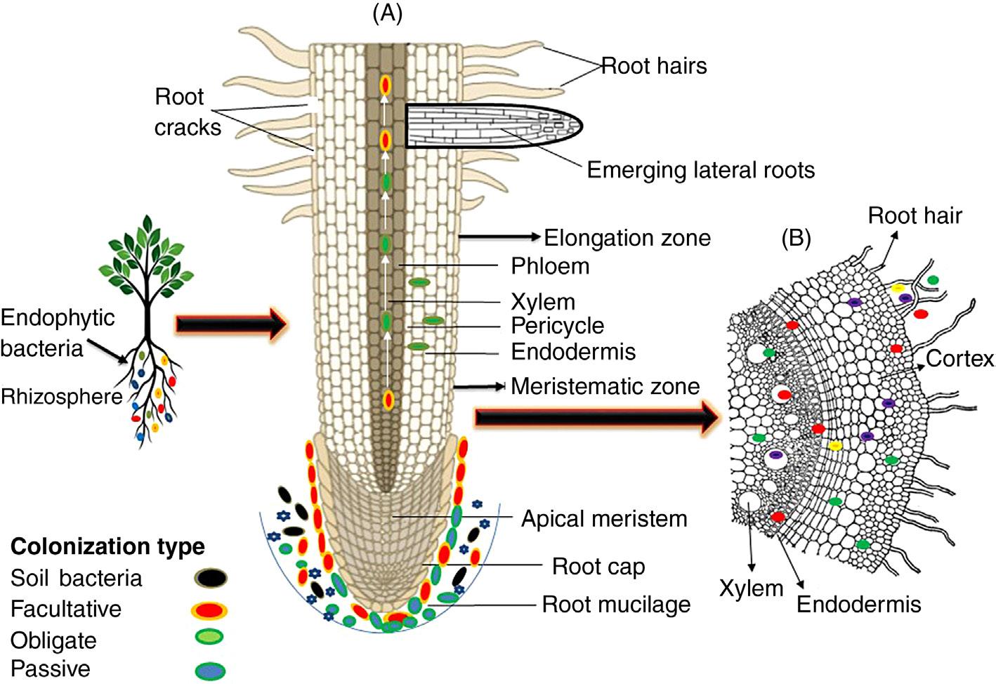
The entry or host tissue colonization by microbes is a complex phenomenon and is controlled by signaling molecules, proteins, and/or the secretory products of the plants as well as microbes. Generally, adhesion of a particular microbial strain to the host surface is considered as the first step of colonization. Subsequently, the microbes migrate toward the host surface in response to root exudates via chemotactic movement that precedes attachment (Begonia and Kremer, 1994) (Fig. 1.1).
The attachment of bacterial cells to the plant surface is one of the most crucial step during endophyte colonization; in this process various structural components, such as flagella, fimbriae, pili, and the secretory products like exopolysaccharides (EPS), lipopolysaccharide (LPS), or cell surface polysaccharides may directly involve in the attachment (Sauer and Camper, 2001). There are various reports available that confirm the role of microbial appendages in surface attachment. Croes et al. (1993) have reported the role of flagella in the primary attachment of Azospirillum brasilense with the root surface of wheat. However, flagella-deficient mutants did not show attachment with the wheat roots. Dörr et al. (1998) have reported the association of type IV pili in the attachment of endophytic strain Azoarcus sp. BH72, with the root surface of rice. These small appendages flagella and pili have also been described to act as propellers leading to movement of microbes toward the plant surface via chemotactic
Figure 1.1 Schematic representation of endophytic bacterial colonization and distribution in the endosphere of a plant root. (A) Invasion of bacteria into a plant using several root zones. White arrows shows translocation of bacteria in to the phloem and xylem and colonization type represented by different colored ovals. (B) Occurrence of endophytes either at the site of entry (indicated in blue) or in the intercellular space of the cortex and xylem vessels (indicated in green). Red and yellow spheres represents rhizospheric bacteria, which are unable to colonize inner plant tissues.
response and developed a weak attractive force to withstand any repulsive barriers that might originate due to electrostatic charges present on the cell envelope (Berne et al., 2015; Zheng et al., 2015).
The bacterially synthesized product EPS could also facilitate bacterial attachment to the host plant surface during early stages of colonization as mentioned by Janczarek et al. (2015) for Rhizobium leguminosarum. Similarly, Meneses et al. (2011) have mentioned the role of EPS secreted by endophyte Gluconacetobacter diazotrophicus in the attachment and colonization of rice root endosphere. However, besides attachment EPS have also been reported to offer other advantages including protection of bacterial cell and host plant from oxidative damage and elevated level of free radicals. Similarly, Marczak et al. (2017) reported the role of EPS secreted by Rhizobium in symbiosis and colonization with the legume plants. Balsanelli et al. (2010) have described the role of LPS secreted by endophytic bacterial strain Herbaspirillum seropedicae in attachment and colonization of maize root. In addition, reports are also available in literature emphasizing the role of LPS N-acetyl glucosamine in binding with lectins present in maize root, and concluded their involvement as an essential in bacterial attachment and subsequent colonization in the host roots (Balsanelli et al., 2013).
Plants respond differentially after attachment of microbial strain with the host surface that leads to significant variation in the pattern of gene expression as reported by Sauer and Camper (2001) in case of Pseudomonas putdia. Further, in depth study was conducted by De Mot and Vanderleyden (1991) pertaining the proteomics of outer membrane porin F (OprF) proteins and their role in attachment and host tissue colonization by P. fluorescens. OprF is a multifunctional outer membrane proteins commonly present on the outer surface of Pseudomonas and helps in the attachment with various surfaces and molecules (Bodilis and Barray, 2006). The function of OprF proteins as adhesive had also been observed in various plant species, such as barley, sunflower, maize (De Mot and Vanderleyden, 1991) cucumber, and tomato roots (Crespo and Valverde, 2009). Similarly, arabinogalactan proteins (AGPs), a glycoprotein present on the plant’s cell wall has also been documented to help in initial colonization of microbes at the different growth stages of the plants (Nguema-Ona et al., 2013). In a similar fashion, the important contribution of flagellin, a globular proteins of flagella during the attachment with host surface as reported in case of A. brasilense strain (RodríguezNavarro et al., 2007) is also evidenced. The responsible genes for the glycosylation of flagellin and LPS are the same, and it had been seen that mutation in these genes results in impairment of the attachment of A. brasilense (Rossi et al., 2016).
1.4 Entry and colonization of plants by bacterial endophytes
After establishing in the rhizosphere and rhizoplane, bacterial endophytes are known to make their way inside the plant root, with subpopulations ranging from 105 to 107 cfu/g fresh weight (Hallmann, 2001). During colonization, pattern and sites are specific for each of the endophytic strain (Zachow et al., 2010). After attachment to the host surface, endophytes start penetrating in order to enter to the host tissue. Endophytic bacteria, however, may prefer various sites to enter the plant tissue; the most preferred entrance path is via root zone, aerial parts of the plants, including stems, leaves, flowers, and cotyledons (Zinniel et al., 2002). The process of penetration into the host can be mediated by passive or active process. The passive penetration occurs at the site of cracks present in the areas of root emergence, root tips that are created by deleterious organisms (Hardoim et al., 2008), whereas active penetration is achieved through attachment and proliferation of EPS, LPS, structural components, quorum sensing, providing considerable help in the movement, and multiplication of endophytes inside the plant tissues (Böhm et al., 2007; Dörr et al., 1998; Duijff et al., 1997; Suárez-Moreno et al., 2010).
There are numerous reports present that have shown different entry modes and colonization patterns of endophytic strains. Apart from this, specialized and frequently studied interaction between nodulating bacteria and legumes is less well-understood. Although not experimentally proven, it has been proposed that endophytic bacteria produce low levels of cell-wall degrading enzymes as compared to phytopathogens that could produce deleteriously high levels of these enzymes and thus endophytes may avoid triggering plant
defence systems (Elbeltagy et al., 2000). Furthermore, another way by which endophytic bacteria escape their detection as a pathogen by host tissue is maintenance of low cell densities (2–6 log cfu/gfw) as compared to pathogenic bacteria.
During entry or colonization, microbial strains prefer the site having thin surfaces such as root hairs, or the elongation zone of the apical root meristem serving as one of the preferred site of rhizoplane. At favorable sites, endophytic microbial strain secretes some lytic enzymes, such as lysozymes, cell wall degrading enzymes, cellulases, facilitating the entry of bacterial strain through hydrolyzing external covering or plant cells (Compant et al., 2005; Reinhold-Hurek et al., 2006; Naveed et al., 2014). Reinhold-Hurek et al. (2006) have reported Azoarcus sp. BH72 species at the entry site having endoglucanase, a kind of cellulase and further confirmed the role of endoglucanase in endophyte colonization by mutant analysis of eglA gene. The mutant endophyte lacking eglA genes was unable to colonize plant tissues, whereas wild type strain invaded and colonized the host surface. Suzuki et al. (2005) have reported a nonspecific wax-degrading enzyme helping in colonization of Streptomyces galbus on the Rhododendron. Taking together, all these previous investigations have shown the ability of bacteria to utilize certain plant metabolites as an essential mechanism for successful establishment as endophyte.
Successful colonization of endophyte involves compatible plant–microbe interactions. As the endophyte invades the host surface, it is recognized by the plant and crosstalk of signaling molecules is initiated (Rosenblueth and Martínez-Romero, 2006; Compant et al., 2010; Brader et al., 2014). The colonization of endophytic microbes depends upon various factors including microbial strains, host genotype, biotic and abiotic factors, nutrients limitation, UV light, etc. and most importantly, the strains better adapted to these factors are comparatively more efficient in getting entry into the plant tissues via various routes like natural opening such as hydathodes, stomata, etc., followed by colonization of host tissue (Hallmann, 2001; Hardoim et al., 2015). To date, numerous reports have presented the details regarding the colonization routes of endophytic microbial strains. In a study, Álvarez et al. (2010) reported the colonization pattern of Ralstonia solanacearum strain and concluded that strains firstly attached to surface followed by invasion of the extension of roots such as root hairs, root tips, lateral roots; however, they may also prefer to enter through mechanical binding during initial colonization. After entering the host tissues, strain may spread themselves upwardly in the plants via xylem vessels. In another study, Compant et al. (2005, 2008) studied the colonization route of strain Paraburkholderia phytofirmans PsJN and reported that endophytic strain entered through the exodermis layer of roots following cortical cells and crossed the barrier of endodermal layer leading to its access to the central zone. From this zone, the endophyte spread toward the upper part of plants through xylem vessels. At the site of xylem colonization, very few bacterial strains are able to cross the endodermal layer. Generally, the endophytic strains prefer unsuberized endodermal cells of the apical root zone to get entry inside host tissues (James et al., 2002; Roncato-Maccari et al., 2003; Compant et al., 2005; Gasser et al., 2011). Studies have demonstrated low concentrations of nutrients in the xylem tissues or plant sap that could be sufficient for the growth of endophytic bacteria (Madore and Webb, 1981; Sattelmacher, 2001; Bacon and Hinton, 2007). At the site of
cortex colonization, once the bacterial strains have crossed the exodermal barrier, they may remain localized at the site of entry (Timmusk et al., 2005) or move deeper into the host system such as cortex of the plant (Roncato-Maccari et al., 2003; Compant et al., 2005; Gasser et al., 2011).
In phyllosphere colonization, bacterial strains are firstly attached to the surface of leaf and randomly distributed throughout. Some of them may enter into the leaf tissue via natural openings such as stomata, hydathodes, and influence their local environment. At this site, bacterial strains multiply and form a thin layer of biofilm, however, some of them may enter into the leaf tissue and start surviving as endophytes (Yaron and Römling, 2014). In a study James et al. (2001) have reported stomata as an entry site during colonization of Gluconobacter diazotrophicus strain in the sugarcane. Currently, various reports have confirmed the utilization of plant nutrient as source of energy by the endophytic microbes (Rasche et al., 2009) and carbon has been reported as the most preferred source for growth and survival of endophytes (Krause et al., 2006; Malfanova et al., 2013). However, Iwai et al. (2003) have reported endophytic pseudomonads isolated from cucumber plants with the ability to utilize l-arabinose as one of the most abundant sugars available in the xylem fluid utilized by endophyte as nutrient source. In another study, Krause et al. (2006) reported alcohol dehydrogenases as an essential component in the colonization of Azoarcus sp. BH72 in waterlogged rice. They also concluded from their study that, in waterlogged rice, alcohol was present abundantly and may be utilized as carbon source by the colonizing bacterial strain Azoarcus sp. BH72.
Some reports have described the local colonization of endophytic strains and further there was no transmission to other parts of the host after successful colonization as documented in the case P. fluorescens strain invading olive plants (Prieto et al., 2011). Similar studies by Moulin et al. (2015) have also reported the colonization of Rhizobium strain only in the symbiotic zone of root nodule of legume. After colonization or entry of endophytic strains into the plant tissue, they may colonize locally or spread systemically (Afzal et al., 2019) to the upper parts of the host tissues. It has been mentioned that 103–104 cfu/gfw population density is established in the ground tissue of root and stem (Compant et al., 2010). The above ground migration of endophytes depends upon their functional and physiological requirements and the strain could move upwardly as above ground tissues are well-adapted for the particular environment and endophytic niche (Hallmann, 2001). The movement of the endophytic strain within the host tissue is however, largely executed by lateral appendages such as flagella, pilli, or the transpiration stream of the plants similar to transport of plant nutrients (Compant et al., 2005; James et al., 2002).
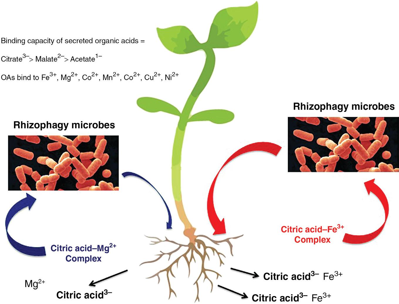
1.5 Plant internalization and extraction of nutrients from microbes in the rhizophagy cycle
Recent studies have shown that plants internalize soil microbes (bacteria and fungi) into plant roots and oxidatively extract nutrients from them in a process that has been termed “rhizophagy” (Paungfoo-Lonhienne et al., 2010, 2013) or “rhizophagy cycle”
(White et al., 2018). In the rhizophagy cycle, plants attract soil microbes to the root tip meristem with root exudates, and then internalize microbes into root meristem cells, which have soft cell walls. The precise mechanism by which microbes are internalized into root meristem cells remains unknown but may involve previously discussed processes. After internalization microbes become situated in the periplasmic space (between cell wall and plasma membrane) of root cells. The root cell plasma membrane secretes superoxide (produced on membrane bound NADPH oxidases) onto microbes and this strips the cell walls from microbes, resulting in formation of microbe protoplasts (White et al., 2018). Superoxide causes microbe protoplasts to become porous and leak nutrients that are absorbed by root cells. Through the action of cyclosis (cytoplasm rotation or streaming) in root cells, microbe protoplasts are circulated around the periphery of root cells and broken into many smaller protoplasts, rapidly replicating the intracellular microbes. Intracellular microbes in root cells accumulate in the tips of root hairs and trigger root hair elongation by an as yet unknown mechanism; without microbes root hair elongation does not occur (Verma et al., 2017). Microbe protoplasts are ejected into the soil through pores that form in the elastic wall at tips of elongating root hairs after a wave of vacuolar expansion propagates from the base of the root hair to the tip. It is unknown what triggers the periodic ejection of microbes from root hairs. Once ejected from root hairs, microbes reform cell walls and move out into the rhizosphere soil to acquire additional nutrients. The rhizophagy cycle appears to occur in all plants that form root hairs, and may be an important mechanism for acquisition of nitrogen and soil micronutrients like iron, zinc, and magnesium (White et al., 2015, 2018). It seems evident that the rhizophagy cycle is a mechanism whereby plants use soil microbes as carriers of difficult to acquire nutrients (Fig. 1.2). Work is still being done to evaluate details of the rhizophagy cycle mechanism and determine its importance to plant growth (Domka et al., 2019).
1.6 Genomic insights into host and endophyte interaction
Comparative genomics studies of close mutualistic or pathogenic endophytic strains have shown very similar genetic contents (Lòpez-Fernàndez et al., 2015; SheibaniTezerji et al., 2015) and this similarity may be used in differentiating strains as a pathogen or beneficial microbe for the host on the basis of genetic analyses. LòpezFernàndez et al. (2015) when comparing the virulence genes in endophytes and other symbiotic bacteria lead to the conclusion that there are only minor differences between endophytes and pathogens and that the similarities between these two groups are set above the species level.
In plant–microbe interactions secretion of protein is a determinant factor and is required for beneficial interaction. The transport of specific proteins for particular functions such as biocontrol is of considerable importance as the immunity of host plant is enhanced multiple orders after transport of effector proteins from microbes to the host, and thus helping in marking a particular microbe as a endophyte or parasite (Jones and
Figure 1.2 Diagrammatic representation of nutrient mining by rhizophagy microbes. Plant roots secrete organic acids (citric, malic, and acetic acids) into the soil. Organic acids complex with metals in the soil (Fe3+, Mg2+, Co2+, Mn2+, Co2+, Cu2+, Ni2, etc.). Rhizophagy cycle microbes possess transporters that bind to these organic acid-metal complexes and absorb them into the microbe cells. Microbes then return to the plant root and enter into root cells where nutrients are extracted from microbes oxidatively.
Dangl, 2006). These effector proteins are recognized by the plant immune system and are demonstrated to participate in activation of effector-triggered immune responses particularly T3SSs and T6SSs genes in the plant (Jones and Dangl, 2006). Interestingly, in the case of mutualistic endophytes, genes for T3SSs are missing (Hardoim et al., 2015; Mitter et al., 2017; Reinhold-Hurek and Hurek, 2011). Reinhold-Hurek and Hurek (2011) have proposed the view that missing T3SSs showed characteristics of an endophytic lifestyle. Iniguez et al. (2005) have also reported a similar observation; mutants of T3SSs of Typhimurium showed increased endophytic colonization in Medicago truncatula. However, some reports are also available that have shown endophytic establishment of Pseudomonas strain in the root even in the presence of T3SS gene (Preston et al., 2001). Endophytic microbes generally contain genes for T6SSs, conferring them with the potential for plant–microbe interaction (Mitter et al., 2013; Reinhold-Hurek and Hurek, 2011). There are various reports in the literature showing the contribution of T6SSs genes in the control of phytopathogens and disease management (Mattinen et al., 2008; Schell et al., 2007). In addition, nod genes are also responsible factors for nodulation and symbiotic association between host and
bacterial strain. Various authors have reported nod genes in the genome sequence of nodule-forming bacteria such as Burkholderia phymatum strain STM815A (Amadou et al., 2008), Bradyrhizobium japonicum USDA110 (Kaneko et al., 2002), and Frankia spp. strain CcI3 (Normand et al., 2007).
1.7 Transmission of endophytes
Plant-associated microbes interact with plants through various ways, and during colonization they may vector horizontally (plant or soil to plant), vertically (parent plant to seed), or in a mixed way (Bright and Bulgheresi, 2010). The transmission mode may also depend upon the ecological and evolutionary relationship between host and microbe. Microbes displaying symbiotic relationship with the host plant generally follow vertical transmission (Moran, 2006) and during transmission, parents (seeds and pollens) fulfill nutrient requirements. In many vertically transmitted symbiosis, the symbiont is obligate and spends its entire life inside the host plant (Bright and Bulgheresi, 2010; Herre et al., 1999). Some of the fungal species are known to prefer vertical mode of transmission via the seeds and is well-documented by various authors (Schardl, 2001; Wilkinson and Sherratt, 2001; Foster and Wenseleers, 2006). Generally, bacterial endophytes employ a horizontal route of transmission and it has been also seen that bacterial count in the soil or in other environment is higher than the seed or the seed grown under artificial conditions (Hardoim et al., 2012). Some of the naturally existing bacteria, after entry to the host tissue, may act as endophytes and thus may be transmitted to the next generation in a similar way as pathogens. Many of the bacterial species are known to infect different plant species through the similar horizontal mode (Ma et al., 2011; Compant et al., 2005; Khan et al., 2012). The horizontal transmission mode of beneficial bacteria appears optimal for the host system, because endophytic strains provide resistance against various biotic and abiotic stresses that may directly influence the plants (Carroll, 1988; Schlaeppi and Bulgarelli, 2015; Bulgarelli et al., 2012; Lundberg et al., 2012; Peiffer et al., 2013; Schlaeppi et al., 2014; Edwards et al., 2015; Verma et al., 2017). Some of the endophytic bacterial strains, however, may employ a mixed mode of transmission and this may depend upon the surrounding environmental conditions. There are various reports available that have confirmed the existence of bacterial inhabitants as endophyte inside the seed or the vertical mode of transmission. In the last few decades, the microbiome of seeds is gaining high importance and attracting researchers to explore their hidden potentials (Verma and White, 2019). The endophytic microbial isolates from different plant seeds have been reported by various authors from hosts such as alfalfa (Charkowski et al., 2001), rice (Hardoim et al., 2012; Cottyn et al., 2001; Bacilio-Jiménez et al., 2001; Kaga et al., 2009; Okunishi et al., 2005; Verma et al., 2017), maize (Liu et al., 2013; Johnston-Monje and Raizada, 2011), tobacco (Mastretta et al., 2009), coffee (Vega et al., 2005), quinoa (Pitzschke, 2016), common bean (López-López et al., 2010), grapevine (Pitzschke, 2016), barley (Zawoznik et al., 2014), and pumpkin (Fürnkranz et al., 2012). Different parts of seeds such as seed coat, endosperm,
and embryonic tissue have been reported to be occupied by various types of bacterial communities (Mitter et al., 2017; Compant et al., 2011; Glassner et al., 2018). Rhizomes of plants may also act as seed and harbour various groups of bacteria as endophyte (Kumar et al., 2016). There are numerous bacterial genera such as Bacillus, Pseudomonas, Klebisella, Burkholderia, Penibacillus, Staphylococcus, Pantoea, Acinetobacter that have been the most commonly reported seed endophytes. Inside seeds, these endophytic bacterial strains mediate various beneficial interactions such as nutrient acquisition, synthesis of growth regulators, along with biotic and abiotic stress management. However, it is not necessary that all the inhabiting seed bacteria colonize the seedlings or are transferred from parent to offspring plants.
The best evidence in support of vertical transfer of endophytes via seed comes from the studies demonstrating overlap in endophyte taxa between seed and seedling (Ferreira et al., 2008; Gagne-Bourgue et al., 2013; Ringelberg et al., 2012; Verma and White, 2019). Other studies have also reported the continued transfer of particular endophytic strains across generations in rice and maize (Mukhopadhyay et al., 1996; Liu et al., 2012), thus supporting vertical transfer. And at least in maize, there is some evidence of long-term conservation in the seed endophytic community; noteworthy, seeds of some genetically related maize hybrids have been found to host similar bacterial taxa (Liu et al., 2012). In an experimental investigation based on terminal restriction fragment length polymorphism (RFLP) of 16S rDNA, the presence of the same genera across several genotypes of maize, including its ancestor teosinte was documented (Johnston-Monje and Raizada, 2011). Further, different bacterial species can colonize the seeds horizontally from the external environment via flowers, fruits, and during seed dispersal.
1.8 Endophytic diversity
In the last few years, exploration and isolation of endophytic microbes have been carried out using new technologies and “omics.” Every plant species, which is growing in the natural environment, has endophytic microbial communities, and it is a peculiar exception, if any plant does not have an endophytic community of microbes (Partida-Martinez and Heil, 2011; Afzal et al., 2019). Currently, more than 16 phyla or 200 genera of bacteria have been reported as endophytes in various plant species. These bacterial genera include both cultivable and uncultivable strains (Malfanova et al., 2013); Proteobacteria followed by Actinobacteria, Firmicutes, and Bacteroidetes (Edwards et al., 2015) are the most dominant phyla, and contain numerous groups of bacteria such as Pseudomonas (Kumar et al., 2016), Bacillus (Deng et al., 2011) Burkholderia (Weilharter et al., 2011), Enterobacter (Taghavi et al., 2010), and Serratia (Taghavi et al., 2009).
There are various reports that show similar types of observations inside roots. Marques et al. (2015) reported Gamma-Proteobacteria (including Enterobacter, Pseudomonas, and Stenotrophomonas genera) was the dominant group in the endosphere of sweet potato. Sun et al. (2008) studied endophytic bacterial diversity of rice roots