Contact your Elsevier Sales Representative for teaching resources, including slides and image banks, for Medical Microbiology, 9e, or request these supporting materials at: http://evolve.elsevier.com/Murray/microbiology/
Medical Microbiology
NINTH EDITION
Patrick R. Murray, PhD, F(AAM), F(IDSA)
Vice-President, Microbiology Sparks, Maryland;
Adjunct Professor, Department of Pathology University of Maryland School of Medicine Baltimore, Maryland
Ken S. Rosenthal, PhD
Professor of Immunology
Augusta University/University of Georgia Medical Partnership Athens, Georgia; Emeritus Professor, Northeastern Ohio Medical University Rootstown, Ohio
Michael A. Pfaller, MD, F(CAP), F(AAM), F(IDSA)
Consultant
JMI Laboratories North Liberty, Iowa
Professor Emeritus
University of Iowa College of Medicine Iowa City, Iowa
No part of this publication may be reproduced or transmitted in any form or by any means, electronic or mechanical, including photocopying, recording, or any information storage and retrieval system, without permission in writing from the publisher. Details on how to seek permission, further information about the Publisher’s permissions policies and our arrangements with organizations such as the Copyright Clearance Center and the Copyright Licensing Agency, can be found at our website: www.elsevier.com/permissions.
This book and the individual contributions contained in it are protected under copyright by the Publisher (other than as may be noted herein).
Notices
Practitioners and researchers must always rely on their own experience and knowledge in evaluating and using any information, methods, compounds or experiments described herein. Because of rapid advances in the medical sciences, in particular, independent verification of diagnoses and drug dosages should be made. To the fullest extent of the law, no responsibility is assumed by Elsevier, authors, editors or contributors for any injury and/or damage to persons or property as a matter of products liability, negligence or otherwise, or from any use or operation of any methods, products, instructions, or ideas contained in the material herein.
ISBN: 978-0-323-67322-8
E-ISBN: 978-0-323-67450-8
Content Strategist: Jeremy Bowes
Content Development Specialist: Joanne Scott
Project Manager: Andrew Riley
Design: Brian Salisbury
Illustration Manager: Paula Catalano
Marketing Manager: Michele Milano
Preface, vii
Acknowledgments, ix
Dedication, xi
SECTION 1
Introduction, 1
1 Introduction to Medical Microbiology, 2
2 Human Microbiome in Health and Disease, 6
3 Sterilization, Disinfection, and Antisepsis, 12
SECTION 2
General Principles of Laboratory Diagnosis, 17
4 Microscopy and In Vitro Culture, 18
5 Molecular Diagnosis, 24
6 Serologic Diagnosis, 30
SECTION 3
Basic Concepts in the Immune Response, 37
7 Elements of Host Protective Responses, 38
8 Innate Host Responses, 49
9 Antigen-Specific Immune Responses, 64
10 Immune Responses to Infectious Agents, 83
11 Antimicrobial Vaccines, 104
SECTION 4
Bacteriology, 113
12 Bacterial Classification, Structure, and Replication, 114
13 Bacterial Metabolism and Genetics, 127
14 Mechanisms of Bacterial Pathogenesis, 142
15 Role of Bacteria in Disease, 152
16 Laboratory Diagnosis of Bacterial Diseases, 161
17 Antibacterial Agents, 169
18 Staphylococcus and Related Gram-Positive Cocci, 178
19 Streptococcus and Enterococcus, 191
20 Bacillus, 210
21 Listeria and Related Gram-Positive Bacteria, 217
22 Mycobacterium and Related Acid-Fast Bacteria, 226
23 Neisseria and Related Genera, 241
24 Haemophilus and Related Bacteria, 250
25 Enterobacteriaceae, 257
26 Vibrio and Related Bacteria, 271
27 Pseudomonas and Related Bacteria, 278
28 Campylobacter and Helicobacter, 286
29 Miscellaneous Gram-Negative Rods, 293
30 Clostridium, 307
31 Non–Spore-Forming Anaerobic Bacteria, 318
32 Treponema, Borrelia, and Leptospira, 327
33 Mycoplasma, 340
34 Rickettsia, Ehrlichia, and Related Bacteria, 343
35 Chlamydia, 353
SECTION 5
Virology, 361
36 Viral Classification, Structure, and Replication, 362
37 Mechanisms of Viral Pathogenesis, 378
38 Role of Viruses in Disease, 388
39 Laboratory Diagnosis of Viral Diseases, 396
40 Antiviral Agents and Infection Control, 403
41 Papillomaviruses and Polyomaviruses, 411
42 Adenoviruses, 421
43 Human Herpesviruses, 428
44 Poxviruses, 450
45 Parvoviruses, 456
46 Picornaviruses, 461
47 Coronaviruses and Noroviruses, 472
48 Paramyxoviruses, 478
49 Orthomyxoviruses, 490
50 Rhabdoviruses, Filoviruses, and Bornaviruses, 500
51 Reoviruses, 507
52 Togaviruses and Flaviviruses, 515
53 Bunyaviridae and Arenaviridae, 527
54 Retroviruses, 533
55 Hepatitis Viruses, 550
56 Prion Diseases, 565
SECTION 6
Mycology, 571
57 Fungal Classification, Structure, and Replication, 572
58 Pathogenesis of Fungal Disease, 578
59 Role of Fungi in Disease, 587
60 Laboratory Diagnosis of Fungal Disease, 589
61 Antifungal Agents, 600
62 Superficial and Cutaneous Mycoses, 612
63 Subcutaneous Mycoses, 622
64 Systemic Mycoses Caused by Dimorphic Fungi, 632
65 Opportunistic Mycoses, 649
66 Fungal and Fungal-Like Infections of Unusual or Uncertain Etiology, 675
SECTION 7
Parasitology, 685
67 Parasitic Classification, Structure, and Replication, 686
68 Pathogenesis of Parasitic Diseases, 693
69 Role of Parasites in Disease, 697
70 Laboratory Diagnosis of Parasitic Disease, 699
71 Antiparasitic Agents, 708
72 Intestinal and Urogenital Protozoa, 716
73 Blood and Tissue Protozoa, 729
74 Nematodes, 750
75 Trematodes, 768
76 Cestodes, 779
77 Arthropods, 791
SECTION 8
78 Microbial Connections by Body System and Disease
BONUS electronic-only chapter. Access via your included activation code
Answers
Index, 809
Preface
Our knowledge about microbiology and immunology is constantly growing, and by building a good foundation of understanding in the beginning, it will be much easier to understand the advances of the future.
Medical microbiology can be a bewildering field for the novice. We are faced with many questions when learning microbiology: How do I learn all the names? Which infectious agents cause which diseases? Why? When? Who is at risk? Is there a treatment? However, all these concerns can be reduced to one essential question: What information do I need to know that will help me understand how to diagnose and treat an infected patient?
Certainly, there are a number of theories about what a student needs to know and how to teach it, which supposedly validates the plethora of microbiology textbooks that have flooded the bookstores in recent years. Although we do not claim to have the one right approach to teaching medical microbiology (there is truly no one perfect approach to medical education), we have founded the revisions of this textbook on our experience gained through years of teaching medical students, residents, and infectious disease fellows, as well as on the work devoted to the eight previous editions.
We have tried to present the basic concepts of medical microbiology clearly and succinctly in a manner that addresses different types of learners. The text is written in a straightforward manner with, it is hoped, uncomplicated explanations of difficult concepts. In this edition, we challenged ourselves to improve the learning experience even more. We are using the new technology on StudentConsult.com (e-version) to enhance access to the material. In the previous edition, we added chapter summaries and learning aids in the beginning of each of the microbe chapters, and on the e-version these are keyed to the appropriate sections in the chapter. In the e-version of the ninth edition, we added an infectious disease chapter that lists the microbes by organ system and disease with hyperlinks to the appropriate chapter in the text. This will facilitate access to the microbes for those in organ-system or disease/case-based curricula.
As in previous editions, there are new and enhanced figures to assist learning. Details are summarized in tabular format rather than in lengthy text, and there are colorful illustrations for the visual learner. Clinical Cases provide the relevance that puts reality into the basic science. Important points are emphasized in boxes to aid students, especially in their review, and the study questions, including Clinical Cases, address relevant aspects of each chapter. Each section (bacteria, viruses, fungi, parasites) begins with a chapter that summarizes microbial diseases, and this also provides review material.
Our understanding of microbiology and immunology is rapidly expanding, with new and exciting discoveries in all areas. We used our experience as authors and teachers
to choose the most important information and explanations for inclusion in this textbook. Each chapter has been carefully updated and expanded to include new, medically relevant discoveries. In each of these chapters, we have attempted to present the material that we believe will help the student gain an interest in as well as a clear understanding of the significance of the individual microbes and their diseases.
With each edition of Medical Microbiology we refine and update our presentation. There are many changes to the ninth edition, both in the print and e-versions of the book. The book starts with a general introduction to microbiology and chapters on the human microbiome and epidemiology of infectious diseases. The human microbiome (that is, the normal population of organisms that populate our bodies) can now be considered as another organ system with 10 times as many cells as human cells. This microbiota educates the immune response, helps digest our food, and protects us against more harmful microbes. Additional chapters in the introductory section introduce the techniques used by microbiologists and immunologists and are followed by chapters on the functional immune system. Recent developments in rapid microbial identification are highlighted. The immune cells and tissues are introduced, followed by an enhanced chapter on innate immunity and updated chapters on antigen-specific immunity, antimicrobial immunity, and vaccines. Each of the sections on bacteria, viruses, fungi, and parasites is introduced by the relevant basic science chapters and then a summary chapter that highlights the specific microbial diseases before proceeding into descriptions of the individual microbes, “the bug parade.”
Each chapter on the specific microbes begins with a summary (including trigger words), which is keyed to the appropriate part of the chapter in the e-version. As in previous editions, there are many summary boxes, tables, clinical photographs, and original clinical cases. Clinical Cases are included because we believe students will find them particularly interesting and instructive, and they are a very efficient way to present this complex subject. Each chapter in the “bug parade” is introduced by relevant questions to excite students and orient them as they explore the chapter. Finally, students are provided with access to the new Student Consult website, which provides links to additional reference materials, clinical photographs, animations, and answers to the introductory and summary questions of each chapter. Many of the figures are presented in step-bystep manner to facilitate learning. A very important feature on the website is access to more than 200 practice exam questions that will help students assess their mastery of the subject matter and prepare for their course and licensure exams. In essence, this edition provides an understandable text, details, questions, examples, and a review book all in one.
To Our Future Colleagues: The Students
On first impression, success in medical microbiology would appear to depend on memorization. Microbiology may seem to consist of only innumerable facts, but there is also a logic to microbiology and immunology. Like a medical detective, the first step is to know your villain. Microbes establish a niche in our bodies; some are beneficial and help us to digest our food and educate our immune system, while others may cause disease. Their ability to cause disease, and the disease that may result, depend on how the microbe interacts with the host and the innate and immune protective responses that ensue.
There are many ways to approach learning microbiology and immunology, but ultimately the more you interact with the material using multiple senses, the better you will build memory and learn. A fun and effective approach to learning is to think like a physician and treat each microbe and its diseases as if it were an infection in your patient. Create a patient for each microbial infection and compare and contrast the different patients. Perform role-playing and ask the seven basic questions as you approach this material: Who? Where? When? Why? Which? What? and How? For example: Who is at risk for disease? Where does this organism cause infections (both body site and geographic area)? When is isolation of this organism important? Why is this organism able to cause disease? Which species and genera are medically important? What diagnostic tests should be performed? How is this infection managed? Each organism that is encountered can be systematically examined.
Use the following acronym to create a clinical case and learn the essential information for each microbe: DIVIRDEPTS.
How does the microbial disease present in the patient and the differential diagnosis?
How would you confirm the diagnosis and identify the microbial cause of disease?
What are the virulence properties of the organism that cause the disease?
What are the helpful and harmful aspects of the innate and immune response to the infection?
What are the specific conditions or mechanisms for replicating the microbe?
What are all the disease characteristics and consequences?
What is the epidemiology of infection?
How can you prevent its disease?
What is its treatment?
What social issues are caused by the microbial infection? Answering the DIVIRDEPTS questions will require that you jump around in the chapter to find the information, but this will help you learn the material.
Get familiar with the textbook and its bonus materials and you will not only learn the material but also have a review book to work from in the future. For each of the microbes, learn three to five words or phrases that are associated with the microbe—words that will stimulate your memory (trigger words, provided in the chapter summary) and organize the diverse facts into a logical picture. Develop alternative associations. For example, this textbook presents organisms in the systematic taxonomic structure (frequently called a “bug parade,” which the authors think is the easiest way to introduce the organisms). Take a given virulence property (e.g., toxin production) or type of disease (e.g., meningitis) and list the organisms that share this property. Pretend that an imaginary patient is infected with a specific agent and create the case history. Explain the diagnosis to your imaginary patient and also to your future professional colleagues. In other words, do not simply attempt to memorize page after page of facts; rather, use techniques that stimulate your mind and challenge your understanding of the facts presented throughout the text and it will be more fun. Use the summary chapter at the beginning of each organism section to review and help refine your “differential diagnosis” and classify organisms into logical “boxes.” No textbook of this magnitude would be successful without the contributions of numerous individuals. We are grateful for the valuable professional help and support provided by the staff at Elsevier, particularly Jeremy Bowes, Joanne Scott and Andrew Riley. We also want to thank the many students and professional colleagues who have offered their advice and constructive criticism throughout the development of this ninth edition of Medical Microbiology.
Patrick R. Murray, PhD, F(AAM), F(IDSA)
Ken S. Rosenthal, PhD
Michael A. Pfaller, MD, F(CAP), F(AAM), F(IDSA)
Acknowledgments
We would like to acknowledge all the editors and staff who helped in the development and production of this text.
To all who use this textbook, that they may benefit from its use as we did in its preparation
and the phenotypic and genotypic properties of the bacteria form the basis for the definitive classification.
We live in a microbial world with microbes in the air we breathe, the water we drink, and the food we eat, many of which are relatively avirulent but some of which are capable of producing life-threatening disease. The human body is inhabited by thousands of different bacterial species, with some living transiently and others living in a permanent parasitic relationship. This population of microbes residing in our intestines and on our skin and other mucoepithelial surfaces (called the “human microbiome”) act almost as an organ of the body. Each of us harbor a unique microbiome, which, similar to a fingerprint, has similarities but individual differences. Although influenced by our genetics and policed by our immune system, the microbiome is sensitive to the environment, our diet, and the antibiotics and other drugs we take. As genetic analysis methods become faster and cheaper, the influences of specific types of microbes within the microbiome on our immune system, metabolism, drug metabolism, behavior, and general health are uncovered. The near future will see increased use of therapeutic manipulation of the intestinal microbiome with fecal transplants beyond the current treatment of recurrent Clostridium difficile colitis to correct inflammatory bowel disease, type 2 diabetesassociated metabolic syndrome, and other diseases.
Bacterial disease can result from the toxic effects of bacterial products (e.g., toxins) or when bacteria invade normally sterile body tissues and fluids. Some bacteria are always pathogenic, expressing virulence factors that cause tissue damage, whereas others cause disease by stimulating inflammation, and many do both. Proper identification of the infecting bacteria allows for prediction of the disease course and appropriate antimicrobial therapy. Unfortunately, inappropriate use of antimicrobials and other factors have led to the selection of multiply antimicrobial-resistant bacteria that cannot be treated.
Fungi
In contrast to bacteria, the cellular structure of fungi is more complex. These are eukaryotic organisms that contain a well-defined nucleus, mitochondria, Golgi bodies, and endoplasmic reticulum. Fungi can exist either in a unicellular form (yeast), which can replicate asexually, or in a filamentous form (mold), which can replicate asexually and sexually. Some fungi have a mold form in the environment and a spherical form in the body at 37° C. These are known as dimorphic fungi and include such organisms as Histoplasma, Blastomyces, and Coccidiodes.
Fungal infections range from benign skin infections to life-threatening pneumonias, sepsis, and disfiguring diseases. Most fungi are effectively controlled by host immunity and can reside within an individual for a lifetime, but these same fungi can cause serious disease in the immunocompromised host. Antimicrobial therapy addresses unique metabolic pathways and structures of the fungi but may be toxic and requires lengthy treatments. As with bacteria, extensive use of antifungal agents in the hospital setting has resulted in the emergence of yeasts and molds that express intrinsic and acquired resistance to several different classes of antifungal agents.
Parasites
Parasites are the most complex microbes. Although all parasites are classified as eukaryotic, some are unicellular and others are multicellular. They range in size from tiny protozoa as small as 4 to 5 μm in diameter (the size of some bacteria) to tapeworms that can measure up to 10 m in length and arthropods (bugs). Indeed, considering the size of some of these parasites, it is hard to imagine how these organisms came to be classified as microbes. Their life cycles are equally complex, with some parasites establishing a permanent relationship with humans and others going through a series of developmental stages in a progression of animal hosts. Parasitic disease is diagnosed by symptoms, a good patient history, and detection of the microbe. Helpful hints are obtained from the travel and dietary history of the patient, because many parasites are unique to different global regions. Therapies exist for some but not all parasites, and the development of resistance to antiparasitic agents complicates the prevention and treatment of many infections involving parasites.
Immunology
It is difficult to discuss human microbiology without also discussing the innate and immune responses to the microbes. Our innate and immune responses evolved to maintain our normal flora microbiome and protect us from infection by pathogens. Physical barriers prevent invasion by the microbe; innate responses recognize molecular patterns on the microbial components and activate local defenses; and specific adapted immune responses target invading microbes for eliminationand block their toxins. Unfortunately, the immune response is often too late or too slow to prevent or limit the spread of the infection. The ensuing war between the host protections and microbial invaders escalates and, even when successful, the inflammatory response that results often contributes to or may be the cause of the symptoms of the disease. To improve the human body’s ability to prevent infection, the immune system can be augmented either through the passive transfer of antibodies present in immunoglobulin preparations or through active immunization with components of the microbes (vaccines). Ultimately, the innate and immune responses are the best prevention and cure for microbial disease.
Diagnostic Microbiology
The clinical microbiology laboratory plays an important role in the diagnosis and control of infectious diseases. Newer molecular, proteomic, and immunologic technologies are being used to enhance the information that the laboratory can provide.
Many of the diagnostic tests require viable samples, and the quality of the results depends on the quality of the specimen collected from the patient, the means by which it is transported from the patient to the laboratory, and the techniques used to demonstrate the microbe in the sample. In addition, the collected specimen must be representative
BOX 1.1 Four Questions Regarding an Infectious Disease Patient
1. Is it an infection?
2. Where is the infection?
3. Which microbe is causing the infection, and how is it causing the disease?
4. Should it be treated and if so, what is the best treatment?
of the site of infection and not contaminated during collection with other organisms that colonize skin and mucosal surfaces. Antimicrobial susceptibility determinations require viable and representative microbes purified from the clinical sample. Knowing the minimal inhibitory or biocidal concentrations for specific drugs is important for prescribing the best treatment.
The procedures for genome and antigen analysis have become less expensive and available for more pathogens. These procedures may not require viable samples. These assays are very sensitive and specific and can speed up the analysis.
Microbiology and Immunology in the Clinic
Relatively few organisms are classified as always pathogenic (e.g., rabies virus, Bacillus anthracis, Shigella, Sporothrix schenckii), whereas some establish disease only under well-defined circumstances or under certain conditions (e.g., opportunistic infections of immunocompromised individuals). Some diseases arise when a person is exposed to organisms from external sources, which is called an exogenous infection (e.g., influenza virus, C. tetani, Neisseria gonorrhoeae, Coccidioides immitis, and Entamoeba histolytica), but most human diseases are produced by organisms from the person’s own microbial flora that spread to normally sterile body sites (e.g., blood, brain, lungs, peritoneal cavity) in which disease can ensue (endogenous infections). Some infections cause a single well-defined disease, which is oftentimes caused by the action of a virulence factor, such as a toxin (e.g., C. tetani [tetanus]), whereas others can cause several manifestations of disease (e.g., Staphylococcus aureus causes endocarditis, pneumonia, wound infections, food poisoning). The same disease can also be caused by different microbes (e.g., meningitis can be caused by viruses, bacteria, fungi, and parasites).
By understanding the characteristics of the microbe and the host’s response to infection, a Sherlock Holmes–like approach can be applied to the microbial villain to solve the clinical infectious disease case. In addition, proper precautions can be taken to protect oneself and others from infection, and a sensible approach to prescribing appropriate therapy can be designed. When approaching a patient with an infectious disease, there are four questions that must be answered (Box 1.1).
Question 1 and the first step in treating an infectious disease is to recognize and distinguish an infection from other maladies. Infections are often accompanied by fever, inflammation, swollen lymph nodes, and other symptoms ( Table 1.1 ). Many of these disease presentations are
TABLE
• Fever
1.1 Indications of an Infection
• High neutrophil count
• Pneumonia
• Diarrhea
• Rash
• Abscess
• Flulike symptoms
• Chills
• Lymphadenopathy
• Enlarged liver or spleen
• Unexplained weight loss
• Sore throat
• “itises”
• Sepsis
• “Hot joint”
caused by the inflammatory response to the infection. These same presentations can be induced by other disease syndromes.
The next question is, where is the infection? Knowing the site of infection can provide clues as to the possible microbes causing the infection and is important in picking an antimicrobial that can reach the infected tissue or site.
The answers to Question 3 are the main subjects of this book: Which microbe is causing the infection and how is it causing the disease? Although the distinction of bacterial, viral, fungal, and parasitic infections can oftentimes be made from the history and physical presentations of the patient, certain laboratory tests can help focus the diagnosis. For example, bacterial infections are often accompanied by increases in serum levels of C-reactive protein and procalcitonin, which are components of an inflammatory response. Once a differential diagnosis (a list of most probable villains) is obtained, then confirmatory tests can identify the disease-causing microbe. Chapters 4 to 6 introduce the different types of tests and their application to each of the microbes to be discussed. In addition to knowing the most appropriate test for a microbe or microbial syndrome, it is also important to know the limitations, sensitivity, and specificity of the tests.
More and more individuals are living with immunodeficiencies caused by treatments for cancer, autoimmune diseases, or infections (e.g., AIDS). These individuals become susceptible to infections caused by less virulent or nonvirulent microbes that do not affect other individuals. The importance of the deficient immune response becomes very apparent for protections against these microbes.
Bacterial disease is usually determined by the microbe’s virulence factors. For some, it is a one–one correspondence, such as for toxin-producing Corynebacterium diphtheriae, Vibrio cholera, and C. botulinum. For others, the disease may result from colonization, toxic by-products, or the immune and inflammatory responses to the microbe. Immune and inflammatory responses are triggered by structures of the microbe. Repetitive microbial structures provide
pathogen-associated molecular patterns that induce innate responses, whereas specific structures are recognized by the immune response. In addition, extracellular bacterial and fungal structures usually trigger the activation of a cascade of soluble proteins of the complement system, which recruits macrophages and neutrophils to the infection site, initiates inflammation, activates antibody production, and generates a molecular membrane pore in the microbe. Intracellular infections, including viruses, bacteria, fungi, and parasites, require a different immune response, and the consequences are also different. Human cells respond to an intracellular microbial infection by shutting down cellular processes and by activating cytolytic cellular responses (natural killer [NK] cell, T cell, and macrophage responses) that kill or wall off the infected cells. Antibody is generated to inactivate toxins, to prevent binding of the microbe, and to facilitate its uptake and clearance by macrophages and neutrophils. The nature of the disease and susceptibility of an individual to a pathogen is determined by how soon the protective response can act on the infection, the efficacy of the response, and the immunopathologic consequences of that response. Inflammation accompanies most immune responses and sometimes it is just as important to treat the inflammation as it is to treat the infection to reduce the severity of the disease.
The fourth question should take considerable thought: Should the microbe be treated and, if so, what is the best treatment? Designing appropriate therapy is necessary for those infections that do not resolve on their own. Although safe, antibiotic treatment can disrupt the normal flora which may allow more pathogenic bacteria or fungi to take their place. Proper therapy requires getting enough of the right antimicrobial drug to a sensitive target within the microbe at the site of infection in the body. The antimicrobial potency and spectrum of activity and the pharmacologic properties of the drug are determined by the structure and mode of action of the drug. Microbes may be naturally resistant, mutate, or acquire genetic information to make them resistant and those that are resistant to antibiotics will be selected and will endure. Initial antimicrobial choices may attempt to cover all possible pathogens, but on identification of the
microbe and its antimicrobial susceptibilities, antibiotics that are more specific, less expensive, easier to administer, and with fewer side effects should be prescribed. Proper antimicrobial stewardship will reduce cost, side effects, and potential development of resistant strains. Antimicrobial drugs are discussed in Chapters 17, 40, 61, and 71.
In addition to the four questions relating to the patient, the care provider must also know how to protect themselves and others from infection. Key questions include: Is there a vaccine? What safety precautions should be taken? How can hands, objects and contaminated surfaces be disinfected? The best means to protect an individual from infection is to prevent exposure or contact, and the second best means is to be immunized against the microbe, by prior infection, or vaccine. Proper sanitation and disinfection techniques are discussed in Chapter 3, and vaccines are discussed in Chapter 11. Restricting access to infected individuals or areas by quarantine helped prevent the spread of the smallpox virus and with an effective vaccine and worldwide vaccination program, it led to the elimination of the virus.
Knowing the epidemiologic characteristics of the microbe helps determine the potential for exposure and identify who is at risk to infection. This includes the means of spread, the vector, if utilized, geographical distribution, and seasonal presence of the microbe, as well as the influence of personal health, genetics, habits, and lifestyle, which increases risk of infection and disease. Asking a patient whether they have traveled recently has become a key question in obtaining a diagnosis and is an indication of the globalization of disease.
Summary
It is important to realize that our knowledge of the microbial world is evolving continually. Just as the early microbiologists built their discoveries on the foundations established by their predecessors, present and future generations will continue to discover new microbes, new diseases, and new therapies. The following chapters are intended as a foundation of knowledge that can be used to build your understanding of microbes and their diseases.
TheHumanMicrobiomeProjectwaslaunchedin2007 withacollectionofsamplesfromthenose,mouth,skin,gut, andvaginafromhealthyadultvolunteers.Themicrobes wereidentifiedbysequencingtargetedregionsofthel6S ribosomalRNAgene,andinformationaboutthegene contentoftheentirepopulationwasdeterminedby sequencingtheentiregenomeofasubsetofspecimens. Theseanalysesshowedthatthereissubstantialvariation inthespeciesandgenecompositionfor,individualsand atdifferentbodysites. For example, bacteria colonizing the gut are different from those in the mouth, skin, and other body sites. The body site with the greatest taxonomic and genetic diversity wasthe intestine, and the vagina was the last complex. Microenvironments such as different regions of the mouth, gut, skin surface, and vagina also had their own unique microbiome (Fig. 2.1).
Microbiota Community of microbes that live in and on an individual; can vary substantiallybetween environmental sites and host
Normal flora
niches in health and disease
Microbiota
Microbiome Aggregate collection of microbial genomes in the microbiota
Core microbiome Commonly shared microbial species among individuals at specific body sites; although typically represented by a limited number of species, these comprise the largest proportion of the microbial population
Secondarymicrobiome Microbial species that contribute to the unique diversity of individuals at specific body sites; typically present in proportionately small numbers
Functional redundancy Required functions (e.g., metabolismof nutrients, regulation of theimmuneresponse) that are provided by the diverse members of the microbiota
Taxonomic diversity
Diverse number of species that comprise the microbiota
Proteomics Study of the protein products of the microbiome population
Metabolomics Study of metabolic activity of the microbiome population
Prebiotic Food ingredient that supports the growth of one or more members of the microbiota
Probiotic Live organism that, when ingested, is believed to provide benefit to the host
Table 2.1 Glossary ofTerms
Glabella
Alar crease
Exter nal auditor y canal
Nare
Manubrium
Axillar y vault
Antecubital fossa
Volar forear m
Hypothenar palm
Interdigital web space
Inguinal crease
Umbilicus
Toe web space
Actinobacter ia
Cor ynebacteriaceae
Propionibacteriaceae
Micrococciaceae
Other Actinobacteria
Bacteroidetes
Cyanobacter ia
Fir micutes
Other Fir micutes
Staphylococcaceae
species are present in the mouth, followed by the nose, intestine, and skin, and the fewest shared species are found in the vagina. Additionally, the small numbers of species that comprise the core microbiome are the most numerous, representing the majority of the total population, whereas the remaining portion of the population (secondary microbiome) consists of small numbers of many species that may not be widely shared by individuals. This would imply that the members of the core microbiome are critically important, providing essential functions that must be retained for normal metabolic and immunologic activities, and the functions provided by the secondary microbiome are also critically important but can be provided by a variety
Retroauricular crease
Occiput
Back
Buttock
Gluteal crease
Popliteal fossa
Plantar heel
Proteobacter ia
Divisions contributing <1%
Unclassified
Sebaceous
Moist
Dr y
of organisms. In other words, although there is tremendous variation of species among individuals, there is less variation in the genetic composition at each site. The taxonomic diversity of a population is great, but the functional properties are highly conserved (functional redundancy) in microbiomes associated with health. This is not surprising if we consider that the microbiome is a community that exists in a symbiotic relationship with its host, providing needed metabolic functions, stimulating innate immunity, and preventing colonization with unwanted pathogens. Thus interpersonal variations of the microbiome can exist in healthy individuals as long as the needed functions are satisfied.
Fig. 2.1 Topographical distribution of bacteria on skin sites. As at other body sites, the distribution of the skin microbiome is dependent on the microenvironment of the sampled site, such as sebaceous or oily (blue circles); moist (green circles); and dry, flat surfaces (red circles). (From Grice, E., Segre, J. 2011. The skin microbiome. Nat. Rev. Microbiol. 9, 244–253.)
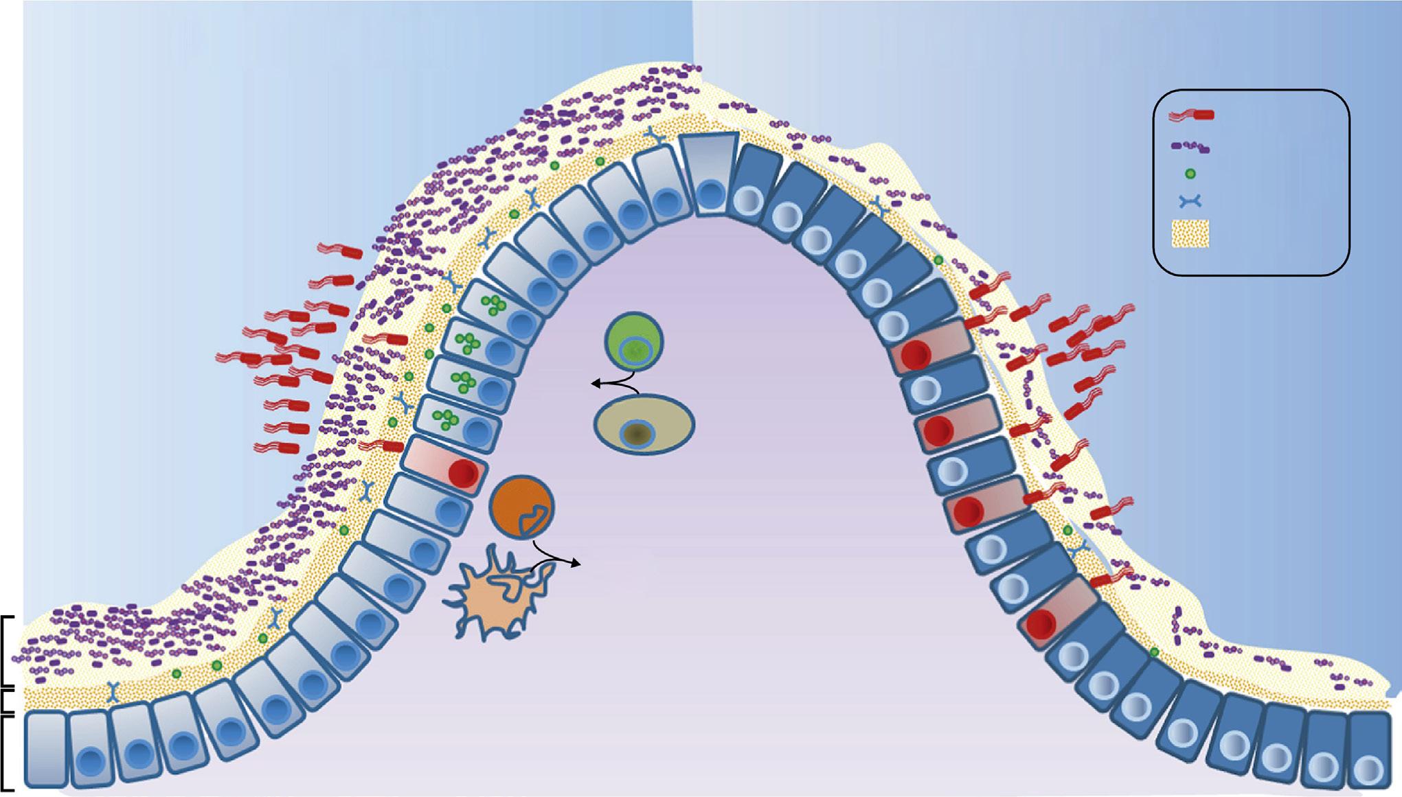
Fig. 2.2 Intestinal microbiota protection against enteric infections. (I) Saturation of colonization sites and consumption of nutrients limit pathogen access to host tissues; (II) the microbiota prime innate immunity by stimulating mucin production, immunoglobulin (Ig)A, and antimicrobial peptides (AMPs); and (III) the microbiota stimulate interleukin (IL)-22 expression, which increases epithelial resistance, and IL-1β production, which promotes recruitment of inflammatory cells. (From Khosravi, A., Mazmanian, S. 2013. Disruption of the gut microbiome as a risk factor for microbial infections. Curr. Opin. Microbiol. 16, 221–227.)
Evolution of the Microbiome and Normal Flora
The normal flora of a particular site of the body consists of a unique community of core and secondary microbiota that evolved through a symbiotic relationship with the host and a competitive relationship with other species. The host provides a place to colonize, nutrients, and some protection from unwanted species (innate immune responses). The microbes provide needed metabolic functions, stimulate innate and regulatory immunity, and prevent colonization with unwanted pathogens (Fig. 2.2). The ability to tolerate the amount of oxygen or lack thereof (redox state) and the pH and salt concentration, as well as to scavenge essential minerals and harvest and metabolize the available nutrients, determines the numbers and nature of the species that populate a site of the body. Anaerobic or facultative anaerobic bacteria colonize most of the sites of the body because of the lack of oxygen in sites such as the mouth, intestine, and genitourinary tract.
The composition of the microbiota is influenced by personal hygiene (e.g., use of soap, deodorants, mouthwash, skin peels, enemas, vaginal douches), diet, water source, medicines (especially antibiotics), and exposure to environmental toxins. Drinking well water versus chlorinated city water or a diet consisting of more or less fiber, sugar, or fats can select for different intestinal bacteria based on their ability to use the essential minerals (e.g., iron) and nutrients. Alteration of the environment with foods or medicines can also alter the microbiota (Fig. 2.3). These changes can
be acceptable if the core microbiome and critical functional properties of the microbiome are maintained but can result in disease if these functions are lost. Historically, the greatest concern with the use of broad-spectrum antibiotics was the selection of resistant bacteria; however, a greater concern should be the disruption of the microbiome and loss of essential functions.
Of the approximately 200 unique species of bacteria that colonize the gut, most are members of Actinobacteria (e.g., Bifidobacterium), Bacteroidetes (e.g., Bacteroides), and Firmicutes (e.g., Eubacterium, Ruminococcus, Faecalibacterium, Blautia). Interestingly, the importance of many of these bacteria was not appreciated before gene sequencing was used to identify and quantitate the gut microbiota. Within the colon, some bacteria wage interspecies warfare to establish their niche with bacteriocins (e.g., colicins produced by Escherichia coli), other antibacterial proteins, and metabolites that deter other species from growing. These molecules also benefit the host by eliminating invading bacteria including Salmonella, Shigella, Clostridium difficile, Bacillus cereus, and other pathogens. The bacteria must also resist antimicrobial peptides and immunoglobulin (Ig) A produced by the host and released into the bowel.
Metabolism of nutrients plays a major role in the symbiotic relationship between the human host and microbe. Bacteria in the human gut are responsible for metabolizing complex carbohydrates (including cellulose) to provide small-chain fatty acids such as acetate, propionate, and butyrate that can be readily transported and used by the cells of our body. These acids also limit the growth of
Bacteroidetes Alistipes
Bacteroidetes Bacteroides
Bacteroidetes Parabacteroides
Firmicutes Blautia
Firmicutes Clostridium XVIII
Firmicutes Enterococcus
Firmicutes Faecalibacterium
Firmicutes Lachnospiraceae incertae sedis
Firmicutes Lactobacillus
Firmicutes Oscillibacter
Firmicutes Streptococcus
Firmicutes Subdoligranulum
Firmicutes uc_Clostridiales
Firmicutes uc_Lachnospiraceae
Firmicutes uc_Ruminococcaceae
Proteobacteria Escherichia/Shigella
Proteobacteria Klebsiella
Proteobacteria uc_Enterobacteriaceae
Others
Actinobacteria Bifidobacterium
Bacteroidetes Alistipes
Bacteroidetes Bacteroides
Bacteroidetes Parabacteroides
Bacteroidetes uc_Bacteroidales
Bacteroidetes uc_Bacteroidetes
Firmicutes Blautia
Firmicutes Enterococcus
Firmicutes Faecalibacterium
Firmicutes Subdoligranulum
Firmicutes uc_Bacilli
Firmicutes uc_Clostridiales
Firmicutes uc_Firmicutes
Firmicutes uc_Lachnospiraceae
Firmicutes uc_Lactobacillales
Firmicutes uc_Ruminococcaceae
Proteobacteria Escherichia/Shigella
Proteobacteria Salmonella
Proteobacteria uc_Enterobacteriaceae
Proteobacteria uc_Gammaproteobacteria
Others
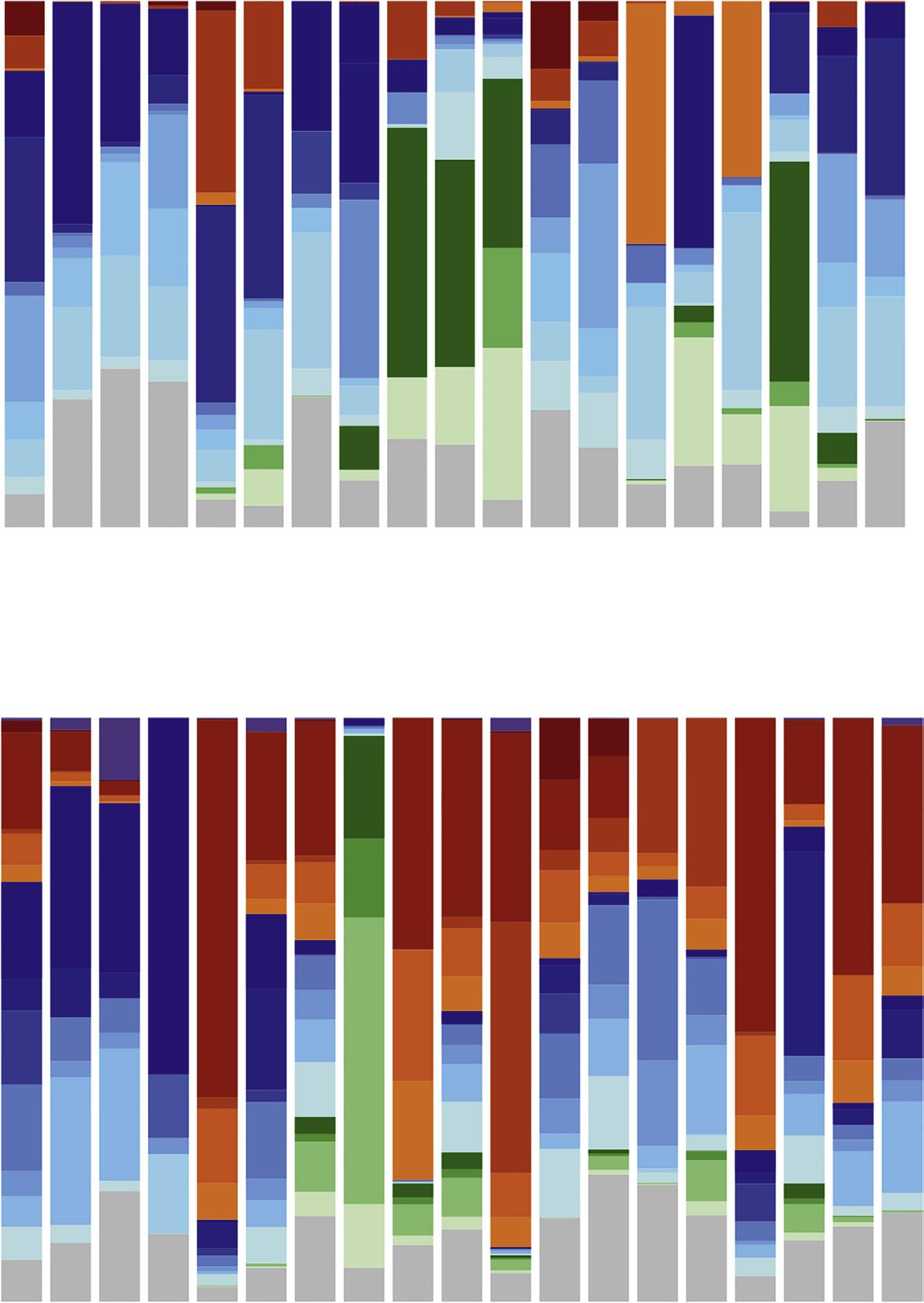
Fig. 2.3 Effect of antibiotics on the gut microbiota. Fecal samples were collected from four patients treated with antibiotics: patient A, moxifloxacin; patient B, penicillin + clindamycin; patient C, cefazolin followed by ampicillin/sulbactam; and patient D, amoxicillin. Fecal samples collected before, during (e.g., 3_D is day 3 of therapy), and after therapy were used to assess the total microbiota. Changes are noted both during therapy and after therapy is discontinued. A, Total microbiota (16S rRNA gene). B, Metabolically active microbiota (16S rRNA transcripts). (From Perez-Cobas, A. E., Artacho, A., Knecht, H., et al., 2013. Differential effects of antibiotic therapy on the structure and function of human gut microbiota. PLoS One 8, e80201.)
undesirable bacteria. Other bacteria graze on the carbohydrates, the mucins that line the epithelium, or the oils released in our sweat. Bacteroidetes and Firmicutes are more efficient than others at breaking down complex carbohydrates, including plant cell wall compounds (cellulose, pectin, and xylan) and host-derived carbohydrates, including those attached to the mucins or chondroitin sulfates of the protective mucous layer of the intestine. Increases in the ratio of these bacteria in the gut microbiome can lead to a higher efficiency in storage of the metabolic by-products. This can be a benefit for malnourished
populations or patients with debilitating diseases such as cancer, or can lead to obesity in well-nourished populations.
Role of the Microbiome in Disease
If the normal microbiome characterizes health, then alterations in the microbiome can signify disease; this is a relationship we are only beginning to understand.
In 1884 Robert Koch and Friedrich Loeffler defined the relationship between an organism and infection. The Koch postulates were based on the concept of one organism:one disease. Microbiome research has introduced a new concept of disease caused by a community of organisms rather than a single species of bacteria, and the influence extends beyond traditional “infectious” diseases to include immunologic and metabolic disorders such as inflammatory bowel disease, obesity, type 2 diabetes, and celiac disease. We are now at the forefront of a new era of redefining the concept of infectious diseases.
Disruption of the normal microflora (commonly referred to as dysbiosis) can lead to disease by the elimination of needed organisms or allowing the growth of inappropriate bacteria. For example, following exposure to antibiotics and suppression of the intestinal normal flora, C. difficile is able to proliferate and express enterotoxins, leading to inflammation of the colon (antibiotic-associated colitis). Another disease of the colon, ulcerative colitis, is associated with an increased level of bacteria producing mucin-degrading sulfatases, leading to degradation of the protective mucosal lining of the intestinal wall and stimulation of inflammatory immune responses. Individuals with an intestinal microbiota that is more efficient at breaking down complex carbohydrates internalize rather than void these nutrients; therefore they are susceptible to obesity and a predisposition to metabolic syndromes such as type 2 diabetes. Not all patients genetically predisposed to celiac disease, which is an immune-mediated enteropathy precipitated by exposure to gluten proteins, are symptomatic. The intestinal microbiota of most individuals is composed of bacteria capable of digesting glutens, which may be sufficient to protect these genetically predisposed individuals. In the absence of these bacteria, disease may occur. Shifts in the skin microbiome are associated with progression to chronic wound infections and episodic exacerbations of atopic dermatitis. Alteration in the vaginal microbiome from relatively few predominant organisms to a heterogeneous mixed population is associated with the progression to vaginitis.
Diagnostics and Therapeutics
An understanding of the influence of dysbiosis on disease pathology can lead to both advanced diagnostic tests and paths for therapeutic intervention. Just as the presence of Salmonella or Shigella signifies disease, changes in the diversity and composition of the fecal microflora can also indicate susceptibility to or onset of disease. The most obvious example is C. difficile disease, which is a clinical disease preceded by a depletion of the normal flora because of antibiotic use. Interestingly, patients with chronic relapsing C. difficile infections are treated successfully by repopulating (some say “repoopulating” ) the intestines with stool transplants from a healthy spouse or close relative, or with artificially created stool specimens consisting of a complex mixture of aerobic and anaerobic fecal organisms.
More subtle alterations in the gut microbiome may predict development of diseases such as necrotizing
enterocolitis (NEC), inflammatory bowel disease, and a predilection for obesity. NEC is a devastating intestinal disease that afflicts preterm infants. Prospectively collected stool samples from infants younger than 29 weeks’ gestational age who develop NEC demonstrate a distinct dysbiosis prior to the development of disease. Infants with early-onset disease have a dominance of Firmicutes (predominantly Staphylococcus), whereas infants with late-onset NEC have a dominance of Enterobacteriaceae.
The effects of microbiome alterations have also been described for the pathogenesis of inflammatory bowel disease and colorectal cancer. Proliferation of bacteria such as Akkermansia muciniphila that produce mucin-degrading sulfatases is responsible for degradation of the intestinal wall lining. Additionally, an increase in members of the anaerobic family Prevotellaceae leads to upregulation of chemokine-mediated inflammation. Enterotoxigenic Bacteroides fragilis can also induce T helper cell–mediated inflammatory responses that are associated with colitis and are a precursor to colonic hyperplasia and colorectal tumors. Finally, Methanobrevibacter smithii, a minor member of the gut microbiome, enhances digestion of dietary glycans by B. thetaiotaomicron and other core intestinal bacteria, leading to accumulation of fat.
Alterations of the microbiome leading to disease may not be characterized by the presence or absence of a specific microbe because more than one organism may provide the needed function. It is likely that future diagnostics will measure for the presence or absence of a specific gene product (proteomics) or metabolic function (metabolomics)
Probiotics
Probiotics are mixtures of bacteria or yeast that when ingested colonize and proliferate, even temporarily, the intestine. Consumers of probiotics believe they act by rebalancing the microbiome and its functions, such as enhancing digestion of food and modulating the individual’s innate and immune response. The most common reason people use over-the-counter probiotics is to promote and maintain regular bowel function and improve tolerance to lactose. Probiotics are commonly gram-positive bacteria (e.g., Bifidobacterium, Lactobacillus) and yeasts (e.g., Saccharomyces). Many of these microbes are found in ingestible capsules and as food supplements (e.g., yogurt, kefir). Probiotics have been used to treat C. difficile–associated diarrhea and inflammatory bowel disease, to provide protection from Salmonella and Helicobacter pylori disease, as therapy for pediatric atopic dermatitis and autoimmune diseases, and even for reduction in dental caries, although the value of probiotics for many of these conditions is unproven. Although probiotics are generally safe dietary supplements, many probiotics are ineffective. The species, mixture of species, and dose and viability of the probiotic organisms within a probiotic formulation influence its potency, efficacy, and therapeutic potential. What is clear is that much like the use of complex artificial mixtures of organisms to treat recurrent C. difficile disease, carefully designed “smart probiotics” will likely be an important adjunct to medical therapy in the future.
In the near future, with faster and cheaper DNA sequencing procedures, analysis of a person’s microbiome may become a routine diagnostic test for predicting and treating a wide range of diseases. However, a number of questions remain to be resolved: Can we predict disease in an individual by monitoring changes in the microbiome? Which changes are most important, taxonomic or genetic function? Can we prevent disease or treat disease by reestablishing a healthy microbiome? Can this be done by prescribing specific replacement microbes (e.g., fecal transplant) or with a universal mixture (probiotic)? Can the use of metabolic supplements (prebiotics) promote a healthy microbiota? Will use of antibiotics be replaced by use of “smart microbiome” therapies? Other questions include: What is the role of the host genome, environmental factors, and our hygienic practices in shaping the microbiome? What will be the informatic requirements for guiding diagnostics or therapeutics? Regardless of the answers to these and other questions, it is certain that we are witnessing the beginning of a new era of microbiology that can radically change our approach to prediction, diagnosis, and treatment of disease.
For questions see StudentConsult.com
Bibliography
Blum, H., 2017. The human microbiome. Adv. Med. Sci. 62, 414–420. Cho, I., Blaser, M.J., 2012. The human microbiome: at the interface of health and disease. Nat. Rev. Genet. 13, 260–270.
Damman, C.J., Miller, S.I., Surawicz, C.M., et al., 2012. The microbiome and inflammatory bowel disease: is there a therapeutic role for fecal microbiota transplantation? Am. J. Gastroenterol. 107, 1452–1459.
David, L.A., Maurice, C.F., Carmody, R.N., et al., 2014. Diet rapidly and reproducibly alters the human gut microbiome. Nature 505, 559–563. Faith, J.J., Guruge, J.L., Charbonneau, M., et al., 2013. The long-term stability of the human gut microbiota. Science 341, 1237439.
Gevers, D., Knight, R., Petrosino, J.F., et al., 2012. The Human Microbiome Project: a community resource for the healthy human microbiome. PLoS Biol. 10, e1001377.
Grice, E., Segre, J., 2011. The skin microbiome. Nat. Rev. Microbiol. 9, 244–253.
Human Microbiome Project Consortium, 2012. A framework for human microbiome research. Nature 486, 215–221.
Human Microbiome Project Consortium, 2012. Structure, function and diversity of the healthy human microbiome. Nature 486, 207–214.
Li, K., Bihan, M., Methé, B.A., 2013. Analyses of the stability and core taxonomic memberships of the human microbiome. PLoS ONE 8, e63139. McDermott, A.J., Huffnagle, G.B., 2014. The microbiome and regulation of mucosal immunity. Immunology 142, 24–31.
Morgan, X.C., Segata, N., Huttenhower, C., 2013. Biodiversity and functional genomics in the human microbiome. Trends Genet. 29, 51–58. Murray, P., 2013. The Human Microbiome Project: the beginning and future status. Ann. Clin. Microbiol. 16, 162–167.
Perez-Cobas, A.E., Artacho, A., Knecht, H., et al., 2013. Differential effects of antibiotic therapy on the structure and function of human gut microbiota. PLoS ONE 8, e80201.
Petrof, E.O., Claud, E.C., Gloor, G.B., et al., 2013. Microbial ecosystems therapeutics: a new paradigm in medicine? Benef. Microbes 4, 53–65.
Petschow, B., Dore, J., Hibberd, P., et al., 2013. Probiotics, prebiotics, and the host microbiome: the science of translation. Ann. N Y Acad. Sci. 1306, 1–17.
Shaffer, M., Armstrong, A., Phelan, V., et al., 2017. Microbiome and metabolome data integration provides insights into health and disease. Transl. Res. 189, 51–64.
Venter, J.C., Adams, M.D., Myers, E.W., et al., 2001. The sequence of the human genome. Science 291, 1304–1351.
Zmora, N., Zilberman-Schapira, G., Mor, U., et al., 2018. Personalize gut mucosal colonization resistant to empiric probiotics is associated with unique host and microbiome features. Cell 174, 1388–1405.
Questions
1. What is the relationship between the human genome and microbiome genetic material?
2. Explain the concepts of taxonomic diversity and genetic diversity.
3. Explain the concept of the core microbiome.
4. Give three examples of alterations of the microbiome (dysbiosis) that are associated with specific diseases.