No part of this publication may be reproduced or transmitted in any form or by any means, electronic or mechanical, including photocopying, recording, or any information storage and retrieval system, without permission in writing from the publisher. Details on how to seek permission, further information about the Publisher’s permissions policies and our arrangements with organizations such as the Copyright Clearance Center and the Copyright Licensing Agency, can be found at our website: www.elsevier.com/permissions
This book and the individual contributions contained in it are protected under copyright by the Publisher (other than as may be noted herein).
Notice
Practitioners and researchers must always rely on their own experience and knowledge in evaluating and using any information, methods, compounds or experiments described herein. Because of rapid advances in the medical sciences, in particular, independent verification of diagnoses and drug dosages should be made. To the fullest extent of the law, no responsibility is assumed by Elsevier, authors, editors or contributors for any injury and/or damage to persons or property as a matter of products liability, negligence or otherwise, or from any use or operation of any methods, products, instructions, or ideas contained in the material herein.
Previous editions copyrighted 2012.
Library of Congress Control Number: 2019943585
Content Strategist: Robin R. Carter
Content Development Specialist: Lisa Barnes
Publishing Services Manager: Deepthi Unni
Project Manager: Beula Christopher
Designer: Renee Duenow
Cover Art: Joe Chovan
Printed in China Last digit is the print number:
This monograph is dedicated to the physicians, surgeons, and researchers who have committed their lives to perfecting mechanical solutions to the failing heart; to the patients so desperately in need of such solutions; and to our families who provide unconditional love and support as we carry on this battle.
James K. Kirklin
Joseph G. Rogers
Larry A. Allen, MD, MHS Professor of Medicine Department of Medicine Division of Cardiology University of Colorado School of Medicine Aurora, Colorado; Colorado Cardiovascular Outcomes Research (CCOR) Consortium Denver, Colorado
Francisco A. Arabia, MD, MBA Director
CSHI Center for Surgical Device Management of Advanced Heart Failure Department of Cardiothoracic Surgery Cedars-Sinai Medical Center Los Angeles, California
Pavan Atluri, MD
Assistant Professor of Surgery Division of Cardiovascular Surgery Department of Surgery University of Pennsylvania Philadelphia, Pennsylvania
J. Timothy Baldwin, PhD Program Director and Deputy Chief Advanced Technologies and Surgery Branch Division of Cardiovascular Sciences National Heart, Lung, and Blood Institute Bethesda, Maryland
Christian A. Bermudez, MD
Associate Professor Division of Cardiovascular Surgery Department of Surgery Hospital of the University of Pennsylvania Perelman School of Medicine Philadelphia, Pennsylvania
Emma Birks, MD, PhD, FRCP, MBBS, BSc Medical Director
Advanced Heart Failure, Transplantation and Mechanical Circulatory Support Department of Medicine Division of Cardiovascular Medicine University of Louisville Louisville, Kentucky
Robin Bostic, BS Global Vice President Health Economics and Reimbursement Department of Health Economics Abbott Windham, New Hampshire
CONTRIBUTORS
Jennifer Cowger, MD, MS
Medical Director of the Mechanical Circulatory Support Program Co-Director of the Cardiac Critical Care Unit Department of Cardiology Henry Ford Hospital Detroit, Michigan
Mani A. Daneshmand, MD Associate Professor Director
Heart Transplant, Lung Transplant, MCS, and ECMO Department of Surgery Emory University Atlanta, Georgia
Walter Dembitsky, MD Director
Mechanical Circulatory Support Program Cardiothoracic and Vascular Surgery Sharp Memorial Hospital San Diego, California
Mary Amanda Dew, PhD Professor of Psychiatry, Psychology, Epidemiology, Biostatistics, Nursing, and Clinical and Translational Science Director
Quality of Life Research, Artificial Heart Program University of Pittsburgh School of Medicine and Medical Center Pittsburgh, Pennsylvania
Stavros G. Drakos, MD, PhD Heart Failure Program Division of Cardiology University of Utah Salt Lake City, Utah
Yakov L. Elgudin, MD, PhD Section Chief Cardiothoracic Surgery Cleveland VA Medical Center; Staff Surgeon Division of Cardiac Surgery University Hospitals Cleveland Medical Center Cleveland, Ohio
Daniel J. Goldstein, MD Professor and Vice Chair Department of Cardiothoracic Surgery Montefiore Medical Center Bronx, New York
Kathleen L. Grady, PhD, RN, MS, FAAN Professor of Surgery and Medicine Department of Surgery, Division of Cardiac Surgery Feinberg School of Medicine Northwestern University; Administrative Director Center for Heart Failure Bluhm Cardiovascular Institute of Northwestern Memorial Hospital Chicago, Illinois
Igor D. Gregoric, MD
Chief of Surgical Division
Memorial Hermann Heart and Vascular Institute; Director Center of Advanced Heart Failure
The University of Texas Health Science Center-Houston; Professor
Department of Cardiothoracic and Vascular Surgery
UT Health McGovern Medical School Houston, Texas
Finn Gustafsson, MD, PhD, DMSci Professor of Cardiology University of Copenhagen Rigshospitalet Copenhagen, Denmark
J. Thomas Heywood, MD Director
Advanced Heart Failure and Mechanical Support Department of Cardiology
Scripps Clinic
La Jolla, California
William L. Holman, MD Professor of Surgery Division of Cardiothoracic Surgery
Department of Surgery
University of Alabama at Birmingham Birmingham, Alabama
Tina Cady Ivovic, BS
Global Senior Director
Field Health Economics and Reimbursement Department of Health Economics Abbott Washington, DC
James K. Kirklin, MD Professor of Surgery Division of Cardiothoracic Surgery, Director
Kirklin Institute for Research in Surgical Outcomes (KIRSO) Department of Surgery University of Alabama at Birmingham Birmingham, Alabama
Robb D. Kociol, MD, MS, FHFSA Director of HF Network Integration
Beth Israel Deaconess Medical Center Boston, MA;
Medical Director, Heart Failure
Beth Israel Deaconess Hospital - Plymouth Plymouth, MA; Assistant Professor of Medicine
Harvard Medical School Boston, MA
Jeff Larose, MSME
Chief Scientific Officer
MCS
Medtronic HeartWare
Miami Lakes, Florida
James W. Long, MD, PhD Director
Nazih Zuhdi Transplant Institute
INTEGRIS Healthcare
Oklahoma City, Oklahoma
Donald A. Middlebrook, AA, BS Consultant
Regulatory and Clinical Affairs
MCS Group/HF Division/Abbott Pleasanton, California
Nader Moazami, MD
Professor of Cardiothoracic Surgery
Department of Cardiothoracic Surgery
New York University New York City, New York
David Luís Simón Morales, MD
Professor of Surgery and Pediatrics
Clark-Helmsworth Chair of Pediatric Cardiothoracic Surgery, Director
Congenital Heart Surgery—The Heart Institute
Cincinnati Children’s Hospital Medical Center
The University of Cincinnati College of Medicine Cincinnati, Ohio
Francis D. Pagani, MD, PhD
Otto Gago MD Endowed Professor of Cardiac Surgery
Cardiac Surgery
University of Michigan Ann Arbor, Michigan
Soon J. Park, MD
Professor of Surgery, Chief, Division of Cardiac Surgery
University Hospitals Cleveland Medical Center; Co-Director
UH Harrington Heart and Vascular Institute; Jay L. Ankeney, MD Chair, Professorship of Surgery Cleveland, Ohio
Sean Pinney, MD Director
Advanced Heart Failure and Transplantation Medicine, Cardiology
Mount Sinai Hospital; Professor of Medicine, Cardiology
Icahn School of Medicine at Mount Sinai New York City, New York
J. Eduardo Rame, MD, M.Phil, FAHA
Associate Professor of Medicine and Surgery
Health System Director of Mechanical Circulatory Support
University of Pennsylvania
Advanced Heart Failure/Cardiac Transplantation and Mechanical Circulatory Support Philadelphia, Pennsylvania
Kyle William Riggs, MD Research Fellow
Department of Cardiothoracic Surgery Cincinnati Children’s Hospital Medical Center Cincinnati, Ohio
David N. Rosenthal, MD
Professor Department of Pediatrics
Stanford University Palo Alto, California
Stuart D. Russell, MD
Regional Director of Heart Failure Professor of Medicine
Department of Medicine/Division of Cardiology
Duke University School of Medicine Durham, North Carolina
Erin M. Schumer, MD, MPH University of Louisville Louisville, Kentucky
Craig H. Selzman, MD, FACS
Dr. Russell M. Nelson and Dantzel W. Nelson Presidential Endowed Chair, Professor and Chief
Division of Cardiothoracic Surgery, Surgical Director
Mechanical Circulatory Support and Heart Transplantation Division of Cardiothoracic Surgery University of Utah School of Medicine Salt Lake City, Utah
Mark S. Slaughter, MD Professor and Chair
Department of Cardiovascular and Thoracic Surgery University of Louisville School of Medicine Louisville, Kentucky
Randall C. Starling, MD, MPH Professor of Medicine
Cleveland Clinic Lerner College of Medicine, Case Western Reserve University
Kaufman Center for Heart Failure Cleveland Clinic Cleveland, Ohio
Lynne Warner Stevenson, MD
Lisa M. Jacobson Professor of Cardiovascular Medicine
Director of Cardiomyopathy
Vanderbilt University Medical Center Nashville, Tennessee
Garrick C. Stewart, MD, MPH
Associate Physician
Brigham and Women’s Hospital; Instructor
Harvard Medical School; Associate Physician
Division of Cardiovascular Medicine
Centre for Advanced Heart Disease
Brigham and Women’s Hospital Boston, Massachusetts
Jeffrey Teuteberg, MD
Section Chief of Heart Failure, Cardiac Transplant and Mechanical Circulatory Support
Stanford University School of Medicine
Falk Cardiovascular Research Center Stanford, California
Daniel Timms, PhD CTO Engineering BiVACOR Houston, Texas
Nir Uriel, MD, MSc
Director of Heart Failure, Heart Transplant and Mechanical Circulatory Support Department of Medicine University of Chicago Chicago, Illinois
Richard Wampler, MD
Research Associate Professor of Surgery Oregon Health Sciences University Portland, Oregon
John T. Watson, PhD Professor of Bioengineering
Jacobs School of Engineering University of California, San Diego San Diego, California
James B. Young, MD
Professor of Medicine and Executive Dean
Lerner College of Medicine of Case Western Reserve University
Cleveland Clinic Cleveland, Ohio
Those who dreamed of a mechanical pump to permanently replace the functioning heart thought not about subsequent transplantation; no, they envisioned a lifelong mechanical heart pump. But the two are inextricably intertwined. When Alexis Carrel was awarded the Nobel Prize in Physiology or Medicine in 1912 “ in recognition of his work on vascular suture and transplantation of blood vessels and organs” at the age of 39, his scientific journey was far from complete. In the 1930’s, Carrel and Lindbergh developed a “perfusion pump” resembling an in vitro artificial heart, designed to support organs removed from small animals for several days. Their work has been credited as foundational for the later artificial heart. In humans, the origins of mechanical circulatory support (MCS) rest with Gibbon’s development and single successful clinical use of a pump oxygenator to perform open heart surgery in 1953. John Kirklin at the Mayo Clinic followed in 1955 with the world’s first successful series of open heart operations using a pump oxygenator, in which four of the first eight patients survived.
As beautifully detailed by Baldwin and Watson in Chapter 1 of this book (Historical Aspects of Mechanical Circulatory Support), an era of excitement about a possible total artificial heart in the 1960s not only consumed a number of leading engineers and physicians (Kolff, Akutsu, DeBakey, Liotta, and Kantrowitz), but also inspired the US Congress to empower the National Heart Advisory Council to target mechanical solutions to heart failure. With an increasing concentration of energy and intellect brought to bear on this challenge, shorter term ventricular assist systems began to outpace the total artificial heart experimental successes. The timeline and challenges of developing these devices between 1963 and 1991 were chronicled in the Institute of Medicine Committee report to “Evaluate the National Heart, Lung, and Blood Institute (NHLBI) Artificial Heart Program” (in which Editor JKK participated as a young surgeon/investigator), which appeared in 1992.
With the formal introduction of cyclosporine immunosuppression into clinical heart transplantation in 1983, a reanimation of the entire field erupted after lying essentially dormant for 25 years. Heart transplantation programs rapidly appeared at many major academic cardiac surgery centers, accompanied by an explosion of transplantation activity and the inevitable donor shortages. As the waiting lists progressively exceeded available donors, patients were dying in large numbers. Researchers and surgeons began to consider a novel use of mechanical devices as “bridge-to-transplant” (BTT) therapy. With reports from Hill and Oyer using the Pierce-Donachy ventricular assist device (VAD) and the Novacor VAD, and Copeland implanting the CardioWest total artificial heart, all as BTT therapy, the field of MCS was awakened in the early 1980’s.
But engineers and MCS proponents were frustrated, not satisfied with a limited application of sophisticated MCS systems that simply supported a dying patient through successful heart transplantation and then were discarded. Everything began to change with the REMATCH Trial, initiated in 1998 and completed in 2001. With FDA approval then Centers for Medicare and Medicaid Services (CMS) coverage in 2002-2003, a new world of interest blossomed in MCS therapy. For the first time, selected patients with terminal heart failure not eligible for heart transplantation could be offered life-saving VAD “destination therapy” (DT). While initial DT experiences were evolving with the bulky, flawed, pulsatile XVE, a new generation of continuous flow (CF) devices was under development and entering clinical trials.
With the prospect of thousands of patients being supported long term on these expensive devices, John Watson and his visionary colleagues at the NHLBI sought to pursue the recommendation from
the 1992 Institute of Medicine Artificial Heart Program report that “… maintaining a registry of MCS recipients should be considered a routine aspect of this care…” Their initiative generated the birth of INTERMACS in 2006, an NHLBI-funded scientific registry to capture the national experience, facilitate the introduction of new devices, identify risk factors to predict good and bad outcomes with various pumps, quantify ongoing improvements in the field, and inform best practices.
As the field rapidly evolved, whole new areas of specialization and expertise emerged. With the clear superiority of current generation CF devices over prior technology, optimism reigned about ambulatory advanced heart failure patients gaining years and quality of life with these compact, durable devices. Later, in the aftermath of the failed REVIVE-IT trial (in which equipoise for a randomized trial of a CF pump in ambulatory heart failure was violated by a high incidence of pump thrombosis, and no surgical patients were entered), understanding the etiology and possible prevention of adverse events during pump support took center stage as a focus of research.
The first edition of the Braunwald Companion Text on Mechanical Circulatory Support was published 7 years ago, and much has evolved since then. In this second edition, existing chapters have been completely updated, some that lack current relevance have been removed, and new chapters of interest have been added. The pathophysiology and risk stratification in advanced heart failure have been updated in Chapters 2 and 3. Candidate selection and decision making has matured over the past 7 years, and this is reflected in Chapter 4. Entirely new chapters on acute (temporary) support and ECMO have been added to address the renewed interest in “bridging” patients in shock to durable devices (Chapters 5 and 6). Given the critical importance of the principles of CF physiology, device hemocompatibility, and the biological response to CF unloading, entirely new chapters (Chapters 7, 8, and 9) have been created. A review of current devices, surgical techniques, and longitudinal patient management has been thoroughly updated in Chapters 10 to 12. Sections on major adverse events and right heart failure have incorporated the most recent information and challenges (Chapters 13 and 14). All relevant clinical trials through 2018 are reviewed in Chapter 15, and health-related quality of life studies are updated in Chapter 16. Entirely new chapters (Chapters 17 and 18) detail the application of CF pumps in pediatric and special populations. The physiology of myocardial recovery during CF pump support and strategies for explant are described in depth in Chapter 19. The newest updates on unique information from registries and the current regulatory landscape provide the content for Chapters 20 and 21. Finally, we paint a prescient picture of future mechanical support in chapter 22.
This comprehensive text is truly a testimony to the phenomenal progress that has accrued with mechanical solutions to the failing heart. Although current devices do not yet rival the expected longevity and quality of life with heart transplantation, the comparative supply is massive and the target population of benefit is huge. After more than 50 years of nearly continuous disruptive innovation, combatants in the field of MCS (with their many associated intellectual peptides) are poised for further scientific and clinical breakthroughs. Yet major challenges remain, and this monograph sets the table for all interested stakeholders, scientists, clinicians, and regulators who embrace the future.
James K. Kirklin
Joseph G. Rogers
FAMILY OF BOOKS
1
Historical Aspects of Mechanical Circulatory Support
J. Timothy Baldwin, John T. Watson
KEY POINTS
Early Mechanical Circulatory Support Devices and Technology Development
EARLY MECHANICAL CIRCULATORY SUPPORT DEVICES AND TECHNOLOGY DEVELOPMENT
Establishing the Concept
In the 1930s, Carrel and Lindbergh1 developed an in vitro artificial heart-like apparatus for keeping organs alive outside the body. They removed the hearts, kidneys, ovaries, adrenal glands, thyroid glands, and spleens of small animals to watch them develop and function over the course of several days.2 Acute animal studies in Russia and the United States followed in the 1940s. However, the meaningful origin of the modern era of mechanical circulation support (MCS) can be traced to the development of the heart-lung machine by Gibbon (Table 1.1) and its first successful clinical use in 1953.3,4 The device was developed for cardiopulmonary bypass so that surgical cardiac procedures that require hours of circulatory support could be performed. The success of the device and the need for prolonged circulatory support for patients who could not be weaned from the heart-lung machine or whose hearts could recover with longer durations of support provided the initial impetus for developing devices that could provide long-term circulatory support. The optimism in the 1950s and 1960s that circulation could be successfully supported for extended periods by an artificial heart spurred its development by pioneers such as Kolff, Akutsu, DeBakey, Liotta, and Kantrowitz.5 In 1963, DeBakey and Lederberg testified before the US Congress on the need for an artificial heart in very different domains: for patients otherwise healthy except for their failed heart and for isolated travelers on long space journeys.6 These hearings coincided with the debate about the implications of the Russian Sputnik Program and unbridled national enthusiasm for taking on large technologic challenges such as the program to put the first man on the moon, which had begun just a few years earlier.
In 1964, with special congressional approval, the National Heart Advisory Council established the mission-oriented Artificial Heart Program (AHP) to design and develop devices to assist a failing heart and to rehabilitate heart failure (HF) patients.7 In the initial planning stages of the program, cardiology and surgery experts recommended that clinical systems be capable of a cardiac output of 10 L/min, be able to maintain normal blood pressure, and be
Ongoing Technology Developments and Devices
Current State of MCS
“biocompatible” (a vague physiological term then and now). These and other physiological parameters represented the defined design goals of the first generation of MCS systems.2 Despite this limited set of design inputs, engineers, biologists, and clinicians created teams and collaborations and used them, when appropriate, as quantifiable engineering design inputs to achieve the physiological goals in the early MCS systems.
Important progress on these early MCS systems resulted from the cooperation and collaboration fostered by the National Heart, Lung, and Blood Institute (NHLBI). In 1977, following a recommendation from the Cardiology Advisory Committee, the NHLBI Devices and Technology Branch (DTB) started the annual Contractors Meeting.4,8 The primary purpose of the meeting was to provide a public forum for showcasing the progress of the contract research projects. The DTB viewed the meeting as an opportunity for gathering the branch grantees and contractors together to share ideas and network with other teams. After a decade of successful annual meetings sponsored by the NHLBI, the meeting was moved to Louisville, Kentucky, as the “Cardiovascular Science and Technology: Basic and Applied” meeting under the leadership of Jack Norman,5 then to Washington, DC, with the guidance of Hank Edmunds,5 and was finally integrated into the Annual Meeting of the American Society of Artificial Internal Organs by then President Bob Eberhart (1993).7 This progression has preserved the spirit of collaboration of the original Contractors Meeting, which also includes the highly regarded Hastings Lecture dedicated to the memory of Dr. Frank Hastings, the first chief of the NHLBI Artificial Heart Program.
The annual meetings emphasized the importance of developing collaborative venues for the field to share results, both positive and negative, and develop a common language across disciplines with procedural guidelines, which improve the comparability of data between research teams.
During the early period of the program, spanning from 1963 to 1980, progress on implantable, long-term ventricular assist systems outpaced similar work on the more widely publicized total artificial heart (TAH) systems.9 In fact, short-term ventricular assist systems were being fabricated for use in initial clinical trials in the late 1970s.10 The Institute of Medicine Committee report to “Evaluate the NHLBI
TABLE 1.1 Mechanical Circulatory Support Milestones
Year Event
1953
1958
1963
1964
1968
1969
1977
1980
1982
1984
1985
1988
First successful use of heart-lung machine for cardiopulmonary bypass (Gibbon)
First successful use TAH in a dog (Kolff and Akutso)
First successful use of LVAD in human (DeBakey)
Artificial Heart Program established at NIH
Six contracts awarded to analyze issues and need for program
First clinical use of intraaortic balloon pump (Kantrowitz)
First artificial heart implant in humans (Cooley)
NHLBI RFPs for blood pumps, energy converters, and energy transmission
NHLBI RFA on blood-material interactions
NHLBI RFP for integration of blood pumps designed for 2-year use
Barney Clark received first TAH implant for destination therapy (DeVries)
NHLBI RFP for 2-year reliability studies
First use of Pierce-Donachy VAD (Thoratec PVAD) as BTT (Hill)
First implant of Novacor VAD
First use of electromechanical VAD (Oyer)
First use of CardioWest TAH as BTT (Copeland)
First use of hemopump in humans (Rich Wampler)—first rotary blood pump used (Frazier)
NHLBI awards four contracts to develop portable, durable TAHs
1989 Manual of operations for Novacor VAD NHLBI clinical trial completed
1991
1994
1996
1998
1999
2000
2001
2002
2003
2004
2006
2007
2008
2009
2010
2012
2014
2017
First HeartMate VE implant (Frazier)
FDA approval for pneumatic HeartMate VE as BTT
NHLBI IVAS contracts awarded for Jarvik 2000, HeartMate II, CorAide VADS
Pilot trial (PREMATCH) for destination therapy begins
NHLBI awards two contracts for TAH Clinical Readiness Program (Abiomed, Penn State)
FDA approval for HeartMate XVE as BTT
FDA approval for Novacor as BTT
REMATCH trial begins
First DeBakey VAD implant (Wieselthaler)
First human implant Arrow LionHeart VAD (first use of TETS) (Korfer)
First HeartMate II implant (Lavee)
First Jarvik 2000 implant (Frazier)
REMATCH trial completed
First implant of the AbioCor TAH (Dowling)
FDA approval of HeartMate XVE as destination therapy
CMS coverage decision for destination therapy
NHLBI pediatric mechanical circulatory support program launched
First implant of DuraHeart VAD (Korfer)
First implant of HeartWare HVAD (Wieselthaler)
First implant of Levacor VAD (Long)
FDA approval of AbioCor TAH (Humanitarian Device Exemption)
INTERMACS registry launched (PI: Kirklin )
First implant of Circulite Synergy device (Meyns); advent of miniature VADs
Peter Houghton dies after a record 2714 days of VAD support
HeartMate II BTT clinical trial completed
FDA approval of HeartMate II for BTT
HeartMate II destination therapy clinical trial completed
850th implant of the CardioWest TAH
FDA approval of HeartMate II for destination therapy
FDA approval of HVAD centrifugal flow pump for bridge-to-transplant therapy
First implant of HM3 (Schmitto)
FDA approval of HVAD for destination therapy
FDA approval of HM3 centrifugal flow pump for bridge-to-transplant and bridge-to-recovery therapy
BTT, Bridge-to-transplant; CMS, Centers for Medicare and Medicaid Services; FDA, Food and Drug Administration; HM3, HeartMate; INTERMACS, Interagency Registry of Mechanically Assisted Circulatory Support for End-Stage Heart Failure; IVAS, Innovative Ventricular Assist System; LVAD, left ventricular assist device; NHLBI, National Heart, Lung, and Blood Institute; NIH, National Institutes of Health; PI, principle investigator; PVAD, paracorporeal ventricular assist device; REMATCH, Randomized Evaluation of Mechanical Assistance for the Treatment of Congestive Heart Failure; RFA, request for application; RFP, request for proposal; TAH, total artificial heart; TETS, transcutaneous energy transfer system; VAD, ventricular assist device; VE, vented electric.
Artificial Heart Program” contains a useful chronology of research and related important events from 1963 to 1991.11,12
The first generation (1980) of implantable MCS systems was designed to meet a 2-year operational goal during benchtop reliability testing.13 Next followed the NHLBI Readiness Program.14 This program aimed to ensure the functional reliability of the MCS systems that demonstrated the greatest promise. Each awarded contractor placed 12 MCS systems on “Mock Circulations” to assess their function during a simulated cycle of daily life for 2 years without interruption or maintenance. Success was completing the test with no more than one system failure at 2 years.
Clinical Application and Evolution of MCS
These early MSC programs created the engineering design basis for the HeartMate XVE (HM XVE) (Fig. 1.1) that was used in the Randomized Evaluation of Mechanical Assistance for the Treatment of Congestive Heart Failure (REMATCH) trial.15 In clinical use, the HM XVE and other systems demonstrated that first-generation implantable systems could achieve meaningful physiological objectives for 2 years and improve quality of life. However, many patients suffered serious adverse events such as bleeding, infection, and device malfunction.
Recognizing the initial success of MCS devices by multidisciplinary teams created by the NHLBI programs, in 1994, the NHLBI released the “Innovative Ventricular Assist System” (IVAS) request for proposals to encourage innovation of totally implantable MCS systems that were designed to achieve at least a 5-year functional lifetime with 90% reliability.16 This program was designed to incorporate the latest advances gained from first-generation MCS systems, the TAH, and in related engineering, clinical, and biology fields.
The IVAS program was crucial to advancing the field of MCS for longer survival and patient quality of life. The program again brought together skilled teams of clinicians, engineers, and
biologists working together and collaborating with other teams toward the same defined physiological goals. As a result, the systems with the most promise exceeded the expectations of the program. These included the HeartMate II (HMII) (see Fig. 1.1 ) and the Jarvik 2000 VAS. 17,18 The HMII, an axial-continuous flow system, became the most clinically successful MCS system. In bench tests, HMII systems met physiological requirements, and some operated indefinitely.
The pivotal clinical trial with the HMII showed significantly improved survival and reduced adverse events for study patients. The unexpected result was underscored in the companion editorial to the release of the multicenter randomized trial. The author compared the REMATCH trial with the HM XVE to the HMII trial using KaplanMeier survival graphs.19 The survival results with HM XVE were exactly the same in 2009 as in 2001, very strongly suggesting that the improved HMII survival was largely due to the engineering design of the systems. This again pointed to the value of the National Institutes of Health/NHLBI initiating the IVAS program.
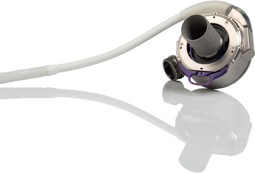
After the HMII trial, thrombus-related device malfunctions increased without explanation.20 There was speculation that the dimensionally tight axial-flow channel of the HMII was a contributing factor in thrombus formation. At the same time, magnetically levitated centrifugal-flow systems were on the drawing board. These systems became technically feasible because of advances in permanent magnets. The potential advantage of the centrifugal-flow pump is the dimensionally wider blood flow channels that may reduce the potential for thrombus formation. The importance of design is also seen in the widened internal flow patterns of the HeartMate 3 (HM3), which are likely responsible for the improved rate of “survival free of disabling stroke or reoperation to replace or remove a malfunctioning device” at 2 years compared to the HMII, despite the rates of disabling stroke being similar.21 The HVAD (Fig. 1.2), also a centrifugal blood pump,
Fig. 1.1 The HeartMate II (A and lower left in B) compared with the Heartmate XVE (B). The HeartMate II was the first rotary ventricular assist device to receive U.S. Food and Drug Administration approval for bridge to transplant and destination therapy. (Courtesy of Abbott.)
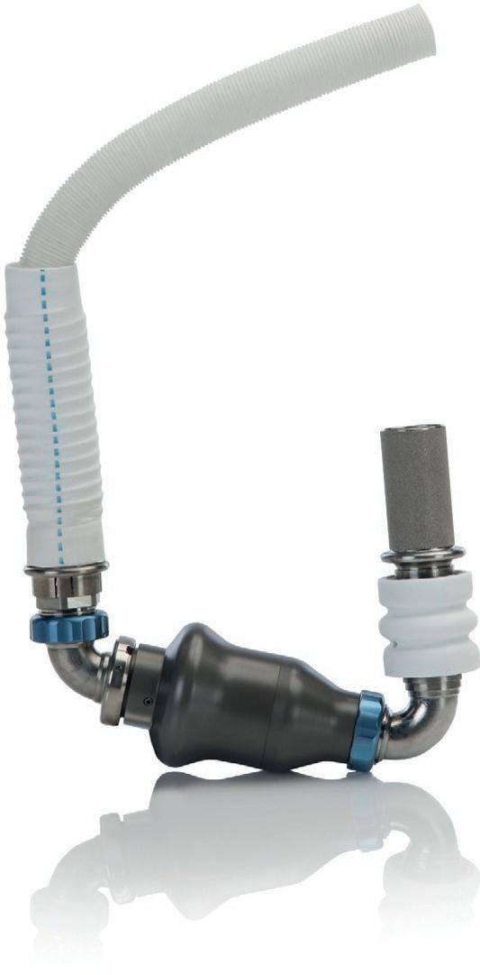
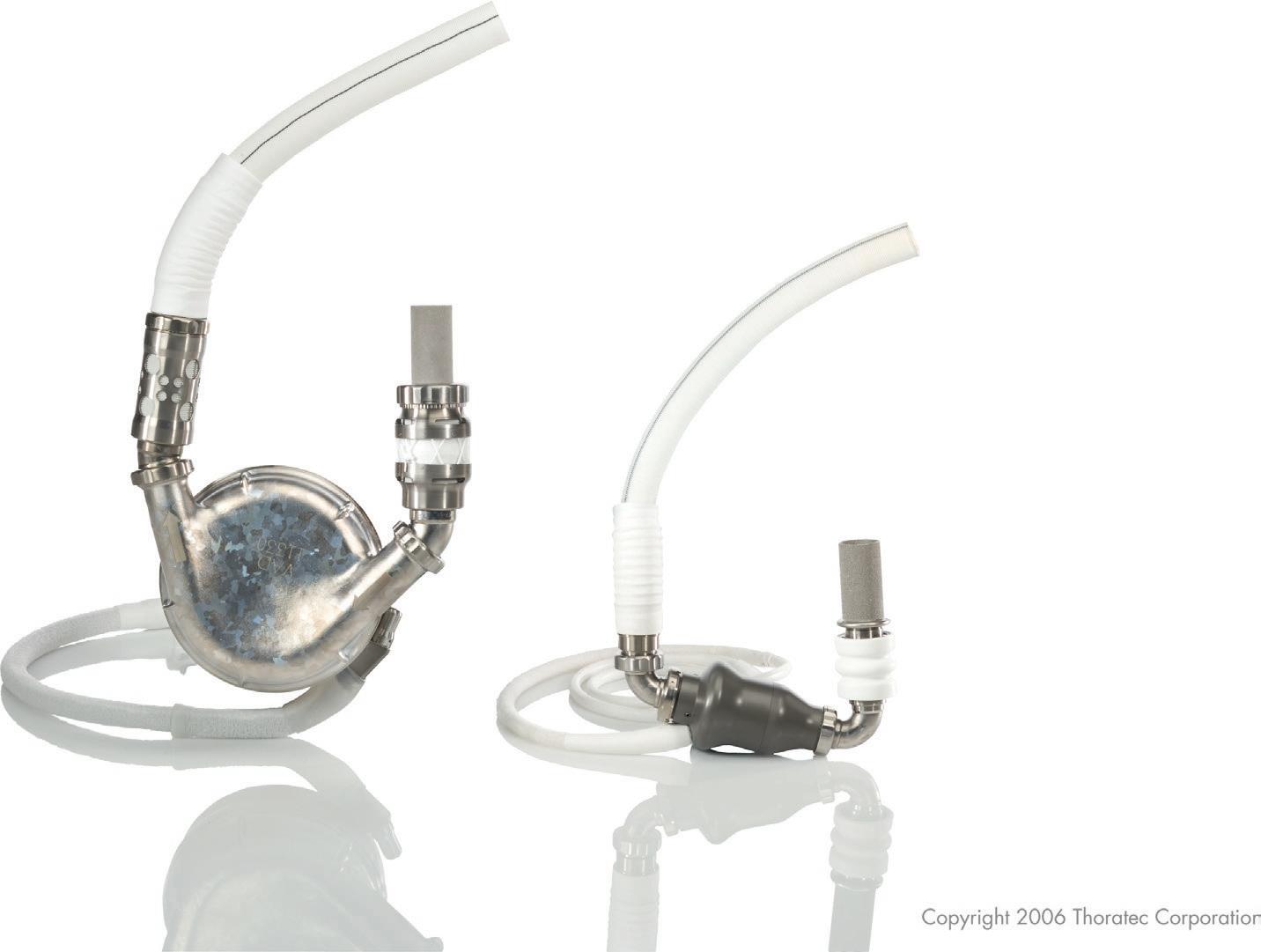
Fig. 1.2 The HeartWare HVAD. (Reproduced with permission of Medtronic, Inc.)
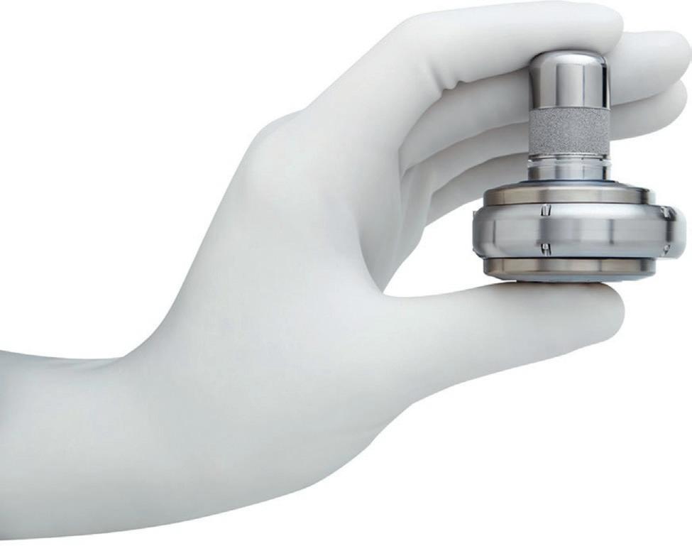
Fig. 1.3 The HeartMate 3, like the HeartWare left ventricular assist device, is a centrifugal pump but, rather than using bearings, has a fully magnetically levitated (Full MagLev) rotor. (Courtesy of Abbot Laboratories, Lake Bluff, IL.)
has different internal flow patterns from the HM3 (Fig. 1.3). In 2017, the Heartware system received Food and Drug Administration (FDA) approval for “Destination” therapy, following prior approval in 2012 as a bridge for cardiac transplantation.22
With the growth of MCS in the 1990s and early 2000s and its profound impact on patient outcomes, it was recommended that the NHLBI create a mechanism to assess various MCS technologies as they entered clinical use.11 To fulfill this vision, the NHLBI elected to develop a registry to collect data to improve patient MCS selection, measure quality of life, meet the FDA regulatory requirements, and inform the Centers for Medicare and Medicaid Services (CMS) regarding reimbursement decisions. This led to a solicitation that resulted in the Interagency Registry of Mechanically Assisted Circulatory Support for End-Stage Heart Failure (INTERMACS).23
The NHLBI provided the necessary financial support for the contract to develop and run the registry, which was awarded to the University of Alabama at Birmingham. This substantially reduced
triagency administrative factors and allowed the three agencies to join together on the steering committee and address questions relevant to their “agency mission” data collection. INTERMACS exceeded expectations for organizing, collecting, and analyzing clinical data and provided Medical Device Reports for adverse events to the FDA and data for CMS payment decisions.
INTERMACS made an early decision to only curate data generated by patients implanted with FDA-approved durable MCS devices (i.e., devices with the potential for patient discharge).24 This requirement added additional rigor to the INTERMACS database as the implants were under design controls and thus not subject to random design modifications that may directly influence clinical outcomes. Fourteen device systems met this standard. In recent years, of the 14 systems, 3 adult ventricular assist systems, 1 pediatric device, and 1 TAH system became the primary MCS devices in clinical use, thus providing essentially all the Bridge and Destination therapy data for INTERMACS.
ONGOING TECHNOLOGY DEVELOPMENTS AND DEVICES
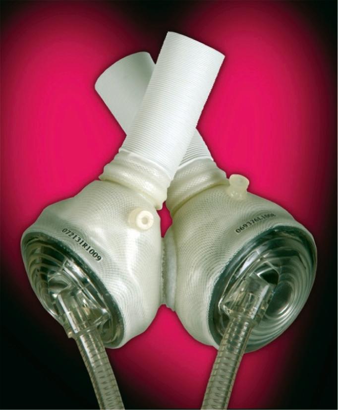
The development of new MCS devices has been spurred by innovation, as well as building on the success of earlier concepts. Substantial activity has focused on the development of novel TAHs. To date, the SynCardia TAH (Fig. 1.4), based on the Jarvik-7 TAH developed in the early 1980s, is the only one that has received substantial use, accounting for over 95% of the more than 1700 worldwide TAH implants since the first TAH in 1969.25 Recent efforts to develop a newer generation of TAH include the CARMAT bioprosthetic TAH,26 the Cleveland Clinic continuous-flow total artificial heart (CFTAH),27 and the BiVACOR TAH.28 The CARMAT TAH, like the SynCardia TAH, is a positive displacement device. However, it utilizes bioprosthetic blood-contacting surfaces, electro-hydraulic pumps to activate the membrane between the two ventricles to product pulsatile flow, and an advanced control system involving implanted sensors to provide flows to meet patient demands. Four patients were implanted with the device in a pilot study, which is anticipated to lead to a pivotal study. The Cleveland Clinic TAH and BiVACOR TAH both involve a single moving part, a rotor with impeller blades on each side, with each side driving the flow in the left and right centrifugal continuous-flow pumps that make up the device. These are considerably smaller than positive displacement TAHs, so small that a pediatric version of the Cleveland Clinical CFTAH is being developed for infants down to 0.3 m2 body surface area (BSA). The continuous-flow TAHs are both at the stage of animal studies.
Novel, advanced ventricular assist devices (VADs) are being designed and developed to address the outstanding issues with adverse events and special populations. These include a minimally invasive intraaortic balloon pump for long-term support (NuPulseCV, Raleigh, NC) and a valveless VAD that uses magnetically driven pistons to create pulsatile flow, known as the TorVAD (Windmill Technologies, Austin, TX).29,30 They also include newer generations of continuous-flow VADs such as a miniature implantable pump platform, the Revolution, in which minor modifications of components can be implemented to adjust the pump performance to support the right or left side of the heart (Vadovations, Inc., Oklahoma City, OK).31,32
Some of the greatest attention has focused on the development of MSC devices for children, specifically small ones. This was spurred on by the NHLBI Pediatric Circulatory Support Program spanning from 2004 to 2009 and the Pumps for Kids, Infants, and Neonates
A
B
Program, which started in 2010.31,32 These programs led to the development of various devices for children less than 20 kg with advanced HF because, when the programs began, the only device available for these patients was the Berlin Heart EXCOR, but only through emergency or compassionate use. Since then, the Berlin Heart received FDA approval and currently is the only FDA-approved MCS device for these small children. However, 29% of patients supported by the device experienced strokes in the pivotal trial.33 Developers hope to lower rates of serious adverse events by incorporating the latest technologies realized in VADs for adults into the new generation of pediatric VADs.
The Jarvik 2015 Ventricular Assist System, which resulted from the NHLBI programs, is poised to begin clinical evaluation.33 Progress continues on other devices that were developed independently of the NHLBI programs or resulted from them. These include a pediatric rotary flow VAD (Vadovations, Inc., Oklahoma City, OK), the Pediatric TorVAD (Windmill Technologies, Inc., Austin, TX), and the Penn State Pediatric Total Artificial Heart (Penn State University, Hershey and University Park, PA).34–36
The development of novel MCS devices like these has been led by a community of multidisciplinary teams. And, like the MCS devices they work on, the MCS community has evolved. Since the days of the DTB Contractors Meetings, the MCS community has continued to grow, collaborate, communicate, and now use other various meetings and organizations to do so such as the Gordon Research Conference on Assisted Circulation, 37 the ITERMACS Registry, 38 the International Society of Heart and Lung Transplantation forum for MCS 39 and the Society of Thoracic Surgeons STS National Database. 40
CURRENT STATE OF MCS
Over the past five decades, MCS has grown substantially from a small community of researchers working to develop early successful devices to meet modest goals and demonstrate the value of the therapy to one that includes significant numbers of research teams and mature industry leaders in medical devices and has helped
thousands of patients with late-stage HF. MCS served as a catalyst for a grassroots coalescing of key disciplines, organizations, and talents dedicated to patient quality of life and the well-being of caretakers. It has attracted clinicians, engineers, and scientists, both senior and recent graduates, who voluntarily committed their careers to MCS research, practice, and patients. Additionally, program coordinators work well beyond their position descriptions to provide 24/7 availability in the management and wellbeing of MCS patients and caregivers. As a benefit, patients have exceeded expectations in the scope of successful daily and recreational activities.
It is difficult to estimate the worldwide utilization of MCS. Based on increasing accrual rate in the INTERMACS Registry, the annual utilization in the United States may be around 3500 implants. Assuming that MCS use outside of the United States is similar,41–43 annual worldwide implants may total 6000–7000 units. Of note is that TAH use has grown worldwide, now totaling well over a thousand patients since its first use.43 With the development of MCS therapy, many patients are living well beyond 5 years, with the longest known patients alive after 15 years. These patients are informing the MCS research community and fellow patients by sharing best practices with their MCS systems through online organizations such as MyLVAD.44
An added research benefit of MCS is that while it extends patient survival, it is, in essence, extending the natural history of their physiological condition. With a growing number of patients living 5 years and longer with a device, there is a growing reservoir of opportunities to gain better understanding of the condition of HF itself. For example, the studies of myocardial recovery with device explant may reveal therapeutic strategies that deserve clinical research and trials involving patients without MCS.45 To this end, the NHLBI held a working group on “Advancing the Science of Myocardial Recovery with Mechanical Circulatory Support” in June 2016.46 Recommendations from this meeting were made to provide some clear directions to advance the science of cardiac recovery in the setting of mechanical circulatory support, and the hope is that research to address important outstanding questions about myocardial recovery will soon follow.
Fig. 1.4 The SynCardia Temporary Total Artificial Heart (A), shown with the wearable driver system (B). (Courtesy of Syncardia Systems, Inc., Tucson, AZ.)
The interdisciplinary teams of clinicians, engineers, and scientists that began in the early days of the Artificial Heart Program and formed the underpinning of the dynamic MCS field have endured and evolved over the decades. These teams continue working on MCS therapy because patients with MCS devices are still beset by serious adverse events associated with the devices. Progress to address them has been modest because, in part, terms for bleeding, stroke, infection, arrhythmias, and right HF have been incompletely quantified. To take MCS devices to the next level and overcome these issues, comprehensive biology, physics, and engineering parameters that define the device design inputs and goals are needed.
DISCLOSURE
Dr. Baldwin is an employee of the NHLBI, NIH. The comments expressed here are those of the authors and do not reflect official positions of the NHLBI or NIH.
FUNDING
No funding sources were used for this review.
REFERENCES
1. Carrel A, Lindbergh CA. The culture of whole organs. Science 1935;81(2112):621–623.
9. DeBakey ME. Development of mechanical heart devices. Ann Thorac Surg. 2005;79(6):S2228–S2231.
10. Norman JC, Dasco CC, Reul GJ, et al. Partial artificial heart (ALVAD) use with subsequent cardiac and renal allografting in a patient with stone heart syndrome. Artif Organs. 1978;2(4):413–420.
11. IOM Committee Report. The Artificial Heart: Prototypes, Policies and Patients; 1991.
12. Hogness JR. Committee to Evaluate the Artificial Heart Program of the National Heart, Lung, and Blood Institute, Division of Health Care Service, Institute of Medicine. The Artificial Heart: prototypes, policies, and patients Washington, DC: National Academy Press; 1991.
13. Request for Proposal RFP NHLBI 80-3, Development of an Implantable Integrated Electrically Powered Left Heart Assist System. January 1980.
14. Request for Proposal RFP NHLBI 84-1, Device Readiness Testing of Implantable Ventricular Assist Systems. October 1983.
15. Rose E, Gelijns A, Moskowitz A, et al. Long-term mechanical left ventricular assistance for end-stage heart failure. NEJM. 2001;345(20):1435–1443.
16. Request for Proposal RFP NHLBI-HV-94-25, Innovative Ventricular Assist System. August 1994.
19. Fang JC. Rise of the machines—left ventricular assist devices as permanent therapy for advanced heart failure. NEJM. 2009;361:2241–2251.
20. RC1 Starling, Moazami N, Silvestry SC, et al. Unexpected abrupt increase in left ventricular assist device thrombosis. N Engl J Med 2014;370(1):33–40.
21. Mehra MR, Goldstein DJ, Uriel N, et al. MOMENTUM 3 Investigators. Two-year outcomes with a magnetically levitated cardiac pump in heart failure. N Engl J Med. Mar 11, 2018. https://doi.org/10.1056/ NEJMoa1800866. [Epub ahead of print].
22. Rogers JG, Pagani FD, Tatooles AJ, et al. Intrapericardial left ventricular assist device for advanced heart failure. N Engl J Med Feb 2 2017;375(5):451–460.
23. Kirklin JK, Naftel DC, Stevenson LW, et al. INTERMACS database for durable devices for circulatory support: first annual report. J Heart Lung Transplant. 2008;27(10):1065–1072.
24. INTERMACS Device List (Approved and Unapproved): Website http:// www.uab.edu/medicine/INTERMACS/INTERMACS-documents
26. Latrémouille C, Carpentier A, Leprince P, et al. A bioprosthetic total artificial heart for end-stage heart failure: results from a pilot study. J Heart Lung Transplant. 2018;37(1):33–37.
27. Fukamachi K, Karimov JH, Sunagawa G, et al. Generating pulsatility by pump speed modulation with continuous-flow total artificial heart in awake calves. J Artif Organs. 2017;20(4):381–385.
28. Timms D, Fraser J, Hayne M, Dunning J, McNeil K, Pearcy M. The BiVACOR rotary biventricular assist device: concept and in vitro investigation. Artif Organs. 2008;32(10):816–819.
29. Costantini H, Juricek C, Kagan V, et al. Management of a counterpulsation device outside of the intensive care unit. J Heart Lung Transplant 2017;36(4):S356–S357.
30. Letsou GV, Pate TD, Gohean JR, et al. Improved left ventricular unloading and circulatory support with synchronized pulsatile left ventricular assistance compared with continuous-flow left ventricular assistance in an acute porcine left ventricular failure model. J Thorac Cardiovasc Surg. 2010;140(5):1181–1188.
31. Wampler R. Heart assist device. United States Patent US 9. August 22, 2017;737(651). United States Patent and Trademark Office.
32. Wampler R. Heart assist device. United States Patent US 9. December 15, 2015;211(368). United States Patent and Trademark Office.
33. Baldwin JT, Borovetz HS, Duncan BW, et al. The National Heart, Lung, and Blood Institute Pediatric Circulatory Support Program. Circulation. 2006;113:147–155.
34. Fraser, C. D., Jr., et al. (2012). Prospective trial of a pediatric ventricular assist device. N Engl J Med. 367(6): 532-541.
35. Snyder TA, Coghill P, Azartash-Namin K, Wu J, Stanfield J, Long JW. Design of an implantable blood pump for mechanical circulatory support in pediatric patients. In: ASME. Frontiers in Biomedical Devices; 2017. Design of Medical Devices Conference.
36. Gohean JR, Larson ER, Hsi BH, Kurusz M, Smalling RW, Longoria RG. Scaling the low-shear pulsatile TORVAD for pediatric heart failure. ASAIO J. 2017;63(2):198–206.
37. Penn State Pediatric TAH (grant reference, but no publications in Pubmedn) n.d.
38. Conferences Gordon Research. https://www.grc.org/find-a-conference/ ?keywords=assisted+circulation; 2003.
39. Interagency Registry for Mechanical Assisted Circulation Support (INTERMACS). https://www.uab.edu/medicine/INTERMACS/
40. International Society for Heart and Lung Transplantation (ISHLT). http:// www.ishlt.org/
41. Society of Thoracic Surgery (STS). https://www.sts.org/
42. Japanese registry for Mechanically Assisted Circulatory Support. First report, The Journal of Heart and Lung Transplantation. October 2017;36(10):1087–1096.
44. Copeland JG. SynCardia total artificial heart: update and future. Tex Heart Inst J. 2013;40(5):587–588.
45. MyLVAD: Living with an LVAD. www.mylvad.com/content/living-lvad
46. Birks EJ. The promise of recovery. JACC:HF. 2016;4(7):577–579.
47. Drakos SG, Pagani FD, Lundberg MS, Baldwin JT. (2017) Advancing the science of myocardial recovery with mechanical circulatory support: a working group of the National, Heart, Lung, and Blood Institute. JACC Basic Transl Sci. 2017; 2(3):335–340, ASAIO J. 2017 Jul/Aug;63(4): 445–449, J Card Fail. 2017 May;23(5):416–421, and J Thorac Cardiovasc Surg. 2017 Jul;154(1):165–170.
Advanced Heart Failure and Cardiogenic Shock
J. Thomas Heywood, James B. Young
KEY POINTS
Introduction
Definition
Etiology of Cardiogenic Shock
Hemodynamic Effects of Cardiogenic Shock
INTRODUCTION
The term shock first appeared in the English medical literature in a translation by John Clarke of a French treatise on gunshot wounds by Henry LeDran in 1740, Traité ou Reflexions Tirées de la Pratique sur les Playes d’armes à feu.1 In his translation, he used the English “shock” to translate the French word saisissement, which at that time might have meant “fright” or “violent emotion.”1 The description only slowly gained acceptance and described more the neurologic reaction, either torpor or agitation, to the trauma of violent injury rather than a physiologic response. The battlefield surgeons of the American Civil War were well acquainted with the condition, “Although the nervous shock accompanies the most serious wounds…. It is recognized by the sufferer becoming cold, faint and pale, with the surface bedewed with a cold sweat; the pulse is small and flickering; there is anxiety, mental depression, with at times incoherence of speech.”2 The measurement of blood pressure, at first invasively and then noninvasively, in the later part of the 19th century added reduced blood pressure to the syndrome.3,4
In the late 19th and early 20th centuries, the etiology of shock was thought to be a neurologic reflex or a result of abnormal blood pooling in the mesenteric vessels. World War I led to an intensification of medical interest on the shock syndrome, with insights added from animal models used to scientifically test models for the initiation of shock.
In August 1917, George Crile led the Lakeside Unit from Cleveland into the war. These were the first US Army troops to enter the conflict. A base hospital was established to treat the wounded troops along the German-Allied lines near Rouen, France, and in Belgium’s Flanders Field. Several other units from academic medical centers were assembled, including the Harvard Unit, led by Harvey Cushing. Prior to the war, Crile developed an interest in hemorrhagic shock, developing during surgical procedures. He created a number of approaches to blood transfusion after visiting the labs of Alexis Carrel in 1902 and is sometimes credited with the first direct human blood transfusion. A poignant event is described in his autobiography when he was called by his dear friend Harvey Cushing to his outpost, also serving as a forward base hospital in the war. William Osler’s only child had been mortally wounded and Crile was called to assist with surgery and to arrange for blood transfusions in a desperate attempt to save his life.
Neurohormonal Response to Cardiogenic Shock
Inflammatory Pathways
End Organ Injury
Conclusion
Sadly, Revere Osler died despite efforts to ameliorate his shock state, which was multifactorial in nature. Crile’s work subsequently led to development of “shock trousers,” which were used in the operating room when shock developed.5 It was also learned during this time that shock from blood loss could be reversed with lactated Ringer’s solution.6
The current understanding, still incomplete, of cardiogenic shock moved forward in 1927, when Alfred Blalock, prior to the creation of his eponymous shunt with Vivien Thomas and Helen Taussig, began pivotal research on the origins and classification of shock.7 He was able to show experimentally that it often was not neurologically driven. He also classified its presentation into five clinical syndromes, which form the foundation for approaching shock today, including cardiogenic shock.
1. Shock due to volume loss
2. Neurogenic shock
3. Vasogenic shock, which includes sepsis and anaphylaxis
4. Cardiogenic shock
5. Unclassified conditions
What is described in the following is a current understanding of the pathophysiology of cardiogenic shock. This is by no means to suggest that cardiogenic shock is “understood,” a point that is underscored by the persistently high mortality of cardiogenic shock. We are standing on the shoulders of giants, but our vision is still woefully incomplete (Table 2.1).
DEFINITION
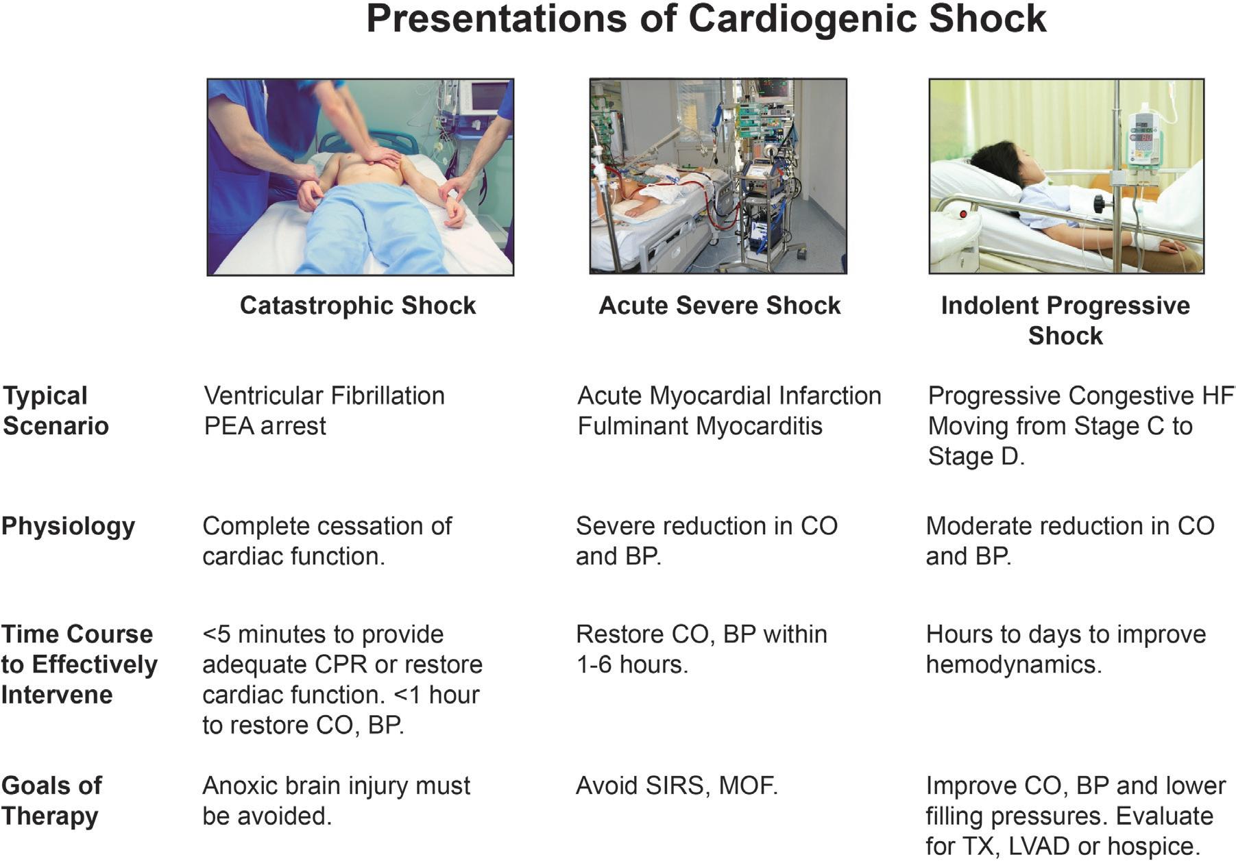
Shock is a clinical syndrome, much like heart failure, that is characterized by signs and symptoms recognized by the clinician. These can be horribly apparent, as when cardiac arrest initiates cardiogenic shock with complete absence of vital signs (Fig. 2.1) or so subtle that the patient drifts into shock over the course of weeks or months and even skilled clinicians miss the transition (gradual-onset cardiogenic shock). In general, contemporary definitions of cardiogenic shock are eerily similar to battlefield depictions of severe trauma in the American Civil War, but with the addition of quantitative parameters of reduced urine output and blood pressure. Cardiogenic shock was defined in a
TABLE 2.1 INTERMACS Profiles Inform the Definitions of Cardiogenic Shock
ADL, Activities of daily living; CHF, congestive heart failure; INTERMACS, Interagency Registry for Mechanically Assisted Circulatory Support; NYHA, New York Heart Association; Rx, medications; sx, symptoms.
Source: Stevenson LW, Pagani FD, Young JB, et al. INTERMACS profiles of advanced heart failure: the current picture. J Heart Lung Transplant 2009;28:535–541.
recent trial evaluating percutaneous intervention in shock as “a systolic blood pressure of less than 90 mm Hg for longer than 30 minutes or the use of catecholamine therapy to maintain a systolic pressure of at least 90 mm Hg, clinical signs of pulmonary congestion, and signs of impaired organ perfusion with at least one of the following manifestations: altered mental status, cold and clammy skin and limbs, oliguria with a urine output of less than 30 mL per hour, or an arterial lactate level of more than 2.0 mm per liter.”8
ETIOLOGY OF CARDIOGENIC SHOCK
Acute myocardial infarction is a common, but by no means the only, cause of cardiogenic shock (Box 2.1), and the infarction can result in shock in a number of ways. It can be the result of a catastrophically large infarct or a relatively small infarction in the setting of an ischemic cardiomyopathy. Infarction of the right ventricle with relative sparing of the left ventricle has unique clinical features, including shock. Severe ischemia even in the absence of myocardial injury can reduce cardiac
BOX 2.1 Etiologies of Cardiogenic Shock
Ischemic
Acute myocardial infarction
Unstable angina with global ischemia
Right ventricular infarction
Complications of ischemic heart disease
Papillary muscle rupture
Acute ventricular septal defect
Myocardial rupture
Valvular
Severe aortic stenosis or insufficiency
Severe mitral regurgitation or stenosis
Severe pulmonic stenosis or regurgitation
Severe tricuspid regurgitation or stenosis
Myocardial disease/unknown
Acute myocarditis
Giant cell myocarditis
Takotsubo stress myocarditis
Substance abuse
Toxins
Chemotherapeutic agents
End-stage ischemic or nonischemic cardiomyopathy
Extracardiac
Cardiac tamponade
Acute aortic dissection with aortic insufficiency, tamponade, or rupture
Large pulmonary embolism
End-stage congenital heart disease
output substantially with reduced blood pressure. Finally, infarction with tissue necrosis can result in papillary muscle rupture, acute ventricular septal defect, or free wall rupture with severe, often fatal, shock.
In the absence of coronary artery disease, acute myocarditis and especially giant cell myocarditis can result in profound cardiogenic shock such that almost cessation of myocardial contraction can be seen and/or incessant malignant arrhythmias requiring ECMO support. Takotsubo cardiomyopathy can mimic acute myocardial infarction in the emergency room and has a reported incidence of cardiogenic shock of 9%.9 Long-standing heart failure that has been stable for decades can devolve rapidly or insidiously into end-stage heart failure with hypotension and multiorgan dysfunction. Similarly, chronic valvular abnormalities, when they become severe, can profoundly impair hemodynamics and become life-threatening. Patients with adult congenital heart disease can sink later in life into a shock state with multiorgan failure after years of relatively normal cardiovascular status following early palliative surgery. Large pulmonary emboli can present with syncope and cardiogenic shock from right heart failure, as can end-stage pulmonary arterial hypertension. Finally, long-standing alcohol abuse and illicit drug use with methamphetamines or other sympathomimetic drugs can be responsible for profound cardiac dysfunction.
HEMODYNAMIC EFFECTS OF CARDIOGENIC SHOCK
Reduced Cardiac Output
In general, cardiac output is reduced in cardiogenic shock, although not universally so. Cardiac output is a continuum, so there is not an absolute number below which a patient is in cardiogenic shock. The shock state exists when the output is not sufficient to meet the metabolic needs of the principle organ systems, including the kidneys, liver, central nervous system, and digestive tract. In terms of cardiac output, shock has
been defined as a cardiac index less than 1.8 to 2.2 L/min/m2.10 That said, not all patients with this reduction in cardiac index are in shock, but it does indicate significant derangement in cardiac function.
Unfortunately, cardiac output requires invasive measurements, and so there are often delays in obtaining this important determinate of shock. Beyond thermodilution estimation of cardiac output, mixed venous saturation can be an important indicator of low output states when corrected for anemia. Mixed venous saturations below 50% or, more frequently, less than 40% are indicative of cardiogenic shock.11 Echocardiographic determination of the left ventricular outflow time velocity integral can be used to calculate stroke volume and, hence, cardiac output, and extremely low values are associated with poor outcome.12 Stroke volume can also be estimated by arterial waveform analysis, although this may be less accurate in severe vasodilation or vasoconstriction states.13
In some instances of cardiogenic shock, cardiac output may be nearly normal but is associated with profound vasodilation.14 Vasodilatory shock or systemic inflammatory response, which will be discussed further in the chapter, can develop quickly or during later stages of cardiogenic shock.15 Calculation of systemic vascular resistance can quickly differentiate between vasodilatory or vasoconstrictive shock versus shock presenting with a normal or increased vascular resistance. These distinctions are vital because the pharmacologic and mechanical support approaches are quite different and initial errors in management can prolong tissue hypoperfusion. Judicious use of vasodilators can result in marked improvement of cardiac output in vasoconstriction, whereas they are absolutely contraindicated in vasodilatory states where vasopressin may be beneficial because of vasopressin depletion.16,17
Hypotension
The maintenance of normal blood pressure and primarily to prevent hypotension during changes in posture and abnormal physiologic states (dehydration, hemorrhage) is a critical physiologic function in humans. In common parlance, shock and hypotension are so closely associated that they are often felt to be synonymous. When hypotension is detected by the carotid baroreceptors, this sets off a cascade of neural and hormonal responses that seek to increase cardiac output by increasing heart rate, normalize blood pressure by intense vasoconstriction, and preserve volume by changes in renal handling of salt and water.18
The degree and duration of hypotension are critical in both the ongoing pathophysiology of cardiogenic shock and its prognosis. Catastrophic shock associated with cardiac arrest must be corrected or at least ameliorated within minutes to prevent cerebral anoxic injury. Severe hypotension, that is, mean blood pressure <50 mm Hg, may not immediately cause severe brain injury but can result in acute tubular necrosis and liver injury; organ failure at this level may be survivable but complicates and exacerbates shock with poorer outcomes. Milder degrees of hypotension may be present for weeks or even months in chronic heart failure, but frequently lead to more extreme derangement.
Increased Filling Pressures
As cardiogenic shock progresses, filling pressures usually rise. If the genesis of the shock is ischemic, then ischemia itself increases diastolic stiffness by interfering with calcium reuptake in the sarcoplasmic reticulum. When myocardial systolic performance is reduced, then systolic emptying falls and filling pressures rise.19 An increase in filling pressures may actually be salutary in that they increase cardiac output by the Frank Starling mechanism but, very quickly, the pressures rise to levels that are detrimental.20 Neurohormonal activation causes increased sodium reabsorption and decreased excretion of
free water. Severe prolonged hypotension can result in acute tubular necrosis, so urine output may actually stop, which exacerbates fluid retention. Fluid can shift from the mesenteric bed to the central circulation, further increasing filling pressures.21 As left atrial pressure rises, fluid moves into the lungs, increasing the work of breathing and reducing gas exchange so that ischemia may be compounded. In primarily right-sided cardiogenic shock, left-sided filling pressures are usually low and may exacerbate systemic hypotension due to inadequate left ventricular preload.
NEUROHORMONAL RESPONSE TO CARDIOGENIC SHOCK
There is a marked activation of the sympathetic nervous system in response to the reduced systemic blood pressure, which is a hallmark of cardiogenic shock. This response is mainly through arterial baroreceptors located in the carotid sinus and the aortic arch.22,23 Indeed, many of the classic clinical signs of shock are mediated via profound activation of this system, whose key neurotransmitter is norepinephrine. Classical physical signs of shock, such as tachycardia, peripheral vasoconstriction, and cool, clammy skin, are the direct results of norephedrine’s effect on the sinus node, vasoconstriction of epithelial arteries near the surface of the skin, and direct effects of the sweat glands. Norepinephrine levels are elevated both in heart failure and in acute myocardial infarction, but the levels are higher and persist longer when cardiogenic shock intervenes.24 The increases in norepinephrine level are important systemic compensatory mechanisms to both protect blood pressure and increase cardiac output via heart rate increase and myocardial contractility, but this can come at the cost of exacerbating myocardial ischemia and promoting further myocardial cell death via the toxic effect of very high levels of norepinephrine on cardiac myocytes via apotosis.25
Plasma renin levels are also elevated with myocardial infarction and acute decompensated heart failure.26 These levels are especially elevated when blood pressure is significantly reduced.27 Elevated renin produces secondary increases in angiotensin II and aldosterone, whose effects include maintenance of blood pressure and sodium retention. Aldosterone levels are elevated in septic shock but have not been reported so in cardiogenic shock, although higher aldosterone levels are associated with worse long-term prognosis following myocardial infarction.28,29 NT proBNP levels may also increase after myocardial infarction, especially if shock intervenes. Extremely high levels of NT proBNP >12,000 are associated with very poor prognosis in cardiogenic shock, especially when coupled with high interleukin 6 (IL-6) levels.30 Although angiotensin II levels are elevated in severe heart failure, a recent trial suggests that pharmacologic doses of angiotensin II may improve outcome in vasodilatory shock.31 In late 2017, this formulation of angiotensin II was approved for clinical use.32
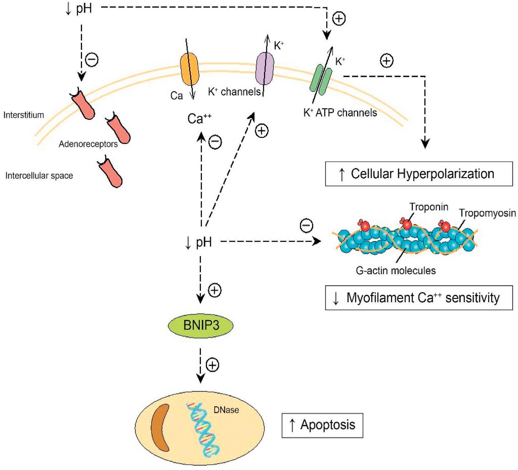
Lactic Acidosis
As a consequence of decreased oxygen delivery to tissue due to hypotension and reduced cardiac output, mitochondrial production of adenosine triphosphate (ATP) is impaired and pyruvate levels increase, resulting in increased levels of lactate, a strong acid (Fig. 2.2). A reduction in intracellular and extracellular pH has important physiologic consequences that exacerbate the shock state.33 Reduced intracellular pH has negative effects on cardiac function by decreasing myofilament sensitivity to Ca++ and to adrenergic agonists.34,35 In addition, it interferes with depolarization by enhancing K+ egress from the cell, resulting in hyperpolization.17 Lactic acidosis also causes adrenoreceptor internalization so that sensitivity to norepinephrine is reduced.36,37
Finally, low intracellular PH stimulates BNIP23, which promotes apoptosis and induces nitric oxide (NO) production, which has detrimental effects.38
In smooth muscle cells, hyperpolarization occurs, so these cells critical to the maintenance of blood pressure cannot constrict normally, thus contributing to hypotension. Acidosis also reduces the sensitivity of vascular smooth muscle cells to the vasoconstrictor effects both of NE and angiotensin II.17 Clearly, in some patients with cardiogenic shock, vasoconstrictive reflexes are still functional. However, as shock is prolonged and/or a vasodilatory state appears, the vascular smooth muscle beds can no longer function normally, and irreversible shock may develop. In patients resuscitated from cardiac arrest out of hospital, presenting lactate levels were highly associated with mortality; 61% of patients survived if the initial lactate levels were <5 mmol/L, whereas only 8% survived if the levels were greater than 10 mmol/L.39
Intravascular pH and lactate levels are important indicators of both developing shock and its severity. Lactate is a key biomarker for the detection of early shock, the severity of shock, and its successful management (Box 2.2). High lactate levels correlate strongly with prognosis in cardiogenic shock and shock from other etiologies.40 Lactate is cleared by both the kidneys and the liver, and as these organs fail, lactate levels remain elevated even when corrective measures have restored blood pressure and cardiac output.41 Thus, lactate may remain elevated for many hours even though pH has been corrected.
INFLAMMATORY PATHWAYS
In concert with the hemodynamic and neurohormonal abnormalities attendant with cardiogenic shock, an inflammatory response via the cytokine system plays an important role in the progressive circulatory deterioration (Fig. 2.3). IL-6 levels are increased significantly in both cardiogenic and septic shock. The actions of IL-6, which is generated and released by monocytes and T cells, contribute to the inflammatory response by an antiapoptotic effect on T cells. Geppert et al. reported that in patients with cardiogenic shock, IL-6 levels were significantly higher than in other intensive care unit patients without shock and that very high levels of IL-6 were associated with multiorgan failure.42 IL-6, -7, -8, and -10 levels are also predictive of increased mortality.43
Tumor necrosis factor-alpha (TNF-α) is elevated in advanced heart failure and is in part responsible for cardiac cachexia; hence, it is also known as cachexin.44 Produced by activated macrophages, its release is one component of the acute phase reaction and induces fever and inhibits viral replication. Inhibition of TNF-α has proven successful in some autoimmune conditions but was not successful when applied to heart failure.45 Peak concentrations of TNF-α were higher in patients with cardiogenic shock compared to patients with acute myocardial infarctions without shock and were higher still in those whose cardiogenic shock and had a vasodilatory component.46 C-reactive protein, like TNF-α, is also involved in the acute phase reactant produced by hepatic cells and is increased in cardiogenic shock.47
While the measurement of most cytokines is not available clinically, procalcitonin is a valuable, routinely used clinical biomarker. It is the precursor of calcitonin, but it has additional biochemical effects that are similar to cytokines.48 Procalcitonin levels may be elevated in sepsis and may be useful in the early initiation of antibiotics. However, procalcitonin levels may be also elevated in cardiogenic shock without apparent bacterial infection and are indicative of a marked inflammatory response.46
Fig. 2.2 Reduction in pH that occurs with lactic acidosis has a significant, negative impact on cellular function. Lower intracellular pH potentiates K+ egress from cells, which results in hyperpolarization. In smooth muscle cells this interferes with normal depolarization and contraction, which exacerbates hypotension. Acidosis also is associated with a decrease in adrenergic receptors. Intracellular acidosis interferes with Ca++ binding of myocytes which impairs contractility. Acidosis also activates the BNIP3 gene, which is involved in apoptosis.
ATP, Adenosine triphosphate. (From Antoine Kimmoun, Emmanuel Novy, Thomas Auchet, Nicolas Ducrocq et al, Bruno Lev., Hemodynamic consequences of severe lactic acidosis in shock states: from bench to bedside., Critical Care201721:40. http://creativecommons.org/licenses/by/4.0/.)
BOX 2.2 Generation and Effects of Lactic Acidosis in Cardiogenic Shock
Lactic acidosis—key biomarker in cardiogenic shock
• Elevated lactate levels indicate that oxygen delivery is inadequate to support normal metabolism.
• Indicates transition from aerobic to anaerobic metabolism.
• Higher lactate levels indicate more reliance on anaerobic metabolism with increased acidosis because lactate is a strong acid.
• Survival declines with higher lactate levels and lower pH.
• Organs with high metabolic needs (brain, heart, and kidney) are more dependent on aerobic metabolism and are thus more sensitive to hypoxia.
• Acidosis has multiple detrimental cellular effects, including reduced sensitivity to norepinephrine, impaired contractility, and hyperpolarization of the smooth muscle exacerbating hypotension.
• Reduction in lactate levels with shock treatment is associated with improved prognosis.
• Lactate levels may remain elevated for some time despite reduced production and clearing acidosis because of ongoing kidney and liver disease, which impairs clearance.
Nitric Oxide
NO is an important signaling molecule produced in endothelial cells, neurons, and other mammalian cell lines. The identification of NO and the elegant proof of its pivotal role in biological signaling was recognized by the awarding of the Nobel Prize to Robert Furchgott, Louis Ignarro, and Ferid Murad in 1998.49,50 It is produced from L-arginine via nitric oxide synthetase (NOS), which is present as several isoforms whose major function in vascular endothelium is to promote vasodilation and inhibit platelet adhesion and aggregation. One isoform of NOS, inducible NOS (iNOS), can be produced in large quantities in sepsis and contribute to hypotension and vasodilatory shock.51 High levels of NO produced via iNOS in addition can interfere with mitochondrial function and suppress myocardial function.52,53 The precise mechanism by which iNOS is expressed at high levels is unknown, but IL-6 may play a role.54
Inhibitors of NO synthetase exist, and early studies showed beneficial effects in cardiogenic shock.55,56 A large randomize trial, Tilarginine Acetate Injection in a Randomized International Study in Unstable MI Patients With Cardiogenic Shock (TRIUMPH), was undertaken to evaluate the effects of tilarginine in patients presenting
Pathophysiology of Cardiogenic Shock
Reduced Cardiac Output
Lactic Acidosis
Systemic Inflammatory Response
Nitric Oxide Interleukins (TNF-α)
Severe Ventricular Valvular Dysfunction
Reduced Systemic Pressure
Neural Hormonal Response
Increased Filling Pressures
Reduced Renal, Liver Function
Multiorgan Failure
DEATH
Increased Vascular Permiability
Pulmonary Edema
ARDS
Peripheral Edema
WORSENS
Fig. 2.3 Cardiogenic shock is a complex physiology response to hypotension and/or reduced cardiac output. The primary effect of this is a shift from aerobic to anaerobic metabolism with the elaboration of lactic acid and reduce pH. Neurohormonal and inflammatory pathways are activated by hypotension and acidosis which cause fluid retention and reduce capillary integrity. Impaired oxygen delivery and volume expansion worsen organ function and can result in multiorgan failure. ARDS, Acute respiratory distress syndrome; TNF-α, tumor necrosis factor-alpha.
with myocardial infarction and cardiogenic shock.57 Although the agent did increase systolic blood pressure in the treatment group, the study was terminated for futility. Tilarginine is a nonspecific inhibitor of NOS, so all isoforms are affected; therefore, important intracellular isoforms may be inhibited along with the inducible form thought responsible for the high extracellular NO levels.58
END ORGAN INJURY
A common criterion included in the clinical definition of shock is reduced urine output. Renal function may be temporarily impaired by the onset of shock or acute tubular necrosis can ensue and renal function may never recover even if the patient survives. Renal dysfunction is common in cardiogenic shock and is a marker for increased mortality.
As reported by Koreny et al. in a cohort of patients with cardiogenic shock following myocardial infarction, mortality was 87% in those who developed renal failure in the first 24 hours versus 53% in those who did not.59 Decreased urine output and worsening renal function are the end result of many factors during the initiation and evolution of shock. Hypotension and reduced cardiac output reduce glomerular filtration pressure, although the preferential shunting of blood to the central organs may forestall this. The effects of hypotension may also be mitigated by increased levels of angiotensin II, which constrict efferent arterioles and thus increase intraglomerular pressure.60 Conversely, high levels of norepinephrine and sympathetic stimulation cause generalized vasoconstriction, which may further diminish renal blood flow and even lead to renal ischemia. If the systemic vascular resistance is high enough to severely reduce cardiac output careful vasodilation
while scrupulously maintaining blood pressure can, in rare instances of early shock, preserve renal function. However, in most cases, blood pressure cannot be supported chemically and mechanical support is required. In any case, preservation of renal function is a critical goal in shock management.
In some cases of cardiogenic shock, a vasodilatory component can occur initially or as shock becomes progressive and unrelenting. Vasodilatory shock makes maintenance of blood pressure extremely difficult as the arteriolar bed becomes resistant to pressors.17,35 Vasopressin can be useful in some instances of vasodilatory shock because stores of vasopressin are depleted as shock continues.17 As noted earlier, intravenous angiotensin II may show some benefit in high output shock.31 Renal failure is almost always seen when vasodilatory shock is present.
Another important component in the setting of worsening renal function is high central venous pressure, which is rarely seen in other forms of shock but is a frequent hemodynamic finding in cardiogenic shock. The increased levels of renin, aldosterone, angiotensin II, and vasopressin all work to increase sodium and free water reabsorption when the kidneys are functioning. When they fail, volume cannot be offloaded except through dialysis and ultrafiltration. The resulting high central venous pressures cause further disruption in renal func tion by increasing interstitial and abdominal pressures. 61–63 Severe tricuspid regurgitation also significantly impairs renal venous outflow and is associated with a marked reduction in survival in heart failure.64 Reduction of filling pressures to normal levels via dialysis and continuous renal replacement therapy help to maintain or restore renal function and are important components of shock management.
The liver may also be severely impacted in cardiogenic shock by similar mechanisms as the kidney, that is, passive congestive and ischemic injury.65 At times, liver failure may be the chief presenting feature of cardiogenic shock; indeed, a cardiac etiology may not be detected initially.66 Passive congestion of the liver without shock can result in elevation of liver enzymes to several times the normal range and mild elevation of bilirubin. If this elevation persists for years, frank cirrhosis can result. In cardiogenic shock, ischemic hepatic necrosis can be seen. Ordinarily, the liver is resistant to hypoxic injury because hepatocytes are capable of extracting almost all the oxygen from the blood to meet its considerable metabolic needs. When shock persists and necrosis ensues, this presents histologically as centrolobular necrosis of zone 3 hepatocytes without inflammation.67 Elevations in aspartate aminotransferase (AST) and alanine aminotransferase (ALT) more than 10 times above normal may be seen with much greater increases in bilirubin. Elevations in bilirubin are an independent predictor of mortality in heart failure. Unlike the kidney, where full-blown ATN develops, the liver can recover from the shock state in a matter of days if the shock is reversed quickly enough. If shock persists, liver failure can progress, with reductions in serum albumin and the development of coagulopathies, which increase the risk of mortality.
Respiratory failure due to increased work of breathing and intractable pulmonary edema is a common complication of cardiogenic shock so that mechanical ventilation is extremely common. Elevated filling pressures play an important role, as does breakdown in capillary integrity, which can result in adult respiratory distress syndrome even if filling pressures are controlled by diuretics or renal replacement therapy.68
Of all the organs in the body, the central nervous system, because of its extremely high oxygen requirements, is most susceptible to hypotension in cardiogenic shock. Ordinarily, in catastrophic shock with
complete cessation of cardiac function, irreversible brain injury can occur unless high-quality cardiopulmonary resuscitation (CPR) is initiated in the first 1 to 2 minutes.
CONCLUSION
Since the introduction of the concept of shock into the medical literature in the 18th century, our understanding of this dreadful and fascinating condition has benefited from the work of physicians on the battlefields, physiologists in the early 20th century, and basic scientists working out complex biochemical effects of neurohormones, cytokines, and apoptosis. As a result of this work, shock can now be diagnosed more quickly and reversed in some cases via well-placed balloons, appropriate pharmaceutical agents, and the rapid application of mechanical pumps. Nonetheless, many more patients are lost to cardiogenic shock than are saved each year. This is a major health care system problem with multiple causes, including inadequate CPR, lack of access to defibrillators, and poor management of shock. On a deeper level, the pathophysiology of shock is still incompletely understood, and thus, the means of reversing its pathophysiologic storm are not yet in our armamentarium.
REFERENCES
1. Millham FH. A brief history of shock. Surgery. 2010;148:1026–1037.
2. Chisolm JJ. A Manual of Military Surgery: For the Use of the Surgeons in the Confederate Army: with an Appendix of the Rules and Regulations of the Medical Department of the Confederate Army (No. 4). Norman Publishing; 1861.
3. Mansell-Moulin CW. On the Pathology of Shock. Dissertation. London: HG Saunders; 1880.
4. Booth J. A short history of the blood pressure measurement. Proc Roy Soc Med. 1977;70:793–799.
5. Crile G. George Crile: An Autobiography. 2 Lippincott; 1947.
6. Shires T, Coln D, Carrico J, Lightfoot ST. Fluid therapy in hemorrhagic shock. Arch Surg. 1964;88:688–693.
7. Blalock A. Shock and hemorrhage. Bull NY Acad of Med. 1936;11: 610–622.
8. Thiele H, Akin I, Sandri M, et al. PCI strategies in patients with acute myocardial infarction and cardiogenic shock. N Engl J Med. 2017;377:2419–2432.
9. Templin C, Ghadri J, Diekmann J, et al. Clinical features and outcomes of Takotsubo (stress) cardiomyopathy. N Engl J Med 2015;373:929–938.
10. Holmes DR Jr, Berger PB, Hochman JS, et al. Cardiogenic shock in patients with and without ST-segment elevation. Circulation 1999;100:2067–2073.
11. Muir AL, Kirby BJ, King AJ, et al. Mixed venous oxygen saturation in relation to cardiac output in myocardial infarction. Br Med J. 1970;4:276–278.
12. Tan C, Rubenson D, Srivastava A, et al. Left ventricular outflow tract velocity. Cardiovasc Ultrasound 2017;15:18–26.
13. Mehta Y, Arora D. Newer methods of cardiac output monitoring. World J Cardiol. 2014;6:1022–1029.
14. Kohsaka S, Menon V, Lowe AM, et al. Systemic inflammatory response syndrome after acute myocardial infarction complicated by cardiogenic shock. Arch Intern Med. 2005;165:1643–1650.
15. Argenziano M, Chen JM, Choudhri AF, et al. Management of vasodilatory shock after cardiac surgery: identification of predisposing factors and use of a novel pressor agent. J Thorac Cardiovasc Surg. 1998;116:973–980.
16. Elkayam U, Janmohamed M, Habib M, et al. Vasodilators in the management of acute heart failure. Crit Care Med. 2008;36(1 suppl):S95–S105.
17. Landry DW, Oliver JA. The pathogenesis of vasodilatory shock. N Engl J Med. 2001;345:588–594.
18. Creager MA. Baroreceptor reflex function in congestive heart failure. Am J Cardiol. 1992;69:10G–16G.