https://ebookmass.com/product/materials-for-biomedicalengineering-hydrogels-and-polymer-based-scaffolds-alexandru-
Instant digital products (PDF, ePub, MOBI) ready for you
Download now and discover formats that fit your needs...
Materials for Biomedical Engineering: Thermoset and Thermoplastic Polymers Valentina Grumezescu
https://ebookmass.com/product/materials-for-biomedical-engineeringthermoset-and-thermoplastic-polymers-valentina-grumezescu/
ebookmass.com
Materials for Biomedical Engineering: Inorganic Micro- and Nanostructures Valentina Grumezescu
https://ebookmass.com/product/materials-for-biomedical-engineeringinorganic-micro-and-nanostructures-valentina-grumezescu/
ebookmass.com
Materials for Biomedical Engineering - Inorganic Microand Nanostructures Valentina Grumezescu
https://ebookmass.com/product/materials-for-biomedical-engineeringinorganic-micro-and-nanostructures-valentina-grumezescu-2/ ebookmass.com
Vector Mechanics for Engineers 12th Edition Ferdinand
Pierre Beer
https://ebookmass.com/product/vector-mechanics-for-engineers-12thedition-ferdinand-pierre-beer/
ebookmass.com
Financial Accounting 7th Canadian Edition Edition Robert Libby
https://ebookmass.com/product/financial-accounting-7th-canadianedition-edition-robert-libby/
ebookmass.com
Sex Matters: Essays in Gender-Critical Philosophy Holly Lawford-Smith
https://ebookmass.com/product/sex-matters-essays-in-gender-criticalphilosophy-holly-lawford-smith/
ebookmass.com
Checkmate to Murder: A Second World War Mystery E.C.R. Lorac
https://ebookmass.com/product/checkmate-to-murder-a-second-world-warmystery-e-c-r-lorac/
ebookmass.com
The Arc Tory Henwood Hoen
https://ebookmass.com/product/the-arc-tory-henwood-hoen/
ebookmass.com
A Sketch of the World After the COVID-19 Crisis: Essays on Political Authority, The Future of Globalization, and the Rise of China 1st ed. Edition Jean-François Caron
https://ebookmass.com/product/a-sketch-of-the-world-after-thecovid-19-crisis-essays-on-political-authority-the-future-ofglobalization-and-the-rise-of-china-1st-ed-edition-jean-francoiscaron/ ebookmass.com
Bookworm: Rugged Mountain Ink (Filthy, Dirty, Small-Town Love) (Rugged Mountain Ink (Filthy, Dirty, Small-Town Sweetness) Book 10) Khloe Summers
https://ebookmass.com/product/bookworm-rugged-mountain-ink-filthydirty-small-town-love-rugged-mountain-ink-filthy-dirty-small-townsweetness-book-10-khloe-summers/
ebookmass.com
Hydrogelsand Polymer-based Scaffolds Hydrogelsand Polymer-based Scaffolds Editedby
Alina-MariaHolban
FacultyofBiology,UniversityofBucharest,Bucharest,Romania
AlexandruMihaiGrumezescu
FacultyofAppliedChemistryandMaterialsScience,University PolitehnicaofBucharest,Bucharest,Romania MaterialsforBiomedicalEngineering
Elsevier Radarweg29,POBox211,1000AEAmsterdam,Netherlands TheBoulevard,LangfordLane,Kidlington,OxfordOX51GB,UnitedKingdom 50HampshireStreet,5thFloor,Cambridge,MA02139,UnitedStates
Copyright © 2019ElsevierInc.Allrightsreserved.
Nopartofthispublicationmaybereproducedortransmittedinanyformorbyanymeans,electronicor mechanical,includingphotocopying,recording,oranyinformationstorageandretrievalsystem,without permissioninwritingfromthepublisher.Detailsonhowtoseekpermission,furtherinformationaboutthe Publisher’spermissionspoliciesandourarrangementswithorganizationssuchastheCopyrightClearance CenterandtheCopyrightLicensingAgency,canbefoundatourwebsite: www.elsevier.com/permissions
ThisbookandtheindividualcontributionscontainedinitareprotectedundercopyrightbythePublisher (otherthanasmaybenotedherein).
Notices Knowledgeandbestpracticeinthisfieldareconstantlychanging.Asnewresearchandexperiencebroaden ourunderstanding,changesinresearchmethods,professionalpractices,ormedicaltreatmentmaybecome necessary.
Practitionersandresearchersmustalwaysrelyontheirownexperienceandknowledgeinevaluatingand usinganyinformation,methods,compounds,orexperimentsdescribedherein.Inusingsuchinformationor methodstheyshouldbemindfuloftheirownsafetyandthesafetyofothers,includingpartiesforwhomthey haveaprofessionalresponsibility.
Tothefullestextentofthelaw,neitherthePublishernortheauthors,contributors,oreditors,assumeany liabilityforanyinjuryand/ordamagetopersonsorpropertyasamatterofproductsliability,negligenceor otherwise,orfromanyuseoroperationofanymethods,products,instructions,orideascontainedinthe materialherein.
BritishLibraryCataloguing-in-PublicationData
AcataloguerecordforthisbookisavailablefromtheBritishLibrary LibraryofCongressCataloging-in-PublicationData
AcatalogrecordforthisbookisavailablefromtheLibraryofCongress ISBN:978-0-12-816901-8
ForInformationonallElsevierpublications visitourwebsiteat https://www.elsevier.com/books-and-journals
Publisher: MatthewDeans
AcquisitionEditor: GwenJones
EditorialProjectManager: EmmaHayes
ProductionProjectManager: DebasishGhosh
CoverDesigner: GregHarris
TypesetbyMPSLimited,Chennai,India
ListofContributors GustavoA.Abraham
ResearchInstituteforMaterialsScienceandTechnology,INTEMA (UNMdP-CONICET),MardelPlata,Argentina
SatishAgrawal
DivisionofPharmaceutics&Pharmacokinetics,CSIR-CentralDrugResearch Institute,Lucknow,India
HafsaAhmad
Pharmacognosy&EthnopharmacologyDivision,CSIR-NationalBotanical ResearchInstitute,Lucknow,India
LuigiAmbrosio
InstituteofPolymers,CompositesandBiomaterials,NationalResearchCouncil ofItaly,Naples,Italy
TakaakiArahira
SectionofBioengineering,DepartmentofDentalEngineering,FukuokaDental College,Fukuoka,Japan
StephanArmyanov
InstituteofPhysicalChemistryRostislawKaischew,BulgarianAcademyof Sciences,Sofia,Bulgaria
AbhishekArya
DivisionofPharmaceutics&Pharmacokinetics,CSIR-CentralDrugResearch Institute,Lucknow,India
PetarA.Atanasov InstituteofElectronics,BulgarianAcademyofSciences,Sofia,Bulgaria
MariaIngridRochaBarbosa
InstituteofBiofabrication,SchoolofChemicalEngineering,Universityof Campinas,Campinas,SaoPaulo,Brazil
RejaneAndradeBatista
InstitutoTecnolo ´ gicoedePesquisasdoEstadodeSergipe,RuaCampodo Brito,Aracaju,Brazil
SusanMichelzBeitel
DepartmentofBiochemistryandMicrobiology,InstituteBioscience,SaoPaulo StateUniversity(UNESP),Sa ˜ oPaulo,Sa ˜ oPaulo,Brazil
AnaCarolinaB.Benatti
InstituteofBiofabrication,SchoolofChemicalEngineering,Universityof Campinas,Campinas,SaoPaulo,Brazil;SchoolofMedicalSciences, SurgeryDepartment,UniversityofCampinas,Campinas,Sa ˜ oPaulo,Brazil
BinayBhushan
DepartmentofPhysics,BirlaInstituteofTechnology,Mesra,PatnaCampus,India
S.Bolshanina
SumyStateUniversity,MinistryofEducationandScienceofUkraine,Sumy, Ukraine
PabloC.Caracciolo
ResearchInstituteforMaterialsScienceandTechnology,INTEMA (UNMdP-CONICET),MardelPlata,Argentina
LucianaFontesCoelho
DepartmentofBiochemistryandMicrobiology,InstituteBioscience,Sa ˜ oPaulo StateUniversity(UNESP),SaoPaulo,SaoPaulo,Brazil
JonasContiero
DepartmentofBiochemistryandMicrobiology,InstituteBioscience,SaoPaulo StateUniversity(UNESP),Sa ˜ oPaulo,Sa ˜ oPaulo,Brazil;AssociateLaboratory IPBEN-UNESP,RioClaro,Sa ˜ oPaulo,Brazil
AnilKumarDwivedi
DivisionofPharmaceutics&Pharmacokinetics,CSIR-CentralDrugResearch Institute,Lucknow,India
PaulaJ.P.Espitia
NutritionandDieteticsSchool,UniversidaddelAtla ´ ntico,Atla ´ ntico,Colombia
RubensMacielFilho
InstituteofBiofabrication,SchoolofChemicalEngineering,Universityof Campinas,Campinas,Sa ˜ oPaulo,Brazil
AlessandraGiuliani
DepartmentofClinicalSciences,Universita ` PolitecnicadelleMarche,Ancona, Italy
VincenzoGuarino
InstituteofPolymers,CompositesandBiomaterials,NationalResearchCouncil ofItaly,Naples,Italy
FlorinIordache
InstituteofCellularBiologyandPathology“NicolaeSimionescu”of RomanianAcademy,Bucharest,Romania;FacultyofVeterinaryMedicine, UniversityofAgronomicSciencesandveterinaryMedicine,Bucharest, Romania
Andre ´ LuizJardini
InstituteofBiofabrication,SchoolofChemicalEngineering,Universityof Campinas,Campinas,Sa ˜ oPaulo,Brazil
AndreasKaasi
InstituteofBiofabrication,SchoolofChemicalEngineering,Universityof Campinas,Campinas,SaoPaulo,Brazil;SchoolofMedicalSciences,Surgery Department,UniversityofCampinas,Campinas,Sa ˜ oPaulo,Brazil; EvaScientificLtda,SaoPaulo,SaoPaulo,Brazil
PauloKharmandayan
InstituteofBiofabrication,Schoolof ChemicalEngineering,Universityof Campinas,Campinas,Sa ˜ oPaulo,Brazil;SchoolofMedicalSciences, SurgeryDepartment,UniversityofCampinas,Campinas,Sa ˜ oPaulo, Brazil
KonstantinKolev
InstituteofPhysicalChemistryRostislawKaischew,BulgarianAcademyof Sciences,Sofia,Bulgaria
RakeshKumar
DepartmentofBiotechnology,CentralUniversityofSouthBihar,Gaya,India
SandraM.London ˜ o-Restrepo
PosgradoenCienciaeIngenierı´adeMateriales,CentrodeFı´sicaAplicaday Tecnologı´aAvanzada,UniversidadNacionalAuto ´ nomadeMe ´ xico,Quere ´ taro, Me ´ xico
NaylaJ.Lores
ResearchInstituteforMaterialsScienceandTechnology,INTEMA (UNMdP-CONICET),MardelPlata,Argentina
AdrianManescu “VictorBabes”UniversityofMedicineandPharmacy,Timisoara,Romania
SerenaMazzoni
DepartmentofClinicalSciences,Universita ` PolitecnicadelleMarche,Ancona, Italy
NikolayN.Nedyalkov
InstituteofElectronics,BulgarianAcademyofSciences,Sofia,Bulgaria
CaioGomideOtoni
NationalNanotechnologyLaboratoryforAgribusiness,Embrapa Instrumentac¸ao,SaoCarlos,Brazil
NidhiPareek
DepartmentofMicrobiology,CentralUniversityofRajasthan,Ajmer,India
KunwarParitosh
CentreforEnergyandEnvironment,MalaviyaNationalInstituteofTechnology, Jaipur,India
AnaFla ´ viaPattaro
InstituteofBiofabrication,SchoolofChemicalEngineering,Universityof Campinas,Campinas,Sa ˜ oPaulo,Brazil
CristianF.Ramirez-Gutierrez
PosgradoenCienciaeIngenierı´adeMateriales,CentrodeFı´sicaAplicaday Tecnologı´aAvanzada,UniversidadNacionalAuto ´ nomadeMe ´ xico,Quere ´ taro, Me ´ xico
AnaAme ´ liaRodrigues
InstituteofBiofabrication,SchoolofChemicalEngineering,Universityof Campinas,Campinas,Sa ˜ oPaulo,Brazil
MarioE.Rodriguez-Garcı ´ a DepartamentodeNanotecnologı´a,CentrodeFı´sicaAplicadayTecnologı ´ a Avanzada,UniversidadNacionalAuto ´ nomadeMe ´ xico,Quere ´ taro,Me ´ xico
AngelRomo-Uribe
Johnson&JohnsonVisionCare,Inc.,AdvancedScience&Technology, Jacksonville,FL,UnitedStates
MonicaSandri
CNR-NationalResearchCouncilofItaly,InstituteofScienceandTechnology forCeramicMaterials(ISTEC),Faenza,Italy
SilviaScaglione
CNR-NationalResearchCouncilofItaly,IEIITInstitute,Genoa,Italy
SimoneSprio
CNR-NationalResearchCouncilofItaly,InstituteofScienceandTechnology forCeramicMaterials(ISTEC),Faenza,Italy
NadyaE.Stankova
InstituteofElectronics,BulgarianAcademyofSciences,Sofia,Bulgaria
AnnaTampieri
CNR-NationalResearchCouncilofItaly,InstituteofScienceandTechnology forCeramicMaterials(ISTEC),Faenza,Italy
MitsuguTodo
ResearchInstituteforAppliedMechanics,KyushuUniversity,Fukuoka,Japan
GiulianaTromba
SincrotroneTriesteS.C.p.A,Trieste,Italy
EugeniaValova
InstituteofPhysicalChemistryRostislawKaischew,BulgarianAcademyof Sciences,Sofia,Bulgaria
HerminsoVillarraga-Go ´ mez
NikonMetrology,Inc.,Brighton,MI,UnitedStates
VivekanandVivekanand
CentreforEnergyandEnvironment,MalaviyaNationalInstituteofTechnology, Jaipur,India
MarianaViteloXavier
InstituteofBiofabrication,SchoolofChemicalEngineering,Universityof Campinas,Campinas,Sa ˜ oPaulo,Brazil;SchoolofMedicalSciences,Surgery Department,UniversityofCampinas,Campinas,SaoPaulo,Brazil
MonikaYadav
CentreforEnergyandEnvironment,MalaviyaNationalInstituteofTechnology, Jaipur,India
A.Yanovska
SumyStateUniversity,MinistryofEducationandScienceofUkraine,Sumy, Ukraine
SeriesPreface Inthepastfewdecadestherehasbeengrowinginterestinthedesignandimplementationofadvancedmaterialsfornewbiomedicalapplications.Thedevelopmentofthesematerialshasbeenfacilitatedbymultiplefactors,especiallythe introductionofnewengineeringtoolsandtechnologies,emergingbiomedical needs,andsocioeconomicconsiderations.Bioengineeringisaninterdisciplinary fieldencompassingcontributionsfrombiology,medicine,chemistry,andmaterialsscience.Inthiscontext,newmaterialshavebeendevelopedorreinventedto fulfilltheneedformodernandimprovedengineeredbiodevices.
Amultivolumeseries, MaterialsforBiomedicalEngineering highlightsthe mostrelevantfindingsanddiscusseskeytopicsinthisimpressiveresearchfield.
Volume1. BioactiveMaterials:PropertiesandApplications,offersanintroductiontobioactivematerials,discussingthemainproperties,applications,and perspectivesofmaterialswithmedicalapplications.Thisvolumereviewsrecently developedmaterials,highlightingtheirimpactintissueengineeringandthedetection,therapy,andprophylaxisofvariousdiseases.
Volume2. ThermosetandThermoplasticPolymers,analyzesthemainapplicationsofadvancedfunctionalpolymersinthebiomedicalfield.Inrecentyears therehasbeenarevolutioninthermoplasticandthermosettingpolymerswith medicalandbiologicaluses,whicharecurrentlybeingdevelopedformedical devices,drugdelivery,tailoredtextiles,packaging,andtissueengineering.
Volume3. AbsorbablePolymers,describesthemaintypesofpolymersofdifferentcompositionswithbioabsorbableandbiodegradableproperties.Thebiomedicalapplicationsofsuchmaterialsarereviewedandthemostinnovative findingsarepresentedinthisvolume.
Volume4. BiopolymerFibers,highlightstheapplicationsofpolymericfibers ofnaturalbiologicalorigininbiomedicalengineering.Suchmaterialsareofgreat utilityintissueengineeringandbiodegradabletextiles.
Volume5. InorganicMicro-andNanostructures andVolume6. Organic Micro-andNanostructures,deal,respectively,withthepreparationandproperties ofinorganicandorganicnanostructuredmaterialswithbiomedicalapplications.
Volume7. HydrogelsandPolymer-BasedScaffolds,discussestherecentprogressmadeinthefieldofpolymericmaterialsdesignedasscaffoldsandtoolsfor tissueengineering.Thetechnologicalchallengesandadvancesintheirproduction, aswellascurrentapplicationsintheproductionofscaffoldsanddevicesfor regenerativemedicinearepresented.
Volume8. BioactiveMaterialsforAntimicrobial,Anticancer,andGene Therapy,offersanupdatedperspectiveregardingnewbioactivematerialswith potentialinthetherapyofseverediseasessuchasinfections,cancer,andgenetic disorders.
Volume9. NanobiomaterialsinTissueEngineering,providesvaluableexamplesofrecentlydesignednanomaterialswithpowerfulapplicationsintissueengineeringandartificialorganapproaches.
Volume10. Nanomaterials-BasedDrugDelivery ,discussesthemostinvestigatedtypesofnanoparticlesandnanoengineeredmaterialswithanimpactindrug delivery.Applicationsfordrug-therapy,andexamplesofsuchnanoscalesystems areincludedinthisvolume.
Thisserieswasmotivatedbytheneedtoofferascientificallysolidbasisfor thenewfindingsandapproachesrelevanttothebiomedicalengineeringfield. Thisscientificresourcecollectsnewinformationonthepreparationandanalysis toolsofdiversematerialswithbiomedicalapplications,whilealsoofferinginnovativeexamplesoftheirmedicalusesfordiagnosesandtherapiesofdiseases. Theserieswillbeofparticularinterestformaterialscientists,engineers,researchersworkinginthebiomedicalfield,clinicians,andalsoinnovativeandestablishedpharmaceuticalcompaniesinterestedinthelatestprogressmadeinthe fieldofbiomaterials.
MichaelR.Hamblin1 andIoannisL.Liakos2 1HarvardMedicalSchool,Boston,MA,UnitedStates 2IstitutoItalianodiTecnologia,Genoa,Italy
Preface Scaffoldscanbecomposedofvariousmaterials,dependingontheirintendeduse. Polymersofvariousoriginsareintensivelyinvestigatedforthedesignofscaffolds andmaterialsfortissueengineering.Infact,thewholefieldofregenerativemedicinereliesonthedevelopmentofpolymericmaterialswithparticularproperties, suchasgreatbiocompatibility,resistance,plasticity,andperspectivesofuse. Naturalandsyntheticpolymericmaterialswererecentlydevelopedorreinvented tofulfilltherequirementsoftheconstantlyexpandingfieldofscaffoldswithbiomedicalapplications.Inthiscontext,hydrogelshavearisenasvaluablealternativestotraditionalpolymericmaterials,offeringuniquepropertiesfortissue engineering.Thisbookaimstorevealthelatesttoolsandapplicationsofscaffolds developedwiththehelpofhydrogelsandpolymericmaterials.Thevolumecontains15chapterspreparedbyoutstandingauthorsfromItaly,theUnitedStates, Mexico,Brazil,India,Romania,Argentina,Ukraine,Bulgaria,andJapan.
Chapter1,entitledInteractionsbetweentissues,cellsandbiomaterials:An advancedevaluationbysynchrotronradiation-basedhigh-resolutiontomography, waspreparedbyAlessandraGiulianietal.Thechapterdiscussesthemostrecent applicationsofsynchrotronradiation basedmicrotomographyinthestudyof interactionsbetweentissues,cells,andbiomaterials.Synchrotronimaginghas proventobeafundamentalcharacterizationtoolforunderstandingthemechanismsofbiologicalbehaviorofbiomaterialsproposedastissuesubstitutes.These innovativetechniquesallownotonlythevisualizationandquantificationofregeneratedtissues,butalsotheeventualpresenceanddistributionofthe neovascularization.
Chapter2,Bioprintedscaffolds,byFlorinIordacheetal.,presentstherecent progressinthefieldofthree-dimensional(3D)bioprintingappliedtofabricate tissue,organs,andbiomedicalpartsthatimitatenaturaltissuearchitecture. Thistechnologycombinescells,growthfactors,andbiomaterialstocreatea microenvironmentinwhichcellscangrowanddifferentiateintissuestructures.
Chapter3,Fundamentalofchitosan-basedhydrogels:Elaborationandcharacterizationtechniques,preparedbyRejaneAndradeBatistaetal.,reviewsthe recentbreakthroughsonhydrogelsfeaturingbiologicalproperties,aswellastheir capabilitiesofcarryingbioactivecompounds.Fundamentalsofhydrogelelaborationandthenatureofbiopolymersusedarepresented,aswellasthefuturetrends regardingtheuseofchitosan-basedhydrogels.
Chapter4,Bioreabsorbablepolymersfortissueengineering:PLA,PGA,and theircopolymers,preparedbyAnaCarolinaBenattietal.,discussesbiomaterials withcharacteristicscompatibletothesitesofmedicalapplicationanddonotpresenttoxicity.Thischapteraimstoconductareviewofdifferentsynthesisroutes, modificationofpolymericstructuresbycopolymerization,andtopresentthecharacteristicsnecessaryforuseindifferentmedicalareasandforapplicationsintissueengineering.
Chapter5,Technologicalchallengesandadvances:Fromlacticacidtopolylactateandcopolymers,preparedbySusanMichelzBeiteletal.,reviewslactic acid,anorganicacidwhichhasbeenextensivelyusedworldwideinavarietyof industrialandbiotechnologicalapplications.Lacticacidcanbeobtainedchemicallyorbymicrobialfermentation.Productionbyfermentationresultsintheformationof D( )or L(1)lacticacid,oraracemicmixture,dependingonthe microorganismsused.PLAhasbeenconsideredasoneofthemostpromisingbiodegradableplasticsbyhavingphysicalcharacteristicssimilartothepolymers derivedfromnonrenewablesources,suchaselasticity,stiffness,transparency, thermoresistance,biocompatibility,andgoodmoldability.
Chapter6,PLGAscaffolds:Buildingblocksforthenewagetherapeutics,preparedbyHafsaAhmadetal.,presentsrecentprogressofbiodegradableinterventionsas3Dscaffoldsfortissueengineering.Theirbiodegradationratecanbe controlledandtheyaresuitableforclinicalapplications.Theyhavealsobeensuccessfullyusedfordeliveryofseveralgrowthfactorsandinvaccineandgene delivery,besidesbeingpopularlyusedassutureandsutureanchorsforsurgeries andasorthopedic-fixationdevices.
Chapter7,Electrospunbiomimetic scaffoldsofbiosynthesizedpoly(β-hydroxybutyrate)fromAzotobactervinelandiistrains.Cellviabilityandbonetissueengineering, preparedbyAngelRomo-Uribeetal.,gives anup-to-dateoverviewonthecharacteristicsoffibrouspolymericscaffoldswhich comprisecapabilitiesofbiomimeticstothe nativetissuearchitectureandarepromising forachievingfunctionaltissue-engineered productswithminimalsurgicalimplantation.InvitrostudiesshowedthatPHBscaffoldsarealsosuitableforbonetissueengineeringbysupportingadhesionandproliferationofnormalhumanosteoblastcells.
Chapter8,Polyurethane-basedstructuresobtainedbyadditivemanufacturing technologies,preparedbyPabloCaraccioloetal.,reviewstheuseofnoveladditivemanufacturing(AM)asmicrofabricationtoolsforbioresorbablesegmented polyurethanes(SPU),elastomers,andSPUcomposites.Currentadvancesin3D printingofSPUaredescribedanddiscussed.Advantagesandshortcomingsofthe currentapproachesaswellasfutureperspectivesareoutlined;avisionofpossible futureresearchonthistopicisalsopresentedhere.
Chapter9,Compositesbasedonbioderivedpolymers:Potentialroleintissue engineering,preparedbyMonikaYadavetal.,reviewstheroleandresearch effortsforbioinspireddesigningofcompositestargetedtowardbiomedicalapplications.Studiesofthestructure functionrelationshipofnaturalbiologicalmaterialshaveinspiredmaterialscientistsforthedevelopmentofbiomimeticdesigns ofnewmaterials.
Chapter10,Compositescaffoldsforboneandosteochondraldefects,prepared byVincenzoGuarinoetal.,offersanoverviewofrecentlydevelopedcomposite porousandnonporousplatformsusedforrepairorregenerationofboneand osteochondraltissue.
Chapter11,Plasmatreatedanduntreatedthermoplasticbiopolymers/biocompositesintissueengineeringandbiodegradableimplants,preparedbyBinay
Bhushanetal.,reviewsthepropertiesandapplicationsofpristineand plasma-treatedPLAandPHAsintissueengineeringandbiodegradableimplants. ThesurfacemodificationofPLAandPHAsthroughplasmatreatmentsarebeing undertakentofittherequirementsofvariousfieldsinbiomedicalengineering. Plasma-assistedsurfacemodificationoffersaverysuitablestrategytoincorporate reactivefunctionalgroupsonpolyestersurfaces.Thedevicesmadefromthese biopolymerscanbeimplantedinhumanbeingswithoutnecessitatingasecond surgerytoremovethedevice.BiocompatibletissuesmadefromPLAandPHAs couldbeemployedtoreplacedamagedordiseasedtissuesinreconstructive surgery.
Chapter12,Thedesignoftwodifferentstructuralscaffoldsusing β-tricalcium phosphate(β-TCP)andcollagenforbonetissueengineering,preparedbyTakaaki Arahiraetal.,describedtwodifferentstructuralscaffoldsfabricatedbyusing β-TCP andcollagen.Oneiscollagen-basedscaffoldswith β-TCPparticlesfabricatedby freeze-dryingmethods.Collagen/β-TCPscaffoldisnamed“particledistributedscaffold”.Theotheris β-TCP-basedscaffoldwithporouscollagenstructuresfabricated usingthepolyurethanetemplatemethodandasubsequentfreeze-dryingmethod. β-TCP/collagenscaffoldisnamed“two-phasestructuralscaffold.”
Chapter13,Compositematerialsbasedonhydroxyapatiteembeddedinbiopolymermatrices:Waysofsynthesisandapplication,preparedbyA.Yanovska etal.,discussescompositematerialsbasedonvariousbiopolymers’combination withHAandeachotherprovideexcellentmechanicalproperties,biocompatibility,osteoconductivity,biodegradability,andcellproliferationandcouldbesuccessfullyusedasmaterialsforbonesubstitution.
Chapter14,Studyofmicrostructural,structural,mechanical,andvibrational propertiesofdefattedtrabecularbovinebones:Naturalsponges,preparedby SandraLondono-Restrepoetal.,dissectedthepropertiesofbovinebones,useful tobeappliedasspongesfortissueengineering.
Chapter15,Laserprocessingofbiopolymersfordevelopmentofmedicaland high-techdevices,preparedbyNadyaStankovaetal.,discussestheprocessing propertiesandapplicationsoflaserprocessingtechnologiesappliedforthedevelopmentofadvanceddevices.Processingbynanosecond,picosecond,orfemtosecondlaserpulsesofmedical-gradepolydimethylsiloxane(PDMS)arediscussed.
Alina-MariaHolban1 andAlexandruMihaiGrumezescu2
1FacultyofBiology,UniversityofBucharest,Bucharest,Romania
2FacultyofAppliedChemistryandMaterialsScience,University PolitehnicaofBucharest,Bucharest,Romania
Interactionsbetween tissues,cells,and biomaterials:anadvanced evaluationbysynchrotron radiation-basedhighresolutiontomography
AlessandraGiuliani1,SerenaMazzoni1,AdrianManescu2 andGiulianaTromba3
1DepartmentofClinicalSciences,Universita` PolitecnicadelleMarche,Ancona,Italy
2“VictorBabes”UniversityofMedicineandPharmacy,Timisoara,Romania 3SincrotroneTriesteS.C.p.A,Trieste,Italy
1.1 CONDUCTION,INDUCTION,ANDCELL TRANSPLANTATIONINTISSUEENGINEERING: THELIMITATIONSOFCROSS-TALKSTUDIESBY CONVENTIONALTECHNIQUES Tissuelossordamageduetocongenitaldefects,disease,andinjuryaremajor clinicalproblems.Themajorityofanatomicaldistrictsarecomprisedofseveral tissuesforwhichthepreferredmethodofreplacementisthroughautologous grafting.Forinstance,autograftsarestandardforbonegraftsbecauseoftheir biocompatibility,immunogeniccharacteristics,andbecausetheyofferallthe essentialpropertiesrequired:theyachieveosteoinductionthroughbonemorphogeneticproteins(BMPs)andothergrowthfactors(GFs);osteogenesis,bymeans ofosteoprogenitorcells;andosteoconduction,becauseautograftsareimplantedin theshapeof3Dporousmatrixes(Aminietal.,2012).
However,thereisofteninsufficienthosttissueforacompleterepairofa defect;orforspecificdiseases,sites,andtissues,thereplacementisclinicallyforbidden.Furthermore,autologoustransplantsentailextremelyexpensiveprocedures,ofteninducingsignificantdonorsiteinjuryandmorbidity(Alsbergetal., 2001;Iezzietal.,2016).
MaterialsforBiomedicalEngineering:HydrogelsandPolymer-basedScaffolds. DOI: https://doi.org/10.1016/B978-0-12-816901-8.00001-8 © 2019ElsevierInc.Allrightsreserved.
Inthiscontext,tissueengineeringaimstorestorefunctiontoorreplacedamagedordiseasedtissuesthroughtheapplicationofbiologicalandengineering rationales(Alsbergetal.,2001).Theapproachesnormallyadopted,aloneorin combination,include:conduction,induction,andcell(normallystemcell)transplantation(LangerandVacanti,1993;PutnamandMooney,1996).
Thechoiceofthecorrectapproachdependsonseveralfactors,includingthe siteandthesizeofthedefect,theavailabilityofcellsinsurroundingareas,cell migrationkinetics,andthepresenceorabsenceofsufficientvascularization.
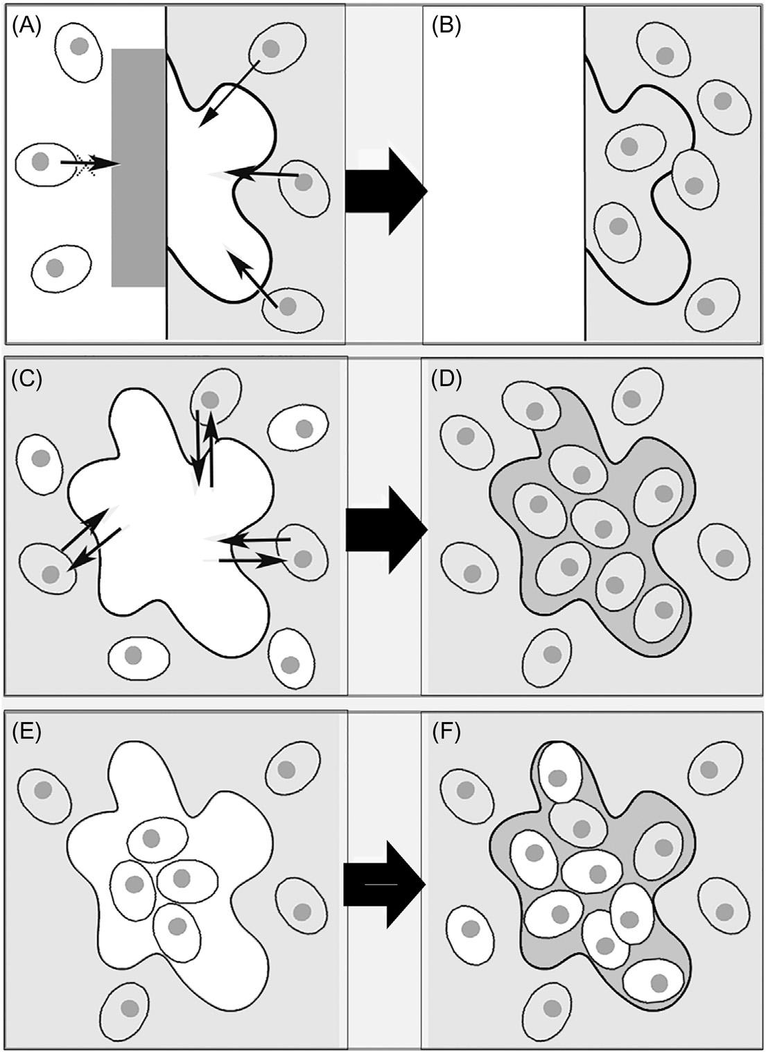
Inconductiveapproaches(Fig.1.1,panelsAandB),thebiomaterialactsasa 3Dmatrixforendogenouscellsmigration,grafting,proliferation,anddifferentiation.Thesecellsformthenewtissuethat,hopefully,willbeintegratedwiththe hosttissueandwiththegraftedbiomaterialthat,dependingonitscomposition, mayormaynotdegradeovertime.
Sometimes,however,endogenouscellmigration,differentiationofthesecells, andthefollowingtissueformationareprocessesthatneedtobecontrolled.Inthis case,aninductiveapproachtotissueengineeringappearstobethebestsolution (Fig.1.1,panelsCandD).Bioactivescaffolds,GFs,drug,orplasmidDNAdeliveryareusedtoinducecellmigrationandcontrolcellularbehavior.
Conductiveandinductivemethodsareusuallyusedincasesoflimiteddefects, asinmostmaxillaryormandibularbonesites,whenthepresenceofoptimized biomaterialspromotecellmigrationfromthehosttissueintothescaffolds.
However,therepairoflargedefectsoftenalsorequiresthedirecttransplantationofcells.Thisapproach(Fig.1.1,panelsEandF)isalsorequiredwhenthere arenotenoughavailablecellsinthetissuesurroundingthedefect,orwhen endogenouscellmigrationtothedamagedsitewouldrequiretoolongatime (Alsbergetal.,2001).Inthesecases,abiopsyisusuallytakenfromadonor source,cellsareisolatedandexpandedinvitro,andthenseededonadulypreparedscaffoldtoproliferateandformnewtissue.Inturn,thistissueisnormally implantedintothedamagedarea.
Autologouscellsgiveinsignificantimmuneresponse,buthavethedisadvantageofrequiringalongtimetoexpandtotheneededquantities.Allogeneiccells, whicharegeneticallydifferentcellsfromthesamespeciesasthepatientand evenmoresoxenogeneiccellsderivedfromadifferentspeciesthanthepatient, presenttheoppositeproblems:theyarereadyavailablebutstronglyincreasethe possibilityofimmunologicalreactions.
Thisisthecase,forexample,whentryingtorepairmuscledamageinDuchenne musculardystrophy(DMD)bytransplantingmyogenicprogenitorsdirectlyintothe muscles.Infact,thisprocedurehasshowntosufferfromtheproblemsoflimitedcell survivalandreducedmigrationofthesecellsinthemuscles(Farinietal.,2012).
Inparticular,celltherapiesconsistoftheuseofstemcellpopulationsthat havebeenpreviouslymanipulatedandculturedinvitro,withtheobjectiveof repairingandregeneratingdamagedtissues.Inthiscontext,thepresenceinthe heartofprimitivecells,abletogeneratethedifferentstructuresofthemyocardium,hasbeenrecentlydocumented(Giuliani,2012).
FIGURE1.1
Descriptionofthethreeapproachestotissueengineering.(AandB)Conduction—A scaffoldcontrolsandselectscellsinfiltratingthedefectsitefromtheoutsideinorderto repairit.Thescaffoldmayberesorbedintimeorsurgicallyremoved.(CandD) Induction—Bioactivemoleculesbindwithselectedhostcells(withreceptorsforthe molecules)thatmigratetothedefectsiteforminganewextracellularmatrix.(EandF) Celltransplantation—Cellsaretransplantedfromadonorsourcetoascaffold.Afterwards, thecell/scaffoldconstructisgraftedintothedefectsitetoachievetissueregenerationin conjunctionwithhostcellsthatmigratetothedefectsite.
Themonitoringofthelongitudinaloutcomesofconduction,induction,and celltherapyrequirestheuseofnondestructivemethodsthatarecapableofidentifyingthelocation,amount,andextentofcellularsurvivalandfate,aswellasof evaluating,qualitativelyandquantitatively,thetissuegrowthunderdifferentconditions.Thistypeofstudyhasbeenfrequentlyperformedintherecentliterature, forinstancetoengineerbone(Appeletal.,2013;Giulianietal.,2011,2014a; Olubamijietal.,2014),cartilage(Olubamijietal.,2014;Zehbeetal.,2010a), andtendon(Giganteetal.,2013).
Inthiscontext,imagingtechniquesareassuminganincreasinglyimportant role,notonlyforarigorouscharacterizationofthepropertiesandfunctionsof biomaterials,butalsotoinvestigatethekineticsoftheirbiologicalbehaviorin conduction,induction,andcelltransplantationprocesses.
Severaladvanced2Dimagingtechnologiesareavailabletocomplementhistologicalevaluationandtostudycomplexbiologicaleventsoccurringattheinterface betweentissuesandbiomaterials(Appeletal.,2013;Nametal.,2015).However, 2Dimagingtechnologiespresentseverallimitations.Infact(Zehbeetal.,2010a), thecurrentmethodsofopticalmicroscopyrequireasectioningofserialsections thattakesalongtimeandtheyareseverelylimitedintheirabilitytoanalyzeopaque3Dbiologicalstructures.Furthermore,althoughelectronmicroscopy(EM) methodsoffergood3Dtopographicrepresentation(byEMscanning),highresolutionimagingisrestrictedtoextremelythinsamples(bytransmissionEM). Moreover,forthepreviouslymentionedtechniques,samplepreparationsometimes causessignificantdamagetothespecimen,practicallyinhibitingthevisualization andquantificationofthecellsandtissuesspatialdistribution,presentandformed withinporousbiomaterialsinvitroandinvivoconditions.
Furthermore,forregenerationofvascularizedtissues,suchasboneormuscle, thereisaneedforimagingtechniquesabletoquantify3Dvascularingrowth,particularlyforrecentinnovativestudiesfocusedonexploringthepotentialto enhanceregenerationviatherapeuticangiogenesisstrategies(Langeretal.,2009; Appeletal.,2013;Giulianietal.,2017).
Theimagingmodalitymostextensivelyappliedforthispurpose,especiallyfor bonetissueengineeringstudies(Giulianietal.,2013;Manescuetal.,2016a; Komlevetal.,2009;Belicchietal.,2009),ishigh-resolutionX-raycomputed tomography(microCT).
Inparticular,theuseofsynchrotron-producedX-rayshasseveraladvantages withrespecttoX-raysproducedbylaboratoryorindustrialsources.
Inthischapter,severalmajorexperiencesapplyingsynchrotronradiation (SR)-basedmicroCTtoexploreregenerationinbone,tendons,cartilage,andskeletalandcardiacmusclesiteswerereviewed.
Conductive,inductive,andcelltransplantationapproacheswereoverviewed, althoughdifferentemphasiswasgiventoeachstrategydependingonthespecified engineeredtissue.
Inaddition,innovativeprotocolswereexploredtotrackdonorcellsaftertransplantationinordertoclarifytheirroleintissueregeneration.
1.2 X-RAYCOMPUTEDMICROTOMOGRAPHY: ACHALLENGINGDIAGNOSTICTOOL Anewconceptofdiagnosticbyimagingwasdevelopedatthebeginningofthe 1970s,whenthefirstequipmentforX-rayscomputedtomography(CT)wasproduced.TheCTtechniqueovercameseverallimitationsofconventionalX-ray radiology,manyofwhichweremainlyduetothe2Dnatureoftheradiological imagesthathadbeenuseduptothatmomentinmedicaldiagnostics.Indeed,conventionalanddigitalX-rayradiologyareimagingmethodswithconstraintslinked totheirtwo-dimensionality:radiographsprovidea2Dimageofa3Dobject,not accuratelyreplicatingtheanatomythatisbeingassessed.Anatomicalstructures maysuperimposecausingmisleadingsignalsforradiographinterpretation. Indeed,2Dradiographsusuallyshowminorstructuredamagescomparedtothose actuallypresentanddonotrevealtheinteractionsbetweensoftandhardtissues (Manescuetal.,2016b).
Forthesereasons,thediagnosticcontributionprovidedbytheCTwaspioneering,allowingforthevisualizationofinternaldetailsofthesamplewithunprecedentedprecision,inanondestructivewayandachievingacontrastupto1000 timesbetterthanconventionalradiography(Claesson,2001).
Tomographydataareacollectionofcross-sectionalimagesobtainedfrom eithertransmissionorreflectionconfigurationsandcollectedbyilluminatingthe samplefrommanydifferentdirections(KakandSlaney,1988).Thefirstapplicationswereindiagnosticmedicine,buttodayCTisacharacterizationtechnique alsousedinseveralnonmedicalimagingapplications.
X-raymicrotomography(microCT)exploitsthesamephysicalprinciplesof conventionalCTusuallyusedinmedicaldiagnostics,but,unlikethis,reachesa spatialresolutionofupto0.1 μm(Weitkampetal.,2010),thatis,aboutthree ordersofmagnitudehigher.
Thenumberofprojectionsandofdatapointsperprojectiondefinethespatialresolution:thus,largedatasetscont ainmoreinformation(i.e.,morepixels inasmallerobjectisequivalenttobette rspatialresolutions).Indeed,the choiceofspatialresolutionversusove rallsamplesizeisacrucialissuein microCT.
Both3DconventionalCTandmicroCTemployX-raystovirtuallyreconstruct thesamplesofinterestbasedontheirattenuationcoefficient.Inanidealsetup, thesamplemustabsorbavalueclosetoandatmostequalto90%oftheincident photons:inthisconfiguration,itisexpectedtoachievethebestsignal-to-noise ratio.
Tomographicimagesarereconstructedstartingfromitsprojections,thatis, theradiographiesacquiredover180 samplerotation(orreciprocalrotationofthe detectionsystemaroundafixedobject).Hundredsof2Dprojectionradiographs aretakenatseveraldifferentangles:eachofthemisaprojectionofabsorption densitydistribution,inthedirectionofthephotons,ontotheplaneperpendicular
tothedirectionofthebeam.Thismeansthat,ifthesampleisimagedindifferent orientations,3D(i.e.,volumetric)informationonthesamplestructurecanbe obtainedinasecondphaseusingcomputeralgorithms.Thissecondphase, referredtoas“tomographicimagereconstruction,”isbasedonsolvinganinverse problem,estimatinganimagefromitslineintegralsondifferentdirections,andis theoreticallyequivalenttotheinversionoftheRadonTransformoftheimage. Indeed,in1917Radonsolvedtheproblemtoreconstructafunctionfromradiographicprojections:thisfindingwasexploitedasHounsfield’sinventionofthe X-raycomputedtomographicscannerforwhichthesameHounsfieldreceivedthe NobelPrizein1972.
Nowadays,therearetwotypesofalgorithmswithdifferentapproaches (Penczek,2010)forthereconstructionphase:transform-basedmethodswhich exploitanalyticinversionformulae;andseriesexpansionmethodsbasedonlinear algebra.
Thetraditionalreconstructionalgorithmusedinmostpracticalapplicationsof microCTisthefilteredbackprojection(FBP),aFourier-basedtechnique.FBPis derivedfromtheFourierslicetheorem,describedindetailelsewhere(Kakand Slaney,1988).TheadvantagesofFBParethatitsimplementationisstraightforwardandexecutionisrelativelyfast.
Anotherapproachisbasedoniterativereconstructionalgorithms:thesegive aninitialguessoftheattenuationcoefficients(Webb,2003)andcomparesuch estimationswiththoseactuallyacquired.Thecorrectionoftheinitialmatrixis madeinaniterativewayforeachprojectioninafirststepandforthewholedatasetinasecondstepuntiltheresidualerrorbetweenthemeasureddataandestimatedmatrixfallsbelowapredesignatedvalue.Iterativeschemesarescarcely usedinstandardCT.
3Drenderingsofthedataobtainedafterthereconstructionareeasily obtainedbystackinguptheslicesandmaybesectionedinarbitrarywaysto betterlocateandquantifythedetails .Indeed,ifthe2Dslicesandthe3D reconstructionsar efascinatingtoolstoperform qualitativeobservationsofthe internalstructureofbiomaterials,cells andtissues,therealbenefitisthequantitativeinformationthatcanbeextractedfrom3Ddatasets( Ohgushietal., 1989 ).
Differentmethodsmaybeappliedtoextra ctquantitativearchitecturalparametersfromtomographicimages.Forinstance,inthefieldofboneresearch, differentprotocolshavebeenproposedforbonemicroarchitecturequantification.The3Dmeaninterceptlengthmethodprovidesagoodestimationoftrabecularthicknessandspacingbasedons tructuralgeometryassumptions,for example,parallelplatemodel( HildebrandandRuegsegger,1997a ).However, using3Dimagessuchassumptionscanbeavoided,allowingfortheachievementofnewmodel-independen tquantitativeparameters(Hildebrandand Ruegsegger,1997b).
1.3 INNOVATIVEAPPROACHESTOHIGH-RESOLUTION TOMOGRAPHYBYSYNCHROTRONRADIATION Byproperlyselectingthephotonenergy,theinteractionbetweenX-raysandbiologicalstructuresprovidessemitransparencyoftissues,allowingpenetrationof evenlargespecimens.Basedonthealgorithmsdescribedinthepreviousparagraph,angularprojectionscanbeusedfortomographicimaging.
Theimagingofcellsinsidebiologicaltissuesischallengingwithconventional microCTdevices,becauseofseveralexperimentalconditionslinkedtothelimited spatialandstructuralresolution.Amajorproblemisthelowdifferenceindensity andabsorptioncontrastbetweencellsandsurroundingtissue.Therefore,amonochromaticX-raybeam,sufficientlyhighphotonflux,andcoherentbeampropertiesarekeyrequirements,currentlyonlyachievedwithsynchrotronlight(Zehbe etal.,2010a).
SRisanelectromagneticlightcreatedwhenchargedparticles(forinstance electrons)areemittedbyanelectrongunandarethenlinearlyacceleratedbyan electricfield.Next,theparticlesarefurtheracceleratedtonearthespeedoflight intwoconnectedrings(thefirst,namedthe“boosterring,”andthesecondthe “storagering”).Inthestoragering,astheelectronstravelroundthering,they passthroughdifferenttypesofmagnetsand,intheprocess,theyproduceX-rays. Indeed,thesemagnetscausetheelectronstochangedirection:thisresultsina changeintheirvelocityvectorand,consequently,intheemissionofSR.Inparticular,whentheelectronismovingfastenough,theemittedenergyisatX-ray wavelength(Giulianietal.,2014b;Olubamijietal.,2014;http://www.esrf.eu/ about/synchrotron-science/synchrotron).
ThepropertiesofSRsignificantlyimprovecontrastsensitivityinX-rayimagingsystems(Canceddaetal.,2007; Giulianietal.,2010),asspecificallydiscussedindetailinthenextparagraphsforthedifferentanatomicaldistricts.
X-raysproducedbysynchrotronfacilitieshaveseveraladvantagescompared tothosedeliveredbydesktoplaboratorysources.Inparticular,theyofferseveral possibilities:
1. togetahighphotonflux,achievingmeasurementswithhighsignal-to-noise ratioaswellashighspatialresolutions;
2. totunetheX-raysource,allowingmeasurementsatdifferentenergies;
3. tosetaspecificmonochromaticX-rayradiation,eliminatingthebeam hardeningeffects;
4. toperformparallelbeamacquisitions,allowingtheuseofspecific tomographicreconstructionalgorithms.
Duetothelistedadvantagesofferedbysynchrotronlightsources,several3D imagingtechniqueshavebeendevelopedinordertoserveandsupportbiomedical research.
Inapureabsorptionexperimentalsetup,thesampleisascloseaspossibleto thedetector,withaminimizationofimageblurring.Atsynchrotronfacilities,parallelphotonbeamsaremadeavailablefromawidecross-sectionalsource;they passthroughthesampleandarecollectedbya2Ddetector.BeingtheX-ray beamparallel,theprojectionofeachsliceoftheobjectonthedetectorisnot dependentofalltheotherslices(Stock,2009).
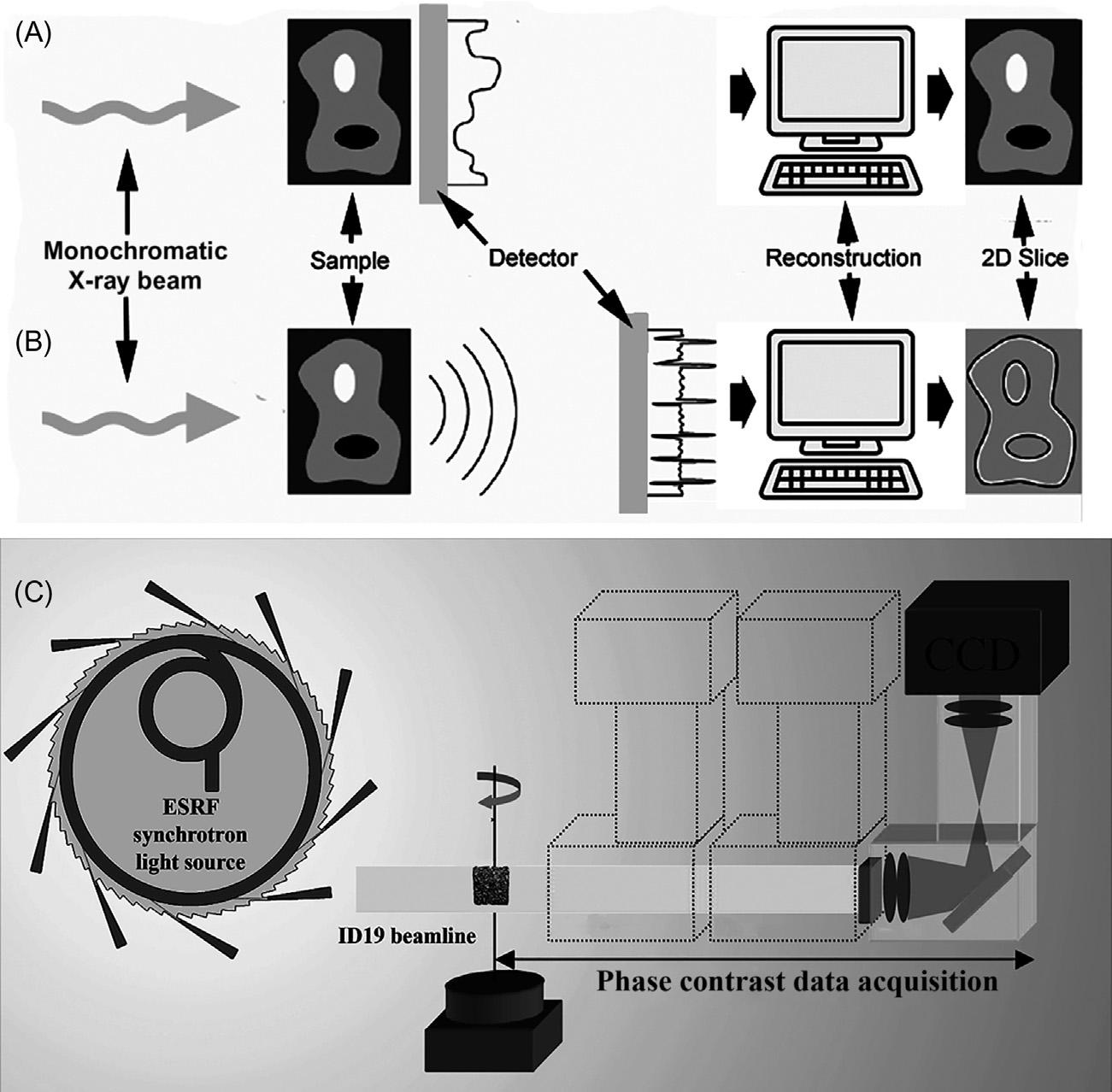
Absorptiontomography(Fig.1.2,panelA)isbasedontheBeer-Lambertlaw, whichdescribesthattheintensityofmonochromaticX-raysthattransmitthe specimendecreasesexponentiallyasafunctionofthelineintegralofthelinear attenuationcoefficientsalongtheX-raypath:
where μ isthelinearabsorptioncoefficientatposition x alongaparticularbeam direction.Tomographysolvestheproblemofassigningthecorrectvaluesof μ to eachpositionalongthephotonpath,knowingonlythevaluesofthelineintegral butforalargenumberofpathsthroughthesample(i.e.,rayssentthroughthe samplefromdifferentangles).Astheypassthroughthedifferentphasesofthe sample,thephotonsinteractintheelectronshellsoftheatomstheypass. AbsorptiontomographyexploitsthattheattenuationofX-raybeamsofagiven energyvarieswiththeatomicelectrondensityoftheimagedmaterialanditsbulk density.ThedifferencesintheX-rayattenuationratewithinthesamplesare representedbydifferentpeaksinthegray-scalehistogram,correspondingtothe differentphases.
However,bymodulatingthesample-to-detectordistance,contrastisalsogeneratedbyphasedifferencesamongthescatteredX-raywaves(Fig.1.2,panelB). Inparticular,thisphase-contrast(PhC)effectputsintoevidencetheinterfaceand edgesbetweentwomaterials,anditisparticularlyusefulwhenmediawithsimilar absorptioncoefficientsshouldbediscriminated(Fiorietal.,2012).Indeed,differentlyfromconventionalX-raytomography,inthePhCapproach,theimagecontrastisnotbasedsolelyonattenuationofthebeam.Inthiscase,theeffectofan X-raybeampenetratingthesampleisdescribedbytherefractiveindex,
where δ istherefractiveindexdecrementand β istheattenuationindex. δ isactuallyproportionaltothemeanelectrondensityofthespecificphase,whichinturn isnearlyproportionaltoitsmassdensity.Moreover,the δ valueismuchlarger thantheimaginarypart β innonmineralizedtissues,suggestingthatthephase approachprovidesgreatersensitivitythantheabsorptionapproachwhenstudying thesetissues.
Themethodsusedforthereconstructionoftherefractiveindex n aretypically basedonatwo-stepapproach:thephaseprojectionsareextractedinthefirststep whiletherefractiveindexdecrement δ isreconstructedbyapplyingaconventionalFBPorsimilaralgorithmsinthesecondstep(Giulianietal.,2014a,b).
FIGURE1.2
(A)Standard(absorption-based)tomography—Thesampleismountedonatranslationrotationstage(standardSR-microCTsetup).Thedetector(madeupofascintillator,light microscopeoptics,andaCCD)ismountedonatranslationstage.Projectionsare acquiredwiththedetectorclosetothesampleand,afterwards,areprocessedwiththe filteredbackprojectionalgorithmforthereconstructionofthe3Dabsorptionindex.(B) Propagation-basedphase-contrastimaging—Theimagingsourceconsists,alsointhis case,ofmonochromatizedSRX-rays.Thelongsourcetosampledistanceyieldsahigh degreeofspatialcoherence.Thedetector,mountedonatranslationstageatalong distancefromthesample,allowsfreespacepropagationofthebeamafterthesample.In thiscase,theapplicationoftheFBPalgorithmproducesanedge-enhancementeffect, whichisproportionaltotheLaplacianoftherefractiveindex.(C)In-linephasetomography (holotomography),fromRef.(Giulianietal.,2013)—Thetomographicacquisitionis performedwiththedetectordistances(D1, D2, , Dn)fromthesample.Foreachrotation angle,aphaseretrievalalgorithmisappliedtoprojectionsacquiredateachdistance, providingthephasemaps,inturn,arethenprocessedwiththeFBPalgorithmtorecover the3Drefractiveindexdecrement.
Forthefirstphase(Langeretal.,2010),twomainclassesofalgorithmscan beidentifiedinliterature:theyarealternativelybasedonalinearizationwith respecttothepropagationdistance,whichyieldswhatisknownasthetransport ofintensityequation(TIE)(Teague,1982a,b),oronalinearizationwithrespect totheobject,whichyieldsthecontrasttransferfunction(Cloetensetal.,1999).
Furthermore(Manescuetal.,2016a,b),phaseretrievalusuallyimpliesthe reconstructionofthetwodifferentreal-valued3Ddistributions, δ and β ;such reconstructiongenerallyrequirestheacquisitionof2Dprojections,atleastattwo differentsample-detectordistancesateachviewangle(Fig.1.2,panelC).
However,insomecases,itcanbeshownapriorithattherealandimaginary partsoftherefractiveindexareproportionaltoeachother,thatis:
wheretheproportionalityconstant ε doesnotdependonthespatialcoordinates. Thisassumptionispossibleonlyforspecialclassesofobjects,suchaspurephase (i.e.,weaklyabsorbing)objects,orhomogeneousobjects,suchasobjectsconsistingpredominantlyofasinglematerial(possiblywithaspatiallyvaryingdensity) (Gureyevetal.,2004,2006,2009).
Moreover(Giulianietal.,2014a,b),Bronnikovsuggestedanalgorithmprovidingadirectreconstructionoftherefractiveindexandavoidingthefirstphase retrievalstep.Itestablishedafundamentalrelationbetweenthe3DRadontransformoftheobjectfunctionandthe2DRadontransformofthephase-contrastprojection(Bronnikov,2000).Thus,areconstructionalgorithmisderivedintheform ofaFBP.
Inrecentyears,therehasbeenincreasinginterestinthementionedapproaches toevaluatedifferentbiomaterials’performancebymeansofSR-microCT.Tissue regenerationderivedfromhostingsitesgraftingwithdifferenttypesofbiomaterials(withorwithoutstemcellsseeding),wasrecentlyexploredusingSRmicroCT(Canceddaetal.,2007; Giulianietal.,2014a,2016; Rominuetal., 2014; Giganteetal.,2013).
Evaluationofnewlyformedtissueisusuallybasedonhistology,byobservation ofoneormoresections;however,conventionalhistologicalevaluationandcorrespondinghistomorphometricmeasurementsprovideonly2Dinformationwiththe consequentriskthattheselectedsectionsdonotproperlyrepresenttheentirebiopsy specimen(Giuliani,2016).Furthermore,iftheinvolvementofneighboringtissues withdifferentmorphology(bone,unmineralizedextracellularmatrix,regenerated vessels,etc.)ontheregenerationprocessofdefectsortissuesisunknownorstill notclearlyverified,3Danalyzingmethods,includinghigh-resolutionSR-microCT, areindicatedtoexplorethedynamicandspatialdistributionofregenerativephenomenaintheseanatomicstructures.Traditionally,absorptionimagingwithSRmicroCTinmedicalapplicationswasperformedwithalmostnodistancebetween sampleanddetector,obtainingsignificantinformationonmorphometricdistribution ofthebioengineeredstructures(Canceddaetal.,2007; Giulianietal.,2014a,2016; Renghinietal.,2013; Pozdnyakovaetal.,2010).However,homogeneousmaterials,
withalowattenuationcoefficient(likecollagen,polymers,thermosetandthermoplasticmatrices,unmineralizedextracellularmatrix,vessels,nerves,etc.),orheterogeneousmaterials,withanarrowrangeofattenuationcoefficients(likethecaseof heterologousbonescaffoldsorgradedmineralizedbone),produceinsufficientcontrastforabsorptionimaging.Forsuchmaterials,theimagingqualitycanbe enhancedthroughtheuseofPhC-microCT,withanincreaseddistancebetween sampleanddetector(Manescuetal.,2016a,b;Giulianietal.,2014b;Albertini etal.,2009).Inaddition,whereasPhC-microCTcanbebasedonasingledistance betweenthedetectorandthesample(forthespecialclassesofobjectspreviously mentioned),holotomography(HT)involvesPhCimagingatseveraldistances (Fig.1.2,panelC),thencombiningthephaseshiftinformationtogenerate3D reconstructions.
HTishelpfulwhenthematerialofinteresthasextremelysmallvariationsin attenuationcoefficients,whichleadtounsatisfactoryimagingresultsevenwith phase-contrasttechniquesonasingle-distanceandthephaseretrievalalgorithms previouslydescribed(Giulianietal.,2013;Giuliani,2016).
1.4 SKELETALTISSUEENGINEERING 1.4.1 BONE Bonestructureshavefundamentalfunctionsinthebody.Whencongenitaldefects, trauma,ordiseasesarepresent,thereisasignificantneedforbonereplacement (Alsbergetal.,2001).
Thecombinationoflivingcells,biologicallyactivemolecules,andastructural scaffoldtoformaconstructabletopromotetherepairandregenerationofbone isthefundamentalconceptunderlyingboneengineering.Thescaffoldplaysacrucialrole,beingexpectedtosupportcellcolonization,migration,growth,anddifferentiation.Inparallelwithboneformation,thescaffoldmayundergo degradation,releasingproductsthathavetobebiocompatibleorthatareeasily excretedorsubjectedtometabolism(Hutmacheretal.,2007).
Inthiscontext,imagingtechniques,includingSR-PhC-microCTinvestigations,wereextensivelyappliedtoinvestigatethepropertiesofseveralbiomaterialsproposedtoactasscaffolds.
Sinceeachbonesiteperformsmultiplefunctionalroles,itisunlikelythata singlescaffoldwouldserveasauniversalsupportfortheregenerationofbonetissue.Theconsiderationsforscaffolddesignare,hence,complexandincludematerialcomposition,architecture,structuralmechanics,surfaceproperties, degradationpropertiesandproducts,togetherwiththecompositionofanyadded biologicalcomponentand,ofcourse,thechangesinallofthesefactorswithtime (Hutmacheretal.,2007).
Allowinganaccurate3Dexaminationofsamples,SR-basedmicroCTwasnot onlyemployedtoreconstruct,athighresolution,thecomplexarchitectureofbone
tissueatdifferentscales(Langeretal.,2012;Peyrinetal.,2014;Giulianietal., 2018a)andindifferentgeneticandenvironmentalconditions(Tavellaetal., 2012;Costaetal.,2013;Cancianietal.,2015;Giulianietal.,2018b),butitis alsoincreasinglybecomingapowerfultoolforengineeredbonecharacterization indifferentskeletalsites.
Inthisscenario,interestingmicroCTstudieshavebeenperformedondifferent biomaterialsthathavepreviouslybeenindicatedasbone-substitute(Olubamiji etal.,2014).
SR-microCTwasexploitedbyYue(Yueetal.,2010)tocharacterizethescaffoldmorphology,themineraldistributionwithinscaffoldpores,andthetissue ingrowthin4-week-oldexplantsofabioactiveglassfoamscaffoldimplanted betweenthemuscleandtibiaofamouse.Similarly,SR-microCTwasusedto successfullyidentifyscaffoldarchitectureandboneingrowthintocell-loaded hydroxyapatitescaffoldsimplantedinimmunodeficientmicefor8weeks (Mastrogiacomoetal.,2004).Theboneingrowthwasestimatedintermsoftotal volumefraction,distribution,andthicknessintheporesoftheimplantandthe scaffoldarchitecturewasanalyzedintermsoftheporosityandspatialdistribution ofwalls.ThesamegroupexploredtheabilityofSR-microCTtoexaminetheprogressiveresorptionofandboneingrowthintoscaffoldsimplantedinimmunodeficientmiceforrepairtimesof8,16,or24weeks(Canceddaetal.,2007; Papadimitropoulosetal.,2007; Komlevetal.,2006).Whenusingahydroxyapatitescaffold(Engipore),asinglepeakwasobservedintheX-rayabsorptionhistogrambeforeimplantation,correspondingtothebiomaterialusedforthe manufacturingofthescaffolditself(Fig.1.3,panelA).Afterimplantation,an additionalpeakwasobservedatlowerX-rayabsorptionvalues,correspondingto thenewlyformedbone(Fig.1.3,panelB).
Itispossibletoobservethatthenewlyformedbonepeakshiftedtohigher valuesoflinearattenuationcoefficientwiththeincreasingoftheimplantation time:thisisexplainedbytheprogressivemineralizationofthebone.
Indentaldistricts,successfulboneregenerationusingbiphasiccalciumphosphatematerialswasreportedinsomeclinicalapplicationsformaxillarysinuselevation(Manganoetal.,2013;Ohayon,2014).Specialmorphologiesof3D scaffolds,intheshapeofgranulesorstructuredblocks,wereshowntorealize promisingscaffoldstobeusedeitherinanacellularstrategy(purescaffoldgraftinganditscolonizationbyendogenouscells)(Manganoetal.,2013)orcombiningthebiomaterialwithcellsinvitro(Barbonietal.,2013).
Whilepreviouslyreportedstudieswereusuallybasedonsingletimepoints, thelong-termkineticsofboneregenerationbeingnotfullyinvestigated,arecent clinicalstudy(Giulianietal.,2016)reportedaquantitativekineticsevaluationof blocksversusgranulesinbiphasiccalciumphosphatescaffoldscarriedoutbySR microCT.Twenty-fourbilateralsinusaugmentationswereperformedandgrafted withHA/β-TCP30/70,12withgranulesand12withblocks.Thesampleswere retrievedat3,5/6and9monthsfromgraftingandwereevaluatedforboneregeneration,graftresorption,neovascularization,andmorphometricparameters.Big


