Northern Rehabilitation and Sports Medicine Associates DeKalb, Illinois
Adjunct Assistant Professor Marquette University Milwaukee, Wisconsin
HISTORY OF MANIPULATION
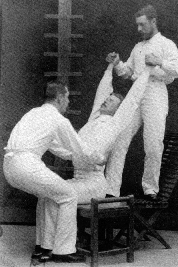
Manipulation in recorded history can be traced to the days of Hippocrates, the father of medicine (460–370 bc). Evidence is seen in ancient writings that Hippocrates used spinal traction methods. In the paper “On Setting Joints by Leverage,” Hippocrates describes the techniques used to manipulate a dislocated shoulder of a wrestler.3 Succussion was also practiced in the days of Hippocrates. The patient was strapped in an inverted position to a rack that was attached to ropes and pulleys along the side of a building. The ropes were pulled to elevate the patient and the rack as much as 75 feet, at which time the ropes were released and the patient crashed to the ground to receive a distractive thrust as the rack hit the ground4 (Figure 1-1). Six hundred years later, Galen (130–200 ad) wrote extensively on exercise and manipulation procedures in medicine.3
Hippocrates’ methods continued to be used throughout the Middle Ages, with little advance in the practice of medicine and manipulation because of the reliance on the church for most healing throughout Europe. 3 In the Renaissance era, Ambroise Paré (1510–1590) emerged as a famous French physician and surgeon 3 who used armor to stabilize the spine in patients with tuberculosis 4 ( Figure 1-2 ). His manipulation and traction techniques were similar to those of Hippocrates, but he opposed the use of succussion. 4
The bonesetters flourished in Europe from the 1600s through the late 1800s. In 1656, Friar Moulton published The Complete Bone-Setter. The book was later revised by Robert Turner.4 No formal training was required for bonesetters;
the techniques were often learned from family members and passed down from one generation to the next. The clicking sounds that occurred with manipulation were thought to be the result of bones moving back into place.4
In 1871, Wharton Hood published On Bone-Setting, the first such book by an orthodox medical practitioner. 5 Hood learned about bonesetting after his father had treated a bonesetter, Richard Hutton. Hutton was grateful for the medical care and offered to teach his practitioner about bonesetting. Instead, it was the practitioner’s son, Wharton Hood, who accepted the offer. Hood thought that the snapping sound with manipulation was the result of breaking joint adhesions. 5 Paget 6 believed that orthodox medicine should consider the adoption of what was good and useful about bonesetting but should avoid what was potentially dangerous and useless.
Osteopathy was founded by Andrew Still (1826–1917) in 1874. In 1896, the first school of osteopathy was formed in Kirksville, Missouri.4 Still developed osteopathy based on the “rule of the artery,” with the premise that the body has an innate ability to heal and that with spinal manipulation to correct the structural alignment of the spine, the blood can flow to various regions of the body to restore the body’s homeostasis and natural healing abilities. Still’s philosophy placed an emphasis on the relationship of structure to function and used manipulation to improve the spinal structure to promote optimal health.7 The osteopathic profession continues to include manipulation in the course curriculum but does not adhere to Still’s original treatment philosophy. Many osteopathic physicians in the United States do not practice manipulation regularly because they are focused on other specialty areas, such as internal medicine or emergency medicine. Osteopathy in many European countries remains primarily a manual therapy profession.
Chiropractic was founded in 1895 by Daniel David Palmer (1845–1913). One of the first graduates of the Palmer School of Chiropractic in Davenport, Iowa, was Palmer’s son Bartlett Joshua Palmer (1882–1961), who later ran the school and
FIGURE 1-2 Ambroise Paré applied manual therapy to the spine in conjunction with spinal traction, similar to Hippocrates’ methods described over 1000 years earlier. (From Paré, Ambroise. Opera. Liber XV, Cap. XVI, pp. 440-441, Paris, 1582.)
promoted the growth of the profession. D. D. Palmer was a storekeeper and a “magnetic healer.” According to legend, in 1895 he used a manual adjustment directed to the fourth thoracic vertebra that resulted in the restoration of a man’s hearing.8 The original chiropractic philosophy is based on the “law of the nerve,” which states that adjustment of a subluxed vertebra removes impingement on the nerve and restores innervation and promotes healing of disease processes.3 The “straight” chiropractors continue to adhere to Palmer’s original subluxation theories and use spinal adjustments as the primary means of treatment. The “mixers” incorporate other rehabilitative interventions into the treatment options, including physical modalities, such as therapeutic ultrasound and exercise.
The origins of physical therapy can be traced to the Royal Central Institute of Gymnastics (RCIG), founded in 1813 by Pehr Henrik Ling (1776–1839) in Stockholm, Sweden 9,10 ( Figure 1-3 ). Ling’s educational system included four branches: pedagogical gymnastics (physical education), military gymnastics (mostly fencing), medical gymnastics (physical therapy), and esthetic gymnastics (philosophy). Ling systematized medical gymnastics into two divisions, massage and exercise, with massage defined as movements done on the body and exercise being movements done with a part of the body. 11,12 Ling may not have been the originator of medical gymnastics or massage, but he systematized these methods and attempted to add contemporary knowledge of anatomy and physiology to support medical gymnastics. 11,12
Graduates of the RCIG earned the title “director of gymnastics” and in 1887 were licensed by Sweden’s National Board of Health and Welfare, where physical therapists continue to use the title sjukgymnast (“gymnast for the sick”).9,13 Throughout the nineteenth century, the RCIG provided its graduates
with a scientific rationale, based on contemporary knowledge of anatomy and physiology, for the benefits of combining specific active, resistive, and passive movements and exercises, including variations of spinal manipulation, traction, and massage.9 “Ling’s doctrine of harmony” purported that the health of the body depended on the balance between three primary forms: mechanics (movement/exercise/manipulation), chemistry (food/medicine), and dynamics (psychiatry), and the Ling physical therapists were trained to restore this harmony through use of manual therapy.
Graduates of RCIG immigrated to almost every major European city, Russia, and North America through the mid to late 1800s to establish centers of medical gymnastics and mechanical treatments.9 Jonas Henrik Kellgren (1837–1916) graduated from the RCIG in 1865, eventually opened clinics in Sweden, Germany, France, and London, and is credited with development of many specific spinal and nerve manipulation techniques.9 In addition, medical doctors from throughout Europe enrolled in the RCIG to add physical therapy methods to their treatment of human ailments and attained joint credentials as physician/physical therapist. Edgar F. Cyriax (1874–1955), the son-in-law of Kellgren and a graduate of RCIG before becoming a medical doctor, published more than 50 articles on Ling’s and Kellgren’s methods of physical therapy in international journals and advocated to include “mechano-therapeutics” in the curriculum and training of medical doctors in Britain.9 In 1899, the Chartered Society of Physiotherapy was founded in England.3 The first professional physical therapy association in the United States, which was the forerunner to the American Physical Therapy Association (APTA), was formed in 1921.1
Between 1921 and 1936, at least 21 articles and book reviews on manipulation were found in the physical therapy literature,14 including the 1921 textbook, Massage and Therapeutic Exercise, by the founder and first president of the APTA, Mary McMillan. McMillan credits Ling and his followers with development and refinement of the methods used to form the physical therapy profession in the United States.11,12 In fact, McMillan devotes a 15-page chapter of her book to specific therapeutic exercise regimes developed by Ling referred to as “A Day’s Order” and states that the term medical gymnastics is synonymous with therapeutic exercise. In a subsequent editorial,11 she wrote of the four branches of physiotherapy, which she identified as “manipulation of muscle and joints, therapeutic exercise, electrotherapy, and hydrotherapy.”12 Titles of articles during this period were quite explicit regarding manipulation, such as “The Art of Mobilizing Joints”15 and “Manipulative Treatment of Lumbosacral Derangement.”16 The articles used phrases such as “adhesion . . . stretched or torn by this simple manipulation”17 and “manipulation of the spine and sacroiliac joint.”18 This usage helps illustrate that manipulation has been part of physical therapy practice since the founding of the profession and through the 1930s.14
From 1940 to the mid-1970s, the word manipulation was not widely used in the American physical therapy literature.3 This omission may have been due in part to the American
FIGURE 1-3 Thoracic traction as performed by graduates of the Royal Central Institute of Gymnastics in the mid-1800s. (Reproduced with permission from Dr. Ottosson, http://www.chronomedica.se.)
Medical Association’s Committee on Quackery, which was formed in the 1960s and was active for the next 30 years in an attempt to discredit the chiropractic profession. The committee was forced to dissolve in 1990 because of Wilk’s “restraint of trade” case, which was upheld in the US Supreme Court.8 Because physical therapy remained within the mainstream medical model, the terms mobilization and articulation were used during this timeframe to separate physical therapy from chiropractic. However, physical therapists continued to practice various forms of manipulation.
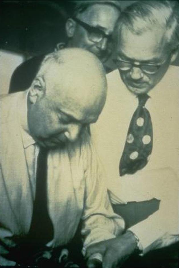
Through the early to mid 1900s, several prominent European orthopaedic physicians influenced the practice of manipulation and the evolution of the physical therapist’s role as a manipulative therapist. Between 1912 and 1935, James Mennell (1880–1957) provided advanced training in manipulation technique for physiotherapist at St. Thomas’s Hospital in London.19 In 1949, James Mennell published his textbook titled the Science and Art of Joint Manipulation. Mennell adapted knowledge of joint mechanics in the practice of manipulation and coined the phrase “accessory motion.”20 James H. Cyriax (1904–1985), son of Edgar Cyriax and grandson of Jonas Henrik Kellgren, published his classic Textbook of Orthopaedic Medicine in 1954. He made great contributions to orthopaedic medicine with the development of detailed systematic examination procedures for extremity disorders, including refinement of isometric tissue tension signs, end feel assessment, and capsular patterns.21 Cyriax attributed most back pain to disorders of the intervertebral disc and used aggressive general manipulation techniques that included strong manual traction forces to “reduce the disc.”21 Cyriax, who also taught and practiced orthopaedic medicine at St. Thomas’s Hospital until 1969 and was the successor of Mennell at St Thomas’s,22 influenced many physiotherapists, including Stanley Paris and Freddy Kaltenborn, to carry on the skills and techniques required to effectively use manipulation.
Alan Stoddard7 (1915–2002) was a medical and osteopathic physician in England who used skillful specific manipulation technique and also mentored many physical therapists, including Paris and Kaltenborn (Figure 1-4). Stoddard authored two textbooks, Manual of Osteopathic Technique (1959) and Manual of Osteopathic Practice (1969), which became the cornerstone of osteopathic teaching in schools around the world.23 Physical therapists, Kaltenborn24 and Paris,25 both believed that the Cyriax approach to extremity conditions was excellent, but they preferred Stoddard’s specific manipulation techniques for the spine.
John Mennell (1916–1992), the son of James Mennell, first practiced orthopaedic medicine in England. In the 1960s, he immigrated to the United States, where he held many educational programs for physical therapists through the 1970s and 1980s to promote manipulation within the physical therapy profession. He published several textbooks, including Joint Pain, Foot Pain, and Back Pain and coined the phrase “joint play.”27 Mennell brought attention to sources of back pain other than the intervertebral disc.
In the 1960s, several physical therapists emerged as international leaders in the practice and instruction of manipulation. Physical therapist Freddy Kaltenborn, originally from Norway, developed what is now known as the Nordic approach. He published his first textbook on spinal manipulation in 1964 and was the first to relate manipulation to arthrokinematics.25 His techniques were specific and perpetuated the importance of biomechanical principles, such as the concave/convex and arthrokinematic rules. Kaltenborn, in collaboration with physical therapist Olaf Evjenth, also developed extensive longterm training programs for physical therapists to specialize in manual therapy first in Norway and later throughout Europe, North America, and Asia.
Australian physical therapist, Geoffrey Maitland (1924–2010), published the first edition of his book Vertebral Manipulation in 1964.28 Maitland was also influenced by the work of Cyriax and Stoddard but further refined the importance of a detailed history and comprehensive physical examination. He also developed the concept of treatment of “reproducible signs” and inhibition of joint pain with use of gentle oscillatory manipulation techniques. Maitland developed the I to IV grading system to further describe oscillatory manipulation techniques.28 Maitland also established long-term manual therapy educational programs affiliated with universities in Australia, which subsequently facilitated the rapid growth of musculoskeletal physical therapy research.
Physical therapist, Stanley Paris, was originally from New Zealand. Early in his career, in 1961 and 1962, he was awarded a scholarship to study manipulation in Europe and the United States.14 He had the opportunity to study with Cyriax, Stoddard, and Kaltenborn during this time and in 1965 published the textbook Spinal Lesion.26 In the late
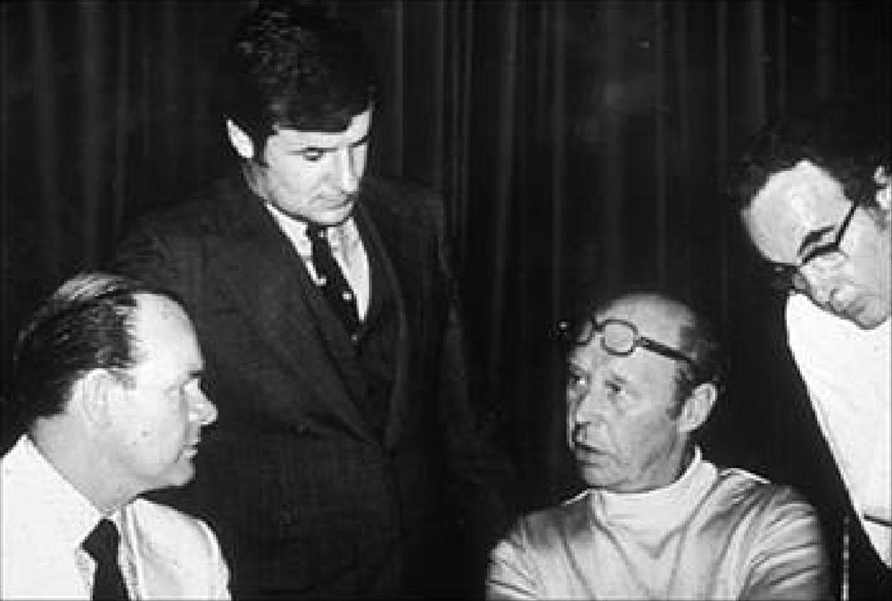
FIGURE 1-4 Cyriax (left) and Stoddard (right) in Norway, 1965. (From Kaltenborn FM: Manual mobilization of the joints: volume II: The spine, Oslo, Norway, 2012, Norli.)
FIGURE 1-5 Photograph was taken in 1974 in Montreal, Canada, at the successful formation of the International Federation of Orthopaedic Manipulative Physical Therapists (IFOMPT). Dr. Paris was Chair of the conference. The other three individuals were consultants to the process and had served in that capacity for 6 years before this event. IFOMPT later became a subsection of the World Confederation for Physical Therapy (WCPT). From left: Geoffrey Maitland, Stanley Paris, Freddy Kaltenborn, and Gregory Grieve. (From Paris SV: 37th Mary McMillan lecture: in the best interest of the patient, Phys Ther 86[11]:1541-1553, 2006.)
1960s, Paris immigrated to the United States, where he eventually completed his doctoral work in neuroanatomy of the lumbar spine and developed extensive educational programs for postprofessional physical therapy education in manual physical therapy and manipulation that eventually resulted in the formation the University of St. Augustine for Health Sciences in St. Augustine, Florida. Paris also played key roles in formation of professional organizations in the United States, including the APTA Orthopaedic Section and the American Academy of Orthopaedic Manual Physical Therapists (AAOMPT), two professional organizations that have played roles in advocating for inclusion of manipulation within the scope of physical therapy practice and that have promoted education, practice, and research in manual physical therapy. Paris worked with physical therapists Maitland, Kaltenborn, and Gregory Grieve of the United Kingdom to form the International Federation of Orthopaedic Manipulative Physical Therapists (IFOMPT; Figure 1-5).
The IFOMPT was founded in 1974 and represents organized groups of manual/manipulative physical therapists around the world that have established stringent postgraduation specialization educational programs in manual/manipulative physical therapy. The federation sets educational and clinical standards and is a subgroup of the World Confederation of Physical Therapy (WCPT). One organization of each WCPT country can be recognized by IFOMPT to represent that country if the organization meets IFOMPT criteria. The IFOMPT educational standards and international monitoring system has allowed physical therapists to be recognized as orthopaedic manual physical therapy (OMPT) specialists in countries beyond the country where they received their training.
The Orthopaedic Section of the APTA represents all aspects of musculoskeletal physical therapy and is open to all members of the APTA, including physical therapist assistants. Before formation of the AAOMPT, no organization in the United States could meet the IFOMPT criteria because no recognized educational system in manual therapy upheld standards of training and examination in manual therapy for physical therapists in the United States. However, by 1990 at least eight active manual therapy fellowship programs were operating independently within the United States.
In 1991, Freddy Kaltenborn invited representatives from these eight manual therapy fellowship programs to meet at Oakland University in Michigan to consider how the United States could develop educational standards in manual therapy and become a member organization of IFOMPT.29 These eight physical therapists, Stanley Paris, Mike Rogers, Michael Moore, Kornelia Kulig, Bjorn Swensen, Dick Erhard, Joe Farrell, and Ola Grimsby, became the founding members of the AAOMPT. The AAOMPT developed a standards document, bylaws, and a recognition process for manual therapy fellowship programs. In 1992, the AAOMPT was accepted as the member organization to represent the United States in IFOMPT.
Although prominent individuals, such as Paris, Kaltenborn, and Maitland, played a large role in development and advancement of manipulation and manual therapy within the physical therapy profession over the last half of the twentieth century, the current practice and the future of the specialty area of OMPT are driven by evidence-based practice and the promotion of OMPT practice through professional associations, such as IFOMPT, AAOMPT, and the APTA. 29 A large and growing body of research evidence supports and guides the practice of manipulation within the scope of physical therapy practice and for other manual therapy practitioners.
IFOMPT defines OMPT as a specialized area of physiotherapy/ physical therapy for the management of neuro-musculo-skeletal conditions, based on clinical reasoning, using highly specific treatment approaches including manual techniques and therapeutic exercises. OMPT also encompasses, and is driven by, the available scientific and clinical evidence and the biopsychosocial framework of each individual patient (see the IFOMPT Constitution 2012 at http://www.ifompt.com/site/ifompt/files/pdf// IFOMPT_Constitution.pdf).
IFOMPT considers the following terms as being interchangeable: orthopaedic manual therapy, orthopaedic manual physical therapy, orthopaedic manipulative therapy, and orthopaedic manipulative physical therapy (per IFOMPT Constitution 2012).
Paris30 described a nine-point “Philosophy of Dysfunction” that summarizes the components of a traditional OMPT
BOX 1-1 Philosophy of Dysfunction as Described by Paris
I. That joint injury, including such conditions referred to as osteoarthritis, instability, and the after effects of sprains and strains, are dysfunctions rather than diseases.
II. That dysfunctions are manifest as either increases or decreases of motion from the expected normal or by the presence of aberrant movements. Thus, dysfunctions are represented by abnormal movements.
III. That where the dysfunction is detected as limited motion (hypomobility), the treatment of choice is manipulation to joint structures, stretching to muscles and fascia and the promotion of activities that encourage a full range of movement.
IV. That when the dysfunction is manifest as increased movement (hypermobility), laxity or instability, the treatment of the joint in question is not manipulation but stabilization by instruction of correct posture, stabilization exercises and correction of any limitations of movement in neighboring joints that may be contributing to the hypermobility.
V. That the primary cause of degenerative joint disease is joint dysfunction. Therefore, it may be concluded that its presence is due to the failure or lack of accessibility to physical therapy.
VI. That the physical therapist’s primary role is in the evaluation and treatment of dysfunction, whereas that of the physician is the diagnosis and treatment of disease. These are two separate but complementary roles in health care.
VII. That since dysfunction is the cause of pain, the primary goal of physical therapy should be to correct the dysfunction rather than the pain. When, however, the nature of the pain interferes with correcting the dysfunction, the pain will need to be addressed as part of the treatment program.
VIII. That the key to understanding dysfunction, and thus being able to evaluate and treat it, is understanding anatomy and biomechanics. It therefore behooves us in physical therapy to develop our knowledge and skills in these areas so that we may safely assume leadership in the non-operative management of neuromusculoskeletal disorders.
IX. That it is the patients’ responsibility to restore, maintain, and enhance their health. In this context, the role of the physical therapist is to serve as an educator, to be an example to the patient, and to reinforce a healthy and productive lifestyle.
treatment philosophy (Box 1-1). Paris defines “dysfunction” as increases or decreases of motion from the expected normal or as the presence of aberrant movements.4 Therefore, the primary focus of the orthopaedic manual physical therapist’s examination is the analysis of active and passive movement. If hypomobility is noted, joint mobilization and stretching techniques are used; if hypermobility is noted, stabilization exercises, motor control, and postural correction are emphasized. If aberrant movements are noted, a motor retraining exercise approach is appropriate. If localization of tissue reactivity and pain are noted, gentle oscillatory techniques as described by Maitland can be used to attempt to inhibit pain.28 To use Guide to Physical Therapist Practice
terminology, this is an “impairment-based approach,” which is a foundation of physical therapy.
Manual physical therapy approaches place an emphasis on application of biomechanical principles in the examination and treatment of spinal disorders. Motion is analyzed with active and passive motion testing with visualization of the spinal mechanics; the motion is best described with standardized biomechanical terminology. Passive forces are applied, with passive accessory intervertebral motion testing and mobilization/ manipulation techniques, along planes of movement parallel or perpendicular to the anatomic planes of the joint surfaces. Therefore, knowledge of spinal anatomy and biomechanics is a prerequisite to learning a manual physical therapy approach for examination and treatment of the spine.
Orthopaedic manual physical therapists use a process of clinical reasoning that includes continual assessment of the patient, followed by application of a trail of manual therapy treatment or exercise, followed by further assessment of the patient’s response to the treatment. This intimate relationship between examination, treatment, and reexamination provides useful clinical data for sound judgments regarding the patient’s response to treatment and the need to modify, progress, or maintain the applied interventions. Use of examination procedures with proven reliability and validity further enhances the clinical decision-making process.
Physical therapists have embraced the principles of evidence-based practice. When research evidence is available to guide clinical decisions, the physical therapist should follow the evidence-based practice guidelines. However, when research evidence is not clear, an impairment-based approach that includes a thorough evaluation and sound clinical decision making should be used, with a focus on restoring function, reducing pain, and returning the patient to functional activities. In fact, a growing body of research evidence demonstrates the effectiveness of an impairmentbased orthopaedic manual physical therapy approach for the treatment of spine and extremity musculoskeletal conditions. 31–39 This textbook attempts to incorporate the best available evidence with an orthopaedic manual physical therapy approach.
The evidence supports use of a classification system to guide the treatment of patients with spinal disorders.40-41 An impairment-based classification system that is linked to the International Classification of Functioning, Disability, and Health (ICF) has been developed by the Orthopaedic Section of the APTA for low back and neck pain conditions.42-43 The ICF impairment-based terminology is incorporated within this textbook where appropriate. The impairment-based classification system recognizes that patients with spinal disorders are a heterogeneous group. However, subgroups of patients can be identified with common signs and symptoms that respond to interventions that can be provided by physical therapists, including manipulation, specific directional exercises, stabilization/neuromuscular control exercises, and traction. A classification of common disorders is described in great detail for each anatomic region covered in this textbook.
Adapted from Paris SV: Introduction to spinal evaluation and manipulation, Atlanta, 1986, Institute Press.
So, for effective treatment of patients with spinal disorders, physical therapists complete a comprehensive physical examination that includes screening for red flags to ensure that physical therapy is appropriate to the patient’s condition. The examination includes procedures with proven reliability and validity, and the results of the examination are correlated with patient questionnaire information and the patient’s history to determine a diagnosis. The diagnosis places the patient in a classification and includes a problem list of noted impairments that affect the patient’s condition. As treatment is implemented, the patient’s condition is continually reassessed to determine the results of treatment and to determine whether modifications in diagnosis and treatment are necessary. The primary emphasis of the treatment is integration of manual therapy techniques and therapeutic exercise with principles of patient education to ultimately allow the patient to selfmanage the condition.
Evidence-Based Practice
Evidence-based practice is defined as the integration of best research evidence with clinical expertise and patient values.44 The research evidence considered in evidence-based practice is meant to be clinically relevant patient-centered research of the accuracy and precision of diagnostic tests, the power of prognostic markers, and the efficacy and safety of therapeutic, rehabilitative, and preventive regimens.44 Clinical experience, the ability to use clinical skills and past experience, should also be incorporated into evidence-based practice to identify each patient’s health state and diagnosis, risks and benefits of potential interventions, and the patient’s values and expectations.44 Patient values include the unique preferences, concerns, and expectations each patient brings to a clinical situation; these values must be integrated into clinical decisions if the therapist is to properly serve the patient.44
Evidence-based principles are incorporated throughout this textbook. When studies are identified to illustrate the accuracy and precision of diagnostic tests, this information is reported in the “notes” section of the examination technique description; when clinical outcome studies that use a specific intervention are identified, this information is included as well. The examination and treatment procedures included in this textbook have been chosen based on the research evidence to support their use, on my clinical experience, and on safety considerations. The decision to use the examination and treatment techniques presented in this textbook should be made based on the clinician’s knowledge of the evidence, competence in application of the intervention, and clinical experience combined with the patient’s values and expectations. Although this textbook can establish a foundation for evidence-based practice for physical therapy management of spinal and temporomandibular disorders, new evidence continues to emerge regarding the best diagnostic and treatment procedures. Therefore, the practitioner’s responsibility is to stay abreast of new developments in research findings and to make appropriate changes in practice to reflect these new findings.
Kappa Coefficient Interpretation
KAPPA STATISTIC
< 0.00
0.00–0.20
0.21–0.40
0.41–0.60
0.61–0.80
0.81–1.00
STRENGTH OF AGREEMENT
Poor
Slight
Fair
Moderate
Substantial
Almost perfect
Data from Landis JR, Koch GG: The measurement of observer agreement for categorical data,Biometrics 33:159-174, 1977.
Many of the examination tests presented in this textbook have been tested for reliability and validity; this information is reported when available. Reliability is defined as the extent to which a measurement is consistent and free of error.45 If an examination test is reliable, it is reproducible and dependable to provide consistent responses in a given condition.45 Validity is the ability of a test to measure what it is intended to measure.45 Both reliability and validity are essential considerations in determination of what tests and measures to use in the clinical examination of a patient.
Reliability is often reported as both interrater and intrarater reliability. Intrarater or intraexaminer reliability defines the stability or repeatability of data recorded by one individual across two or more trials.45 Interrater reliability defines the amount of variability between two or more examiners who measure the same group of subjects.45 For the statistical analysis of interval or ratio data, the intraclass correlation coefficient (ICC) is the preferred statistical index, because it reflects both correlation and agreement and determines the amount of variance between two or more repeated measures.45,46 For ordinal, nominal, or categorical data, percent agreement can be determined and the kappa coefficient (k) statistic applied, which takes into account the effects of chance on the percent agreement.46-47 Landis and Koch48 have established a general guideline for interpretation of kappa scores (Table 1-1). Because the effect of chance is not affected by prevalence, the kappa coefficient can be deflated if the prevalence of a particular outcome of the test or measure is either very high or very low.44 “Acceptable reliability” must be determined by the clinician who uses the specific test or measure and should be based on which variable is tested, why a particular test is important, and on whom the test is to be used.49
Results of validity testing examination procedures are reported as sensitivity (Sens), specificity (Spec), positive likelihood ratio (+LR), and negative likelihood ratio (−LR). Sensitivity is the test’s ability to obtain positive test results when the target condition is really present, or a true positive.45 The 2 × 2 contingency table (Table 1-2) is used to calculate the sensitivity and specificity. “SnNout” is a useful acronym to remember that tests with high sensitivity have few false negative results; therefore, a negative result rules out the condition.44 Specificity is the
TABLE 1-1
2 × 2 Contingency Table*
Test positive True positive A False positive B
Test negative False negative C True negative D
Sensitivity
Adapted from Sackett DL, Straus SE, Richardson WS, et al.: Evidence-based medicine: how to practice and teach EBM, ed 2, Edinburgh, 2000, Churchill Livingstone.
*Table is used to compare results of reference standard with results of test under investigation; used to calculate sensitivity and specificity.
test’s ability to obtain negative test results when the condition is really absent, or a true negative.45 “SpPin” is a useful acronym to remember that tests with high specificity have few false positive results; therefore, a positive result rules in the condition.44
Likelihood ratios dictate the degree of the shift from the pretest probability that a patient has or does not have a condition to the posttest probability. A positive likelihood ratio is equal to Sensitivity/(1 − Specificity) and represents the amount of increase in odds favoring the condition if the test results are positive.46 Positive likelihood ratios (+LR) of greater than 10 generate a large and often conclusive shift in probability; ratios of 5 to 10 generate moderate shifts in probability; and ratios of 2 to 5 generate small but sometimes important shifts in probability.50 A likelihood ratio nomogram can be used to draw a line from the pretest probability through the likelihood ratio score and continue in a straight line to end at the posttest probability (Figure 1-6).
A negative likelihood ratio (−LR) is equal to (1 − Sensitivity)/ Specificity and represents the decrease in odds favoring the condition if the test results are negative.46 Negative likelihood ratios of less than 0.1 generate large and often conclusive shifts in probability; ratios of 0.1 to 0.2 generate moderate shifts in probability; and ratios of 0.2 to 0.5 generate small but sometimes important shifts in probability (Table 1-3).50
The quality assessment of diagnostic accuracy studies (QUADAS) tool is an evidence-based tool.51 It consists of a set of 14 items, phrased as questions, each of which should be scored as yes, no, or unclear (Table 1-4). The tool was developed for systematic reviews of research studies that assess the diagnostic accuracy of physical examination tests. The tool primarily assesses the studies bias, which limits the validity of the study results, and variability, which may affect the generalizability of study results; additional questions assess the quality of reporting.51 The original intent of the QUADAS tool was to provide a qualitative assessment of the studies on diagnostic accuracy and not to provide a quality score.51 However, many authors have interpreted use of the tool with QUADAS scores of 7 to 14 “yeses” to indicate a high-quality diagnostic accuracy study and a score of less than 7 as indicative of low quality.52 Other authors have suggested that a score of 10 or more “yeses” is required to consider a study design as one of high quality.53 Systematic reviews of diagnostic accuracy studies
FIGURE 1-6 Likelihood ratio monogram. (From Sackett DL, Straus SE, Richardson WS, et al.: Evidence-based medicine: how to practice and teach EBM, ed 2, Edinburgh, 2000, Churchill Livingstone.)
Interpretation of Positive and Negative Likelihood Ratios
Alters posttest probability of a diagnosis by a moderate degree
More than 10 Alters posttest probability of a diagnosis by a large degree Less than 0.1
Adapted from Jaeschke R, Guyatt GH, Sackett DL: How to use an article about a diagnostic test. B. What are the results and will they help me in caring for my patients? JAMA 271(9):703-707, 1994.
that use the QUADAS tool must incorporate the judgment of at least two independent reviewers, and disagreements between reviewers must be resolved by a third qualified individual or by discussion and consensus between the reviewers.54 For this reason, only QUADAS scores that have been developed through a published systematic review are reported in this textbook. Clinical prediction rules (CPRs) may be used to enhance the clinician’s accuracy in predicting a diagnosis or in determining appropriate treatment strategies.46 The rule is developed by applying an intervention to a group of patients and then identifying common characteristics in the
TABLE 1-3
QUADAS Tool
1 Was the spectrum of patients representative of the patients who will receive the test in practice?
2 Were selection criteria clearly described?
3 Is the reference standard likely to correctly classify the target condition?
4 Is the time period between reference standard and index test short enough to be reasonably sure that the target condition did not change between the two tests?
5 Did the whole sample or a random selection of the sample, receive verification using a reference standard of diagnosis?
6 Did patients receive the same reference standard regardless of the index test result?
7 Was the reference standard independent of the index test (i.e., the index test did not form part of the reference standard)?
8 Was the execution of the index test described in sufficient detail to permit replication of the test?
9 Was the execution of the reference standard described in sufficient detail to permit its replication?
10 Were the index test results interpreted without knowledge of the results of the reference standard?
11 Were the reference standard results interpreted without knowledge of the results of the index test?
12 Were the same clinical data available when test results were interpreted as would be available when the test is used in practice?
13 Were uninterpretable/intermediate test results reported?
14 Were withdrawals from the study explained?
Adapted from Whiting P, Rutjes AWS, Reitsma JB, et al.: The development of QUADAS: a tool for the quality assessment of studies of diagnostic accuracy included in systematic reviews, BMC Med Res Methodol 3:25, 2003. QUADAS, Quality assessment of diagnostic accuracy studies.
of patients who responded favorably to the intervention through calculation of positive and negative likelihood ratios. After the CPR is developed, it must be validated with an investigation of the accuracy of the CPR in a new group of patients with clinical tests or interventions performed by a different group of clinicians other than those who developed the rule.45,55 Validation should also occur in multiple settings to enhance the rule’s generalizability, and an impact study should be completed to determine what effect the rule has had on changing clinical behaviors and to assess whether economic benefits have resulted.44,53
The highest level of evidence to support interventions is based on the recommendations of systematic reviews and clinical practice guidelines, and clinicians should start their search to answer clinical management questions with identification of applicable systematic reviews.44 A systematic review is a summary of the medical literature that uses explicit methods to systematically search, critically appraise, and synthesize the world literature on a specific issue.44 The quality of systematic reviews is dependent on the quality of the randomized controlled trials (RCTs) that have been done to investigate the effectiveness of
the interventions being studied. Sackett et al.44 describe the essential questions to ask when reviewing the validity of RCTs:
1. Was the assignment of patients to treatment randomized? Was the randomization list concealed?
2. Was follow-up of patients sufficiently long and complete?
3. Were all patients analyzed in the groups to which they were randomized (even those who did not follow through on the prescribed treatment)?
4. Were patients and clinicians kept blind to treatment?
5. Were groups treated equally, apart from the experimental therapy?
6. Were the groups similar at the start of the trial?
If these questions are answered favorably, the results of the RCT can be used to assist with clinical decision making as long as the patient under consideration fits within the parameters of the patient population studied in the RCT.
Lower levels of evidence, such as case reports or case series, are useful for developing a hypothesis of the effect of a treatment approach, but a true cause and effect from the treatment used in the case reports and case series cannot be assumed without a control group. Often case series studies are used to
support the need for an RCT and assist with development of the RCT methodology.
The literature is reviewed in each chapter related to the classification categories for subgrouping disorders commonly treated by physical therapists. One goal of this textbook is to promote an increase in the number of physical therapists, physicians, and other health professionals who follow the recommendations of high-quality clinical practice guidelines and systematic reviews for management of spinal disorders and to provide the necessary background and instructional information to assist in skill development to effectively implement the treatment recommendations related to manual therapy and exercise.
HOW TO USE THIS BOOK
The textbook has been organized by anatomic region as a useful and easy to use reference resource for students and clinicians. However, when this textbook is used as a resource to teach a course, students should be taught the principles and procedures of a detailed spinal examination and the clinical decision making required to appropriately classify and diagnose spinal disorders before learning the motor skills of
Definitions of Terms from the Guide to Physical Therapist Practice
Arthrokinematic: The accessory or joint play movements of a joint that cannot be performed voluntarily and that are defined by the structure and shape of the joint surfaces, without regard to the forces producing motion or resulting from motion.
Assessment: The measurement or quantification of a variable or the placement of a value on something. Assessment should not be confused with examination or evaluation.
Diagnosis: Diagnosis is both a process and a label. The diagnostic process includes integrating and evaluating the data that are obtained during the examination to describe the patient/ client condition in terms that will guide the prognosis, the plan of care, and intervention strategies. Physical therapists use diagnostic labels that identify the impact of a condition on function at the level of the system (especially the movement system) and at the level of the whole person.
Evaluation: A dynamic process in which the physical therapist makes clinical judgments based on data gathered during the examination.
Examination: A comprehensive screening and specific testing process leading to diagnostic classification or, as appropriate, to a referral to another practitioner. The examination has three components: the patient/client history, the systems review, and tests and measures.
Functional limitation: The restriction of the ability to perform, at the level of the whole person, a physical action, task, or activity in an efficient, typically expected, or competent manner.
Impairment: A loss or abnormality of anatomical, physiological, mental, or psychological structure or function. Secondary impairment: Impairment that originates from other, preexisting impairments.
Intervention: The purposeful interaction of the physical therapist with the patient/client and, when appropriate, with other individuals involved in patient/client care, using various physical therapy procedures and techniques to produce changes in the condition.
Joint integrity: The intactness of the structure and shape of the joint, including its osteokinematic and arthrokinematic characteristics.
spinal manipulation. The advantage of teaching students the examination procedures before teaching manipulation techniques includes facilitation of safe application of the treatment procedures, and many of the passive intervertebral motion (PIVM) tests used in the spinal examination are converted to manipulation techniques. Therefore, the process of learning the PIVM tests facilitates the motor skills required for proper performance of the manipulation techniques. The more proficient students become in the examination procedures, the easier the manipulation techniques are to learn.
The video clips can be used to assist the instructor in demonstration of the examination and manipulation techniques. Two or three cameras were used to film each technique, which provides unique angles of perspective and viewing that an individual viewing a demonstration in a large group of students cannot have. A live demonstration is still valuable, and the best use for the video clips may be for a second viewing or review of the technique during practice sessions. In addition, because all students have access to the video clips with the textbook, they can check the proper performance of the technique during practice sessions.
Joint mobility: The capacity of the joint to be moved passively, taking into account the structure and shape of the joint surface in addition to characteristics of the tissue surrounding the joint.
Manual therapy techniques: Skilled hand movements intended to improve tissue extensibility; increase range of motion; induce relaxation; mobilize or manipulate soft tissue and joints; modulate pain; and reduce soft tissue swelling, inflammation, or restriction.
Mobilization/manipulation: A manual therapy technique comprising a continuum of skilled passive movements to the joints and/or related soft tissues that are applied at varying speeds and amplitudes, including a small-amplitude/highvelocity therapeutic movement.
Osteokinematics: Gross angular motions of the shafts of bones in sagittal, frontal, and transverse planes.
Passive accessory intervertebral motion (PAIVM) tests: A type of passive joint mobility assessment that uses passive joint play motions of the spine to induce spinal segment passive motion. The therapist judges the degree of passive mobility at the targeted spinal motion segment by sensing the amount of resistance to the passive joint play movement. Joint mobility, irritability, and end feel can be assessed with these procedures.
Passive intervertebral motion (PIVM) tests: A type of passive segmental joint mobility assessment of the spine that might include either passive accessory intervertebral motion tests or passive physiological intervertebral motion tests. The therapist will make judgments of segmental passive motion, end feel, and pain provocation (i.e., irritability) assessment based on these procedures.
Passive physiological intervertebral motion (PPIVM) tests: A type of passive joint mobility assessment that uses passive osteokinematic motions of the spine to induce spinal segment passive motion, which is palpated by the therapist to judge the degree of passive mobility at the targeted spinal motion segment.
Adapted from American Physical Therapy Association: Guide to physical therapist practice, Phys Ther 81:9-746, 2001.
Additional Definitions of Manual Therapy Terminology
Accessory motion: Those motions that are available in a joint that may accompany the classical movements or be passively produced isolated from the classical movement. Accessory movements are essential to normal full range of motion and painless function.
Component motion: Motions that take place in a joint complex or related joint to facilitate a particular active motion.
Close-packed position: Position of maximum congruency of a joint that is locked and statically efficient for load bearing but dynamically dangerous.
Joint dysfunction: A state of altered mechanics, either an increase or decrease from the expected normal, or the presence of an aberrant motion.
Joint play: Movements not under voluntary control that occur only in response to an outside force.
Kinematics: The study of the geometry of motion independent of the kinetic influences that may be responsible for the motion. In biomechanics, the two divisions of kinematics are osteokinematics and arthrokinematics.
Loose-packed position: Position of a joint where the capsule and ligaments are their most slack, which is unlocked, statically inefficient for load bearing, and dynamically safe.
Data from Paris SV, Loubert PV: Foundations of clinical orthopaedics, St Augustine, FL, 1990, Institute Press.
References
1. APTA: American Physical Therapy Association. Available at http:// www.apta.org/StateIssues/Manipulation/. Accessed December 17, 2014.
2. APTA CAPTE, editor: Evaluative criteria for the accreditation of education programs for the preparation of physical therapists, Alexandria, VA, 2005, APTA.
3. Paris S V , Loubert P V : Foundations of clinical orthopaedics , St Augustine, FL, 1990, Institute Press.
4. Paris SV: A history of manipulative therapy, JMMT 8(2):66–77, 2000.
5. Hood W: On the so-called “bone-setting”: its nature and results, Lancet 1:336–338, 1871.
6. Paget J: Clinical lecture on cases that bone-setters cure, Br Med J 1(314):1–4, 1867.
7. Stoddard A: Manual of osteopathic practice, London, 1969, Hutchinson.
8. Peterson DH, Bergmann TF: Chiropractic technique: principles and procedures, ed 2, St Louis, 2002, Mosby.
9. Ottosson A: The manipulated history of manipulations of spines and joints? Rethinking orthopaedic medicine through the 19th century discourse of European mechanical medicine, Med Studies 3:83–116, 2011.
11. McMillan M: Change of name [editorial], PT Rev 5(4):3–4, 1925.
12. McMillan M: Massage and therapeutic exercise, ed 2, Philadelphia, 1925, WB Saunders.
13. Kaltenborn KM: Traction-manipulation of the extremities and spine. Basic thrust techniques, vol. 3. Oslo, Norway, 2008, Norli.
14. Paris SV: 37th Mary McMillan lecture: in the best interest of the patient, Phys Ther 86(11):1541–1553, 2006.
15. Herman RF: The art of mobilizing joints, Phys Ther Rev 16: 94–95, 1936.
16. Thornhill MC: Manipulative treatment of lumbosacral derangement: report of a series of cases treated with technic described by Dr. B. S. Troedsson, Phys Ther Rev 18:65–67, 1938.
17. Tait R: The place of manipulation and gymnastics in treatment, Am J Phys Ther 240–242, 1929.
18. Swenson LL: Study of the intervertebral disc: with special reference to rupture of the nucleus pulposus and its relation to low back pain and to sciatica, Phys Ther Rev 21:179–184, 1941.
19. Pettman E: A history of manipulative therapy, JMMT 15(3): 165–174, 2007.
20. Mennell J: The science and art of joint manipulation, London, 1949, Churchill.
21. Cyriax J: Textbook of orthopaedic medicine, ed 2, vol. 1. London, 1957, Cassell.
22. Cyriax J: Textbook of orthopaedic medicine, ed 11, vol. 2. London, 1984, Baillierre Tindall.
23. Flint I, Hague S: Alan Stoddard, BMJ 325(7375):1305, 2002.
24. Huijbregts P: Orthopaedic manual physical therapy: history, development and future opportunities. Historical paper, J Phys Ther 1:11–24, 2010.
25. Kaltenborn FM: The spine basic evaluation and mobilization techniques, Oslo, Norway, 1964, Olaf Norlis Bokhandel.
26. Paris SV: Spinal lesion, Christchurch, New Zealand, 1965, Pegasus.
27. Mennell J McM: Joint pain, Boston, 1964, Little Brown.
29. Olson KA: President’s message: history is on our side, Articulations 11(2):1–3, 8, 2005.
30. Paris SV: Introduction to spinal evaluation and manipulation, Atlanta, 1986, Institute Press.
31. Bang MD, Deyle GD: Comparison of supervised exercise with and without manual physical therapy for patients with shoulder impingement syndrome, J Orthop Sports Phys Ther 30(3): 126–137, 2000.
32. Bergman GJ, Winters J, Croesier KH, et al.: Manipulative therapy in addition to usual medical care for patients with shoulder dysfunction and pain: a randomized, controlled trial, Ann Intern Med 141(6):432–439, 2004.
33. Cleland JA, Fritz JM, Kulig K, et al.: Comparison of the effectiveness of three manual physical therapy techniques in a subgroup of patients with low back pain who satisfy a clinical prediction rule: a randomized clinical trial, Spine 34(25):2720–2729, 2009.
34. Deyle GD, Allison SC, Matekel RL, et al.: Physical therapy treatment effectiveness for osteoarthritis of the knee: a randomized comparison of supervised clinical exercise and manual therapy procedures versus a home exercise program, Phys Ther 85(12):1310–1317, 2005.
35. Hoeksma HL, Dekker J, Ronday HK, et al.: Comparison of manual therapy and exercise in osteoarthritis of the hip: a randomized clinical trial, Arthritis Rheum 51(5):722–729, 2004.
36. Hoving JL, Koes BW, de Vet HCW, et al.: Manual therapy, physical therapy, or continued care by a general practitioner for patients with neck pain: a randomized controlled trial, Ann Intern Med 136:713–722, 2002.
37. Walker MJ, Boyles RE, Young BA, et al.: The effectiveness of manual physical therapy and exercise for mechanical neck pain: a randomized clinical trial, Spine 33(22):2371–2378, 2008.
38. Whitman JM, Flynn TW, Childs JD, et al.: A comparison between two physical therapy treatment programs for patients with lumbar spinal stenosis: a randomized clinical trial, Spine 31(22):2541–2549, 2006.
39. Vermeulen HM, Rozing PM, Obermann WR, et al.: Comparison of high-grade and low-grade mobilization techniques in the management of adhesive capsulitis of the shoulder: randomized controlled trial, Phys Ther 86(3):355–368, 2006.
40. Brennan GP, Fritz JM, Hunter SJ, et al.: Identifying subgroups of patients with acute/subacute “nonspecific” low back pain: results of a randomized clinical trial, Spine 31(6):623–631, 2006.
41. Fritz JM, Delitto A, Erhard RE: Comparison of a classificationbased approach to physical therapy and therapy based on clinical practice guidelines for patients with acute low back pain: a randomized clinical trial, Spine 28:1363–1372, 2003.
42. Delitto A, George SZ, Van Dillen L, et al.: Low back pain, J Orthop Sports Phys Ther 42(4):A1–A57, 2012.
43. Childs JD, Cleland JA, Elliott JM, et al.: Neck pain: clinical practice guidelines linked to the international classification of functioning, disability, and health from the orthopaedic section of the American Physical Therapy Association, J Orthop Sports Phys Ther 38(9):A1–A34, 2008.
44. Sackett DL, Straus SE, Richardson WS, et al.: Evidence-based medicine: how to practice and teach EBM, ed 2, Edinburgh, 2000, Churchill Livingstone.
45. Portney LG, Watkins MP: Foundations of clinical research applications to practice, ed 2, Upper Saddle River, NJ, 2000, Prentice Hall.
46. Cleland JA: Orthopaedic clinical examination: an evidence-based approach for physical therapists, Carlstadt, NJ, 2005, Icon Learning Systems.
47. Cohen J: A coefficient of agreement for nominal scales, Educ Psychol Meas 20(1):37–46, 1960.
48. Landis JR, Koch GG: The measurement of observer agreement for categorical data, Biometrics 33:159–174, 1977.
49. Rothstein JM, Echternach JL: Primer on measurement: an introductory guide to measurement issues, Alexandria, VA, 1999, American Physical Therapy Association.
50. Jaeschke R, Guyatt GH, Sackett DL: How to use an article about a diagnostic test. B. What are the results and will they help me in caring for my patients? JAMA 271:703–707, 1994.
51. Whiting P, Rutjes AWS, Reitsma JB, et al.: The development of QUADAS: a tool for the quality assessment of studies of diagnostic accuracy included in systematic reviews, BMC Med Res Methodol 3:25, 2003.
52. De Graf I, Prak A, Bierma-Zeinstra S, et al.: Diagnosis of lumbar spinal stenosis: a systematic review of the accuracy of diagnostic tests, Spine 31:1168–1176, 2006.
53. Cook C, Hegedus E: Diagnostic utility of clinical tests for spinal dysfunction, Man Ther 16:21–25, 2011.
54. Simoneau G, editor-in-chief of J Orthop Sports Phys Ther: Personal communication, June 19, 2014.
55. McGinn T, Guyatt GH, Wyer P, et al.: Users’ guides to the medical literature XXIIa: how to use articles about clinical decision rules, JAMA 284:79–84, 2000.
CHAPTER 2
Spinal Examination and Diagnosis in Orthopaedic Manual Physical Therapy
CHAPTER OVERVIEW
The purpose of this chapter is to provide a framework for completion of a comprehensive spinal examination, including systems medical screening, patient interview, disability assessment, and tests and measures. In addition, evaluation of the examination findings and principles involved in a diagnosis and plan of care are included. The tests and measures presented in this chapter are the basic examination procedures used in screening the spine, or they are techniques used across anatomic regions to complete a comprehensive spinal examination. Additional special tests and manual examination procedures, such as passive intervertebral motion tests, are presented in detail in subsequent chapters that focus on each anatomic region of the spine.
OBJECTIVES
□ Describe the components of a comprehensive spinal examination.
□ Perform a medical screening as part of a spinal examination.
□ Describe common red flags and yellow flags that must be evaluated as part of a comprehensive spinal examination.
□ Explain the components of a patient interview, and provide interpretation of common responses to interview questions.
□ Use and interpret relevant questionnaires for pain, function, and disability.
□ Perform common tests and measures used in a spinal examination.
□ Explain the reliability and validity of common tests and measures used in a spinal examination.
□ Describe the process used in the evaluation of clinical findings, diagnosis, and treatment planning for common spinal disorders, utilizing the current best evidence with an impairment-based approach.
To view videos pertaining to this chapter, please visit www.olsonptspine.com
DIAGNOSIS IN PHYSICAL THERAPY PRACTICE
Physical therapy diagnostic classifications are based on clusters of patient signs and symptoms that guide treatment decisions. Because physical therapy interventions are designed for correction of physical impairments such as hypomobility or instability, the physical therapy diagnostic classifications are based on impairments that can be treated with physical therapy interventions. Other physical therapy diagnostic classifications may describe symptom location and behavior if these are the primary focus of the physical therapy interventions.
Medical diagnostic classifications focus on identification of disease and are determined by physicians. Although the
physical therapist does not make a medical diagnosis, the physical therapist must determine whether the patient’s condition is appropriate for physical therapy or whether the patient should be immediately referred for further medical diagnostic assessment. The physical therapist may also identify signs of conditions that warrant further medical consultation but that may not be severe or progressive in nature so that physical therapy can still proceed while the patient seeks further medical assessment. The patient may also have medical conditions that have been diagnosed and are being appropriately managed. In this situation, physical therapy can proceed, but the condition should be monitored or taken into consideration as physical therapy treatment is implemented.
BOX 2-1 Red Flags for the Cervical Spine
Cervical Myelopathy
• Sensory disturbance of hand
• Muscle wasting of hand intrinsic muscles
• Unsteady gait
• Hoffmann reflex
• Inverted supinator sign
• Babinski sign
• Hyperreflexia
• Bowel and bladder disturbances
• Multisegmental weakness or sensory changes
• Age > 45 years
Neoplastic Conditions
• Age > 50 years
• History of cancer
• Unexplained weight loss
• Constant pain; no relief with bed rest
• Night pain
Upper Cervical Ligamentous Instability
• Occipital headache and numbness
• Severe limitation during neck AROM in all directions
• Signs of cervical myelopathy Inflammatory or Systemic Disease
• Temperature > 37° C
• Blood pressure > 160/95 mm Hg
• Resting pulse > 100 bpm
• Resting respiration > 25 bpm
• Fatigue Vertebral Artery Insufficiency
• Drop attacks
• Dizziness
• Lightheadedness related to head movements
• Dysphasia
• Dysarthria
• Diplopia
• Cranial nerve signs
Adapted from Childs JD, Fritz JM, Piva SR, et al.: Proposal of a classification system for patients with neck pain, J Orthop Sports Phys Ther 34(11):686-696, 2004. AROM, Active range of motion; bpm, beats per minute.
MEDICAL SCREENING
Medical screening is the evaluation of patient examination data to help determine whether a patient’s referral to a medical practitioner is warranted.1 Box 2-1 and Table 2-1 list common red flags for which patients must be screened before initiation of physical therapy. With any signs or symptoms characteristic of red flags, patients should be referred to the appropriate medical practitioner for further diagnostic tests. Some comprehensive resources can assist in training clinicians to screen for medical conditions that need to be further assessed by a physician.1,2 Conditions such as gastrointestinal (GI) disease, psychosocial issues, or cardiovascular disease are cause for caution. If these conditions have not been diagnosed and treated by a physician, a referral is warranted. If these conditions are being medically managed, the physical therapist can proceed with evaluation and treatment while continuing to monitor these conditions.
TABLE 2-1
Red Flags for Low Back Region
CONDITION RED FLAGS
Back-related tumor
• Age > 50 years
• History of cancer
• Unexplained weight loss
• Failure of conservative therapy
Back-related infection (spinal osteomyelitis)
• Recent infection (e.g., urinary tract or skin)
• Intravenous drug user/abuser
• Concurrent immunosuppressive disorder
• Urine retention or incontinence
• Fecal incontinence
• Saddle anesthesia
• Global or progressive weakness in lower extremities
• Sensory deficits in feet (i.e., L4, L5, and S1 areas)
• Ankle dorsiflexion, toe extension, and ankle plantarflexion weakness
Spinal fracture
• History of trauma (including minor falls or heavy lifts for individuals who have osteoporosis or are elderly)
• Prolonged use of steroids
• Age > 70 years
WG: Primary care for the physical therapist: examination and triage, Philadelphia, 2005, Saunders.
Evidence-based screening strategies for serious conditions like cancer, fractures, and abdominal pain that is nonmusculoskeletal in nature are completed by identification of a cluster of history and physical examination findings.3 For example, low back pain (LBP), advanced age, corticosteroid use, or pain caused by a traumatic incident may not be a concern when each finding is considered in isolation, but when these factors are clustered in an individual with back pain, they are highly predictive of a fracture.3,4 Life-threatening conditions, such as fracture or malignant disease, are important conditions for identification; if suspected, these conditions warrant an immediate referral to the appropriate physician.
The results of a systematic review for assessment of the accuracy of clinical features and tests used to screen for malignant disease in patients with LBP found the prevalence rate of malignant disease ranged from 0.1% to 3.5%.3 A history of cancer (positive likelihood ratio [+LR] = 23.7), an elevated erythrocyte sedimentation rate (ESR; +LR = 18.0), a reduced hematocrit level (+LR = 18.2), and overall clinician judgment (+LR = 12.1) increased the probability of identification of a malignant disease.5 A combination of age of 50 years or more, history of cancer, unexplained weight loss, and no improvement after 1 month of conservative treatment showed a sensitivity of 100% for identification of malignant disease.5 Therefore, cancer as the cause of LBP can be ruled out with 100% sensitivity if the patient is less than 50 years old, does not exhibit unexplained weight loss, does not have a history of cancer, and is responding to conservative intervention.6 Malignant disease is a rare cause of LBP, and the most useful features and tests to evaluate for it
Cauda equine syndrome
From Boissonnault
are a history of cancer, an elevated ESR, a reduced hematocrit level, and clinician judgment.5
A medical intake form is an essential component of a comprehensive initial patient examination. See Figure 2-1 for an example of a medical intake form. Symptoms of medical
conditions, such as increased muscle tone and pain, may mimic symptoms of musculoskeletal dysfunctions. In addition, identification of risk factors for certain medical conditions affects the precautions to and progression of physical therapy interventions. For instance, a patient with cardiovascular disease
To ensure you receive a complete and thorough evaluation, please provide us with important background information on the following form. All information is considered confidential and will be released only to your physician unless prior written authorization is given. Thank you
Name:
Occupation:
Are you seeing any of the following for your current condition? (Check box.)
Physician (MD, DO)
Dentist
Psychiatrist/psychologist Attorney
therapist Physical
Have you EVER been diagnosed as having any of the following conditions?
Cancer. If YES, describe what kind:
Heart problems
Pacemaker
Circulation problems
High blood pressure
Lung disease
Asthma
Diabetes
Rheumatoid arthritis
Other arthritic conditions
Osteoporosis
Kidney disease
Thyroid problems
Stroke
Chiropractor
Prostate problems
Epilepsy/seizure disorders
Depression
Sexually transmitted diseases
Fibromyalgia
Chemical dependency (i.e., alcoholism)
Anemia
Ulcers
Liver disease
Tuberculosis
Allergies
Latex allergy
Other:
Please list any surgeries or other conditions for which you have been hospitalized within the past few years, including the approximate date of the surgery or hospitalization:
Date
Surgery/hospitalization
Please describe any injuries for which you have been treated (including fractures, dislocations, and/or sprains) within the past few years and the approximate date of injury:
Injury
Date No Yes
months? 12 past the within fallen you Have
During the past month, have you often been bothered by feeling down, depressed, or hopeless? Yes No
During the past month, have you often been bothered by little interest or pleasure in doing things? Yes No
How much coffee or caffeine-containing beverages do you drink a day?
How many packs of cigarettes do you smoke a day?
If one drink equals one beer or glass of wine, how much alcohol do you drink in a week?
How are you able to sleep at night? Fine Moderate difficulty Only with medications
On the scale below, please circle the number that best represents the average level of pain you have experienced over the past 48 hours:
Aggravating factors: Identify up to three important activities that you are unable to do or are having difficulty with as a result of your problem. List them below:
Therapist Use
Unable to perform 0 1 2 3 4 5 6 7 8 9 10
Able to perform activity at same level as before
Body chart: Please mark your present symptoms on the body chart.
Below for the therapist:
Rating:
Rating:
Rating:
AVG:
Please list any PRESCRIPTION medication you are currently taking (INCLUDING pills, injections, and/or skin patches):
Which of the following OVER-THE-COUNTER medications have you taken in the past week? (Check box.)
risk factors, such as hypertension, needs close monitoring as therapeutic exercise programs are initiated and progressed. However, if the patient’s hypertension is managed with beta blocker therapy, which lowers heart rate and dampens or eliminates the pulse response to exercise, the pulse rate is not an effective means for monitoring the patient’s response to exercise.7 Instead, perception of the patient’s level of exertion needs to be used to monitor patients who exercise while undergoing beta blocker therapy. Likewise, a diagnosis of osteoporosis is a precaution to excessive strain through the skeletal system with strong stretching or manipulation procedures. However, skilled gentle manual therapy and soft tissue techniques used with precautions to protect the skeletal system and gradual progressive loading of the skeletal system with a monitored exercise program benefit patients with osteoporosis.
FIGURE
A complete list of medications that the patient is taking is also an important component of the medical screening. This information can provide insights into the medical conditions for which the patient is undergoing treatment, and the therapist may find that the combination of prescription and over-the-counter medications is causing an overdosage situation that could result in medical complications. A common example is the use of antiinflammatory drugs. Boissonnault and Meek8 found that 79% of 2433 patients who were treated in a sample of outpatient physical therapy clinics reported use of antiinflammatory drugs during the week before the survey. Nearly 13% of these patients had two or more risk factors for development of GI disease, and 22% reported combined use of aspirin and another antiinflammatory drug.8 The risk factors for development of GI complications from nonsteroidal antiinflammatory drugs (NSAIDs) include advanced age (> 61 years), history of peptic ulcer disease, use of other drugs known to damage or exacerbate damage to the GI tract, consumption of or high doses of multiple antiinflammatory drugs or aspirin, and serious systemic illness, such as rheumatoid arthritis.9
The physical therapist should review the medical intake form with the patient for follow-up questions regarding medical conditions and medications to obtain greater detail concerning the nature of each condition. This review can also provide insight into the level of understanding the patient has of the medical conditions and medications. The physical therapist can assist the physician in identification of patient needs regarding further education on the medical management of the patient’s conditions; the physical therapist also can make referrals for further consultation regarding identified risk factors for medical complications that may inhibit the rehabilitation process.
Psychosocial issues or yellow flags, as listed in Box 2-2, are indications that the rehabilitation approach should be modified.10 Fear-avoidance beliefs associated with chronic LBP have been shown to be effectively treated with an active exercise program monitored by a physical therapist combined with a behavior modification program that provides positive reinforcement for functional goal attainment.11 A gradual introduction of activities that the patient fears in a monitored therapeutic environment has yielded favorable results in patients with chronic LBP.11
Patients with chronic whiplash-associated disorder (WAD) with moderate to severe ongoing symptoms have been shown to have higher levels of unresolved posttraumatic stress and high levels of persistent fear of movement and reinjury.12 Heightened anxiety levels in patients after a whiplash injury have been associated with a greater likelihood of long-term pain and a poorer prognosis.12 When these factors are identified in a patient with acute WAD, an early psychological consultation is indicated.12
Heightened anxiety and fear-avoidance beliefs should not prevent a physical therapist from providing interventions to address the physical impairments identified with these patients but should elevate the clinician’s awareness that an active exercise approach combined with psychologically informed pain
BOX 2-2 Clinical Yellow Flags That Indicate Heightened Fear-Avoidance Beliefs
Attitudes and Beliefs
• Belief that pain is harmful or disabling, which results in guarding and fear of movement
• Belief that all pain must be abolished before return to activity
• Expectation of increased pain with activity or work; lack of ability to predict capabilities
• Catastrophizing; expecting the worst
• Belief that pain is uncontrollable
• Passive attitude toward rehabilitation
Behaviors
• Use of extended rest
• Reduced activity with significant withdrawal from daily activities
• Avoidance of normal activity and progressive substitution of lifestyle away from productive activity
• Reports of extremely high pain intensity
• Excessive reliance on aids (braces, crutches, and so on)
• Sleep quality reduced after onset of pain
• High intake of alcohol or other substances with an increase since onset of back pain
• Smoking
Data from Childs JD, Fritz JM, Piva SR, et al.: Proposal of a classification system for patients with neck pain, J Orthop Sports Phys Ther 34(11):686-696, 2004; Kendall NAS, Linton SJ, Main CJ: Guide to assessing psychosocial yellow flags in acute low back pain: risk factors for long-term disability and work loss, Wellington, New Zealand, 2002, Accident Rehabilitation and Compensation Insurance Corporation of New Zealand and the National Health Committee.
management strategies (see Chapter 4) should be incorporated into the treatment plan.
Depression can also affect the health status and the rehabilitation potential of patients. Clinicians have demonstrated a poor ability to identify depressive symptoms in patients being treated for LBP.13 To enhance clinicians’ ability to properly identify the signs of depression, the medical intake form should include the following two questions to screen for depression:
• During the past month, have you often been bothered by feeling down, depressed, or hopeless?
Yes No
• During the past month, have you often been bothered by little interest or pleasure in doing things?
Yes No
If the patient answers “yes” to these two questions, the follow-up “help” question should be asked:
• Is this something with which you would like help?
Yes Yes, but not today No
Arrol et al.14 reported a sensitivity of 79% and a specificity of 94% for detection of major depression with the two screening questions with the “help” question, for a positive predictive value of 41% and a negative predictive value of 98.8%.14 If the patient answers “yes” to all three questions, the patient should be referred for further assessment and treatment of the depression as an adjunct to the physical therapy treatment. Use of these questions are an effective, valid means of screening for depressive symptoms, has correlated well with a more comprehensive
screening tool for depression (DASS-21), and should be incorporated into the initial physical therapist examination.13
Major clinical depression has a lifetime prevalence rate of 10% to 25% for women and 5% to 12% for men.15 Up to 15% of people with major clinical depression commit suicide.11 In addition, depression is common in patients with chronic back and neck pain, and a multidisciplinary approach that includes counseling, medical management, and exercise is needed to successfully treat these conditions. Wideman et al.16 tracked depressive symptoms in a group of patients with work-related musculoskeletal injuries and depressive symptoms who received 7 weeks of physical therapy and found that depressive symptoms resolved in 40% of the patients. The patients whose depressive symptoms did not resolve during physical therapy were more likely to have had elevated levels of depressive symptoms, pain catastrophizing at pretreatment, and lack of improvement in pain and depressive symptoms at midtreatment.16
Disability and Psychosocial Impact Questionnaires
Disability, function, and pain indexes have been shown to be more accurate measures of response to treatment for spinal disorders than impairment measures.17 Disability index questionnaires, such as the Fear-Avoidance Beliefs Questionnaire (FABQ), the Modified Oswestry Disability Index (mODI), and the Neck Disability Index (NDI), assist in quantification of a patient’s perception of disability, the psychosocial impact of the disability, and the prognosis for recovery. The PatientSpecific Functional Scale (PSFS) and the Numeric Pain Rating Scale (NPRS) can also assist in quantification of a patient’s level of perceived functional limitations and pain perception. These instruments can be used to track outcomes and determine the level of success of a treatment approach for both clinical practice and research situations.
Waddell et al.18 have stated that fear of pain and what we do about it may be more disabling than the pain itself. Individuals react to pain on a continuum from confrontation to avoidance. Confrontation is an adaptive response in which an individual views pain as a nuisance and has a strong motivation to return to normal levels of activity.19 An avoidance response may lead to a reduction in physical and social activities, excessive fear avoidance behaviors, prolonged disability, and adverse physical and psychological consequences.19
The FABQ was developed and tested by Waddell and colleagues18 as a way to quantify a patient’s fear of physical activity, work, and risk of reinjury and their beliefs about the need to change behavior to avoid pain (Figure 2-2). The questionnaire consists of 16 statements that the patient rates on a scale from 0 (completely disagree) to 6 (completely agree). The FABQ work (FABQW) subscale is calculated by adding items 6, 7, 9, 11, 12, and 15. The FABQ physical activity subscale is calculated by adding items 2, 3, 4, and 5. Test-retest reliability when used with patients with chronic LBP and sciatica had a kappa score of 0.74; all results reached a 0.001 level of significance.19 The Pearson product-moment correlation coefficients for the two scales
were 0.95 and 0.88.19 The FABQ was found to correlate with levels of psychological distress, and the FABQW subscale was strongly related to work loss from LBP over a 1-year period, even with a control for pain intensity and location.19
Fear of movement and activity is suspected to be a primary factor in the transition from acute LBP to chronic long-term disability associated with LBP. Fritz19 found that fear-avoidance beliefs were present in patients with acute LBP and were a significant predictor of disability and work status at a 4-week follow-up. In other words, Fritz19 found that patients with higher levels of fear of work (FABQW > 34; sensitivity = 55%; specificity = 84%; +LR = 3.33; negative likelihood ratio [-LR] = 0.54) at the initial evaluation were less likely to return to full work status after 4 weeks of treatment for the LBP condition. Higher scores on the FABQ are an indication to use an active exercise–based approach in which the feared activities are gradually introduced to the patient in a controlled environment to assist the patient in overcoming fears.20 Low scores for the work subscale (FABQW < 19) have been associated with an improved likelihood to succeed with lumbopelvic spinal manipulation.21 In a cohort of patients without work-related LBP, the FABQW subscale was a better predictor of 6-month outcomes, compared with the FABQ physical activity subscale, with the FABQW subscale scores of greater than 20 demonstrating an increased risk of reporting no improvement with 6-month Oswestry Disability Index (ODI) scores.22 Therefore, the FABQ should be completed at the intake of all patients with LBP-related conditions to assist in guiding treatment decisions. The FABQ has also been validated and can be used for patients with musculoskeletal conditions of the neck, upper extremity, and lower extremity with slight modifications for the appropriate anatomic location.23,24
The mODI ( Figure 2-3 ) is a region-specific disability scale for patients with LBP. The modified scale substitutes the Employment/Homemaking category for the Sex Life category in the original scale. 25,26 The mODI has been used in numerous LBP studies. The questionnaire consists of 10 items that address different aspects of function and disability, each scored from 0 to 5, with higher values representing greater disability. The total score is obtained with a sum of the responses, which are then expressed as a percentage (range, 0%–100%). For example, 25/50 = 50%. If all items are answered, the point total can be doubled to obtain the percentage score (i.e., 25 × 2 = 50%).
The purpose of the mODI is assessment of change of perceived disability over time, and the reliability over a 4-week period has been reported as quite good (intraclass correlation coefficient [ICC] = 0.90; 95% confidence interval [CI] = 0.78–0.96). 19 Validity and responsiveness are good for construct and content. 19,27 The minimal clinically important difference (MCID) is 6 percentage points (sensitivity = 0.91; specificity = 0.83) and is defined as the amount of change that best distinguishes between patients who have improved conditions and those whose conditions remain stable. 19 The minimal detectable change (MDC) for the
Name: Date:
Here are some of the statements that other patients have made to us about their pain. For each statement, please circle a number from 0 to 6 to describe how much physical activities (such as, bending, lifting, walking, or driving) affect or would affect your back pain.
1. My pain was caused by physical activity.
2. Physical activity makes my pain worse.
3. Physical activity might harm my back.
4. I should not do physical activities that (might) make my pain worse.
5. I cannot do physical activities that (might) make my pain worse.
The following statements are about how your normal work affects or would affect your back pain.
6. My pain was caused by my work or by an accident at work.
7. My work aggravated my pain.
8. I have a claim for compensation for my pain.
9. My work is too heavy for me.
10. My work makes or would make my pain worse.
11. My work might harm my back.
12. I should not do my normal work with my present pain.
13. I cannot do my normal work with my present pain.
14. I cannot do my normal work until my pain is treated.
15. I do not think that I will be back to my normal work within 3 months.
16. I do not think that I will ever be able to go back to my normal work.
FIGURE 2-2 The Fear-Avoidance Beliefs Questionnaire (FABQ).
mODI has been reported as 10.5 percentage points, which would be the amount of change that should be seen in an individual patient in order to be 90% confident that real change has occurred. 28 The mODI is easy to administer and easy to score. The mODI was developed primarily for patients with acute LBP, and the properties may differ for patients with chronic LBP.
The NDI (Figure 2-4) is a condition-specific questionnaire that has been shown to be reliable and valid with patients with neck pain.29 This scale has been used in numerous neck pain studies and is structured and scored similarly to the mODI. The questionnaire consists of 10 items that address different aspects of function and disability, each scored from 0 to 5, with higher values representing greater disability. The total score is obtained with a sum of the responses, which are then expressed as a percentage (range, 0%–100%). For example, 25/50 = 50%. If all items are answered, the point total can be doubled to obtain the percentage score (i.e., 25 × 2 = 50%).
The NDI has also been tested for reliability and responsiveness for patients with cervical radiculopathy.24 Cleland et al.24 reported test-retest reliability as moderate, with an ICC of 0.68 and a 95% CI of 0.30 to 0.90. The MDC for the NDI is 10.2 percentage points, and the MCID for the NDI was
7.0 percentage points. Sterling et al.12 used data from whiplash clinical studies to define patients who had recovered as having NDI scores of less than 8%, those with mild disability as having scores of 10% to 28%, and those with moderate to severe disability as having scores of greater than 30%. A systematic review of the NDI suggested use of a MDC of 10% and concluded that the NDI has acceptable reliability for use with patients with neck pain and cervical radiculopathy.30
Cleland et al.31 found that a PSFS exhibited superior reliability, construct validity, and responsiveness in a cohort of patients with cervical radiculopathy compared with the NDI. The PSFS has also been found to be the most responsive measurement of disability in patients with chronic whiplash compared with four other disability measures.32
The PSFS is a patient-specific outcome measure for investigation of functional status with the patient asked to nominate activities (up to three) that are difficult to perform because of their condition and then to rate the level of limitation for each activity on a 0- to 10-point scale (see Figure 2-1). The ratings are averaged for the three activities. The PSFS has been shown to be valid and responsive to change for patients with several different clinical conditions, including neck pain, cervical radiculopathy, knee pain, upper extremity musculoskeletal
Section 1: To be completed by patient
Date: Age:
Name: episode) (this pain: back of days of Number
Occupation:
Section 2: To be completed by patient
This questionnaire has been designed to give your therapist information as to how your back pain has affected your ability to manage in everyday life. Please answer every question by placing a mark on the line that best describes your condition today. We realize you may feel that two of the statements may describe your condition, but please mark only the line that most closely describes your current condition
Pain intensity
The pain is mild and comes and goes.
The pain is mild and does not vary much.
The pain is moderate and comes and goes.
The pain is moderate and does not vary much.
The pain is severe and comes and goes.
The pain is severe and does not vary much
Personal care (washing, dressing, etc.)
I do not have to change the way I wash and dress myself to avoid pain.
I do not normally change the way I wash or dress myself even though doing these tasks causes some pain.
Washing and dressing increase my pain, but I can do these tasks without changing how I do them
Washing and dressing increase my pain, and I find it necessary to change the way I do these tasks.
Because of my pain I am partially unable to wash and dress without help.
Because of my pain I am completely unable to wash or dress without help.
Lifting
I can lift heavy weights without increased pain.
I can lift heavy weights but doing so causes increased pain
Pain prevents me from lifting heavy weights off of the floor, but I can manage if they are conveniently positioned (e.g., on a table, etc.).
Pain prevents me from lifting heavy weights off of the floor, but I can manage light to medium weights if they are conveniently positioned.
I can lift only very light weights.
I cannot lift or carry anything at all.
Walking
I have no pain when walking.
I have pain when walking, but I can still walk my required normal distances.
Pain prevents me from walking long distances.
Pain prevents me from walking intermediate distances.
Pain prevents me from walking even short distances.
Pain prevents me from walking at all.
Sitting
Sitting does not cause me any pain
I can sit as long as I like provided that I have my choice of seating surfaces.
Pain prevents me from sitting for more than 1 hour.
Pain prevents me from sitting for more than a half hour.
Pain prevents me from sitting for more than 10 minutes.
Pain prevents me from sitting at all.
Standing
I can stand as long as I want without increased pain.
I can stand as long as I want, but my pain increases with time.
Pain prevents me from standing for more than 1 hour.
Pain prevents me from standing for more than a half hour.
Pain prevents me from standing for more than 10 minutes.
I avoid standing because it increases my pain right away.
Sleeping
I get no pain when I am in bed.
I get pain in bed, but it does not prevent me from sleeping well.
Because of my pain, my sleep is only 3/4 of my normal amount.
Because of my pain, my sleep is only 1/2 of my normal amount.
Because of my pain, my sleep is only 1/4 of my normal amount.
Pain prevents me from sleeping at all.
Social life
My social life is normal and does not increase my pain
My social life is normal, but it increases my level of pain.
Pain prevents me from participating in more energetic activities (e.g., sports, dancing, etc.).
Pain prevents me from going out very often
Pain has restricted my social life to my home
I have hardly any social life because of my pain.
Traveling
I get no increased pain when traveling.
I get some pain while traveling, but none of my usual forms of travel make the pain any worse.
I get increased pain while traveling, but the pain does not cause me to seek alternative forms of travel.
I get increased pain while traveling, and the pain causes me to seek alternative forms of travel.
My pain restricts all forms of travel except that which is done while I am lying down.
My pain restricts all forms of travel.
Employment/homemaking
My normal job/homemaking activities do not cause pain.
My normal job/homemaking activities increase my pain, but I can still perform all that is required of me
I can perform most of my job/homemaking duties, but pain prevents me from performing more physically stressful activities (e.g., lifting, vacuuming).
Pain prevents me from doing anything but light duties.
Pain prevents me from doing even light duties.
Pain prevents me from performing any job or homemaking chores.
Section 3: To be completed by physical therapist/provider
Score: or % (SEM 11, MDC 16) Initial FU weeks discharge
Female Male Gender: sessions: treatment of Number
Diagnosis
Adapted from Hudson-Cook N, Tomes-Nicholson K, Breen A: A revised Oswestry disability questionnaire. In Roland M, Jenner J, editors: Back pain: new approaches to rehabilitation and education, New York, 1989, Manchester University Press. [Prepared May 1999]
FIGURE 2-3 The Modified Oswestry Disability Index (mODI).
Name:
Date:
This questionnaire has been designed to give your therapist information as to how your neck pain has affected you in your everyday life activities. Please answer each section, marking only ONE box that best describes your status today.
Section 1 — Pain Intensity
I have no pain at the moment.
The pain is very mild at the moment. The pain is moderate at the moment. The pain is fairly severe at the moment. The pain is very severe at the moment. The pain is the worst imaginable at the moment.
Section 2 — Personal Care (washing, dressing, etc.)
I can look after myself normally without causing extra pain. I can look after myself normally but doing so causes me extra pain. It is painful to look after myself, and I am slow and careful.
I need help every day in most aspects of self-care.
I do not get dressed, wash with difficulty, and stay in bed.
Section 3 — Lifting
I can lift heavy weights without extra pain.
I can lift heavy weights but doing so gives extra pain.
Pain prevents me from lifting heavy weights off the floor, but I can manage light to medium weights if they are conveniently positioned.
I can lift only very light weights.
I cannot lift or carry anything at all.
Section 4 — Reading
I can read as much as I want, with no pain in my neck.
I can read as much as I want, with slight pain in my neck.
I can read as much as I want, with moderate pain in my neck.
I cannot read as much as I want because of moderate pain in my neck.
I can hardly read at all because of severe pain in my neck.
I cannot read at all.
Section 5 — Headache
I have no headache at all.
I have slight headaches, which come infrequently.
I have moderate headaches, which come infrequently.
I have moderate headaches, which come frequently.
I have severe headaches, which come frequently.
I have headaches almost all the time.
Section 6 — Concentration
I can concentrate fully when I want, with no difficulty.
I can concentrate fully when I want, with slight difficulty.
I have a fair degree of difficulty in concentrating when I want to.
I have a lot of difficulty in concentrating when I want to.
I have a great deal of difficulty in concentrating when I want to.
I cannot concentrate at all.
Section 7 — Work
I can do as much as I want.
I can only do my usual work but no more.
I can do most of my usual work but no more.
I cannot do my usual work.
I can hardly do any work at all.
I cannot do any work at all.
Section 8 — Driving
I can drive my car without any neck pain.
I can drive my car as long as I want, with slight pain in my neck.
I can drive my car as long as I want, with moderate pain in my neck.
I cannot drive my car as long as I want because of moderate pain in my neck.
I can hardly drive at all because of severe pain in my neck.
I cannot drive my car at all.
Section 9 — Sleeping
I have no trouble sleeping.
My sleep is slightly disturbed (less than 1 hour sleep loss).
My sleep is mildly disturbed (1-2 hours sleep loss).
My sleep is moderately disturbed (2-3 hours sleep loss).
My sleep is greatly disturbed (3-5 hours sleep loss).
My sleep is completely disturbed (5-7 hours sleep loss).
Section 10 — Recreation
I am able to engage in all my recreational activities, with no neck pain at all.
I am able to engage in all my recreational activities, with some pain in my neck.
I am able to engage in most but not all of my usual recreational activities because of pain in my neck.
I am able to engage in a few of my usual recreational activities because of pain in my neck.
I can hardly do any recreational activities because of pain in my neck.
I cannot do any recreational activities at all.
FIGURE 2-4 The Neck Disability Index (NDI).
problems, and LBP.33–37 For patients with cervical radiculopathy, the test-retest reliability was high for the PSFS with an ICC of 0.82 and a 95% CI of 0.54 to 0.93.24 The MDC for the PSFS was 2.1, and the MCID was 2 on a 0 to 10 scale.24 The PSFS can be used for patients with many different conditions, whereas the mODI is intended to be used with patients with lumbar conditions and the NDI is designed for patients with cervical spine and cervical radiculopathy conditions.
A pain drawing on a body chart is a helpful clinical assessment tool. The patient is advised to complete a body chart as part of a medical screening form (see Figure 2-1), and the therapist should also complete one as part of the initial interview. Patients may draw symptoms in anatomic areas on the body diagram that were not included in the initial medical diagnosis; these symptoms need to be further explored by the therapist to determine whether the symptoms are from a visceral or somatic structure and to determine whether the multiple pain complaints are linked to the same underlying condition or are separate. In addition, patients may express extreme emotional reactions with their pain symptoms by drawing in pain markings across the entire body or by circling the entire body. In these cases, other questionnaires, such as the FABQ, should be completed by the patient to further quantify the psychosocial components of the patient’s symptoms, and a multidisciplinary approach that includes both active exercise physical therapy and psychological counseling may be necessary for patient rehabilitation.
The 11-point NPRS is a measure of pain in which patients rate pain ranging from 0 (no pain) to 10 (worst imaginable pain); this scale has been shown to have concurrent and predictive validity as a measure of pain intensity (see Figure 2-1).38-40 In clinical situations, it is informative to have the patient consider a 48-hour time frame and rate their pain on three NPR scales based on consideration of the worst, best, and current level of pain. Responsiveness refers to the ability of a measure to detect change accurately when it has occurred.41 The NPRS shows adequate responsiveness for use in both a clinical and a research setting. A two-point change on the NPRS represents a clinically meaningful change in a patient’s perceived level of pain that exceeds the bounds of measurement error.41
Patient Interview and History
The purpose of the initial patient interview is to develop a rapport with the patient, establish a chronology of events, screen for red flags, establish whether physical therapy is appropriate for the patient, develop hypotheses regarding the cause of the patient’s symptoms, and begin to narrow down the appropriate impairment classification or diagnosis for the patient. Expert physical therapist clinicians spend a greater amount of time on the interview portion of the examination than novice clinicians. In fact, experts tend to split their time equally between the subjective examination and the physical examination, which is in contrast to novices who tend to spend more than twice as much time on the physical examination as they spend on the patient interview and history.42 The experts tend to generate the majority of their hypotheses during the
subjective examination and tend to have a clearer idea of the patient’s problems before starting the physical examination compared with novices.42 These are skills that can best be developed through clinical mentoring in clinical internship, residency, and fellowship experiences.
In the beginning of the interview, open-ended questions should be asked, such as the following:
• “When did you first notice this problem?”
• “Where did the pain start?”
• “Explain how this problem started.”
Next, the location and character of the symptoms should be determined. The therapist should use a body chart to mark interpretation of the pain location, to indicate the focal point of the pain, and to mark where the pain tends to spread. Notes can be made on the body chart regarding the nature of the symptoms, such as sharp pain, burning, numbness, or tingling.
Next, the symptom behavior is determined. The therapist should ask questions such as, “What makes your pain worse?” and “What makes you pain better?” The symptoms associated with common musculoskeletal conditions typically are intensified with certain positions or activities and are relieved with other positions and activities. If the patient is unable to identify positions or activities that affect the intensity and nature of the symptoms, either a strong psychosocial component exists with the pain symptoms, or an underlying visceral condition may be causing the symptoms. On occasion, however, the patient is simply a poor historian. These questions also assist with medical screening. For instance, if the patient has throbbing midthoracic pain that intensifies in frequency and intensity with exertion (such as shoveling snow or climbing stairs), a cardiovascular condition (such as an aortic aneurysm) may be suspected and should be further evaluated by a physician. In response to these open-ended questions, more specific follow-up questions should be asked to further outline the symptom behavior as possible diagnostic hypotheses are considered. For instance, with lumbar spinal stenosis, lower extremity symptoms are commonly provoked with standing and walking and relieved with sitting. In contrast, lumbar radicular symptoms caused by a lumbar herniated disc are commonly provoked with standing and sitting. Specific follow-up questioning to make this distinction can assist in development of the diagnosis.
Another important question is, “How does your pain vary through the course of the day and night?” Most musculoskeletal conditions can be relieved with rest and the use of recumbent positions. If the pain wakes the patient at night, the therapist should inquire whether the patient can quickly return to sleep by changing positions or whether the pain is unremitting regardless of position. The latter answer is a red flag and warrants further medical investigation in most circumstances, because malignant diseases can cause intense unremitting night pain. Generally speaking, most musculoskeletal-related pain should improve with rest. However, the patient may feel stiff in the morning, and with activity, a reduction in stiffness is commonly reported. Severe multiple-joint morning stiffness is common with rheumatoid arthritis. If the back pain intensifies
before mealtime and is relieved after eating, a gastric ulcer may be suspected; or if shoulder girdle or thoracic pain is intensified after a heavy meal, a gallbladder problem may be evident.
Determination of functional limitations and establishment of functional goals can assist with documentation and with measurement of progress. Development of a gauge of the level of normal functional activity and how these activities are limited by the current condition can assist in development of the treatment plan, especially regarding duration of treatment. For instance, if the patient wants to return to heavy work or vigorous exercise and currently is very inactive because of a spinal condition, the duration of treatment might be longer than that of a patient who has lesser physical goals.
Inquiries about past treatments for the current condition may assist in development of a treatment plan as well. For instance, if a patient with LBP has received extensive chiropractic “adjustments” for back pain symptoms with minimal benefit, a stabilization exercise program may be indicated, especially if signs and symptoms of instability (i.e., movement coordination impairments) are noted.
A neurologic screen can also start with the initial interview, with asking the patient about tingling, numbness, or loss of skin sensation. If peripheral symptoms are present, a full neurologic examination is warranted, including deep tendon reflexes, sensation, and myotomal strength testing (see Boxes 2-7, 2-8, and 2-9). In addition, saddle paresthesia or numbness is an indication of a central spinal lesion caused by neurologic involvement of the S4 nerve. Presence of this symptom is a red flag and warrants further diagnostic testing, such as magnetic resonance imaging (MRI) for assessment of the integrity of the cauda equina. Follow-up questions regarding bladder function are also indicated with the presence of saddle paresthesia or numbness. Isolation of the specific nerve root that is affected cannot be reliably determined by the location of a patient’s reports of peripheral pain or paresthesia. Even in patients with proven nerve root compression caused by a prolapsed intervertebral disc in the lumbar spine, more than 50% of the patient’s peripheral symptoms will fall outside the corresponding dermatome in more than 85% of the patients.43 The dermatomes map out areas of sensation that correspond with spinal level nerve roots. Dermatomes are not intended for interpretation of pain patterns. The function of the nerve roots is best determined by interpretation of a cluster of neurologic screening findings, including sensation, deep tendon reflexes, and myotomal strength testing. Diagnostic testing, such as electromyogram (EMG) and MRI, can further clarify spinal nerve root involvement and function.
Inquiry about history of similar conditions can provide insight into the underlying diagnosis. For instance, instability and discogenic conditions tend to recur with intermittent flare-ups reported over many years. Simple muscle and joint sprains and strains are more likely to be a result of a first-time episode of acute back pain.
Medical history can be explored by asking the patient an open-ended question such as, “Other than this problem, how is your overall health?” In addition, the medical intake form should be reviewed with the patient, and follow-up questions should be asked for each condition and medication listed to gain further insight into the patient’s health status and to screen each system.
Lastly, the patient should be asked to establish functional therapy goals and asked one last open-ended question such as, “Is there anything else you would like to tell me before I begin the examination?” These questions give the patient another opportunity to provide pertinent medical history that may have been previously missed.
TESTS AND MEASURES
Postural Inspection
Visual inspection of the patient from anterior, posterior, oblique, and lateral views can assist the therapist in determination of postural deviations that may contribute to spinal impairments (Box 2-3). The anterior and posterior views can provide clues of asymmetries in leg length or pelvic height or scoliosis. The lateral view shows alterations in anterior to posterior curves and head, shoulder, and pelvic positions. Kendall’s plumb line assessment of posture can be used as a reference standard against which to describe deviations from ideal posture.44 The oblique views are also important for further analysis of spinal contour. Areas of excessive muscle tone and guarding may also be noted as signs of underlying instability or tissue irritation. Visual assessment should precede structural examination and palpation.
Structural Examination
Structural examination is an extension of the visual inspection but involves palpation of bony landmarks for assessment of alteration in symmetry or positioning of the bony structures of the spine and pelvis. Structural examination findings have greater significance in the diagnostic process if the findings can be correlated with other positive examination findings, such as limitations in active and passive motion and positive pain provocation testing.
BOX 2-3 Postural Inspection
FIGURE 2-5 A, Posterior view visual inspection. B, Posterior view visual inspection with lateral shift to left.