Acknowledgements
Acknowledgements from Charles Nduka
I would like to thank the thousands of patients affected by synkinesis who have allowed me to be part of their care team. They have provided me with important insights and inspiration. I am especially grateful to the Facial Palsy UK community of patients, their families, and supporters who have taught me so much about the impact that facial paralysis has on their lives.
I am grateful to my multidisciplinary colleagues at the Queen Victoria Hospital in East Grinstead, including facial therapists Trina Neville, Tamsin Gwynn, and Karen Young, psychological therapist Beth Jordan, and my plastic surgery colleague Ruben Kannan. I am also grateful for the ongoing support of the oculoplastic team led by Raman Malhotra and André Litwin, and the facial nerve team at Guy’s Hospital in London. Without the encouragement, patience, and support of Veronika Watkins, Jessica McCool, and the Elsevier team, this book would not have been possible.
Acknowledgements from Babak Azizzadeh
My personal journey as a physician has been so rewarding, with my family, patients, mentors, and colleagues having an integral role in my passion for treating patients with facial nerve disorders. During the very early part of my residency training, one of my wife’s closest friends developed facial paralysis due to a benign brain tumor and that started my deep curiosity into this most challenging of all medical and surgical disorders. During my early days as a resident in Head & Neck Surgery at UCLA, Drs. Rinaldo Canalis, Tom Calcatterra, Keith Blackwell, and Gerald Berke were integral in my training in microsurgery, in-depth
understanding of the facial nerve anatomy, and the complexity of treating cranial nerve disorders. I was privileged to train in facial plastic and reconstructive surgery at the Massachusetts Eye & Ear Infirmary which completely changed the trajectory of my career. During my fellowship at Harvard Medical School, I was fortunate enough to be trained by Dr. Mack Cheney who had himself dedicated his life to microsurgery, facial plastic surgery, and facial paralysis and, along with Drs. Mark Varvares and Daniel Deschler, gave me the necessary medical and surgical tools to further develop my skills. After returning to Los Angeles, I was privileged to be surrounded by a passionate team of subspecialists who share the same desire, allowing us to look at facial nerve disorders from a multidisciplinary approach. Drs. Babak Larian, Guy Massry, Bill Slattery, Randy Sherman, Greg Lekovic, and Jackie Diels have been inspirational and I have learned so much from each of them almost every single day. I have also been fortunate to have met so many pioneers of the field such as Dr. Douglas Harrison who have mentored and inspired me. I also want to thank my contemporaries Drs. Tessa Hadlock and Patrick Byrne who continue to push the envelope in this emerging field of study. I would be remiss not to share how lucky I have been to have Dr. Babak Larian as the most compassionate and talented partner who has been with me every step of the way for over two decades. He and I have operated together, dreamed together, devised new approaches, and managed the most challenging medical and surgical cases. Finally, this book would not have become reality without Jessica McCool, Veronika Watkins, and the rest of the Elsevier team. They have been the ultimate professionals in their own field.
Dedication from Charles Nduka
To my wife Jules for her continued understanding and patience.
Dedication from Babak Azizzadeh
I dedicate this book to all my patients who have entrusted me to help them in their own journeys.
Contributors
Babak Azizzadeh MD
Associate Clinical Professor, David Geffen School of Medicine at UCLA
Diplomat, American Board of Facial Plastic & Reconstructive Surgery
Fellow, American Academy of Facial Plastic & Reconstructive Surgery Fellow, American College of Surgeons
Carien Beurskens PhD Doctor Physiotherapy
Radboud University Medical Center Nijmegen, Netherlands
Wendy Blumenow Bsc (Hons), RCSLT, HCPC
Principal Speech & Language Therapist
Alder Hey Children’s NHS Foundation Trust Liverpool, UK
Kofi Boahene MD Professor Department of Otolaryngology, Head and Neck Surgery
Johns Hopkins University School of Medicine Baltimore MD, USA
Patrick Byrne MD Chairman
The Cleveland Clinic Head and Neck Institute Cleveland OH, USA
Scott Chaiet MD, MBA Assistant Professor
Division of Otolaryngology, Department of Surgery University of Wisconsin School of Medicine & Public Health Madison WI, USA
H. Jacqueline Diels OT
Facial Retraining Specialist
Rehabilitation
Facial Retraining LLC Madison WI, USA
Adel Fattah PhD, FRCS(Plast)
Consultant Plastic Surgeon
Regional Paediatric Burns and Plastic Surgery Service
Alder Hey Children’s NHS Foundation Trust Liverpool, UK
Orlando Guntinas-Lichius MD Professor and Chairman
ENT Department
Jena University Hospital
Jena, Germany;
Facial Nerve Center
Jena University Hospital
Jena, Germany;
Medical Faculty, Friedrich-Schiller-University
Jena, Germany
Helen Hartley MSc, BSc
Highly Specialist Paediatric Physiotherapist
Physiotherapy Department
Alder Hey Children’s NHS Foundation Trust Liverpool, UK
Laura Hetzler MD, FACS
Associate Professor and Program Director
Department of Otolaryngology – Head and Neck Surgery
Louisiana State University New Orleans LA, USA
Ruben Yap Kannan MB, PhD, FRCS(Plast), DOHNS, DLM Consultant
Plastic & Reconstructive Surgery, (Facial Palsy)
Queen Victoria Hospital East Grinstead West Sussex, UK
Julia Kerolus MD
Assistant Professor
Otolaryngology – Head and Neck Surgery
University of Illinois at Chicago Chicago IL, USA
G. Nina Lu MD
Assistant Professor
Otolaryngology
University of Washington
Seattle WA, USA
Sara MacDowell PT, DPT
Doctor of Physical Therapy
Our Lady of the Lake Hearing and Balance Center Baton Rouge LA, USA
Guy Massry MD
Ophthalmic Plastic & Reconstructive Surgery Beverly Hills, California, CA, USA; Clinical Professor of Ophthalmology University of Southern California, Keck School of Medicine, CA, USA; Diplomat, American Board of Ophthalmology; Fellow, American Society of Ophthalmic Plastic and Reconstructive Surgery
Oliver Mothes MSc Computer Vision Group Friedrich Schiller University Jena Thuringia, Germany
Charles Nduka MB BS, MA(OXON), MD, FRCS, FRCS(Plas)
Consultant Plastic Surgeon, Facial Palsy Unit Department of Plastic & Reconstructive Surgery Queen Victoria Hospital East Grinstead, UK
Amy Patel Jain MD Oculoplastics
Cedars-Sinai Los Angeles CA, USA
Ietske Siemann MSc Medical Psychology Radboud University Medical Center Nijmegen, Netherlands
Jovanna Thielker MD ENT Department
Jena University Hospital Jena, Germany; Facial Nerve Center
Jena University Hospital Jena, Germany; Medical Faculty, Friedrich-Schiller-University Jena, Germany
Gerd Fabian Volk Priv. Doz. Dr. med. habil.
Senior Consultant
ENT Department
Jena University Hospital Jena, Germany; Head Facial Nerve Center
Jena University Hospital Jena, Germany; Medical Faculty, Friedrich-Schiller-University Jena, Germany
Yao Wang MD Fellow Physician
Ophthalmology and Surgery
Cedars-Sinai Medical Center Los Angeles CA, USA
Stephanie Warrington MD
Department of Otolaryngology – Head Neck Surgery
LSU Health Sciences Center New Orleans LA, USA
Rebecca Williams BSc (Hons)
Specialist Paediatric Physiotherapist
Alder Hey Children’s NHS Foundation Trust Liverpool, UK
Mike Zein MB, MSc
Bascom Palmer Eye Institute University of Miami Miami FL, USA
Facial Nerve and Muscle Anatomy
INTRODUCTION
The facial nerve, also known as the seventh cranial nerve, combines motor, general sensory, special sensory, and autonomic (visceral) components. The intricate course of the facial nerve as it runs intracranially, intratemporally, and extratemporally is essential for any surgeon operating in the head and neck to understand. The facial nerve innervates the muscles derived from the second branchial arch and carries sensory and parasympathetic fibers of the nervus intermedius. The facial muscles, also known as mimetic muscles, function to protect the eye, maintain the nasal airway, provide oral continence, and articulate speech. Equally as important, these mimetic muscles provide humans the means for emotional expression and interpersonal communication. This chapter details an anatomic description of the facial nerve and musculature with a focus on anatomy and physiology as they relate to facial nerve disorder and treatment.
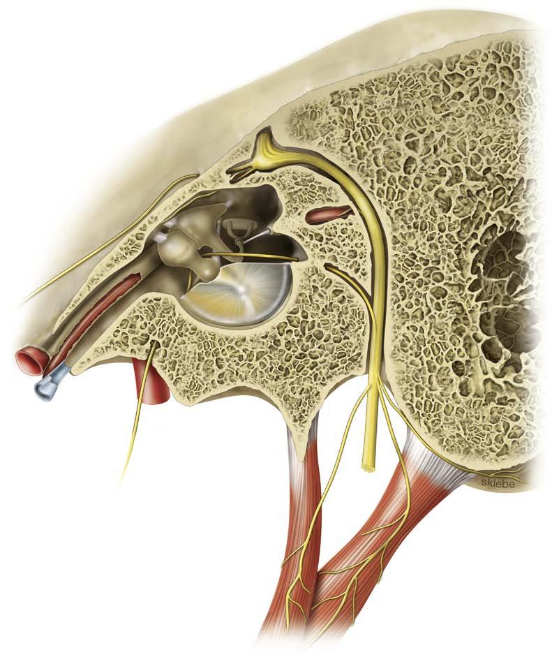
FACIAL NERVE
The facial nerve provides a diverse range of functions via efferent and afferent innervation to structures of the second branchial arch. The most well-associated function of the facial nerve is its innervation of striated muscles of facial expression. These functions will be the focus of this chapter. However, additional efferent motor fibers provide innervation to the stapedius muscle, the stylohyoid muscle, and the posterior belly of the digastric. Collectively, these motor fibers represent the special visceral efferents comprising the majority of the facial nerve fibers. The remaining efferent fibers of the facial nerve are general efferent fibers and form autonomic contributions. These autonomic nerves travel via the greater superficial petrosal nerve (GSPN) to the lacrimal gland and seromucinous glands of the nasal cavity and via the chorda tympani to the submandibular and sublingual glands. Traveling with this autonomic system, visceral afferent fibers supply visceral sensation to the mucosa of the nose, pharynx, and palate. Additional afferent inputs include special sensory fibers and somatic sensory fibers. Special sensory fibers for taste to the anterior two-thirds of the tongue, tonsillar fossa, and palate originate from the geniculate
ganglion cell bodies and travel within the chorda tympani and GSPN. Somatic sensory fibers provide touch sensation to the external auditory canal and conchal skin of the auricle as well as proprioceptive information from the facial muscles (Fig. 1.1).
INTRACRANIAL
Facial muscle movement starts with a conscious or unconscious impulse in the motor cortex of the precentral gyrus of the cerebral hemispheres. The majority of cerebral fibers then project to the contralateral facial motor nucleus via the corticobulbar tract through the thalamus along the posterior limb of the internal capsule. However, a subset of cerebral fibers controlling movement of the frontalis and upper orbicularis oculi project to both the ipsilateral and contralateral facial motor nuclei. A central lesion will thus cause frontalis-sparing facial paralysis due to the bilateral contributions to the upper face. The precentral gyrus fibers synapse in the facial motor nucleus, residing in the pontine tegmentum. Postsynaptic fibers then travel dorsally within the brainstem, loop around the floor of the fourth ventricle at the facial colliculus, and exit the ventrolateral aspect of the brainstem at the caudal border of the pons in the cerebellopontine angle (Fig. 1.2).
Collectively known as the nervus intermedius, preganglionic fibers of the superior salivatory nucleus and sensory fibers from the nucleus of tractus solitarius exit the brainstem directly lateral to the facial nerve motor root. Together, the facial nerve motor root and nervus intermedius enter the internal auditory canal (IAC) at the porus acusticus. For the purposes of this chapter, the facial motor root and nervus intermedius will be collectively referred to as the facial nerve.
INTRATEMPORAL
Meatal
The facial nerve travels through the temporal bone via a bony channel, the fallopian canal. The first segment of the fallopian canal is the meatal segment or IAC. The facial nerve travels in the superior anterior aspect of the IAC. The transverse (falciform) crest separates the facial nerve from the cochlear nerve inferiorly and Bill’s bar, a lateral ridge of bone, and separates the facial nerve from the superior vestibular nerve posteriorly. Within the IAC, the nerve lacks a fibrous sheath
G. Nina Lu, Patrick J. Byrne
Geniculate ganglion
Gustator y por tion of the solitar y nucleus
Superior salivator y nucleus
Ner vus inter medius
Greater petrosal ner ve
Pter ygopalatine ganglion
Lacrimal gland
Nasal mucous glands
Solitar y nucleus
Spinal nucleus of trigeminal ner ve
Facial colliculus
Abducens nucleus
Facial nucleus
Superior salivator y nucleus
Taste fibers
Submandibular and sublingual glands
Submandibular ganglion
Chorda tympani
Main facial ner ve
Fig. 1.1 The Nervus Intermedius Branches and Functional Targets. Blue and pink arrows indicate the direction of nerve impulse. (From Mtui E, Gruener G, Dockery P. Fitzgerald’s Clinical Neuroanatomy and Neuroscience. 7th ed. Oxford: Elsevier; 2016:218-221.)
or endoneurium and is surrounded by a thin layer of arachnoid.1
Labyrinthine
Exiting the IAC, the facial nerve turns gently anteriorly within the otic capsule bone between the cochlea and superior semicircular canal into the labyrinthine segment. This portion is on average 3 to 6 mm in length—the shortest and narrowest section of the fallopian canal. In this segment, the facial nerve is at its thinnest and particularly vulnerable to injury. The vascular supply is a watershed between the terminal arterioles of the vertebrobasilar system (via the labyrinthine branch of the anterior inferior cerebellar artery [AICA]) and the external carotid artery (via the petrosal branch of the middle meningeal artery). There is also a lack of epineurium vascular plexus along the labyrinthine segment.
At the distal end of the labyrinthine segment, the nerve travels superior to the cochlea and opens into the geniculate fossa. The geniculate fossa is separated from the middle fossa floor by a thin layer of bone, which is dehiscent in about 25% of cases. Here the first
Cor ticopontine and cor ticospinal fibers
Main facial ner ve
Fig. 1.2 Transverse section of the brainstem showing the intrapontine course of the facial nerve from the facial motor nucleus, superior salivatory nucleus, and solitary nucleus. NI, Nervus intermedius. (From Mtui E, Gruener G, Dockery P. Fitzgerald’s Clinical Neuroanatomy and Neuroscience. 7th ed. Oxford: Elsevier; 2016:218-221.)
fibers leave the facial nerve as the GSPN. The GSPN carries preganglionic parasympathetic innervation to the lacrimal gland and nasal mucosa. These fibers join the deep petrosal nerve from the carotid plexus to become the Vidian (or pterygoid) nerve. The thin bone above the geniculate and anterior attachment of the GSPN exerting intraneural traction place the nerve at increased risk of traumatic injury at this location. After the geniculate fossa, the facial nerve makes an acute posterior and slightly inferior turn, nearly 180 degrees, to enter the tympanic segment or horizontal segment.
Tympanic (Horizontal)
The tympanic segment is located along the medial wall of the anterior attic, travels superomedial to the cochleariform process, and forms the superior wall of the oval window niche posteriorly. On average 8 to 11 mm in length, dehiscence of the horizontal fallopian canal occurs in up to 20% of patients, with 80% above the oval window and 12% anterior to the cochleariform process.2 At the pyramidal eminence, the facial nerve makes a second genu anteroinferior to the lateral semicircular canal and enters the mastoid or vertical segment.
Mastoid (Vertical)
Greater anatomic variability is seen in the vertical segment prior to exiting the stylomastoid foramen than any other segment of the fallopian canal. In this
Facial nerve [VII]
Geniculate ganglion
Stapedius, tendon
Stapes
Tensor tympani, tendon
Incus
Lesser petrosal nerve
Greater petrosal nerve
Fig. 1.3 Intratemporal Course and Branches of the Facial Nerve. (From Paulsen F, Waschke J. Sobotta Atlas of Human Anatomy, Vol. 3. 15th ed. Munich, Germany: Urban & Fischer; 2012:133-160; Gleeson M. External and middle ear. In: Standring S, et al., eds. Gray’s Anatomy. 41st ed. Oxford, UK: Elsevier; 2016:624-640.e1.)
segment, the facial nerve becomes more superficial as it travels within the mastoid bone. Between 9 and 18 mm in length, the mastoid segment contains the chorda tympani and stapedial branches (Fig. 1.3). The chorda tympani has a variable take off from the mastoid segment but typically exits 6 mm above the stylomastoid foramen. Coursing superiorly nearly parallel to the facial nerve, the chorda angles anteriorly as it enters the mesotympanum. It exits the temporal bone via the petrotympanic fissure and joins the lingual nerve to convey taste information to the anterior two-thirds of the tongue and supplies preganglionic parasympathetic innervation to the submandibular, lingual, and minor salivary glands. This final segment ends at the
stylomastoid foramen, which is lined by the aponeurosis of the posterior belly of the digastric muscle. This aponeurosis directly supplies blood to the facial nerve as it exits the fallopian canal.
EXTRATEMPORAL
The facial nerve exits the stylomastoid foramen covered in tough connective tissue. At the stylomastoid foramen, the posterior auricular branch exits the facial nerve and supplies general sensory innervation to the posterior ear canal and concha, as well as motor innervation to the auricular muscles and occipitalis muscle. The digastric branch supplying motor innervation to the posterior belly of the digastric muscle and the
Nerve to stapedius
Stapedius
Mastoid process
Facial nerve [VII]
Chorda tympani
Digastric branch
Posterior auricular nerve
Stylohyoid
Digastric, posterior belly
Stylohyoid branch
Stylomastoid foramen
Tympanic membrane
Chorda tympani
Petrosphenoidal fissure
Auditory tube, bony par t
Auditory tube, car tilaginous part
Inter nal carotid ar ter y
Canal for tensor tympani
Tensor tympani
Malleus
Pyramidal process
Fig. 1.4 Branching Patterns of the Extratemporal Facial Nerve. (From Holmes S. Face and scalp. In: Standring S, et al., eds. Gray’s Anatomy. 41st ed. Oxford, UK: Elsevier; 2015:475-506.)
stylohyoid branch to the stylohyoid muscle also exit the facial nerve prior to its entrance into the parotid gland. Within the parotid, the facial nerve branches into a larger zygomaticotemporal division and a smaller cervicomandibular division at the pes anserinus and terminates in five classic branches: temporal, zygomatic, buccal, mandibular, and cervical. In reality, the branching pattern of the facial nerve is highly variable with multiple communications and the majority of variations existing among the zygomaticobuccal divisions.3–5 Through the parotid gland, the nerve runs at the level of the retromandibular vein and separates the superficial and deep lobe of the parotid gland. As it exits the parotid, the nerve has on average 8 to 15 branches making up the five divisions6 (Fig. 1.4). The nerve runs deep to the superficial musculoaponeurotic system (SMAS) and innervates the majority of facial muscles from their deep surface. The mentalis, buccinator, and levator anguli oris are innervated on their superficial surface as these are the deepest layer of facial muscles.
Temporal Division
The temporal division, also known as the frontal division or frontotemporal branch, consists of three to four branches traveling obliquely in between the temporoparietal fascia and the superficial layer of the deep temporal fascia.7 Branches entering the frontalis muscle at the level of the supraorbital ridge are located up to 3 cm above the lateral canthus. Branches entering
the upper orbicularis oculi run along the undersurface for 3 to 4 mm before entering the muscle to innervate it. One common estimate of the frontotemporal branch is Pitanguy’s line, defined by a line draw from 0.5 cm inferior to the tragus to 1.5 cm superior and lateral to the eyebrow. Numerous cadaveric studies have attempted to further define the course and branching patterns. Ishikawa demonstrated that three to four branches of the FTN were consistently found between 3.8 and 6.0 cm posterior to the lateral canthus along the superior zygomatic arch in both straight-line and curved trajectories.8
Zygomaticobuccal Division
The zygomaticobuccal division consists of five to eight branches with significant overlap of muscle innervations. These nerves innervate the midfacial muscles spanning the lip elevators to the lower orbicularis oculi. Connections between the lower branches and marginal mandibular division also exist. Dorafshar et al. describe “Zucker’s point” as a reliable surface landmark for identifying the zygomaticobuccal nerve as it exits the parotid.9 The authors found that the midpoint of a line drawn from the root of the helix to the oral commissure predicted the nerve location within an average of 2.3 mm.
Marginal Mandibular Division
The marginal mandibular division consists of one to three branches beginning up to 2 cm below the ramus
of the mandible and arcing upward to cross the mandible halfway between the angle and mental protuberance. Branches lie on the deep surface of the platysma and cross superficial to the facial vessels 3.5 cm from the parotid edge. There are separate branches to the depressor angularis, depressor labii inferioris (DLI), and mentalis, and variable superior ramus supplying upper platysma and lower orbicularis oris.
Cervical Division
The cervical division consists of one branch leaving the parotid and running on the deep surface of the platysma. The point of entry into the muscle is 2 to 3 mm caudal to the platysma muscle branch of the facial vessel. It enters the muscle at the junction of its cranial and middle thirds.
MICROSCOPIC ANATOMY
The microanatomy of the facial nerve reveals a heterogenous organization that explains in part the difficulty of facial nerve repair. As described by Captier et al., the facial nerve lacks fascicular organization as well as epineural or perineural covering from its exit at the brainstem to the geniculate ganglion.10 Prior to the geniculate ganglion, the nerve is surrounded only by an arachnoid sheath. A thin epineural sheath arises within the tympanic segment and thickens as the nerve travels towards the stylomastoid foramen. The perigeniculate facial nerve demonstrates one or two fascicular bundles that increase in number and decrease in size as the nerve travels distally through the fallopian canal. The number of fascicles and their spatial organization changes every 2 mm within the fallopian canal. At the stylomastoid foramen the facial nerve has on average 11 fascicles and up to 16. As individual fibers emerge, they are surrounded by perineurium and endoneurium. In the horizontal segment, the upper motor division has been reported to be more superficial (or lateral), whereas the lower motor division has been reported to lie deeper (or medial).11 The nerve bundles have a spatial organization as they traverse the mastoid segment: branches to the lower division lie within the anterior portion of the nerve and branches to the upper division course posteriorly within the epineurium.11 From the brainstem to the mastoid segment, an average of 7800 myelinated nerve fibers are maintained. A single neuron can innervate up to 25 muscle fibers.
VASCULAR ANATOMY
The intracranial (brainstem and IAC) facial nerve receives blood supply from branches of the AICA arising from the vertebrobasilar system. The labyrinthine artery, a branch of the AICA, provides vascular input within the IAC segment. However, the remaining intratemporal segments receive blood supply through the middle meningeal artery, a branch of the maxillary artery from the external carotid. As described
previously, the transition zone of vascular supply between the AICA and the external carotid centers on the labyrinthine segment and creates a watershed or weak zone of arterial supply.
The extrinsic vascular network consists of one or two main arterial trunks and accompanying venae comitantes running between the periosteum of the fallopian canal and the epineural sheath of the nerve. An intrinsic vascular network also exists within the epineurial sheath consisting of small arterioles, capillaries, and venules.12
FACIAL MUSCLES
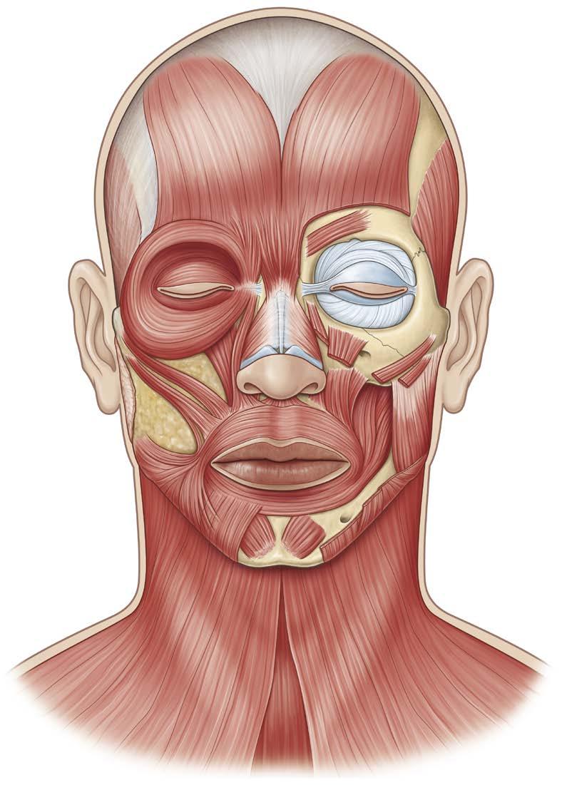
The striated muscles of facial expression derive from the second branchial arch mesoderm and reside within the SMAS layer. The SMAS layer continues superiorly with the galea aponeurotica and inferiorly with the platysma muscle. A total of 17 paired and 1 unpaired sphincter muscle constitute the facial musculature. These originate from the periosteum of the facial bones and insert into the skin, allowing for a limitless number of facial expressions (Fig. 1.5).
The facial muscles possess a three-dimensional relationship to one another and exist in four layers based on muscle origin as demonstrated by Freilinger et al.13 The orbicularis oculi, depressor anguli oris (DAO), and superficial aspect of the zygomaticus minor are most superficially located as the first layer. The platysma, risorius, zygomaticus major, deeper portion of the zygomaticus minor, and levator labii superioris alaeque nasi compose the second layer. The levator labii superioris and orbicularis oris represent the third layer. The deepest layer is composed of the levator anguli oris, the mentalis, and the buccinator—these three muscles are also the only muscles innervated from their superficial surface, as explained by this relationship.
Distinct from most skeletal muscles, facial muscles are flat, strap-like muscle sheets with interdigitations to the skin, short or absent tendons, and an absent fascial covering. A comprehensive list and description of facial muscles are detailed in Table 1.1. The most clinically relevant facial muscles are described in further detail in the following sections.
FRONTALIS
The frontalis muscle is a broad, thin, bilateral muscle originating from the galea aponeurotica near the coronal suture and inserting onto the superciliary ridge of the frontal bone and interdigitating with fibers of the orbicularis oculi, procerus, and corrugator supercilia. Densely adherent to the overlying skin, the frontalis glides over the underlying periosteum to provide brow elevation. The resting tone of the frontalis also prevents brow descent and ptosis. Left- and right-sided frontalis bellies fuse in the midline caudally, often as a fibrous junction. The frontalis and occipitalis bellies can be described as distinct muscle bellies or as parts of a single
Epicranial aponeurosis
Occipitofrontalis, frontal belly
Depressor supercilii
Orbicularis oculi, palpebral par t
Levator labii superioris alaeque nasi
Orbicularis oculi, orbital par t
Levator labii superioris
Zygomaticus minor
Zygomaticus major
Parotid gland
Orbicularis oris, marginal par t
Buccal fat pad
Risorius
Depressor anguli oris
Depressor labii inferioris
Mentalis
Platysma
Procerus
Corrugator supercilii
Levator labii superioris alaeque nasi
Nasalis
Levator labii superioris
Zygomaticus minor
Zygomaticus major
Levator anguli oris
Depressor septi nasi
Buccinator
Masseter, superficial part
Orbicularis oris, labial par t
Depressor anguli oris
Depressor labii inferioris
ed. Philadelphia: Elsevier; 2018:329-357.e2.)
occipitofrontalis muscle connected by an intermediate tendon within the galea aponeurotica.
ORBICULARIS OCULI
The orbicularis oculi muscle is a paired sphincter important in eyelid closure. The pretarsal, preseptal, and orbital subdivisions of the orbicularis oculi are defined by the anatomic level of the muscle. The pretarsal portion is closely adherent to the pretarsal skin, covers the tarsal plate and provides reflexive blink
movement. The preseptal portion covers the orbital septum and is under more voluntary control. It is less closely adherent to the skin other than at the medial and lateral canthi. Both the preseptal and pretarsal components function together during blink. The orbital component forms a ring over the bony orbital margin and is recruited during forceful eye closure as well as brow depression. This component originates medially from the superomedial orbital margin, the maxillary process of the frontal bone, the medial
Fig. 1.5 Facial Musculature (Frontal View). (From Zuker RM, Gur E, Hussain G, Manktelow RT. Facial paralysis. In: Neligan PC, ed. Plastic Surgery: Volume 3: Craniofacial, Head and Neck Surgery and Pediatric Plastic Surgery. 4th
Facial Muscles.
MUSCLE ACTION
MUSCLES OF THE SCALP
Frontalis Occipitalis
Auricularis Anterior
Auricularis Superior
Auricularis Posterior
Raises forehead and eyebrow Draws scalp backwards
Draws auricle forward and upward Draws auricle upwards Draws auricle backwards
Temporoparietalis Tightens scalp, raises auricle and temple
MUSCLES OF THE EYELIDS
Orbicularis Oculi Sphincter muscle of eyelid, eye closure
Corrugator Supercilii Draws eyebrow inferior and medial (vertical forehead wrinkles)
Depressor Supercilii
Depressor of eyebrow
MUSCLES OF THE NOSE
Procerus Draws medial eyebrow downwards (transverse wrinkles of nose)
Nasalis Transverse: depresses cartilaginous part of nose and draws ala toward the septum Alar: enlarges nasal aperture
Depressor Septi Nasi Draws ala downwards, constricts aperture
MUSCLES OF THE MOUTH
Levator Labii Superioris Raises the medial aspect of upper lip
Levator Labii Superioris Alaeque Nasi Raises the medial aspect of upper lip and dilates the nostril
Levator Anguli Oris Elevates angle of the mouth
ORIGIN/INSERTION
O: Galea aponeurotica near the coronal suture
I: Superciliary ridge of the frontal bone (frontalis) and occipital bone (occipitalis)
O: Galea aponeurotica
I: Auricular cartilage
O: Galea aponeurotica
I: Auricularis muscles
O: Frontal bone, maxillary bone, medial palpebral ligament
I: Lateral horizontal raphae
O: Medial orbital rim and frontal bone
I: Frontalis, orbicularis oculi and skin20
O: Frontal process of maxilla above medial canthal tendon
I: Dermis superior to the medial canthal tendon
O: Caudal nasal bone
I: Skin of glabella and medial brow
O: Maxilla over lateral incisor I: Procerus/Glabellar skin and lower lateral cartilage
O: Incisive fossa of maxilla I: Nasal septum
O: Inferior orbital rim and maxilla I: Orbicularis oris of upper lip
O: Medial angle of orbital rim I: Levator labii superioris
O: Maxillary canine fossa I: Orbicularis oris of upper lip and modiolus
DESCRIBED DIMENSION
Width: 60.9 +/- 8.7 mm19
Height: 31.7 +/- 7.5 mm19
Zygomaticus Major Elevates angle of the mouth upward and backward
O: Zygomatic Arch I: Modiolus
Length: 65 +/- 5.6 mm15
Width: 60 +/- 9.6 mm15
Length: 38–53 mm15
Zygomaticus Minor Elevates upper lip backward, upward, and outward
Risorius
Retracts angle of the mouth laterally
O: Zygomatic Arch I: Modiolus
O: Parotid fascia I: Modiolus
Length: 47 +/- 7.5 mm15
Males: 33.67 +/- 4.13 mm13
Females: 35.5 +/- 6.69 mm13
61.6 +/- 7.6 mm13
Width: 7.2 +/- 1.7 mm15
Length: 42 +/- 2.5 mm15
Male length: 37.83 +/- 4.38 mm13
Female length: 38.33 +/- 8.02 mm13
Length: 65.6 +/- 3.8 mm15
Male length: 70.67 +/- 6.32 mm13
Female length: 69.50 +/- 6.48 mm13
Length: 51.8 +/- 7.4 mm15
Depressor Labii Inferioris Draws lower lip downward and laterally
Depressor Anguli Oris Depresses angle of the mouth
Mentalis Raises and protrudes lower lip, wrinkles skin of the chin
Orbicularis oris Sphincter function of lips, closure of lips
Buccinator Compresses/flattens the cheek
Platysma Tightening of the neck, downward pull on lower lip
I, Insertion; O, Origin; SMAS, superficial musculoaponeurotic system.
O: Mandible inferior to mental foramen
I: Modiolus, orbicularis oris, skin of lower lip
O: Mandible inferior to mental foramen
I: Modiolus, orbicularis oris
O: Anterior mandible
I: Chin skin
Neither bony nor tendinous origin, integration with perioral muscles
O: Maxilla, pterygomandibular raphe, mandible
I: Modiolus
O: Supraclavicular skin
I: Continuous with SMAS
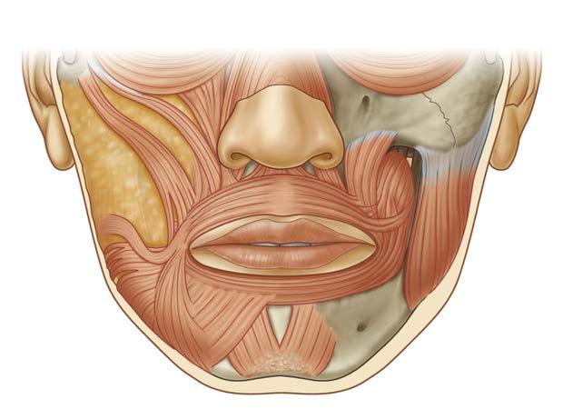
Levator labii superioris alaeque nasi
Levator anguli oris
Length: 29 +/-4.9 mm15
Length: 48 +/- 5.1 mm15
Length: 56 +/- 7.4 mm15
Levator labii superioris
Zygomaticus minor
Zygomaticus major
Modiolus
Risorius
Platysma
Depressor anguli oris
Depressor labii inferioris
Mentalis
Buccinator
Orbicularis oris
Fig. 1.6 Periorbital Muscles Including Lip Elevators and Depressors. (From Drake R, Vogl W, Mitchel A. Gray’s Basic Anatomy. 2nd ed. Philadelphia: Elsevier; 2018:413-596.)
canthal tendon, the frontal process of the maxilla, and the inferomedial margin of the orbit. Within the most superficial layer of facial muscles, the orbital subdivision laterally overlies the temporalis fascia, the origins of the zygomaticus major, and levator labii muscles.
LIP ELEVATORS
The perioral muscles contributing to lip elevation and smile are of particular interest to patients and surgeons in facial rehabilitation. The main lip elevators are the zygomaticus major, the levator labii superioris, and levator anguli oris (Fig. 1.6).
The zygomaticus major is the most superficial of the three and originates from the zygomatic bone in front of the zygomaticotemporal suture. It runs to the angle of the mouth where superficial fibers form the modiolus together with the DAO, the risorius, the orbicularis oris, the buccinator, and the levator anguli oris. The modiolus represents the interdigitation of all the perioral muscles. The deeper insertion of the zygomaticus major fuses with the levator anguli oris and the medial fibers line on the buccinator muscle. The caudal fibers continue into the DAO. The zygomaticus muscle elevates the commissure superiorly and laterally at an approximately 45-degree angle.
The levator labii superioris arises from the lower margin of the orbit above the infraorbital foramen, more medially located compared to the zygomaticus major. Its medial and upper insertion helps form the nasolabial sulcus with partial insertion into this crease. Its lateral fibers descend superficial to the orbicularis oris and deeper fibers interdigitate with the modiolus. Medial levator labii superioris fibers reach the upper lip philtral columns and insert into the medial vermilion border as far as the peak of Cupid’s bow. The levator labii superioris elevates the lip vertically and laterally to expose the upper teeth and deepen the nasolabial fold (Video 1.1 ).
The levator anguli oris originates from the maxillae below the infraorbital foramen, fills the canine fossa, and runs vertically. It inserts into the modiolus and is in the deepest layer of facial muscles. The levator anguli oris serves to elevate the commissure vertically and medially.
The origins and insertions for the zygomaticus major, levator labii superioris, and levator anguli oris determine the vectors of pull. The relative strength of each muscle interdigitating in the modiolus and orbicularis oris determines the smile configuration unique to each patient.
ORBICULARIS ORIS
The orbicularis oris is the only unpaired muscle of facial expression and serves as a sphincter of the mouth essential for oral competence in both speech and the oral phase of swallow. Superficially, portions of the muscle fibers insert into the philtra columns as well as along the vermilion border to form the white roll. Superficial components of the muscle can contract independently to provide nuanced expression of the lips and the deeper component functions as the main constrictor. Upper and lower lip depressors interdigitate with the orbicularis oris and work in concert for lower facial expression.
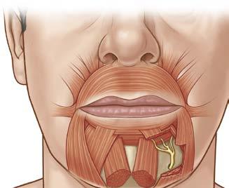
LIP DEPRESSORS
The lower lip musculature is as functionally complex as the upper lip. An intricate three-dimensional assembly of muscular sheets control the shape of the mouth and position of the lips. The DLI, DAO, and platysma comprise the lower lip depressors (Fig. 1.7).
The DLI originates along the alveolar bone of the mandible inferior and lateral to the mental foramen. It travels laterally to insert into the orbicularis oris muscle along its deep surface and attaches to the vermilion and middle third lip skin through fibrous septa.14,15 The DLI acts to depress and lateralize the lower lip, evert the vermilion, and produce lower dental show.
The DAO arises from the mandible lateral and superficial to the DLI origin. It inserts directly into the skin at the labiomandibular crease medially and into the modiolus laterally. Its main action is to draw the corner of the mouth down and medial during frowning.
Fig. 1.7 Lower Lip Depressors. Nerve depicted here is the mental nerve, a branch of the third division of the trigeminal nerve. (From Zuker RM, Gur E, Hussain G, Manktelow RT. Facial paralysis. In: Neligan PC, ed. Plastic Surgery: Volume 3: Craniofacial, Head and Neck Surgery and Pediatric Plastic Surgery. 4th ed. Philadelphia: Elsevier; 2018:329-357.e2.)
When acting in conjunction with the platysma muscle, the DLI and DAO depress the entire lower lip producing a full denture smile. The mentalis muscle similarly originates from around the mental foramen but passes medially or inferomedially. The mentalis muscle is the deepest layer of the lower lip muscles and acts to raise the chin with an indirect action of lower lip elevation.
MICROARCHITECTURE
Most facial muscles exhibit muscle fiber density equal to other skeletal muscles such as the biceps and tibialis anterior.16 Fiber density varies between 1000 and 1400 fibers/mm2 for the majority of facial muscles, with the orbicularis oculi and oris having the highest density (over 2000 fibers/mm2). The total number of muscle fibers varies between 5400 fibers (zygomaticus minor) and 9630 fibers (DAO). Muscle fiber size varies between facial muscles. The frontalis, orbicularis oris, and orbicularis oculi contain smaller muscle fibers (less than 400 μm2). The zygomaticus major, DAO, buccinator, zygomaticus minor, DLI, and levator labii superioris contain intermediate muscle fibers (between 400 and 500 μm2). The platysma, levator anguli oris, and risorius contain large muscles (greater than 500 μm2).
Facial muscles also demonstrate a great variability in fast- versus slow-twitch fiber composition, likely related to the multitude of functions they exhibit. Predominantly tonic or slow-twitch muscles contain a high percentage of type 1 fibers and predominantly phasic or fast-twitch muscles contain a high percentage of type 2 fibers. Facial muscle compositions range from 14% to 67% type 1 muscle fibers.17 The most phasically composed facial muscle is the orbicularis oculi and the most tonically composed facial muscle is the buccinator (Table 1.2).
Facial muscles have a unique microarchitecture compared to other skeletal muscles in the body and a complex pattern of innervation. Most human skeletal muscles consist of muscle fibers up to 18 cm in length
Tablev 1.2
Fiber Composition of Facial Muscles.
Orbicularis oculi 15/85
Zygomaticus major 28/72
Orbicularis oris 29/71
Levator labii superioris 31/69
Depressor anguli oris 37/63
Corrugator superioris 41/59
Depressor labii inferioris 46/54
Occipitofrontalis 57/43
Buccinator 67/33
Adapted from Freilinger G, Happak W, Burggasser G, Gruber H. Histochemical mapping and fiber size analysis of mimic muscles. Plast Reconstr Surg 1990;86(3):422-428.
with a single motor end plate (MEP) in the middle of the fiber.15 These MEPs form a narrow band that traverses across the central zone of the skeletal muscles, known as a motor band. Facial muscles differ significantly from skeletal muscles in innervation pattern, location of motor bands, and arrangement of MEPs. These differences may be partially attributed to the elaborate branching pattern of the facial nerve. Within the parotid gland, the facial nerve divides into multiple branches with an elaborate network of anastomosis and interlaced branches before entering the facial muscles. Each facial muscle is innervated by multiple terminal branches of the facial nerve without an accompanying vascular pedicle. Happak et al. demonstrate that upon entrance into the muscle, round or oval-shaped clusters of MEPs are found in the immediate vicinity of the nerve entrance, described as motor zones. Unlike the central motor band architecture of skeletal muscles, motor zones are spread over facial muscles in a de-centralized fashion with considerable variability determined by pattern of facial nerve branching. The orbicularis oris, orbicularis oculi, and buccinator muscles demonstrate a great number of small MEP clusters spread evenly over the muscle. The zygomaticus major demonstrates a dominant motor zone with two to three additional smaller motor zones. The levator labii superioris contains two to four large motor zones in eccentric positions.
In experimental studies, facial muscle fibers demonstrate a “multiply innervated muscle fiber pattern” with multiple MEPs within a distance ranging from 25 to 500 μm on a single muscle fiber. This pattern of innervation is also found in laryngeal muscles, perhaps pointing towards a system capable of fine adjustments.18 It is not yet known if multiple end plates on a single facial muscle fiber are innervated mono- or polyneuronally. The plexus of the facial nerve and high branching pattern suggest a polyneuronal innervation could be present. Further studies are needed to understand the mechanisms of emotional expression and neural control.
CONCLUSION
Facial nerve and muscle anatomy elegantly demonstrates the complexities of the head and neck region. Comprehensive understanding of the anatomic course and contributions of the facial nerve is critically important for physicians treating facial nerve disorders both surgically and medically. Further research and understanding of nerve and muscle microarchitecture is critical in understanding the nuances of facial movement and emotional expression.
REFERENCES
1. Miehlke A, Fisch U, Eneroth C-M Surgery of the Facial Nerve 2nd ed. Philadelphia: Saunders; 1973.
2. Moody MW, Lambert PR. Incidence of dehiscence of the facial nerve in 416 cases of cholesteatoma. Otol Neurotol 2007;28(3):400–404.
3. Tzafetta K, Terzis JK. Essays on the facial nerve: Part I. Microanatomy. Plast Reconstr Surg. 2010;125(3):879–889.
4. Katz AD, Catalano P. The clinical significance of the various anastomotic branches of the facial nerve. Report of 100 patients. Arch Otolaryngol Head Neck Surg. 1987;113(9):959–962.
5. Davis RA, Anson BJ, Budinger JM, Kurth LR. Surgical anatomy of the facial nerve and parotid gland based upon a study of 350 cervicofacial halves. Surgery, Gynecology & Obstetrics. 1956;102(4):385–412.
6. Zuker RM, Gur E, Hussain G, Manktelow RT. Facial paralysis. In: Neligan PC, ed. Plastic Surgery: Volume 3: Craniofacial, Head and Neck Surgery and Pediatric Plastic Surgery. 4th ed. Philadelphia: Elsevier; 2018:329–357.e2.
7. Pitanguy I, Ramos AS. The frontal branch of the facial nerve: the importance of its variations in face lifting. Plast Reconstr Surg. 1966;38(4):352–356.
8. Ishikawa Y. An anatomical study on the distribution of the temporal branch of the facial nerve. J Cranio-Maxillo-Fac Surg. 1990;18(7):287–292.
9. Dorafshar AH, Borsuk DE, Bojovic B, et al. Surface anatomy of the middle division of the facial nerve: Zuker’s point. Plast Reconstr Surg. 2013;131(2):253–257.
10. Captier G, Canovas F, Bonnel F, Seignarbieux F. Organization and microscopic anatomy of the adult human facial nerve: anatomical and histological basis for surgery. Plast Reconstr Surg. 2005;115(6):1457–1465.
11. May M. Anatomy of cross section of facial nerve in the temporal bone: clinical application. In: Fisch U, ed. Facial Nerve Surgery. Amstelveen, The Netherlands: Kugley Medical Publications; 1977:40–46.
12. Bagger-Sjoback DGM, Thomander L. The intratemporal vascular supply of the facial nerve: a light and electron microscopic study. In: Graham MD, House WF, eds. Disorders of the Facial Nerve: Anatomy, Diagnosis, and Management. New York: Raven Press; 1982:17–31.
13. Freilinger G, Gruber H, Happak W, Pechmann U. Surgical anatomy of the mimic muscle system and the facial nerve: importance for reconstructive and aesthetic surgery. Plast Reconstr Surg. 1987;80(5):686–690.
14. Hur MS. Anatomical relationship of the inferior bundle of the incisivus labii inferioris with the depressor labii inferioris and the platysma. J Craniofac Surg. 2017;28(7):1861–1864.
15. Happak W, Liu J, Burggasser G, Flowers A, Gruber H, Freilinger G. Human facial muscles: dimensions, motor endplate distribution, and presence of muscle fibers with multiple motor endplates. Anat Rec. 1997;249(2):276–284.
16. Ito J, Moriyama H, Shimada K. Morphological evaluation of the human facial muscles. Okajimas Folia Anatomica Japonica. 2006;83(1):7–14.
17. Freilinger G, Happak W, Burggasser G, Gruber H. Histochemical mapping and fiber size analysis of mimic muscles. Plast Reconstr Surg. 1990;86(3):422–428.
18. Rossi G. From the pattern of human vocal muscle fibre innervation to functional remarks. Acta Oto-laryngologica Supplementum. 1990;473:1–10.
19. Jeon A, Kim SD, Han SH. Morphological study of the occipital belly of the occipitofrontalis muscle and its innervation. Surg Radiol Anat. 2015;37(9):1087–1092.
20. Hwang K, Lee JH, Lim HJ. Anatomy of the corrugator muscle. J Craniofac Surg. 2017;28(2):524–527.