MacSween’s Pathology of the Liver
SEVENTH EDITION
Alastair D. Burt, BSc, MBChB, MD, FRCPath, FRCPA, FRCP, FRSB, FAHMS
Executive Dean Faculty of Health and Medical Sciences University of Adelaide Adelaide, Australia
Linda D. Ferrell, MD Emerita Professor Anatomic Pathology University of California San Francisco San Francisco, California
Stefan G. Hübscher, MBChB, FRCPath
Leith Professor and Professor of Hepatic Pathology Institute of Immunology and Immunotherapy University of Birmingham Birmingham, United Kingdom
extensively updated, as has the chapter on vascular disorders. Tumours and tumour-like lesions are more comprehensively covered than ever before and recent developments in clinical management and pathological assessment in the field of hepatic transplantation are reflected in an updated section on this importance topic. As always we have attempted to ensure the use of contemporary terms throughout. In recent years the term “primary biliary cholangitis” has replaced the term “primary biliary cirrhosis”. This is a change that we fully support and have therefore adopted the new terminology in this edition.
For each of us, the editing of this book has been a labour of love; we are honoured to be able to continue the fine tradition that Roddy started almost 40 years ago. We are grateful for the ongoing support and encouragement of our respective partners, Alison, Rick, and Dawn, during the preparation of the book. Thanks also to all of the contributors and the team at Elsevier for keeping us on track.
Alastair D. Burt
Linda D. Ferrell
Stefan G. Hübscher
Kay Washington, MD, PhD
Professor of Pathology
Vanderbilt University Medical Center Nashville, Tennessee
Aileen Wee, MB, BS, FRCPath, FRCPA
Professor of Pathology and Senior Consultant
Department of Pathology
Yong Loo Lin School of Medicine, National University of Singapore; National University Hospital Singapore
Matthew M. Yeh, MD, PhD
Associate Professor
Director of Gastrointestinal and Hepatic Pathology
Pathology
University of Washington School of Medicine Seattle, Washington
Sherif R. Zaki, MD, PhD
Chief, Infectious Diseases Pathology Branch
Centers for Disease Control and Prevention Atlanta, Georgia
Yoh Zen, MD, PhD, FRCPath
Professor of Pathology
Department of Diagnostic Pathology
Kobe University Graduate School of Medicine Kobe, Japan
The practice of hepatopathology requires a clear understanding of liver anatomy and physiology, as a prelude to understanding the expression of pathological processes in the liver. As an anatomical entity, the liver is deceptively simple. It is large, representing about 2% of the total body mass of an adult human and occupying most of the right upper quadrant of the abdomen. It has a roughly triangular profile, with incomplete clefts helping to define the different ‘lobes’ of the liver. It has only one point of vascular inflow, the porta hepatis. Blood exits through several venous orifices into the inferior vena cava, which traverses a deep groove in the dorsum of the liver. There are no ‘moving parts’ of the liver, with the exception of daily secretion of several litres of bile into the common hepatic duct, which exits from the porta hepatis.
Belying its macroscopic simplicity, the liver is home to biosynthetic and biodegradative metabolic pathways of unequaled complexity, generating enough metabolic heat to be a prime source of core homeostatic temperature maintenance. This chapter considers the embryology, macroanatomy and microanatomy of the liver and its basic response to injury, concluding with the appearance of ‘normal’ liver in biopsy and autopsy specimens.
Development of the liver
General features
In human embryos the liver first appears at the end of the third week of development. The liver bud, or hepatic diverticulum, arises as a hollow midline outgrowth of endodermal tissue from the ventral wall of the future duodenum. The connective tissue framework of the liver into which the endodermal bud grows is of mesenchymal origin and develops from two sources: (1) the septum transversum, a transverse sheet of mesenchymal cells that incompletely separates the pericardial and peritoneal cavities and is the primordium for both the diaphragm and the liver, and (2) cells derived from the mesenchymal lining of the associated coelomic cavity, which actively invade the septum transversum. The confluence of endodermal cells from the hepatic diverticulum growing into the mesenchymal primordium creates the solid organ destined to become the liver (Fig. 1.1).
During the fourth week, buds of epithelial cells within the mesenchymal stroma extend radially out from the hepatic diverticulum. Between the epithelial cords, a plexus of vascular hepatic sinusoids develops. As the epithelial buds grow into the septum transversum, they break up into thick, anastomosing epithelial sheets that meet and enmesh vessels of the hepatic sinusoidal plexus, forming the primitive hepatic sinusoids (Fig. 1.2 A). The intimate relation between hepatocytes and sinusoidal capillaries, so characteristic of the adult organ, is therefore already anticipated in the 4-week-old embryo. The hepatic diverticulum remains as a tether between the developing liver primordium and the duodenum, ultimately becoming the extrahepatic biliary tree.
Once established, the liver grows rapidly during the fetal period, to become the largest single visceral organ (by mass) for the remainder of gestation. It bulges into the peritoneal cavity on each side of the midline, as right and left lobes, which are initially symmetrical. It also grows ventrally and caudally into the mesenchyme of the anterior abdominal wall, extending down to the umbilical ring. Associated with these changes, the stomach and duodenum, which were initially in broad contact with the septum transversum, draw away from it, thus producing a midsagittal sheet of mesoderm, the ventral mesogastrium or future lesser omentum. As the duodenum withdraws from the septum transversum, the stalk of the original hepatic diverticulum is also drawn out to form, within the lesser omentum, the epithelial elements of the extrahepatic bile ducts. Simultaneously, the cephalad aspect of the liver becomes partly freed from its originally broad contact with the septum transversum by extensions of the peritoneal cavity and its visceral and parietal mesothelial surfaces so that, in the adult, direct contact of the liver with the diaphragm persists only as the ‘bare area’ of the liver. This is bounded by the attachments of peritoneal reflections, which form the coronary and falciform ligaments.
Vascular arrangements
By the fifth week of development (in embryos of 5 mm), the liver parenchyma consists of anastomosing sheets of liver cells, each sheet being several cells in thickness. Coursing between the liver cells is the vascular ‘sinusoidal plexus’. Initially, the afferent hepatic blood supply is through the symmetrically arranged vitelline veins returning from the abdominal region of the embryo. Blood is also received from the laterally placed right and left umbilical veins, which run in the body wall and carry oxygenated blood from the placenta directly to the paired horns of the sinus venosus of the cardiac primordium. Both the vitelline and the umbilical sources of blood enter into the hepatic sinusoidal plexus through a developing branching vascular network. Blood draining from the sinusoidal plexus passes through symmetrical right and left hepatocardiac channels, to enter the sinus venosus through this same network1 (Fig. 1.2 B). This network, along with the mesenchyme through which it passes, constitutes the early portal tract system within the liver parenchyma.
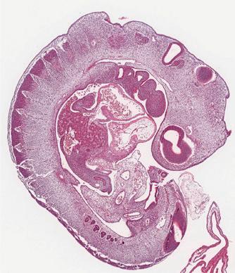
1.1 Photomicrograph of hepatic primordium in human embryo at 25 days’ gestation. The lower-power view shows the organs of the coelomic cavity. The higher-power inset shows cords of endodermal cells within the mesenchyme of the septum transversum, thus forming the hepatic primordium.
Once these vascular connections are made (Fig. 1.2 C), the circulatory pattern within the liver changes rapidly. The left umbilical vein becomes the principal source of blood entering the liver, partly because it comes to carry all the blood returning from the placenta when the right umbilical vein withers and disappears (generating the ‘double-artery/single-vein’ umbilical cord of the term fetus), and partly because the initial volume of blood returning from the gut in the vitelline veins is small. The definitive vascular pattern of the fetal liver is already established by the seventh week in embryos, about 17 mm long (Fig. 1.2 D).2 The originally paired vitelline veins have given way to a single portal vein that, on entering the liver, divides into right and left branches. Blood in the left umbilical vein
Forebrain Heart Liver
Hepatic trabeculae trabeculae
Figure
Mesenchymal lining of coelomic tract
Left hepatocardiac channel
Left umbilical vein
Sinus venosus
Right hepato-cardiac channel
Hepatic sinusoidal plexus
Middle hepatic vein
Hepatic sinusoidal plexus
Left umbilical vein
Origin of hepatic bud
Portal vein
Figure 1.2 A, Section through region of the hepatic bud in a human embryo of 25 somites (~26 days). B, Vascular channels associated with the developing liver, in a human embryo of 30 somites. C, Vascular channels in the human liver at a slightly later stage, showing the further extensive development of the hepatic sinusoidal plexus. D, Scheme of the portal hepatic circulation, in a human embryo of 17 mm (~7 weeks). (A and B redrawn from and C based on Streeter GL. Contributions to embryology of the Carnegie Institution of Washington, 1942;30:213–44; D redrawn from Lassau JP, Bastian D. Organogenesis of the venous structure of the human liver: a haemodynamic theory. Anat Clin 1983;5:97–102.)
traverses a venous extension in the falciform ligament and has a choice of three routes: (1) through the liver in branches that enter the sinusoidal plexus of the left half of the liver; (2) through the sinusoidal plexus of the right half of the liver, by retrograde flow through its connection with the left branch of the portal vein; and (3) through the ductus venosus traversing the short space between the porta hepatis to the inferior vena cava, to enter directly into the systemic venous circulation. By these routes, the converging bloodstreams from the definitive portal vein and indirectly from the umbilical vein enter the rapidly enlarging hepatic primordium through the porta hepatis. Intrahepatic mesenchyme condenses around the intrahepatic branching portal venous system, making up the ramifying portal tracts of the liver (see later discussion).
The hepatic artery is derived from the celiac axis. Arterial sprouts grow into the hepatic primordium from the porta hepatis along the mesenchyme of the portal tract system, spreading to the organ periphery as the fetal liver enlarges. These arterial sprouts appear to be the organizing element for formation of the intrahepatic biliary tree. The hepatic arterial system continues to proliferate and grow after birth, reaching an adult form only at 15 years of age.3–6 In the adult, about four arteries supply the largest intrahepatic bile ducts.7 At the level of the terminal portal tracts, there is a uniform 1 : 1 pairing of hepatic arteries and terminal bile ducts, and approximately two artery/bile duct pairs per single portal vein.8 The most terminal portions of the portal tree lose their portal veins, leaving only residual hepatic artery/ bile duct dyads, which themselves disappear into the parenchyma.8,9
Common cardinal vein
Left umbilical vein
Inferior vena cava
Ductus venosus (Dia. = 200 µm)
Left hepatic vein
Left umbilical vein (Dia. = 600 µm)
Right hepatic vein
Septum transversum
Liver
Right and left vitelline veins
Right umbilical vein
Sinus venosus Gut
Vitelline veins
(Dia. = 100 µm)
To complete this discussion, the rapid changes in hepatic circulation at birth must be considered. A sphincteric mechanism closes the ductus venosus at its proximal end, blood flow ceases in the umbilical vein, and the left side of the liver receives blood that now flows from right to left through the left branch of the portal vein. The closed segment of the umbilical vein between the umbilicus and the liver regresses to form the ligamentum teres; the ductus venosus undergoes fibrosis and becomes the ligamentum venosum
Hepatocyte ontogeny
Primitive hepatocytes are derived exclusively from the endodermal outgrowths of the hepatic diverticulum. Hepatocellular synthesis of alpha fetoprotein (AFP) begins at the earliest stage of liver differentiation, about 25–30 days after conception, and continues until birth. Glycogen granules are present in fetal hepatocytes at 8 weeks; the maximal glycogen reserve is achieved at birth, but the rapid onset of glycogenolysis over 2–3 days postpartum depletes the storage to approximately 10% of term levels. Hepatocellular haemosiderin deposits appear early in development, predominantly in periportal hepatocytes, and become more marked as hepatic haematopoiesis decreases (see later). Hepatocellular bile acid synthesis begins at about 5–9 weeks and bile secretion at about 12 weeks.10 Canalicular transport and hepatic excretory function, however, are still immature at birth and for 4–6 weeks postpartum, and therefore exchange of biliary solutes across the placenta (especially bilirubin) is important in the fetus. Within the sinusoids, endothelial cells, Kupffer cells and hepatic stellate cells appear at 10–12 weeks.11 When the adult liver is ultimately formed, hepatocytes constitute 80% of the cells in the normal liver. Of the remaining 20%, bile duct epithelial cells (cholangiocytes) comprise 1–3%, sinusoidal endothelial cells 10%, Kupffer cells 4%, and lymphocytes 5%.12
Bile duct system
The extrahepatic and intrahepatic biliary system is best understood if the liver is regarded as an exocrine gland. The endodermal cells of the hepatic primordium give rise not only to the epithelial parenchyma—the future hepatocytes—but also to the epithelial lining of the intrahepatic bile duct system. The extrahepatic bile ducts are derived from the caudal portion of the hepatic diverticulum, the
portion that does not invade the septum transversum but remains as a stalk connecting the foregut to the developing liver.13 The caudal part of this tethering diverticulum forms a secondary bud, constituting the epithelial primordium of the cystic duct and gallbladder. The epithelial lining of the extrahepatic bile ducts is continuous at its caudal end with the duodenal epithelium and at the cephalic end with the primitive hepatic sheets.
The intrahepatic ducts develop from the limiting plate of hepatoblasts that surround the mesenchyme of the primordial portal tracts. This has been known since the 1920s14 and was confirmed using immunohistochemical methods and monoclonal antibodies to (cyto) keratins and cell surface markers.15,16 Specifically, normal adult hepatocytes express keratins 8 and 18 (K8 and K18), whereas intrahepatic bile ducts, in addition, express K7 and K19. During the first 7–8 weeks of embryonic development, no intrahepatic bile ducts are evident, and the primordial epithelial cells express K8, K18 and K19. At about 9–10 weeks (27–30 mm embryos), primitive hepatocytes (hepatoblasts) surrounding large portal tracts near the liver hilum express these cytokeratins more intensely and form a layer of cells (Fig. 1.3) that ensheaths the mesenchyme of the primitive portal tracts to form the so-called ductal plate. This is followed by development of a second but discontinuous layer of epithelial cells which show a similar phenotypic change, and thus a network of segmentally double-layered bile duct precursor structures is formed.13,17 This process is recapitulated centrifugally along the length of the portal tract tree, from the hilum outward to the periphery.
Further remodelling of the plate then occurs. Invading connective tissue separates it from the liver parenchyma, and the tubular structures become incorporated into the mesenchyme surrounding the portal vein branches. The patterning of intrahepatic biliary tree development is harmonized with that of hepatic arterial development,18 suggesting that molecular signals emanating from the ductal plate help drive hepatic arterial growth. These signals include expression by cholangiocyte precursors of hepatocyte nuclear factor 6 (HNF6) or HNF1β growth factors, and vascular endothelial growth factor A (VEGF-A).19,20 This molecular coordination of bile duct formation and hepatic artery formation21 explains the pairing of hepatic arteries and terminal bile ducts observed in the adult human liver.8
The endodermal liver cells that are not contiguous to portal tracts lose K19 expression, instead maturing into hepatocytes within the parenchymal liver cell plates. Expression of the SRY-related HMG
Figure 1.3 Development of the ductal plate and of intrahepatic bile ducts. A, Increased expression of keratins in primitive hepatocytes at the interface with the mesenchyme of the primitive portal tracts; human fetus of 12 weeks’ gestation. B, Later stage showing a discontinuous double-layered plate of epithelial cells at the mesenchymal interface; note the formation of tubular structures (upper right) within this plate. Human fetus of 14 weeks’ gestation (immunoperoxidase staining); antibody (5D3) to low-molecular-weight keratins.
B A
box transcription factor 9 (SOX9) in a subset of ductal plate cells marks their differentiation into biliary precursor cells,22 with cholangiocyte precursors on the portal side of primitive ductal structures expressing SOX9, and those on the parenchymal side being negative for SOX9. These primitive ductal structures become surrounded by extracellular matrix and mesenchyme, 23 in the process acquiring mature cholangiocyte morphology and function around their full circumference. Ductal plate cells not expressing SOX9 transdifferentiate into periportal hepatocytes (forming the ‘limiting plate’, hepatocytes abutting portal tracts) and to hepatocytes lining the canals of Hering.24 The timing of biliary differentiation and morphogenesis is coordinated by SOX9, with SOX4 also playing a role.25 Key downstream molecular signals guiding cholangiocyte differentiation then include Notch, Wnt, transcription growth factor β, Hippo-Yap and fibroblast growth factor (FGF) signalling pathways.25,26
As the terminal bile ducts mature within the portal tract mesenchyme, epithelial tethers remain as a connection with the hepatic parenchyma.13 While traversing the portal tract mesenchyme, these structures are bile ductules, circumferentially lined by bile duct epithelial cells (cholangiocytes) derived from the ductal plate. As these channels impale the parenchymal interface, they become hemilunar, with half the circumference as bile duct epithelial cells and the other half as hepatocytes. These are the canals of Hering, which may penetrate the parenchyma for up to one-third the zonal distance to the terminal hepatic vein or may skirt the portal tract interface as a residual short canal.27 The canals of Hering are thought to harbor resident stem cells throughout life, serving as a source for a robust proliferative response after liver injury28,29 (Fig. 1.4).
Bile canaliculi between hepatocytes are first seen in human embryos at the sixth week, long before bile production begins at 12 weeks. They develop from membrane foldings between junctional complexes and appear as intercellular spaces within sheets of presumptive hepatocytes, thereby constituting an ‘apical’ luminal channel between hepatocytes. The bile canaliculi drain centripetally from the perivenous zonal region toward the periportal zone, discharging their fluid into the hemilunar canals of Hering, where the hepatocyte:bile duct epithelial cell connection occurs.13 At the level of hepatocytes, and in addition to the fluid pressure generated by active secretion of biliary solutes and fluid, 30 contraction of the hepatocellular subapical pericanalicular actin network provides a contractile mechanical force for propulsion of newly formed bile downstream.31
The entire process of intrahepatic bile duct development progresses centrifugally from the porta hepatis and also from the larger to the smaller portal tracts. However, this process may not be complete at 40 weeks’ gestation, and full expression of K7 is not found until
Portal vein Portal tract
Hepatic artery
about 1 month postpartum. Thus the intrahepatic bile duct system is still immature at birth.13 Indeed, the liver doubles in size in the first year of life and continues to grow incrementally until adulthood, arriving at a mass 10 times that at birth. Formation of the intrahepatic biliary tree is therefore not fully complete until many years after birth. Failure of remodelling and resorption during fetal liver development produces the ductal plate malformation,32,33 which is significant in the production of various congenital malformations of the intrahepatic biliary tree. Failure of remodelling has been observed in HNF6 and HNF1β knockout mice, indicating that these transcription factors in tandem play a role in normal remodelling of the ductal plate34 (see later). Furthermore, agenesis, injury to or destruction of the ductal plate in utero may be a factor in the development of intrahepatic biliary atresia32 (see Chapter 3).
Despite their common ancestry, mature hepatocytes and cholangiocytes of ductal epithelium in the adult liver are considered as distinct cell types. The epithelium of the terminal twigs of the biliary tree—the canals of Hering—includes typical hepatocytes and typical ductal cells, but no forms intermediate between the two.35 However, the adult liver retains multiple stem cell niches, including hepatic stem/progenitor cells in or near canals of Hering and biliary tree stem/progenitor cells within peribiliary glands along the larger intrahepatic bile ducts.36 When there is significant injury to either hepatocytes or cholangiocytes at the interface between portal tracts and parenchyma, a profound ductular reaction can occur.35 On repair of the injury, hepatocytes and cholangiocytes will return to expressing their normal respective keratins (K8, K18 for hepatocytes; K7, K19 for cholangiocytes).37
The limiting plate merits further comment.35,38,39 Referring back to embryological development, once the ductal plate has involuted, only canals of Hering remain at the portal tract–parenchymal interface as elements containing bile duct epithelial cells. The remainder of the interface is rimmed by mature hepatocytes, directly abutting the portal tract mesenchyme and representing the limiting plate. When liver injury occurs at the interface, involving destruction of hepatocytes and influx of inflammatory cells, the limiting plate is compromised or destroyed; this process is termed interface hepatitis. The canal of Hering–bile ductular compartment proliferates in response, giving rise to ductular reaction at the interface.
Haematopoiesis
Canal of Hering
Intralobular (variably present)
Intraportal
Bile ductule
Terminal bile duct
Figure 1.4 Schematic diagram of relationships among bile ducts, ductules and canals of Hering. (Data from Roskams TA et al. Nomenclature of the finer branches of the biliary tree: canals, ductules, and ductular reactions in human livers. Hepatology. 2004;39:1739–45.)
Hepatic haematopoiesis is a feature of the embryonic and fetal liver of mammals, including humans. The yolk sac is the initial site of haematopoiesis from primitive progenitor cells. Colonization of the liver by definitive erythroid-myeloid progenitor cells begins at about 6 weeks (10 mm embryo).40,41 Foci of haematopoietic cells appear extravascularly alongside the sheets of hepatocytes, and by the 12th week the liver is the main site of haematopoiesis, having superseded the yolk sac. Hepatic haematopoietic activity begins to subside in the fifth month of gestation, when the bone marrow becomes haematopoietic, and has normally ceased within a few weeks after birth. Parenchymal haematopoiesis is largely erythropoietic; haematopoiesis within portal tracts tends more toward granulocytes, megakaryocytes and monocytes.
Molecular control of liver development
As previously noted, the first morphological indication of development of the liver is an endodermal proliferation in the ventral part of the foregut, just caudad to the cardiac mesoderm and septum transversum, at the 18th postfertilization day in the human embryo. Ventral foregut endoderm is marked by expression of the homeobox transcription factor gene, HHEX42, and over the ensuing days is subdivided into
Parenchyme
hepatic (HNF1β)-expressing or pancreatic homeobox factor 1 (PDX1)expressing progenitor domains, a process called specification. 43 Beginning about the 23rd day of gestation, the cardiogenic mesoderm provides an FGF signal that is important for proliferation of the precursor endodermal cells44; both FGF1 and FGF2 appear to be involved.45 These precursor cells proliferate and invade as hepatoblasts into the surrounding septum transversum, forming the liver bud proper. Liver bud outgrowth occurs through about the 56th day of gestation, without further differentiation of the hepatoblasts. Beginning about days 56–58 in the fetal liver, terminal differentiation of hepatoblasts begins, continuing for the remainder of pregnancy as the mature structure of the liver is laid down.
Many of the molecules and receptors involved in regulation of the hepatoblasts and subsequent hepatocyte and cholangiolar differentiation have now been identified.46–49 It has become increasingly apparent that cellular interactions with nonparenchymal cells play a key role in early hepatic development.47,50,51 The sheets of hepatoblasts that invade the septum transversum in the developing mouse liver express the transcription factors HNF1 β and HNF4α, while the surrounding mesenchyme expresses GATA447; the migratory properties of the hepatoblasts appear to require a homeobox gene PROX1 GATA6 also appears to be essential in the formation of the early liver bud, as do FoxA1 and FoxA2 (under the induction of HNF1β).52–54 Some of these factors (e.g. GATA4) appear to be important in early stimulation of hepatocyte-specific gene expression, including AFP, transthyretin and albumin; this occurs before morphological change toward a hepatocyte or a cholangiocyte phenotype.47
Expression of bone morphogenic protein (BMP) by the septum transversum activates the expression of GATA4.47,55 Wnt signalling is now also known to play a role in liver induction; liver development requires its repression by secretion of Wnt inhibitors by the endoderm.56 Studies utilizing embryonic stem cells and RNA technology have reinforced the role of FoxA2 in hepatocytic differentiation.57 The myriad of transcription factors and signalling molecules identified thus far that may be involved in early liver induction are summarized elsewhere.46–48 Vasculogenic cells (angioblasts) are also critical for these earliest stages of organogenesis, before blood vessel formation. In the mouse embryo, angioblasts were found as a loose necklace of cells interceding between the thickening, hepatically specified endoderm and the mesenchyme of the septum transversum. This mesenchymal-epithelial interaction precedes the emergence of the liver bud and persists throughout further liver development.
During later fetal liver development there is continued expansion of the parenchymal cell mass. This involves both stimulatory signals and protection from tumour necrosis factor alpha (TNFα)-mediated apoptosis; these phenomena involve the AP-1 transcription factor c-Jun, hepatoma-derived growth factor (HDGF), the Wnt signalling pathway, the nuclear factor-κB pathway and the hepatocyte growth factor (HGF)–c-met pathway, among others.47,58 In the final step of differentiation, PROX1 and HNF4α direct hepatoblasts to a hepatocyte phenotype, whereas HNF6, SOX9 and HNF1β guide the hepatoblast to a cholangiocyte phenotype.48 Notch signalling is important for creating the proper balance in the numbers of hepatocytes and cholangiocytes.59,60 Development and maintenance of hepatocytic differentiation and function are under the control of HNF4 α 61 Maturation of primitive bile ductular structures into mature bile ducts is promoted by LKB1, a tumor suppressor encoded by the STK11 gene,62 and by the transcription factors SOX4 and SOX9.63
Macroanatomy of the liver
The liver lies almost completely under the protection of the rib cage, projecting below it and coming into contact with the anterior abdominal wall only below the right costal margin and the
xiphisternum. The liver is moulded to the undersurface of the diaphragm, the muscular part of which separates it on each side from the corresponding lung and pleural sac. The liver is separated by the central tendon of the diaphragm from the pericardium and the heart. The anterior dome of the liver and its medial, ventral and lateral aspects are covered by the Glisson capsule, the connective tissue sheath of the liver with its glistening peritoneal surface. The posterior surface of the liver is the least accessible, and its relationships are of some clinical importance. It includes the following, from right to left:
1. The ‘bare area’, which is surrounded by the reflections of peritoneum that form the superior and inferior layers of the coronary ligaments. It lies in direct contact with the diaphragm, except where the inferior vena cava (IVC), the right adrenal gland and the upper part of the right kidney intervene.
2. The caudate lobe, which lies between the IVC on the right and, on the left, the fissure of the ligamentum venosum and the attachment of the lesser omentum. The caudate lobe projects into the right side of the superior recess of the lesser sac and is covered by peritoneum; behind it lies the right crus of the diaphragm, between the IVC and the aorta.
3. A small area on the left posterior surface, covered by peritoneum and apposed to the abdominal oesophagus.
The traditional division of hepatic anatomy into right and left lobes (delineated by the midline falciform ligament) and caudate and quadrate lobes is of purely topographical significance. A more useful and important subdivision is made on the basis of the branching pattern of the hepatic artery, portal vein and bile ducts. As these are followed into the liver from the porta hepatis, each branches in corresponding fashion, accompanied by a branching tree of connective tissue, derived from the original mesenchyme of the developing liver. On this basis of vascular anatomy, the liver is divided into right and left ‘physiological’ lobes of about equal size. The plane of separation between these two ‘hemilivers’ corresponds, on the visceral surface of the liver, to a line extending from the left side of the sulcus for the IVC superiorly, to the middle of the fossa for the gallbladder inferiorly. This parasagittal plane lies approximately 2–3 cm right of the midline. Each lobe has been further subdivided into portobiliary-arterial segments.
Within each hemiliver, the primary branches of the portal vein divide to supply two main portal segments, each of which is further divided horizontally into superior and inferior segments. According to this scheme, there are thus eight segments, or nine if the dorsal bulge of the liver between the groove of the IVC and midline—the caudate lobe—is separately designated. Using the Couinaud system for designating segments64 (Fig. 1.5), the numerical assignments are caudate lobe (I); left lobe: medio-superior (II), medio-inferior (III), latero-superior (IVa) and latero-inferior (IVb); and right lobe: medioinferior (V), latero-inferior (VI), latero-superior (VII) and mediosuperior (VIII). Because segment IVb lies between the falciform ligament medially and the gallbladder fossa and groove for the inferior vena cava laterally, this region also is designated the quadrate lobe. Numerology aside, the caudate lobe stands at the watershed between right and left vascular and ductal territories; its right portion in particular may be served by right or left vessels and ducts, although its left part is almost invariably supplied by the transverse portion of the left branch of the portal vein.
These nine hepatic segments are separate in the sense that each has its own vascular pedicle (arterial, portal venous and lymphatic) and biliary drainage. Although there is substantial opportunity for vascular anastomoses between the different hepatic segments, there are no major intrahepatic vascular connections between the right and left hepatic arteries or portal vein systems. The virtually
and terminal portal venules have been observed, but the frequency of these in normal human livers is uncertain.79,80 By the level of the terminal portal veins, the arteriolar plexus is absent.
A peribiliary arterial-derived plexus supplies all the intrahepatic bile ducts. Around the larger ducts the peribiliary plexus is two layered, with a rich inner, subepithelial layer of fine capillaries and an outer, periductular venous network which receives blood from the inner layer. The smallest, terminal bile ducts have only a single layer of fine capillaries. Ultrastructural studies have shown that the capillaries are lined by fenestrated endothelium.85 The peribiliary plexus drains principally into hepatic sinusoids. The peribiliary plexus develops in parallel with the development of the intrahepatic bile ducts, spreading from the hepatic hilum to the peripheral area of the liver and becoming fully developed with the full maturation of the biliary system.86 In addition to providing the vascular supply to bile ducts, the peribiliary vascular plexus is thought to be involved in reabsorption of bile constituents (including bile acids) taken up apically by bile duct epithelial cells and secreted across their basal plasma membrane into the portal tract interstitium, constituting a ‘cholehepatic’ circulation.87 The cholehepatic reuptake of biliary substances may play a key adaptive role during times of downstream bile duct obstruction, because these solutes may be ‘dumped’ into the systemic circulation for disposal by the kidneys.88
Terminal hepatic arterioles have an internal elastic lamina and a layer of smooth muscle cells, and they open into periportal sinusoids via arteriosinus twigs. Although some mammalian species exhibit hepatic arterioles that penetrate deeply into the parenchyma before entering sinusoids near to the hepatic veins, reports of such penetrating arterioles in humans have been disputed.89 Regardless, isolated parenchymal arterioles may sometimes be seen in liver biopsies. Ekataksin77 has suggested that these vessels supply isolated vascular beds in the parenchyma.
Venous drainage
Having perfused the parenchyma through the sinusoids, blood enters the terminal hepatic venules (‘central veins’ of classic lobules). Scanning electron microscopy (SEM) has clearly demonstrated in the walls of terminal hepatic veins the fenestrations through which the sinusoids open,90 and the astute observer can see these sinusoidal openings into terminal hepatic veins in histological sections. The terminal vein branches unite to form intercalated veins, which in turn form larger hepatic vein branches, whose macroanatomy is described earlier. The venous anatomy does not strictly parallel the distribution of the portal system, because hepatic veins traverse between portal system-defined lobules as the venous system exits the liver. This is understandable; ultimately the hepatic veins need to exit through the dorsum of the liver, whereas the portal system enters the liver ventrally. The hepatic venous system also does not ramify as extensively as the portal system, so there is a slight preponderance of terminal portal tracts to terminal hepatic veins throughout the liver.
Regulation of hepatic microcirculation
Total hepatic blood flow in normal adults under resting conditions is between 1500 and 1900 mL/min, or approximately 25% of cardiac output.91 Of this, about two-thirds is supplied by the portal vein and the remainder by the hepatic artery. Because of variations in splanchnic blood flow, portal vein blood flow increases after feeding and decreases during exercise and sleep,92 the so-called hepatic arterial buffer response (HABR).93 Direct external regulation of hepatic blood flow is mediated primarily through the hepatic artery, influenced by such vascular mediators as angiotensin II.94 During times of reduced
portal venous blood flow, as during acute occlusion or in cirrhosis, the HABR also helps maintain hepatic perfusion so as to support its core metabolic functions, although it cannot fully compensate for lost portal perfusion.95
Intrinsic regulation of blood flow within the liver is quite complex. This account is based on the work of McCuskey96 but derives ultimately from the pioneering studies of Knisely et al.,97 using quartz rod transillumination of living liver. Anatomical arterioportal relationships are summarized in Fig. 1.7, which shows a terminal (penetrating) portal venule from which a series of sinusoids originates, as well as a hypothetical accompanying terminal hepatic arteriole (internal diameter ~10 µm). As noted, whether this arteriole actually penetrates the parenchyma is disputed. Regardless, various connections exist between arteriole and sinusoid, with all found in the periportal areas and all of internal diameter no greater than the diameter of an erythrocyte.
In accordance with macroscopic blood flow, approximately twothirds of the intrahepatic blood supply ultimately comes from the portal venules, whose inlets are controlled by sphincters—afferent or inlet sphincters—composed of sinusoidal endothelial cells. For the remaining third of blood supply, arterial blood flow to the sinusoids is intermittent and determined by independently contractile smooth muscle sphincters in the walls of hepatic arterioles and their arteriolosinusoidal branches. Blood flowing into a group of sinusoids could therefore be arterial, venous or mixed, depending on sphincteric activity of entering venular and arteriolar channels and the distance of the originating sinusoid from the portal tract along the penetrating venule.
There also is heterogeneity in the blood flow through the sinusoids. In the upstream zone the sinusoids form an interconnecting polygonal network. Downstream, however, the sinusoids become organized as convergent parallel channels which drain into the terminal hepatic venule, a convergent architecture that is evident histologically at medium power. In this downstream region, short intersinusoidal channels connect adjacent parallel sinusoids. Blood entering the hepatic venules passes through efferent or outlet sphincters which, like the inlet sphincters, are composed of sinusoidal endothelial cells.
Figure 1.7 Schematic showing relationships of hepatic arteriole (HA), portal venule (PV), sinusoids (S) and hepatic venule (HV). Arteriolosinusoidal branches (AS) may open in various ways into the sinusoids, and blood flowing to a group of sinusoids could therefore be arterial, venous or mixed. Blood flow through the sinusoids is determined by the activity of ‘sphincters’ (SP) in the arteriolar wall and of sphincteric mechanisms at the inlet and outlet of the sinusoids. (Based on McCuskey RS. Hepatic microcirculation. In: Bioulac-Sage P, Balabaud C, editors. Sinusoids in human liver: health and disease. Rijswijk: Kupffer Cell Foundation, 1988.)