No part of this publication may be reproduced or transmitted in any form or by any means, electronic or mechanical, including photocopying, recording, or any information storage and retrieval system, without permission in writing from the publisher. Details on how to seek permission, further information about the Publisher’s permissions policies and our arrangements with organizations such as the Copyright Clearance Center and the Copyright Licensing Agency, can be found at our website: www.elsevier.com/permissions
This book and the individual contributions contained in it are protected under copyright by the Publisher (other than as may be noted herein).
Notices
Practitioners and researchers must always rely on their own experience and knowledge in evaluating and using any information, methods, compounds or experiments described herein. Because of rapid advances in the medical sciences, in particular, independent verification of diagnoses and drug dosages should be made. To the fullest extent of the law, no responsibility is assumed by Elsevier, authors, editors or contributors for any injury and/or damage to persons or property as a matter of products liability, negligence or otherwise, or from any use or operation of any methods, products, instructions, or ideas contained in the material herein.
ISBN: 978-0-323-43040-1
Printed in China
Last digit is the print number: 9 8 7 6 5 4 3 2 1
Content Strategist: Russell Gabbedy/Lotta Kryhl
Content Development Specialist: John Leonard/Louise Cook
Project Manager: V Apoorva
Design: Ashley Miner
Illustration Manager: Karen Giacomucci
Illustrator: Richard Tibbits
Marketing Manager: Kristin Koehler
Preface
The primary goal of this sixth edition has not changed from that of the first edition; it is to facilitate dermatologic diagnosis through a morphologic approach to skin disease. Unlike most other introductory manuals, each chapter in our text is based on the appearance of the primary skin process (e.g., pustules) rather than on the etiology (e.g., infection). This arrangement helps to reflect the way in which most patients present in the clinical setting.
We are grateful to our many students and residents who have used our previous editions and provided us with thoughtful feedback over the years. Their suggestions have been incorporated into this new sixth edition of Lookingbill and Marks’ Principles of Dermatology. It includes more color illustrations, more diseases, key points, differential diagnoses, and updated initial and alternative treatments that should be of use to medical students, primary care physicians, and physician extenders who peruse this book.
Preface to the First Edition
Skin diseases affect virtually everyone sometime during life. Because changes in the skin are so easily recognized by the patient, medical attention is frequently sought. Skin reacts in a limited number of ways, but the neophyte is often bewildered by the appearance of rashes that superficially look alike. This text is meant to be an introduction to cutaneous disorders. It is aimed toward medical students so that they may develop a logical approach to the diagnosis of common cutaneous diseases with an understanding of the underlying clinicopathologic correlations. It has been a most rewarding experience for us to see students on our clinical service at the M.S. Hershey Medical Center grasp the basic principles of skin disease in the short time they spend with us. Their questions, learning experience, and suggestions have been incorporated into this book. We have purposely not tried to make this an encyclopedia of skin diseases, but have chosen those diseases that are most commonly seen. Uncommon diseases are discussed only to illustrate dermatologic principles or important diseases that should not be missed. There are several up-to-date large textbooks available for those who want to delve into the field more deeply.
We are grateful for the contribution of artwork and clinical slides from audiovisual programs in the series A Brief Course in Dermatology, produced and distributed by the Institute for Dermatologic Communication and Education, San Francisco, California, as follows: From Skin Lesions Depicted and Defined, Part One, Primary Lesions, and Part Two, Secondary and Special Lesions, by Richard M. Caplan, M.D., Alfred W. Kopf, M.D., and Marion B. Sulzberger, M.D., and from Techniques for Examination of the Skin, by David L. Cram, M.D., Howard I. Maibach, M.D., and Marion B. Sulzberger, M.D.
We wish to acknowledge those people whose efforts contributed greatly to producing this book. Our secretaries, Dianne Safford, Joyce Zeager, and Sharon Smith, spent many hours typing the drafts and final manuscript. Nancy Egan, M.D., and Ronald Rovner, M.D., proofread much of the book and gave many worthwhile suggestions. Schering Corporation supported the cost of the illustrations which were so handsomely drawn by Debra Moyer and Daniel S. Beisel. Lastly, and most importantly, our families gave us the support and time necessary to write this volume.
Acknowledgements
We acknowledge our families for again giving us the time to produce this book.
To Donald P. Lookingbill, MD, friend, colleague, mentor, and inspiration for this text.
Key Points
1. Many outpatient visits are for dermatologic complaints
2. The patient’s chief complaint can be divided into two diagnostic skin diseases: growths and rashes
Skin diseases are common and a significant number of outpatient visits are for dermatologic complaints. A minority of these patients are seen by dermatologists; most of the remainder are seen by primary care physicians and physician extenders. In a survey of the family practice clinic at the Pennsylvania State University College of Medicine, we found that dermatologic disorders constituted 8.5% of diagnoses. The incidence is higher in a pediatric practice, in which as many as 30% of children are seen for skin-related conditions.
Although thousands of skin disorders have been described, only a small number account for most patient visits. The primary goal of this text is to familiarize the reader with these common diseases. Some uncommon
Introduction 1
and rare skin disorders are covered briefly in this book to expand the readers’ differential diagnosis.
Our diagnostic approach divides skin diseases into two large groups: growths and rashes. This grouping is based on both the patient’s presenting complaint (often a concern about either a skin growth or a symptom from a rash) and the pathophysiologic process (a growth represents a neoplastic change and a rash is an inflammatory reaction in the skin). Furthermore, the correlation between the clinical appearance of the disorder and the pathophysiologic processes responsible for the disease facilitates making the diagnosis and selecting the proper treatment.
Growths and rashes are then subdivided according to the component of skin that is affected. Growths are divided into: epidermal, pigmented, and dermal proliferative processes. Rashes are divided into those with and those without an epidermal component. We also have chapters dedicated to ulcers, disorders of the hair, nails, mucous membranes, and skin signs of systemic disease. A selfassessment chapter at the end of the text provides the learner an opportunity to reinforce diagnostic and treatment principles.
WHAT IS IT? OR
Structure and Function of the Skin
Chapter Contents
● Epidermis
● Structure
● Other Cellular Components
● Dermal–Epidermal Junction – The Basement Membrane Zone
● Dermis
● Skin Appendages
● Subcutaneous Fat
Key Points
1. The major function of the skin is as a barrier to maintain internal homeostasis
2. The epidermis is the major barrier of the skin
ABSTRACT
The skin is a large organ, weighing an average of 4 kg and covering an area of 2 m2. Its major function is to act as a barrier against an inhospitable environment – to protect the body from the influences of the outside world. The importance of the skin is well illustrated by the high mortality rate associated with extensive loss of skin from burns.
The major barrier is provided by the epidermis. Underlying the epidermis is a vascularized dermis that provides support and nutrition for the dividing cells in the epidermis. The dermis also contains nerves and appendages: sweat glands, hair follicles, and sebaceous glands. Nails are also considered skin appendages. The third and deepest layer of the skin is the subcutaneous fat. The functions of all these components are listed in Table 2.1
Components of skin:
1. Epidermis
2. Dermis
3. Skin appendages
4. Subcutaneous fat
Skin disease illustrates structure and function. Loss of or defects in skin structure impair skin function. Skin disease is discussed in more detail in the other chapters.
EPIDERMIS
Key Points
1. Keratinocytes are the principal cell of the epidermis
3. Basal cells are undifferentiated, proliferating cells
4. Stratum spinosum contains keratinocytes connected by desmosomes
5. Keratohyalin granules are seen in the stratum granulosum
6. Stratum corneum is the major physical barrier
7. The number and size of melanosomes, not melanocytes, determine skin color
8. Langerhans cells are derived from bone marrow and are the skin’s first line of immunologic defense
9. The basement membrane zone is the substrate for attachment of the epidermis to the dermis
10. The four major ultrastructural regions of the basement membrane zone include the hemidesmosomal plaque of the basal keratinocyte, lamina lucida, lamina densa, and anchoring fibrils located in the sublamina densa region of the papillary dermis
The epidermis is divided into four layers, starting at the dermal junction with the basal cell layer and eventuating at the outer surface in the stratum corneum. The dermal side of the epidermis has an irregular contour. The downward projections are called rete ridges, which appear threedimensionally as a Swiss cheeselike matrix with the holes filled by domeshaped dermal papillae. This configuration helps to anchor the epidermis physically to the dermis. The pattern is most pronounced in areas subject to maximum friction, such as the palms and soles.
The cells in the epidermis undergo division and differentiation. Cell division occurs in the basal cell layer, and differentiation in the layers above it.
Cell division occurs in the basal cell layer.
TABLE 2.1 Skin Functions
Function
Responsible Structure
Barrier Epidermis
● physical
● Light
● immunologic
Tough flexible foundation
Temperature regulation
● Stratum corneum
● melanocytes
● Langerhans cells
Dermis
Blood vessels
Eccrine sweat glands
Sensation Nerves
Grasp Nails
Decorative Hair
Unknown
insulation from cold and trauma
Calorie reservoir
STRUCTURE
Basal Cell Layer
Sebaceous glands
Subcutaneous fat
Subcutaneous fat
The basal cells are the undifferentiated, proliferating cells. Skin stem cells are located in the basal layer in the interfollicular epidermis, and they give rise to keratinocytes. For normal skin homeostasis, daughter cells from the basal cell layer migrate upward and begin the process of differentiation. In normal skin, cell division does not take place above the basal cell layer. It takes about 2 weeks for the cells to migrate from the basal cell layer to the top of the granular cell layer, and a further 2 weeks for the cells to cross the stratum corneum to the surface, where they finally are shed. Injury and inflammation increase the rate of proliferation and maturation (Fig. 2.1).
Stratum Spinosum
The stratum spinosum lies above the basal layer and is composed of keratinocytes, which differentiate from the basal cells beneath them. The keratinocytes produce keratin, a fibrous protein that is the major component
of the horny stratum corneum. The stratum spinosum derives its name from the “spines,” or intercellular bridges, that extend between keratinocytes and which are visible with light microscopy. Ultrastructurally, these are composed of desmosomes, which are extensions from keratin within the keratinocyte; functionally, they hold the cells together (Fig. 2.2).
Keratinization begins in the stratum spinosum.
Stratum Granulosum
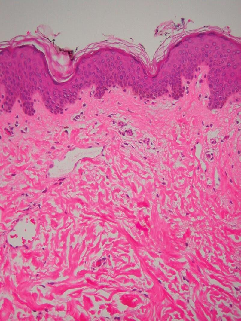
The process of differentiation continues in the stratum granulosum, or granular cell layer, in which the cells acquire additional keratin and become more flattened. In addition, they contain distinctive dark granules, seen easily on light microscopy, that are composed of keratohyalin. Keratohyalin contains two proteins, one of which is called profilaggrin, the precursor to filaggrin. As its name suggests, filaggrin plays an important role in the aggregation of keratin filaments in the stratum corneum. The other protein is called involucrin (from the Latin for “envelope”), and plays a role in the formation of the cell envelope of cells in the stratum corneum. Ichthyosis vulgaris (ichthys, Greek for “fish”) is an inherited dry skin condition secondary to deficient filaggrin production, as noted on light microscopy of a skin biopsy by a reduced or absent granular layer (Fig. 2.3).
Granular cells also contain lamellar granules, which are visualized with electron microscopy. Lamellar granules contain polysaccharides, glycoproteins, and lipids that extrude into the intercellular space and ultimately are thought to help form the “cement” that holds together the stratum corneum cells. Degradative enzymes also are found within the granular cells; these are responsible for the eventual destruction of cell nuclei and intracytoplasmic organelles.
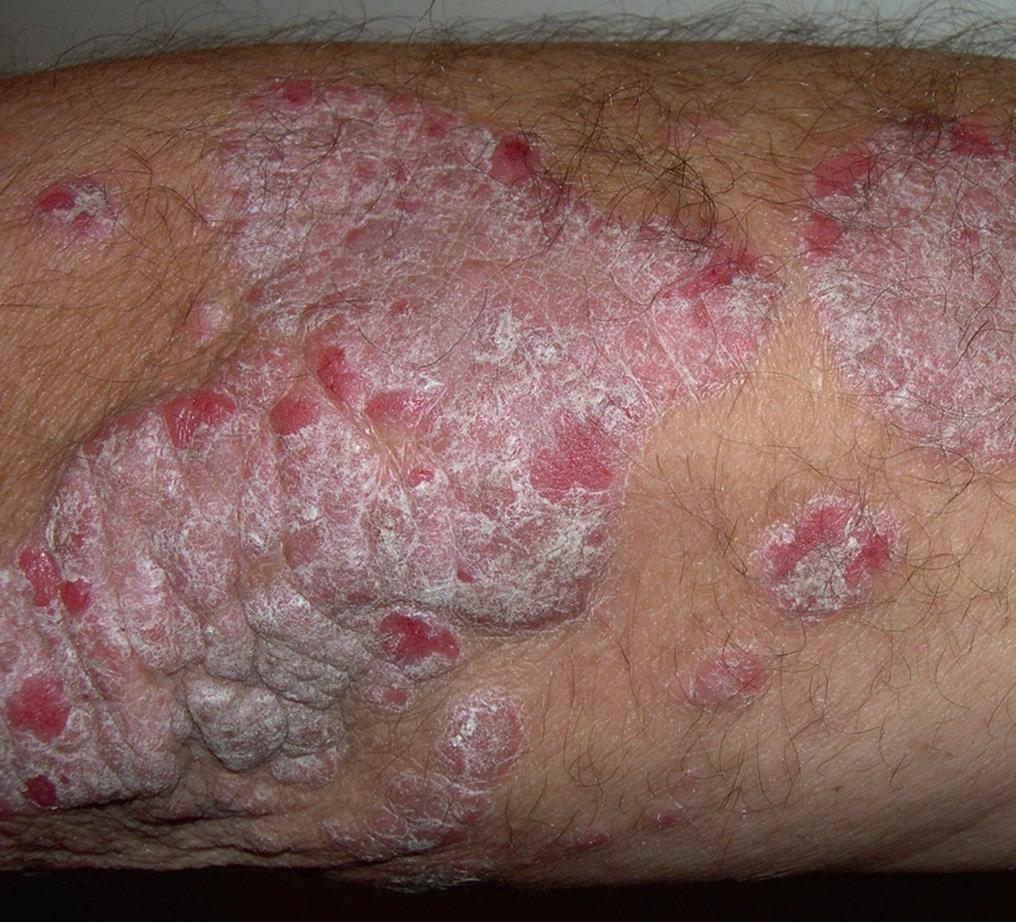
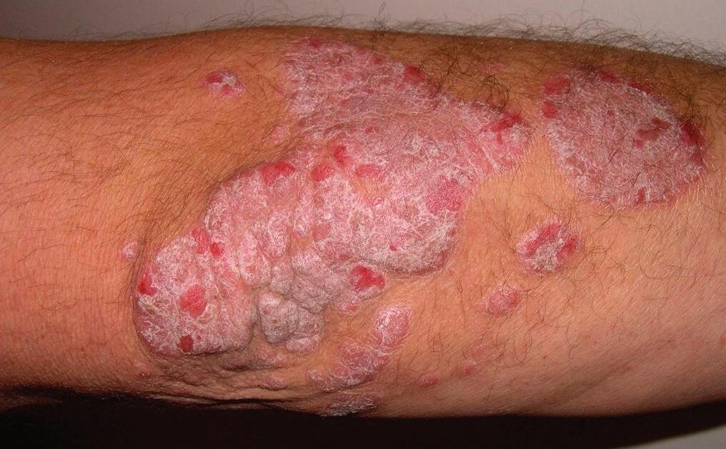
FIGURE 2.1 Psoriasis – an autoimmune disorder characterized by thickened epidermis and increased scale.
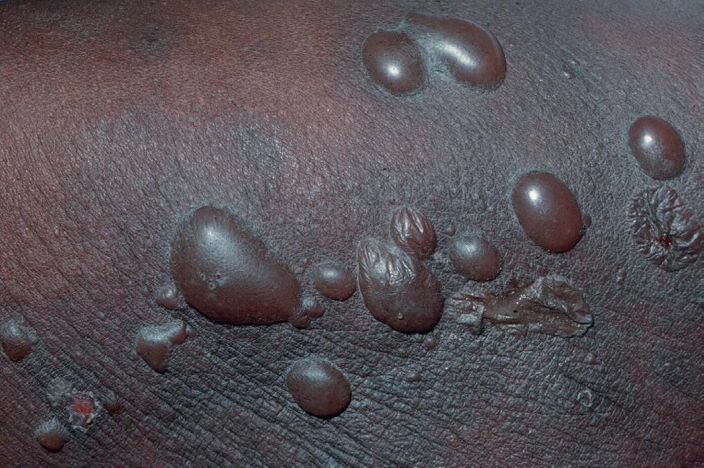
FIGURE 2.2 Pemphigus vulgaris – an autoimmune blistering disease wherein antibodies directed against desmosomes result in keratinocyte separation in stratum spinosum.
Stratum Corneum
A remarkably abrupt transition occurs between the viable, nucleated cells at the top of the granular cell layer and the dead cells of the stratum corneum (Fig. 2.4). The cells in the stratum corneum are large, flat, polyhedral, platelike envelopes filled with keratin. They are stacked in vertical layers that range in thickness from 15 to 25 layers on most body surfaces to as many as 100 layers on the palms and soles. The cells are held together by a lipidrich cement in a fashion similar to “bricks and mortar.” The tightly packed, keratinized envelopes in the stratum corneum provide a semiimpenetrable layer that constitutes the major physical barrier of the skin.
The stratum corneum is the major physical barrier.
The skin microbiome could be considered another outermost layer of the epidermis. With the better sequencing and metagenomics technologies, the role of the microbiome in human health and disease states is being actively investigated. It plays an active role in modulating the host’s immune response to pathogens.
The epidermis, then, is composed of cells that divide in the basal cell layer (basal cells), keratinize in the succeeding layers (keratinocytes), and eventuate into the devitalized, keratinfilled cells in the stratum corneum.
OTHER CELLULAR COMPONENTS
In addition to basal cells and keratinocytes, two other cells are located in the epidermis: melanocytes and Langerhans cells.
Melanocytes
Melanocytes are dendritic, pigmentproducing cells located in the basal cell layer (Figs. 2.4 and 2.5). They protect the skin from ultraviolet radiation. Individuals with little or no pigment develop marked sun damage and numerous skin cancers. The dendrites extend into the stratum spinosum and serve as conduits, through which pigment granules are transferred to their neighboring keratinocytes. The granules are termed melanosomes, and the pigment within is melanin, which is synthesized from tyrosine. Melanosomes are preferentially situated above the nucleus to protect the DNA.
People of all races have a similar number of melanocytes. The difference in skin pigmentation depends on (1) the number and size of the melanosomes and (2) their dispersion in the skin. In darkly pigmented skin, melanosomes are larger in size and more numerous compared with melanosomes in lightly pigmented skin. Sunlight stimulates melanocytes to increase pigment production and disperse their melanosomes more widely.
Langerhans Cells
Langerhans cells are dendritic cells in the epidermis that have an immunologic function (Fig. 2.4). They are
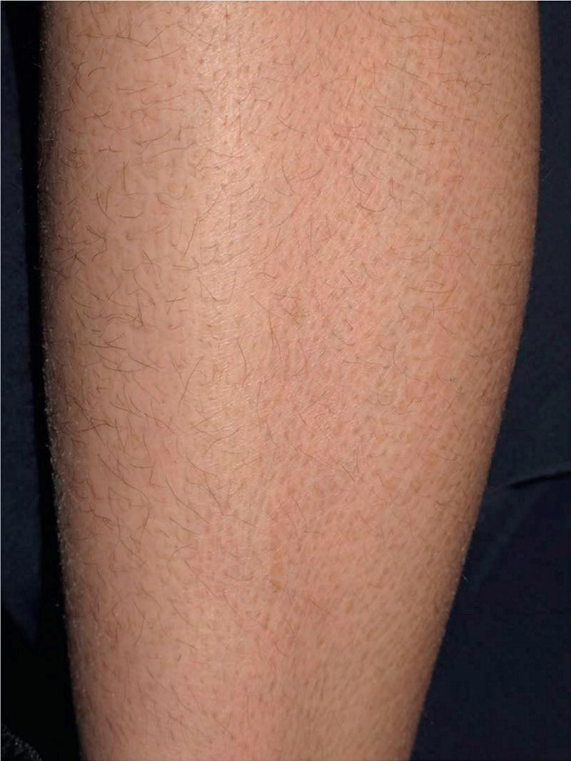
FIGURE 2.3 Ichthyosis vulgaris – a common autoimmune, inherited dry skin condition secondary to deficient filaggrin production. Note “fishlike” scale on the anterior shin.
Granular cells contain keratohyalin and lamellar granules.
FIGURE 2.4
derived from the bone marrow and constitute about 5% of the cells within the epidermis. On electron microscopic examination, characteristic “tennis racket”shaped granules are seen. Langerhans cells are identical to tissue macrophages and present antigens to lymphocytes, with which they interact through specific surface receptors. As such, Langerhans cells are important components of the immunologic barrier of the skin.
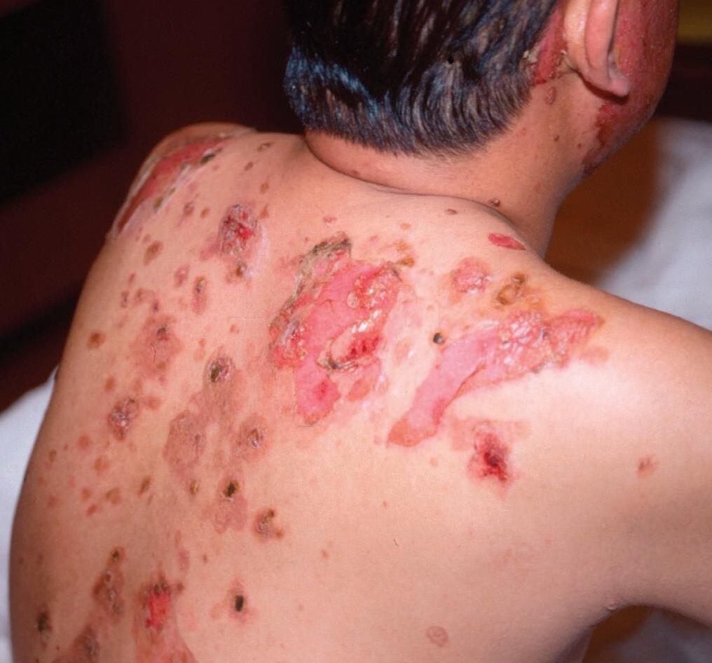
FIGURE 2.6 Bullous pemphigoid – the most common autoimmune blistering disease in the elderly secondary to immune disruption of the hemidesmosome. Note bullae on inner thigh, a characteristic location.
microscopic examination reveals four regions: (1) keratin filaments in the basal keratinocytes attach to hemidesmosomes (electrondense units), which in turn attach to anchoring filaments in (2) the lamina lucida. The lamina lucida is a relatively clear (lucid) zone traversed by delicate anchoring filaments that connect hemidesmosome of basal cells to (3) the lamina densa; the lamina densa is an electrondense zone composed primarily of type IV collagen derived from epidermal cells and (4) anchoring fibrils, which are thick fibrous strands, composed of type VII collagen, and located in the sublamina densa region of the papillary dermis. The basement membrane zone serves as the “glue” between the epidermis and dermis, and is the site of blister formation in numerous diseases (Fig. 2.6). Hence, its structure, composition, and immunologic makeup continue to be investigated intensely.
DERMIS
Key Points
1. provides structural integrity and is biologically active
2. The primary components of the dermal matrix are collagen, elastin, and extrafibrillar matrix
3. Collagen, the principal component of the dermis, represents 70% of skin’s dry weight
Merkel Cells
Merkel cells are located in the basal cell layer. They are more numerous on the palms and soles and are connected to keratinocytes by desmosomes. Merkel cells function as mechanoreceptors. Merkel cell carcinoma is a rare skin cancer with a high mortality rate, as discussed in Chapter 5.
Dermal–Epidermal Junction – The Basement Membrane Zone
The interface between the epidermis and dermis is called the basement membrane zone. With light microscopy, it is visualized only as a fine line. However, electron
The dermis is a tough, but elastic, support structure that contains blood vessels, nerves, and cutaneous appendages. It provides structural integrity and is biologically active by interacting and regulating the functions of cells (i.e., tissue regeneration). The dermis ranges in thickness from 1 to 4 mm, making it much thicker than the epidermis, which in most areas is only about as thick as this piece of paper (Fig. 2.7). The dermal matrix is composed primarily of collagen fibers (principal component), elastic fibers, and ground substance (now called extrafibrillar matrix), which are synthesized by dermal fibroblasts. Collagen accounts for 70% of the dry weight of skin. Collagen and elastic fibers are fibrous proteins
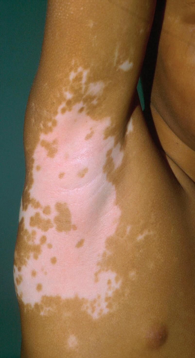
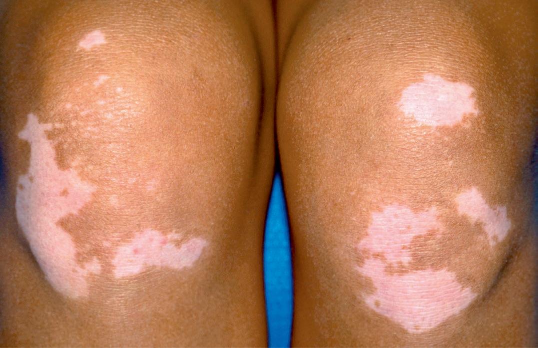
FIGURE 2.5 Vitiligo – an autoimmune disease that results in loss of melanocytes.
Langerhans cells are the first line of immunologic defense in the skin.
that form the strong, yet compliant skeletal matrix. In the uppermost part of the dermis (papillary dermis), collagen fibers are fine and loosely arranged. In the remainder of the dermis (reticular dermis), the fibers are thick and densely packed (Fig. 2.8). Elastic fibers are located primarily in the reticular dermis, where they are thinner and more loosely arranged than collagen fibers. The extrafibrillar matrix fills the space between fibers. It is a nonfibrous material made up of several different mucopolysaccharide molecules, collectively called proteoglycans or glycosaminoglycans. The extrafibrillar matrix imparts to the dermis a more liquid quality, which facilitates movement of fluids, molecules, and inflammatory cells.
Structural components of the dermis:
1. Collagen
2. Elastic fibers
3. Extrafibrillar matrix
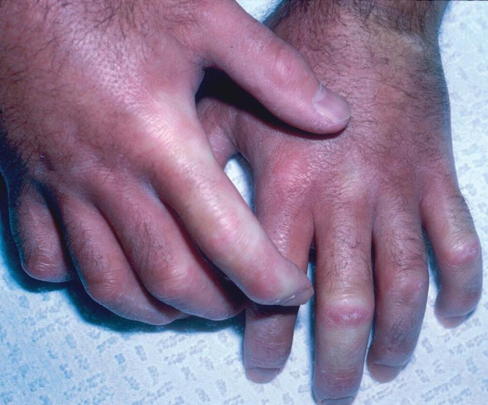
Systemic scleroderma – an increase in the number and activity of fibroblasts produces excessive collagen and results in dermal thickening.
Nerves and blood vessels course through the dermis, and a layer of subcutaneous fat lies below it (Fig. 2.9).
Free nerve endings are the most important sensory receptors.
Nerves
The skin is a major sensory receptor. Without the sensations of touch, temperature, and pain, life would be less interesting and more hazardous. Sensations are detected in the skin by both free nerve endings and more complicated receptors that are corpuscular in structure. The free nerve endings are the more widespread and appear to be more important. The nerve supply of the skin is
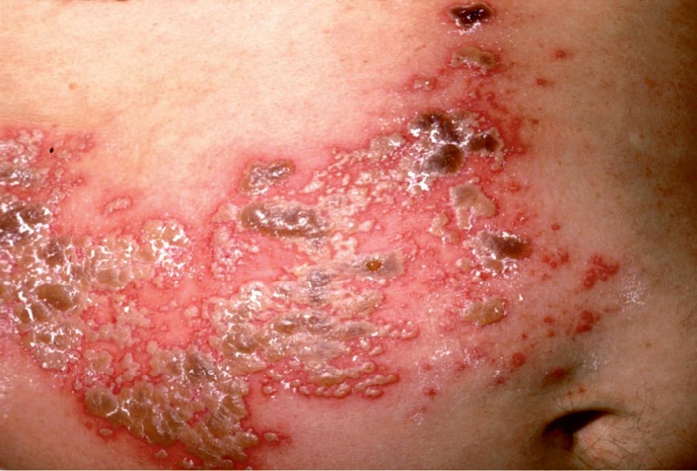
FIGURE 2.10 Herpes zoster – reactivation of varicellazoster virus in sensory nerve ganglia results in a painful, vesicular, dermatomal eruption.
dermis. Temperature regulation is achieved through shunts between the plexuses. Increased blood flow in the superficial plexus permits heat loss, whereas shunting of blood to the deep plexus conserves heat.
SKIN APPENDAGES
Key Points
1. Eccrine glands help to regulate body temperature
2. Apocrine sweat glands depend on androgens for their development
3. The stem cells of the hair follicle reconstitute the nonpermanent portion of the cycling hair follicle
4. Sebaceous glands are under androgen control
5. Nails, like hair, are made of keratin
The skin appendages are the eccrine and apocrine sweat glands, hair follicles, sebaceous glands, and nails. They are epidermally derived but, except for nails, are located in the dermis.
FIGURE 2.11 Sweat gland, apocrine gland, and hair follicle with sebaceous gland.
segmental (dermatomal), with considerable overlap between segments (Fig. 2.10).
Blood Vessels
The blood vessels in the skin serve two functions: nutrition and temperature regulation. The epidermis has no intrinsic blood supply and therefore depends on the diffusion of nutrients and oxygen from vessels in the papillary dermis. Blood vessels in the dermis also supply the connective tissue and appendageal structures located therein.
Functions of blood vessels:
1. To supply nutrition
2. To regulate temperature
The vasculature of the skin is arranged into two horizontal plexuses that are interconnected. The superficial plexus is located at the lower border of the papillary dermis, and the deep plexus is located in the reticular
For physically active individuals and for people living in hot climates, the eccrine sweat glands are physiologically the most important skin appendage. They are activated by emotional and thermal stimuli. Cholinergic innervation is responsible for physiologic eccrine secretion. Botulinum toxin type A (Botox) injected intradermally can treat axillary hyperhidrosis by blocking acetylcholine action. Eccrine sweat glands help to regulate body temperature by excreting sweat onto the surface of the skin, from which the cooling process of evaporation takes place. Two to three million eccrine sweat glands are distributed over the entire body surface, with a total secretory capacity of 10 L of sweat per day. The secretory portion of the sweat apparatus is a coiled tubule located deep in the dermis. The sweat is transported through the dermis by a sweat duct, which ultimately twists a path through the epidermis (Fig. 2.11). Sweat secreted in the glandular portion is isotonic to plasma but becomes hypotonic by the time it exits the skin as a result of ductal reabsorption of electrolytes. Hence, the sweat apparatus is similar to the mechanism in the kidney, that is, glandular (glomerular) excretion is followed by ductal reabsorption.
Eccrine sweat glands help to regulate temperature and are under cholinergic innervation.
Apocrine Sweat Glands
In humans, apocrine sweat glands are androgen dependent for their development and serve no known useful function, although they are responsible for body odor. The odor actually results from the action of surface skin bacteria on excreted apocrine sweat, which itself is odorless. Apocrine sweat glands are located mainly in the axillary
and anogenital areas. The secretory segment of an apocrine gland is also a coiled tubule located deep in the dermis. However, unlike in eccrine glands, in which the secretory cells remain intact, in apocrine glands the secretory cells “decapitate” their luminal (apical) portions as part of the secretory product (see Fig. 2.11). The apocrine duct then drains the secreted sweat into the midportion of a hair follicle, from which it ultimately reaches the skin surface.
Bacterial action on apocrine sweat causes body odor.
HAIR FOLLICLE
In most mammals, hair serves a protective function, but in humans it is mainly decorative.
Hair follicles are distributed over the entire body surface, except the palms and soles. Hair comes in two sizes: (1) vellus hairs, which are short, fine, light colored, and barely noticeable; and (2) terminal hairs, which are thicker, longer, and darker than the vellus type. Terminal hairs in some locations are hormonally influenced and do not appear until puberty (e.g., beard hair in males, and pubic and axillary hair in both sexes).
Types of hair:
1. Vellus (light and fine)
2. Terminal (dark and thick)
A hair follicle can be viewed as a specialized invagination of the epidermis (see Fig. 2.11), with a population of cells at the bottom (hair bulb) that are replicating even more actively than normal epidermal basal cells. These cells constitute the hair matrix. As with basal cells in the epidermis, the matrix cells first divide and then differentiate, ultimately forming a keratinous hair shaft. Melanocytes in the matrix contribute pigment, the amount of which determines the color of the hair. As the matrix cells continue to divide, hair is pushed outward and exits through the epidermis at a rate of about 1 cm per month. Hair growth in an individual follicle is cyclical, with a growth (anagen) phase, a transitional (catagen) phase, and a resting (telogen) phase. The lengths of the phases vary from one area of the body to another. On the scalp, for example, the anagen phase lasts for about 3 years, the catagen phase for about 3 weeks, and the telogen phase for about 3 months. The length of the anagen phase varies from individual to individual, explaining why some persons can grow hair longer than others.
Hair growth cycles through growth (anagen), transitional (catagen), and resting (telogen) phases.
At the end of the anagen phase, growth stops and the hair follicle enters the catagen and telogen phase, during which the matrix portion and lower twothirds of the hair follicle shrivels and the hair within the follicle is shed. Subsequently, through mesenchymal interaction with the
hair follicle stem cells, a new hair matrix is formed at the bottom of the follicle, and the cycle is repeated (Fig. 2.12). At any time, 80% to 90% of scalp hair is in the anagen phase and 10% to 20% is in the telogen phase, thus accounting for a normal shedding rate of 25 to 100 hairs per day.
Normally, 25 to 100 hairs are shed from the scalp each day.
As shown in Fig. 2.11, the hair follicle is situated in the dermis at an angle. Not shown is an attached arrector pili muscle. When this muscle contracts, the hair is brought into a vertical position, giving a “goose flesh” appearance to the skin. The stem cells of the hair follicle are located in the “bulge” area of the follicle, where the arrector pili muscle inserts into the hair follicle, and in the dermal papilla. The stem cells are important for reconstituting the nonpermanent portion of the cycling hair follicle and play a role in reconstituting the epidermal cells.
Sebaceous Glands
Sebaceous glands produce an oily substance termed sebum, the function of which is unknown. In fact, the skin of children and the palmar and plantar skin of adults function well without sebum.
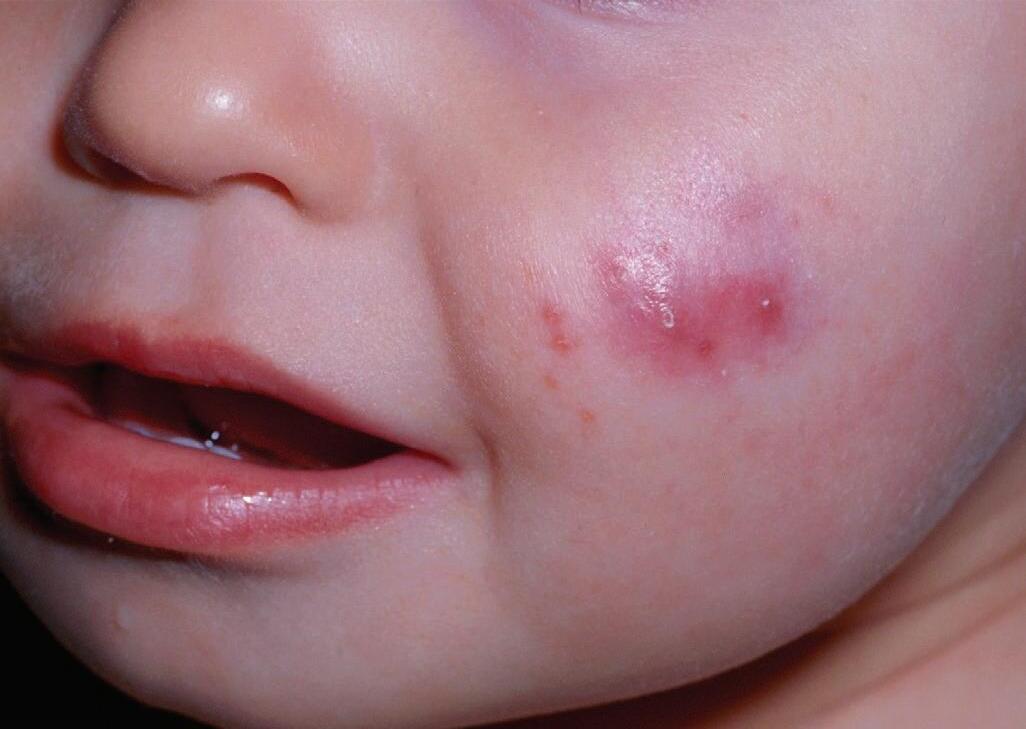
Sebaceous glands are part of the pilosebaceous unit and so are found wherever hair follicles are located. In addition, ectopic sebaceous glands are often found on mucous membranes, where they may form small yellow papules called Fordyce spots. In the skin, sebaceous glands are most prominent on the scalp and face, and are moderately prominent on the upper trunk. The size and secretory activity of these glands are under androgen control. The sebaceous glands in newborns are enlarged owing to maternal hormones, but within months, the glands shrink (Fig. 2.13). They enlarge again in preadolescence from stimulation by adrenal androgens and reach full size at puberty, when gonadal androgens are produced.
Sebaceous glands are androgen dependent.
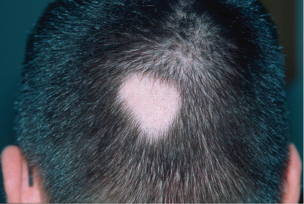
FIGURE 2.12 Alopecia areata – autoimmune condition resulting in nonscarring circular patches of alopecia.
The lipidladen cells in the sebaceous glands are wholly secreted (holocrine secretion) to form sebum. Triglycerides compose the majority of the lipid found in sebaceous gland cells. From the sebaceous glands, sebum drains into the hair follicle (see Fig. 2.11), from which it exits onto the surface of the skin.
Nails
Nails, like hair, are made of keratin, which is formed from a matrix of dividing epidermal cells (Fig. 2.14). Nails, however, are hard and flat, and lie parallel to the skin surface. Located at the ends of fingers and toes, they facilitate fine grasping and pinching maneuvers.
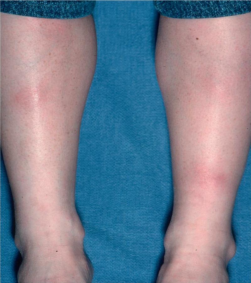
Erythema nodosum. Subcutaneous nodules most commonly seen on shins of women after starting birth control pills; results from inflammation concentrated in the fibrous septa that separate the aggregated fat cells or lobules.
Fingernails grow at a continuous rate of about 0.1 mm/ day, and toenails at a slightly slower rate.
Four epithelial zones are associated with the nail:
1. The proximal nail fold helps to protect the matrix. The stratum corneum produced there forms the cuticle.
The nail plate is a hard, translucent structure composed of keratin. It ranges in thickness from 0.3 to 0.65 mm.
2. The matrix produces the nail plate from its rapidly dividing, keratinizing cells. Most of the matrix underlies the proximal nail fold, but on some digits (especially the thumb) it extends under the nail plate, where it is grossly visible as the white lunula. The most proximal portion of the matrix forms the top of the nail plate; the most distal portion forms the bottom of the nail plate (Fig. 2.15).
FIGURE 2.13 Infantile acne – a common disorder affecting the pilosebaceous unit. maternal androgens are influential.
Proximal nail fold
FIGURE 2.14 Normal nail.
Nail is made of keratin produced in the matrix.
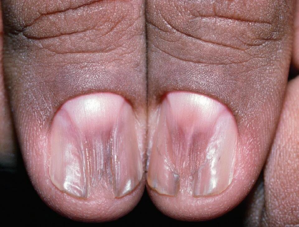
FIGURE 2.15 Lichen planus – an inflammatory condition that normally affects the skin and mucous membranes, but can affect the nail matrix and cause dystrophic nails.
FIGURE 2.16
3. The epithelium of the nail bed produces a minimal amount of keratin, which becomes tightly adherent to the bottom of the nail plate. The pink color of a nail is due to the vascularity in the dermis of the nail bed.
4. The epidermis of the hyponychium underlies the free distal edge of the nail plate. Stratum corneum produced there forms a cuticle to seal the junction of the distal nail bed and nail plate.
Subcutaneous Fat
A layer of subcutaneous fat lies between the dermis and the underlying fascia. It helps to insulate the body from cold, cushions deep tissues from blunt trauma, and serves as a reserve source of energy for the body. Biologically active fat cells play a role in hormone messaging, as
evidenced by metabolic disturbances in obese children and adolescents with peripheral insulin resistance. Recent evidence supports the role of adiposederived stem cells in wound healing, hair follicle support/growth, and protection against photoaging. Within the subcutaneous fat layer, aggregates of fat cells (lipocytes) are separated by fibrous septa that are traversed by blood vessels and nerves (Fig. 2.16).
Subcutaneous fat:
1. insulates
2. Absorbs trauma
3. is a reserve energy source
4. is biologically active
Principles of Diagnosis 3
Chapter Contents
● History
● Preliminary History
● Follow-Up History
● Physical Examination
● Terminology of Skin Lesions
● Clinicopathologic Correlations
● Growths
● Rashes
● Miscellaneous Conditions
● Configuration of Skin Lesions
● Distribution of Skin Lesions
Key Points
1. Morphologic appearance is critical in making the diagnosis
2. Skin diseases can be divided into growths and rashes
ABSTRACT
The approach to a patient with skin disease does not differ markedly from the approach to any other patient. Data are collected from a history and physical examination (and sometimes from the laboratory), a differential diagnosis is generated, and the best diagnosis is selected.
Steps in dermatologic diagnosis:
1. History
2. Physical: identify the morphology of basic lesion
3. Consider clinicopathologic correlations
4. Configuration or distribution of lesions (when applicable)
5. Laboratory tests
In history taking, a modified format is suggested. Instead of beginning with an exhaustive interrogation, it is more efficient to divide the history into a preliminary and a followup format. You should sit, face the patient, let the patient talk, listen, show empathy, and then clarify with questions (location, duration, symptoms, and prior treatment)
The most important part of the physical examination is inspection. Dermatology is a visual specialty, and diagnosis rests heavily on skin inspection. Unfortunately, although the skin is the most visible organ of the body, in a routine physical examination it often is the one most overlooked. Skin lesions need to be looked for, not at Just as the examiner hears only the subtle heart sounds for which he or she listens, so will a clinician see on the skin only the lesions for which he or she searches. We need to train our eyes to see the skin lesions before us and ultimately be able to recognize them.
Dermatologic diagnosis depends on the examiner’s skill in skin inspection.
We have divided skin disorders into two broad categories: growths and rashes. A growth is a discrete lesion resulting from proliferation of one or more of the skin’s components. A rash is an inflammatory process that usually is more widespread than a growth. For both skin growths and rashes, the most important task is to characterize the clinical appearance of the basic lesion, that is, to identify its morphology. The pathophysiologic processes responsible for the clinical lesion must then be considered. These clinicopathologic correlations are emphasized in the diagnostic approach presented in this book. For skin rashes, important diagnostic information can sometimes also be obtained by noting the manner in which the lesions are arranged or distributed.
After the history and physical examination have been completed, laboratory tests may be indicated. In dermatology, these are usually simple office procedures that can provide valuable information needed either to confirm or to establish a diagnosis in selected disorders.
HISTORY
Key Points
1. Establish rapport.
2. Let the patient talk uninterruptedly in the beginning
3. Clarify location, duration, symptoms, and prior treatment
4. Expand the history based on the differential diagnosis
In medicine, the traditional approach is to take the history before performing the physical examination. Some dermatologists prefer to reverse this order. We find it most useful to ask questions both before and after the examination. With this approach, a preliminary history is taken, in which several general questions are asked of all patients. Depending on the physical findings, more selective questions may be asked subsequently. For example, a history of sexual contacts would be inappropriate for an 82yearold invalid complaining of an itching scalp, but would be indicated for a patient with an indurated ulcer on the penis.
PRELIMINARY HISTORY
In addition to its diagnostic value, a preliminary history also helps to establish rapport with the patient. The shortcut of examining the skin without expressing an interest in the person will often be found wanting, especially by the patient. This initial history is composed of two parts that correlate with the chief complaint and the history of the present illness in the standard history format.
The initial history can be abbreviated by asking four general questions:
1. How long?
2. Where affected?
3. Does it itch or other symptoms?
4. How have you treated it?
CHIEF COMPLAINT
In eliciting the chief complaint, one can often learn much by asking an openended question, such as, “What is your skin problem?” This is followed by four general questions regarding the history of the present illness.
HISTORY OF THE PRESENT ILLNESS
The general questions concern onset and evolution of the condition, distribution, symptoms, and treatment to date.
Onset and Evolution
“When did it start? Has it gotten better or worse?” Answers to these questions determine the duration of the disorder and how the condition has evolved over time. For most skin conditions, this is important information.
Symptoms
“Does it bother you?” is an openended way of asking about symptoms. For rashes, the most common symptom is itching. If the patient does not respond to the general symptom question, you may want to ask specifically, “Does it itch?” Questions concerning systemic symptoms (e.g., “How do you feel otherwise?”) are not applicable for most skin diseases and are more appropriately reserved until after the physical examination.
Treatment to Date
The question, “How have you treated it?” results in an incomplete response from almost all patients. For skin disease, one is particularly interested in learning what
topical medications have been applied. Many patients do not consider overthecounter preparations important enough to mention. The same applies for some systemic medications. Providing the patient with specific examples of commonly used topical and systemic medications, such as calamine lotion and aspirin, may jog a patient’s memory enough to recall similar products that he or she may have used. It is important to inquire about medications, not only because they cause some conditions, but also because they may aggravate many others. For example, contact dermatitis initially induced by poison ivy may be perpetuated by contact allergy to an ingredient in one of the preparations used in treatment.
After the skin examination, one may need to return to the treatment question if any suspicion exists that a medication is causing or contributing to the disorder. Interestingly, a patient often recalls using pertinent medication only when he or she is asked the question again.
Persistence is often required in eliciting a complete medication history.
Finally, at the end of the visit, when one is ready to prescribe medications for the patient, it is helpful to know what medications have already been used. This approach avoids the potentially awkward situation in which a patient replies to your enthusiastic recommendation of your favorite therapy with, “I’ve already tried that and it didn’t work!”
FOLLOW-UP HISTORY
After the initial history and physical examination, it is hoped that a diagnosis, or at least a differential diagnosis, has been formulated. With a diagnosis in mind, more focused questions may be necessary. This questioning may include obtaining more details about the history of the present illness or may be directed toward eliciting specific information from other categories of the traditional medical history, including past medical history, review of systems, family history, and social history. The following serve only as examples for the use of focused questions.
PAST MEDICAL HISTORY
After the physical examination, one may want to learn more about the patient’s general health. For example, in a patient with suspected herpes zoster, a past history of chickenpox would be of interest. We have discussed how topically applied and systemically administered medications often contribute to skin conditions. Skin findings may encourage further pursuit of these possibilities. For example, in a patient with a generalized erythematous rash or hives, systemic drugs should be high on the list of possible causes. Because drugs can cause virtually any type of skin lesion, it is useful to consider drug eruptions in the differential diagnosis of almost any skin disease. It may also be helpful to ascertain whether the patient has any known allergies, in order to determine whether any medications are currently being used that could produce a crossreaction.
Drugs can cause all types of skin rash.
REVIEW OF SYSTEMS
In a patient with a malar rash, a diagnosis of systemic lupus erythematosus should be considered, and the examiner will want to question the patient further for symptoms of additional skin or other organ involvement, including Raynaud’s phenomenon, photosensitivity, hair loss, mouth ulcers, and arthritis. In a patient with a generalized maculopapular eruption, the two most common causes are drugs and viruses, so the physician will want to inquire about both medication use and viral symptoms, such as fever, malaise, and upper respiratory or gastrointestinal symptoms.
FAMILY HISTORY
In certain cutaneous conditions, some knowledge of the family history may help in diagnosis. Innumerable inherited disorders have dermatologic expression. The following serve only as examples:
● In a child with a chronic itching eruption in the antecubital and popliteal fossae, atopic dermatitis is suspected. A positive family history for atopic diseases (atopic dermatitis, asthma, hay fever) supports the diagnosis.
● In a youngster with multiple caféaulait spots, a diagnosis of neurofibromatosis is considered. A positive family history for this disorder, substantiated by examination of family members, helps to support the diagnosis of this dominantly inherited disease.
inherited disorders have numerous skin findings.
Knowledge of the family’s present health is also important when considering infectious diseases. For example, impetigo can occur in several family members, and this knowledge may help in considering the diagnosis; it would certainly be important for treatment. Likewise, in a patient with suspected scabies, it is important to know, for both diagnostic and therapeutic purposes, whether other family members are itching.
SOCIAL HISTORY
In some disorders, knowledge of the patient’s social history may be important. For example, a chronic skin ulcer from persistent herpes simplex infection is a sign of immunosuppression, particularly acquired immune deficiency syndrome (AIDS). Therefore, a patient with such an ulceration should be asked about highrisk factors for acquiring AIDS, including sexual behavior, intravenous drug abuse, and exposure to blood products.
Another common occasion for probing into a patient’s social history is when the patient is suspected of having contact dermatitis; this aspect of the social history could be subtitled the skin exposure history. Patients encounter potentially sensitizing materials both at work and at play. Industrial dermatitis is a leading cause of workers’ disability. For chronic hand dermatitis, questions about occupational exposure are important and should be directed particularly to materials and substances the patient contacts either by handling or by immersion. Similarly, a patient presenting with an acute eruption characterized by streaks of vesicles should be queried regarding recent outdoor activities resulting in exposure to poison ivy or poison oak. Contact dermatitis is a common and challenging problem. On the part of the physician, it often requires painstaking efforts in a detectivetype search to elicit from the patient an exposure history that fits the dermatitis.
A complete “skin exposure history” is required whenever contact dermatitis is suspected.
Some harbor the misconception that in dermatology, one needs only to glance at the skin to arrive at a diagnosis and that talking with the patient is superfluous. Although this is occasionally true, we hope that the previous examples serve to illustrate that this frequently is not the case. In fact, in some instances (and contact dermatitis is a good example), detailed historical information is essential to establish a diagnosis.
PHYSICAL EXAMINATION
Key Points
1. Complete skin examination is recommended at the first visit
2. good lighting is critical
3. Describe the morphology of the eruption
The physical examination follows the preliminary history. For the skin to be inspected adequately, three essential requirements must be met: (1) an undressed patient, clothed in an examining gown; (2) adequate illumination, preferably bright overhead fluorescent lighting; and (3) an examining physician prepared to see what is there.
one should do hand hygiene prior to and after touching the patient.
for persistent skin infections, consider the possibility of AiDS.
Examine the entire mucocutaneous surface, but patients will be more firmly convinced of your sincere interest in their particular problems if you start by examining the affected areas before proceeding with the more complete examination.
At least for the initial examination, the patient needs to be disrobed so that the entire skin surface can be examined. Busy physicians who tend to overlook this rule will miss much. An occasional patient may be reluctant to comply, saying, “My skin problem is only on my hands; why do you need to look at the rest of my skin?” We tell such patients that we have at least two reasons:
1. Other lesions may be found that “go along with” the lesions on the hands, and help to confirm the diagnosis. For example, in a patient with sharply demarcated plaques on the palms, the finding of a few scaling plaques on the knees or a sharply marginated intergluteal plaque will help to substantiate a suspicion of psoriasis.
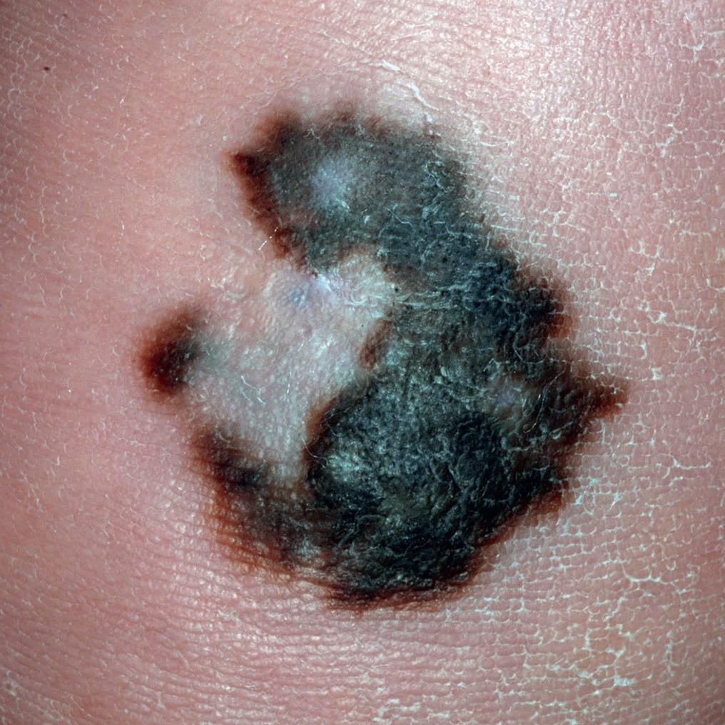
2. An important incidental skin lesion may be found. The finding of a previously undetected malignant melanoma on a patient’s back is an example. We studied the yield from a complete skin examination in 1157 consecutive new dermatology patients and found an incidental skin malignancy in 22. Some 20 of these patients had basal cell carcinoma, one had melanoma, and one had Kaposi’s sarcoma that served as the presenting manifestation of AIDS. A subsequent study of 874 patients reported an incidental skin cancer detection rate of 3.4%.
The entire skin surface is examined for:
1. Lesions that may accompany the presenting complaint
2. Unrelated, but important incidental findings
For the skin to be examined adequately, it must be properly illuminated. Natural lighting is excellent for this purpose, but is difficult to achieve in most offices and hospital rooms. The alternative is bright overhead fluorescent lighting, supplemented with a movable lamp that is usually wall mounted. One additional illuminator that is often useful is a simple penlight. Either this or the movable lamp can be used as side lighting to detect whether a lesion is subtly elevated. For this technique, the light is directed onto the lesion from an angle that is roughly parallel to the skin. If the lesion is elevated, a small shadow will be thrown, and the relief of the skin will be appreciated. The penlight also is useful for examining the mouth, an area that is sometimes overlooked but in which one may detect lesions that are helpful in diagnosing a cutaneous disorder.
Another piece of examination equipment that is occasionally useful is the Wood’s light, a longwavelength ultraviolet “black” light. Contrary to some popular misconceptions, this light does not enable one to diagnose most skin fungal infections; it detects fluorescence of affected hairs only in some, now uncommon, types of tinea capitis. The Wood’s light is, however, still used to accentuate pigmentary alterations in the skin, such as vitiligo.
Except for provision of adequate illumination, minimal equipment is needed for examining the skin. A simple handheld lens can be helpful. Enlarging the image may
improve diagnostic accuracy. However, on some occasions, such as clarifying a burrow in scabies or detecting Wickham’s striae in a lesion of lichen planus, a handheld lens can be useful. For diagnosing pigmented growths, dermatologists often employ a dermatoscope. This is an illuminated handheld magnifying device intended to help the clinician to diagnose melanoma and other growths clinically.
A dermatoscope is useful in diagnosing growths, especially melanoma.
An adequate examination of the skin should actually be called a mucocutaneous examination so that one is reminded to include an examination of the mouth. Similarly, the scalp and nails should not be overlooked. Because both cutaneous and systemic diseases may be expressed in the nails and nail beds as well as in the mouth, these areas should be inspected in every cutaneous examination.
The scalp, mouth, and nails should not be overlooked.
Physical examination depends largely on inspection, but one should not neglect the opportunity to palpate the skin as well. The two major purposes for this are (1) to assess the texture, consistency, and tenderness of the skin lesions; and (2) to reassure patients that we are not afraid to touch their skin lesions–that they do not have some dreadful contagious disease. Nothing is more disquieting to a patient than to be cautiously approached with a gloved hand. For anogenital, mucosal, and all weeping lesions, gloving is necessary and expected, but for most other lesions, the physician learns more and the patient is less frightened if the touching is done without gloves. Palpation is the major method by which we evaluate not only the consistency (e.g., softness, firmness, fluctuance) but also the depth of a lesion.
Palpation helps to:
1. Assess texture and consistency
2. Evaluate tenderness
3. Reassure patients that they are not contagious
After the patient is properly gowned and perfectly illuminated, for what do we inspect and palpate? The first and most important step is to characterize the appearance (i.e., identify the morphology) of each skin lesion. After the morphology of a lesion is identified, its clinicopathologic correlation can be considered.
The most important task in the physical examination is to characterize the morphology of the basic lesion.
TERMINOLOGY OF SKIN LESIONS
Key Points
1. Primary lesions include macule, patch, papule, plaque, nodule, cyst, vesicle, pustule, ulcer, wheal, telangiectasia, burrow, and comedo
2. Secondary lesions include scale, crust, oozing, lichenification, induration, fissure, and atrophy
A special vocabulary is used in describing the morphologic appearances of skin lesions. These terms are illustrated and defined in Fig. 3.1.
CLINICOPATHOLOGIC CORRELATIONS
Key Points
1. Envisioning the gross and microscopic morphology together helps to make the diagnosis
2. Rash or growth?
3. Epidermal, dermal, or subcutaneous?
TABLE 3.1 Clinicopathologic Correlations
Skin Component
Epidermis
Stratum corneum
Subcorneal epidermis
Melanocytes
Dermis
Blood vessels
nerves
Connective tissue
Dermal appendages
Pilosebaceous units
Sweat glands
Subcutaneous fat
Pathologic Alteration
Hyperkeratosis
Hyperplasia
Hyperplasia
Disruptive inflammatory changes
Dried serum/blood
increased number or function
Decreased number or function
Hyperplasia or inflammation
Vasodilation
Hemorrhage
Vasodilation with edema
Hyperplasia
Hyperplasia
Loss of epidermis
Loss of epidermis and dermis
Hyperplasia
Atrophy
Hyperplasia or inflammation
Hypersecretion
Hyperplasia or inflammation
Hyperplasia or inflammation
The lesions defined in Fig. 3.1 result from alterations in one or more of the skin’s structural components. For clinical diagnostic purposes, we try to envision what pathologic changes are associated with each clinical lesion (Table 3.1). Scale, lichenification, vesicles, bullae, pustules, and crusts represent epidermal alterations, whereas erythema, purpura, and induration reflect changes in the dermis. Such clinicopathologic correlations form the basis of the diagnostic approach. For example, scaling of a nodule suggests hyperkeratosis of the stratum corneum and, thus, an epidermal growth.
Determine which of the skin components are involved in the clinical lesion.
Table 3.2 presents an algorithm for this approach and outlines the organization of the remainder of this book. Most skin disorders can be categorized first as proliferative “growths” (neoplasms) or inflammatory “rashes” (eruptions). The growths and rashes are then subdivided, depending on how they appear clinically and which structural component is involved pathologically.
growths are hyperplastic lesions; rashes are inflammatory.
Clinical Manifestation
Scale
Lichenification
Papules, plaques, and nodules
Vesicles, bullae, and pustules
Crusts
Pigmented macules, papules, and nodules
White spots
Macules, papules, and nodules
Erythema
Purpura
Wheals
Papules, nodules
induration, papules, nodules, and plaques
Erosion
Ulceration
Hirsutism
Alopecia
Comedones, papules, nodules, and cysts
Hyperhidrosis
Vesicles, papules, pustules, and cysts
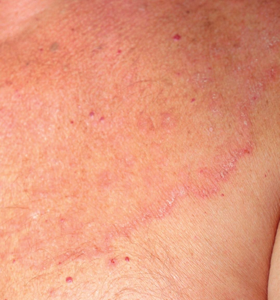
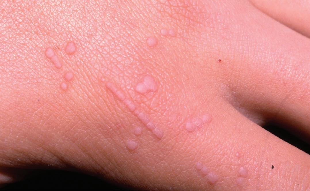
induration and nodules
Skin lesions. A. Vitiligo – macule. A flat skin lesion recognizable because its color differs from that of the surrounding normal skin. The most common color changes are white (hypopigmented), brown (hyperpigmented), and red/purple (erythematous and purpuric). B. Tinea corporis – patch. A macule with some surface change, either slight scale or fine wrinkling. C. flat warts – papules. Small elevated skin lesions <0.5 cm in diameter.