technical assistance: email expertconsult.help@elsevier.com call 1-800-401-9962 (inside the US) call +1-314-447-8200 (outside the US)
Liu, Volpe, and Galetta’s Neuro-Ophthalmology: Diagnosis and Management
Liu, Volpe, and Galetta’s NeuroOphthalmology
Diagnosis and Management
Grant T. Liu, MD
Professor of Neurology and Ophthalmology and Raymond G. Perelman Endowed Chair in Pediatric Neuro-Ophthalmology; Division of Neuro-Ophthalmology; Departments of Neurology and Ophthalmology; Hospital of the University of Pennsylvania and the Children’s Hospital of Philadelphia; Perelman School of Medicine at the University of Pennsylvania, Philadelphia, PA, USA
Nicholas J. Volpe, MD
George and Edwina Tarry Professor of Ophthalmology; Chairman, Department of Ophthalmology, Northwestern Memorial Hospital, Northwestern University Feinberg School of Medicine, Chicago, IL, USA
Steven L. Galetta, MD
Philip Moskowitz Professor and Chairman, Department of Neurology, NYU Langone Medical Center, NYU School of Medicine, New York, NY; and Professor Emeritus, Perelman School of Medicine, Philadelphia, PA, USA THIRD
No part of this publication may be reproduced or transmitted in any form or by any means, electronic or mechanical, including photocopying, recording, or any information storage and retrieval system, without permission in writing from the publisher. Details on how to seek permission, further information about the Publisher’s permissions policies and our arrangements with organizations such as the Copyright Clearance Center and the Copyright Licensing Agency, can be found at our website: www.elsevier.com/permissions.
This book and the individual contributions contained in it are protected under copyright by the Publisher (other than as may be noted herein).
Notices
Practitioners and researchers must always rely on their own experience and knowledge in evaluating and using any information, methods, compounds or experiments described herein. Because of rapid advances in the medical sciences, in particular, independent verification of diagnoses and drug dosages should be made. To the fullest extent of the law, no responsibility is assumed by Elsevier, authors, editors or contributors for any injury and/or damage to persons or property as a matter of products liability, negligence or otherwise, or from any use or operation of any methods, products, instructions, or ideas contained in the material herein.
ISBN: 978-0-323-34044-1
Content Strategist: Russell Gabbedy
Content Development Specialist: Trinity Hutton
Project Manager: Julie Taylor
Design: Christian Bilbow
Illustration Manager: Nichole Beard
Marketing Manager: Melissa Fogarty
Video Contents, vi
Dedication, ix
Foreword, x
Preface, xi
Acknowledgments, xii
List of Contributors, xiii
PART ONE HISTORY AND EXAMINATION
1 The Neuro-Ophthalmic History, 3
GRANT T. LIU, NICHOLAS J. VOLPE, and STEVEN L. GALETTA
2 The Neuro-Ophthalmic Examination, 7
GRANT T. LIU, NICHOLAS J. VOLPE, and STEVEN L. GALETTA
PART TWO VISUAL LOSS AND OTHER DISORDERS OF THE AFFERENT VISUAL PATHWAY
3 Visual Loss: Overview, Visual Field Testing, and Topical Diagnosis, 39
GRANT T. LIU, NICHOLAS J. VOLPE, and STEVEN L. GALETTA
4 Visual Loss: Retinal Disorders of Neuro-Ophthalmic Interest, 53
MADHURA A. TAMHANKAR
5 Visual Loss: Optic Neuropathies, 101
STACY L. PINELES and LAURA J. BALCER
6 Optic Disc Swelling: Papilledema and Other Causes, 197
MELISSA W. KO
7 Visual Loss: Disorders of the Chiasm, 237
ROBERT A. AVERY
8 Retrochiasmal Disorders, 293
SASHANK PRASAD
9
Disorders of Higher Cortical Visual Function, 341
VICTORIA S. PELAK
10 Transient Visual Loss or Blurring, 365
MADHURA A. TAMHANKAR
11 Functional (Nonorganic) Visual Loss, 379
GRANT T. LIU, NICHOLAS J. VOLPE, and STEVEN L. GALETTA
12 Visual Hallucinations and Illusions, 395
GRANT T. LIU, NICHOLAS J. VOLPE, and STEVEN L. GALETTA
PART THREE EFFERENT NEUROOPHTHALMIC DISORDERS
13 Pupillary Disorders, 417
LAURA J. BALCER
14
Eyelid and Facial Nerve Disorders, 449
HEATHER E. MOSS
15 Eye Movement Disorders: Third, Fourth, and Sixth Nerve Palsies and Other Causes of Diplopia and Ocular Misalignment, 489
MADHURA A. TAMHANKAR
16
17
Eye Movement Disorders: Conjugate Gaze Abnormalities, 549
DANIEL R. GOLD
Eye Movement Disorders: Nystagmus and Nystagmoid Eye Movements, 585
DANIEL R. GOLD
18 Orbital Disease in Neuro-Ophthalmology, 611
KENNETH S. SHINDLER
PART FOUR OTHER TOPICS
19 Headache, Facial Pain, and Disorders of Facial Sensation, 661
MELISSA W. KO and SASHANK PRASAD Index, 685
Video Contents
PART ONE: HISTORY AND EXAMINATION
Chapter 2 The Neuro-Ophthalmic Examination
Video 2.1 Examination. Afferent System. Confrontation Visual Fields – Nicholas J. Volpe, Stacy L. Pineles, and Heather E. Moss
Video 2.2 Examination. Efferent System. Swinging Flashlight Test to Compare Pupillary Reactivity – Nicholas J. Volpe, Stacy L. Pineles, and Heather E. Moss
Video 2.3 Afferent Pupillary Defect, Right Eye – Grant T. Liu
Video 2.4 Examination. Efferent System. Eye Movements and Assessment of Ocular Alignment – Nicholas J. Volpe, Stacy L. Pineles, and Heather E. Moss
Video 2.5 Convergence – Daniel R. Gold
Video 2.6 Smooth Pursuit and Suppression of the VestibuloOcular Reflex (VOR) – Daniel R. Gold
Video 2.7 Saccades – Daniel R. Gold
Video 2.8 Vestibulo-Ocular Reflex (VOR) and Head Impulse Test (HIT) – Daniel R. Gold
Video 2.9 Optokinetic Nystagmus (OKN) Testing With a Flag –Daniel R. Gold
Video 2.10 Esotropia Demonstrated by Alternate Cover Technique – Grant T. Liu
PART TWO: VISUAL LOSS AND OTHER DISORDERS OF THE AFFERENT VISUAL PATHWAY
Chapter 3 Visual Loss: Overview, Visual Field Testing and Topical Diagnosis
Video 3.1 Laser Pointer Visual Field Testing –Steven L. Galetta and Grant T. Liu
Chapter 6 Optic Disc Swelling: Papilledema and Other Causes
Video 6.1 Optic Nerve Sheath Fenestration –Nicholas J. Volpe
Chapter 7 Visual Loss: Disorders of the Chiasm
Video 7.1 Post-Fixation Blindness – Grant T. Liu
Chapter 8 Retrochiasmal Disorders
Video 8.1 Left Homonymous Hemianopia – Grant T. Liu
Chapter 9 Disorders of Higher Cortical Visual Function
Video 9.1 Right Parietal Syndrome – Grant T. Liu
Video 9.2 Balint Syndrome – Grant T. Liu
Chapter 12 Visual Hallucinations and Illusions
Video 12.1 Palinopsia – Grant T. Liu
PART THREE: EFFERENT NEUROOPHTHALMIC DISORDERS
Chapter 13 Pupillary Disorders
Video 13.1 Pupillary Light-Near Dissociation in Parinaud Syndrome – Grant T. Liu
Video 13.2 Aberrant Regeneration of the Third Nerve – Steven L. Galetta and Grant T. Liu
Video 13.3 Physiologic Anisocoria – Grant T. Liu
Video 13.4 Horner Syndrome, Left Eye – Nicholas J. Volpe
Video 13.5 Paradoxical Pupils – Imran Jivraj and Grant T. Liu
Chapter 14 Eyelid and Facial Nerve Disorders
Video 14.1 Eyelid Signs in Ocular Myasthenia – Grant T. Liu
Video 14.2 Marcus Gunn Jaw Winking Phenomenon – Robert A. Avery and Grant T. Liu
Video 14.3 Marcus Gunn Jaw Winking Phenomenon in an Infant – Grant T. Liu
Video 14.4 Eyelid Opening Apraxia – Grant T. Liu
Video 14.5 Left Facial Palsy (Central) – Grant T. Liu
Video 14.6 Left Facial Palsy (Peripheral) – Sashank Prasad and Grant T. Liu
Video 14.7 Blepharospasm in Meige Syndrome –Grant T. Liu
Video 14.8 Hemifacial Spasm – Grant T. Liu
Video 14.9 Ocular Myasthenia and Positive Edrophonium Test – Joel Glaser and Grant T. Liu
Video 14.10 Myasthenic Ptosis, Left Eye, Rest Test –Grant T. Liu
Video 14.11 Congenital Myasthenic Syndrome – Grant T. Liu
Video 14.12 Chronic Progressive External Ophthalmoplegia –Grant T. Liu
Chapter 15 Eye Movement Disorders: Third, Fourth, and Sixth Nerve Palsies and Other Causes of Diplopia and Ocular Misalignment
Video 15.1 Internuclear Ophthalmoplegia, Right –Grant T. Liu
Video 15.2 Internuclear Ophthalmoplegia (INO) With Intact Convergence – Grant T. Liu
Video 15.3 Bilateral Internuclear Ophthalmoplegia (INO) –Grant T. Liu
Video 15.4 Convergence Spasm – Grant T. Liu
Video 15.5 IIIrd Nerve Palsy, Right Eye – Grant T. Liu
Video 15.6 IIIrd Nerve Palsy, Right Eye – Grant T. Liu
Video 15.7 IIIrd Nerve Palsy With Aberrant Regeneration, Left Eye – Grant T. Liu
Video 15.8 Cyclic Oculomotor Spasms – Grant T. Liu
Video 15.9 IVth Nerve Palsy, Right EyeThree Step Test – Grant T. Liu
Video 15.10 IVth Nerve Palsy, Left Eye – ductions –Grant T. Liu
Video 15.11 VIth Nerve Palsy, Right Eye – Grant T. Liu
Video 15.12 Duane’s Syndrome Type 1, Left Eye –Grant T. Liu
Video 15.13 Duane’s Syndrome Type 2, Right Eye –Grant T. Liu
Video 15.14 Duane’s Syndrome Type 3, Both Eyes –Grant T. Liu
Video 15.15 Brown’s Syndrome, Left Eye – Grant T. Liu
Video 15.16 Pseudo-Bilateral Internuclear Ophthalmoplegia due to Ocular Myasthenia – Grant T. Liu
Video 15.17 Pseudo-One-and-a-Half Syndrome due to Ocular Myasthenia – Grant T. Liu
Video 15.18 Inferior Rectus Myotoxicity After Cataract Surgery and Retrobulbar Anesthesia, Left Eye – Nicholas J. Volpe
Video 15.19 Neuromyotonia – Grant T. Liu
Chapter 16 Eye Movement Disorders: Conjugate Gaze Abnormalities
Video 16.1 Unilateral Horizontal Gaze Palsy From a Right Pontine Infarct – Grant T. Liu
Video 16.2 Bilateral Horizontal Gaze Palsy – Grant T. Liu
Video 16.3 Horizontal One-and-a-Half Syndrome –Steven L. Galetta and Larry Gray
Video 16.4 Ipsilateral Gaze Deviation and Lateropulsion in Wallenberg Syndrome –Grant T. Liu
Video 16.5 Saccadic Dysmetria – Grant T. Liu
Video 16.6 Supranuclear Gaze Palsy and Lid Retraction – Steven L. Galetta and Grant T. Liu
Video 16.7 Saccadic Palsy After a Type A Aortic Dissection Repair – Grant T. Liu
Video 16.8 Congenital Ocular Motor Apraxia –Grant T. Liu
Video 16.9 Slow Saccades in Spinocerebellar Ataxia 3 –Grant T. Liu
Video 16.10 Vertical Gaze Paresis and Convergence Retraction Saccades in Parinaud’s Syndrome –Grant T. Liu
Video 16.11 Vertical Gaze Paresis and Convergence Retraction Saccades in Parinaud’s Syndrome –Grant T. Liu
Video 16.12 Progressive Supranuclear Palsy – Grant T. Liu
Video 16.13 Saccadic Palsy due to Neimann Pick C –Grant T. Liu
Video 16.14 Benign Paroxysmal Tonic Gaze of Infancy –Grant T. Liu
Chapter 17 Eye Movement Disorders: Nystagmus and Nystagmoid Eye Movements
Video 17.1 Inability to Cancel the Vestibulo-Ocular Reflex (VOR) – Grant T. Liu
Video 17.2 Congenital Nystagmus (Early) – Grant T. Liu
Video 17.3 Congenital Nystagmus (Later) – Grant T. Liu
Video 17.4 Latent Nystagmus – Grant T. Liu
Video 17.5 Spasmus Nutans (Benign) – Robert A. Avery and Grant T. Liu
Video 17.6 Spasmus Nutans (Tumor-Related) – Grant T. Liu
Video 17.7 Vertical Oscillations Associated With Monocular Vision Loss – Grant T. Liu
Video 17.8 Benign Paroxysmal Positional Vertigo Involving the Right Posterior Canal – Grant T. Liu
Video 17.9 Right Dix-Hallpike Maneuver – Daniel R. Gold
Video 17.10 Supine Roll Testing to Evaluate for Horizontal (Lateral) Canal BPPV – Daniel R. Gold
Video 17.11 Epley Maneuver to Treat Right Posterior Canal BPPV – Daniel R. Gold
Video 17.12 Semont Maneuver to Treat Right Posterior Canal BPPV – Daniel R. Gold
Video 17.13 BBQ Roll to Treat Right Geotropic Horizontal Canal BPPV – Daniel R. Gold
Video 17.14 Gufoni Maneuver to Treat Right Geotropic Horizontal Canal BPPV – Daniel R. Gold
Video 17.15 Gufoni Maneuver to Treat Left Apogeotropic Horizontal Canal BPPV – Daniel R. Gold
Video 17.16 Deep Head Hanging Maneuver to Treat Right or Left Anterior Canal BPPV – Daniel R. Gold
Video 17.17 Gaze-Evoked Nystagmus – Grant T. Liu
Video 17.18 Brun’s Nystagmus – Grant T. Liu
Video 17.19 Rebound Nystagmus – Grant T. Liu
Video 17.20 Periodic Alternating Nystagmus – Grant T. Liu
Video 17.21 Downbeat Nystagmus due to a Chiari Malformation – Grant T. Liu
Video 17.22 Downbeat Nystagmus – Grant T. Liu
Video 17.23 Downbeat Nystagmus – Grant T. Liu
Video 17.24 Upbeat Nystagmus – Grant T. Liu
Video 17.25 Torsional Nystagmus – Grant T. Liu
Video 17.26 Asymmetric Nystagmus in Multiple Sclerosis –Grant T. Liu
Video 17.27 Oculopalatal Tremor – Grant T. Liu
Video 17.28 Oculopalatal Tremor – Grant T. Liu
Video 17.29 Oculopalatal and Facial Tremor – Grant T. Liu
Video 17.30 Oculomasticatory Myorhythmia –Steven L. Galetta
Video 17.31 Seesaw Nystagmus – Steven L. Galetta
Video 17.32 Seesaw Nystagmus in Joubert Syndrome –Grant T. Liu
Video 17.33 Hemi-Seesaw-Jerk Nystagmus – Grant T. Liu
Video 17.34 Voluntary Nystagmus – Grant T. Liu
Video 17.35 Square Wave Jerks – Grant T. Liu
Video 17.36 Macro-Square Wave Jerks – Grant T. Liu
Video 17.37 Macrosaccadic Oscillations – Grant T. Liu
Video 17.38 Ocular Flutter – Steven L. Galetta and Larry Gray
Video 17.39 Opsoclonus/Myoclonus due to Neuroblastoma –Grant T. Liu
Video 17.40 Post-Infectious Opsoclonus in a Child –Grant T. Liu
Video 17.41 Post-Infectious Opsoclonus in an Adult –Steven L. Galetta and Grant T. Liu
Video 17.42 Opsoclonus due to West Nile Virus Encephalitis –Grant T. Liu
Video 17.43 Superior Oblique Myokymia – Grant T. Liu
Video 17.44 Superior Oblique Myokymia – Grant T. Liu
Video 17.45 Ocular Bobbing – Jamie Adams
Video 17.46 Ocular Dipping – Iga Gray
Chapter 18 Orbital Disease in Neuro-Ophthalmology
Video 18.1 Pulsatile Enophthalmos – Grant T. Liu
Dedicated to our teachers, mentors, and friends who taught us the science and shared with us their art: Joel S. Glaser, Norman J. Schatz, and Lawton J. Smith; and Simmons Lessell and Joseph F. Rizzo III
Foreword
I am deeply honored that the authors asked me to write the foreword to the third edition of Liu, Volpe, and Galetta’s NeuroOphthalmology: Diagnosis and Management. That there is a third edition speaks volumes about the extent to which this book has been welcomed by neurologists, ophthalmologists, neurosurgeons, and others who evaluate and treat patients with neuro-ophthalmic disorders. This magnificent text is a monumental tribute to the Penn Neuro-Ophthalmology service founded by the authors.
In 1991, when I came to Penn as Professor and Chair of Ophthalmology and Director of the Scheie Eye Institute, I was disappointed that neuro-ophthalmology consults at Scheie were provided by two part-time physicians, each of whom was on site just one afternoon a week. Having worked at Hopkins with Dr. Frank B. Walsh, the founder of neuroophthalmology, and Dr. Neil Miller, his successor as director of neuro-ophthalmology at Hopkins, I knew that an academic department which aspired to national stature needed a firstclass neuro-ophthalmology unit staffed by full-time faculty. The existing situation pleaded for prompt remediation. There was one bright light. I heard about one young faculty member in the Department of Neurology named Steve Galetta who would on occasion see patients in consultation at Scheie. I was told that he was an excellent diagnostician and teacher but that he was overwhelmingly busy as the only neuroophthalmologist at Penn. I met with Steve and his department chair, Dr. Donald Silberberg, and was pleased to learn that they would welcome my recruiting an ophthalmology-trained neuro-ophthalmologist. However, the demands on Steve’s time were such that a second neuro-ophthalmologist had to be recruited as soon as possible. In fact, Don and Steve had already identified a candidate who had completed his neurology residency at Harvard-Longwood and was about to enter a neuro-ophthalmology fellowship at Bascom Palmer. His name was Grant Liu. While I was somewhat disappointed that the next neuro-ophthalmologist would not have been trained as an ophthalmologist, I knew as soon as I met Grant that he would be an excellent addition to the faculty.
Steve and Grant agreed that recruiting an ophthalmologytrained neuro-ophthalmologist would add an important and valuable dimension to Penn Neuro-ophthalmology. Grant volunteered, “I know just the right person; his name is Nicholas Volpe; but you’ll have to wait a year until he completes his chief residency at Mass Eye and Ear.” Cutting to the chase, I called Nick, invited him to Penn, offered him a position, which he accepted, and the rest is history. So Grant Liu and Nick Volpe both joined the faculty in 1993, and thus was the founding of Penn Neuro-Ophthalmology.
Steve, Grant, and Nick were all exceptionally well-educated, and all were excellent clinicians and gifted teachers both in the clinic and in the lecture hall. Importantly, they all got along and supported each other. Within just a few years,
they recruited additional neuro-ophthalmologists with complementary sub-specialty interests and established the largest neuro-ophthalmology service in the country, and one which arguably was second to none. Among the many notable achievements were the establishment of a joint rotation for both neurology and ophthalmology residents, creation of a joint fellowship and a joint medical student elective, and establishment of continuing education courses both at Penn and at the annual meeting of the American Academy of Ophthalmology. Last but surely not least, their collaboration produced a highly respected textbook, Neuro-Ophthalmology: Diagnosis and Management, whose first edition was published in 2000.
In 1972, when Dr. A. Edward Maumenee offered me a position as Assistant Professor of Ophthalmology at Johns Hopkins, he mentioned that his philosophy in recruiting faculty was “to pick good people and then stay out of their way.” When I came to Penn as Chair of Ophthalmology in 1991, I resolved to embrace that philosophy. I’d like to believe that the magnificent and world-renowned Penn neuro-ophthalmology service led by Drs. Galetta, Liu, and Volpe exemplifies the result of following that philosophy.
In the preface, the authors have described how they were able, since publication of the second edition in 2010, to add valuable new information, describe new diagnostic tests, and update references without increasing the size of the volume. I commend them for succeeding in this effort. It is heartwarming that several of their former fellows, now highly respected neuro-ophthalmologists at leading academic institutions, have authored several chapters in this highly readable third edition of what has become a classic text. If you liked the first and second editions, you’ll love the third edition. As the saying goes, “plus ça change, plus c’est la même chose.”
In closing, let me say how proud I am of the academic and professional accomplishments of the three authors and how pleased I am to number them among valued and admired friends. As their mentor, Norm Schatz, wrote in the foreword to the first edition, quoting from the Talmud, “Let the honor of your student be as dear to you as your own.” Steve Galetta, Grant Liu, and Nick Volpe are the best of the best. I wish them continued success in all their professional and academic endeavors and continued happiness and fulfillment in their personal lives.
Stuart L. Fine, MD Clinical Professor of Ophthalmology, University of
Colorado School of Medicine
Emeritus Professor and Chair of Ophthalmology, University of Pennsylvania
Emeritus Director, Scheie Eye Institute, University of Pennsylvania
Preface
The fear that books are going away is over. People are still buying books. Despite the wealth of information available on the internet, both electronic and print books are still popular. A medical textbook with useful guidelines, illustrations, and references carefully organized within a one volume text still seems helpful.
We wrote in the second edition’s preface that our book could not be encyclopedic, but instead we could offer thoughtful approaches to the diagnosis and management of neuroophthalmic disorders. We were glad to see that the book, like the first edition, was well-received and widely used. In our travels we have noticed copies of both editions on bookshelves in clinics and offices throughout the world. Our colleagues continue to tell us that they recommend our book as essential reading for their fellows, residents, and students.
In order for a third edition to maintain this success, however, the book had to grow. The second edition benefitted from incorporating the colored fundus photos and anatomical drawings within the text and from the addition of supplemental videos. For the third edition, it would have been insufficient merely to update the references. Also, our numerous additional administrative responsibilities have limited our time for academic pursuits, so we needed help. Thus, in an attempt to expand the depth and breadth of the book, we asked some of the former Penn Neuro-Ophthalmology fellows to update (but not rewrite) many of the chapters and add their expertise. These individuals had been our trainees but had gone on to establish themselves as independent leaders and have distinguished themselves in neuro-ophthalmology. These authors are listed in the table of contents and at the beginning of the individual chapters to which they contributed. The three of us are now editors of the book as well as authors, and the book title now has revised with an apostrophe added: “Liu, Volpe, and Galetta’s Neuro-Ophthalmology: Diagnosis and Management.”
The field has expanded rapidly, and we have tried to incorporate all the developing trends including the proliferation of new technologies, such as optical coherence tomography, which have become essential to all our practices. But at the same time we made every effort to preserve the true fundamentals of neuro-ophthalmology: history, examination, differential diagnosis, and management. Therefore, the format
of the book, with four parts and 19 chapters, remains the same as it was in the first and second editions. The figures on the book cover reflect how our diagnosis and management today depends upon our examination, neuroimaging, and new technologies.
Because the book is still designed for nonexperts evaluating neuro-ophthalmology patients, we agreed with our publisher that the size of the third edition should not increase in order to maintain its usefulness, popularity, and cost. Therefore, we set the goal of keeping the word, reference, table, and figure counts relatively similar to second edition’s. This task was difficult for our contributors and us, as we had to eliminate the same number of words and references that we added. On the other hand we were forced to identify and remove outdated text and articles (particularly those published before 2000 unless classic), streamline wordy passages, and replace substandard and previously published figures with more illustrative and original ones. It was also our job as editors having multiple contributors to maintain consistency between the chapters in terms of style, format, and content and to keep redundancy to a minimum.
Because video examples of eye movement and eyelid disorders, pupillary abnormalities, and examination techniques dynamically enhance the text, and having the videos online instead of on a DVD gave us a bit more flexibility, this edition features almost twice the number of videos as the previous one. We also replaced some of the older videos that had poor lighting, movement, or background noise with better quality ones. We are sure the readers will enjoy viewing examples of these common and uncommon neuro-ophthalmic abnormalities.
We remember with great admiration the words of wisdom from our mentors—advice we hear as daily whispers in our ears from Drs. Glaser, Schatz, Smith, Lessell and Rizzo, to whom we dedicate this edition.
Despite our lack of geographic proximity, this book has kept the three of us together. It binds us. This third edition still reflects our views and shared editorial thoughts about the practice of neuro-ophthalmology.
Grant T. Liu, MD
Nicholas J. Volpe, MD
Steven L. Galetta, MD
Acknowledgments
Since the publication of the second edition of this book, Nick and Steve have left the University of Pennsylvania to become department chairmen at other institutions. The three of us had been colleagues at Penn for 17 years, then Steve and I for two more. Therefore, I am so glad that we have this book as an excuse for continued frequent emails, phone conversations, and meetings in person. I am grateful to Nick and Steve for their continued friendship and collaboration. I still learn from them. I was absolutely thrilled to have our former fellows collaborate with us to update the chapters. While we keep in touch with all our former fellows, it was rewarding to have many of them work with us in a collective effort that hopefully will continue for many more editions. I would like to thank them for their hard work and for putting up with our editing and my endless, repetitive demands for endnote perfection.
I also want to thank my chiefs Drs. Frances Jensen, Monte Mills and Joan O’Brien for their unwavering support of my career and the neuro-ophthalmology services at Penn and the Children’s Hospital of Philadelphia.
I appreciate having Elsevier’s Russell Gabbedy, who was with us for the second edition, again lead us during the planning and writing of the third edition. Trinity Hutton, our Content Development Specialist at Elsevier, deserves a medal for tolerating our missed deadlines. I would also like to thank her for working with me to get the brightness and the contrast of the radiology figures exactly the way I wanted. In addition, I would like to recognize Julie Taylor, our Project Manager at Elsevier, for her and her team’s extraordinary attention to detail (ie, catching all our mistakes and inconsistencies) during the typesetting and proof stages.
My wife Geraldine continues to be my greatest supporter, and I can’t thank her enough. My daughter Alex and son Jonathan, featured prominently in the examination figures in Chapter 2 when they were young children, incredibly have now both graduated college. Time flies.
Meanwhile, my golf game is getting worse.
Grant T. Liu, MD
My professional life and the products of my academic endeavours have benefitted from the wisdom, encouragement, mentoring, guidance and friendship of my family, mentors, and colleagues. My parents Nick and Lydia and brothers Russell and Robert initially set the stage for a lifetime of achievement and learning and defined success through hard work and passion. They continue to encourage, comfort and inspire me, and they are always there for me. I am the best person and neuro-ophthalmologist I can be because of Simmons Lessell. I miss him but constantly hear his voice. Joseph Rizzo taught (teaches) me so much, continues the tradition of neuro-ophthalmic excellence and inspires me in all of my academic endeavours. My colleagues at Penn
and now at Northwestern have taught me so much and made the practice of ophthalmology so thrilling. My students, residents and fellows define and teach me, and along with this book are really all that I am most proud of as an academic ophthalmologist. My true and dear friend Stuart Fine, who believed in me and taught me so much, has been a tireless supporter of all of my academic and administrative triumphs, and got me through all of the tribulations. Eric Neilson, my “newest” friend and mentor, continues to support me as Dean and leads Northwestern with the academic missions of education and discovery in the forefront. Thank you all for your constant support and commitment to my success.
Although we no longer practice and teach together, Grant and Steve will always be the very essence of my academic neuro-ophthalmology career. I would not be where I am without having them (and Mark Moster) as colleagues and friends. I am so pleased that the distance between us is vastly shortened by our efforts around “the book.” Once again, the book would not have happened without Grant, and I am thrilled to include the finest of the next generation of neuroophthalmologists, our fellows, as authors.
Finally, my family, my children Nick, Matt, Lena and Tessa make me so proud, inspire me to achieve, and make my life simply wonderful. My dear wife Francesca literally makes everything I do possible, never wavers in her support and love, and constantly cheers me on. They are what really matters and for what I am most grateful.
Nicholas J. Volpe, MD
I would like to thank those individuals who have had a major impact in my life: My parents Louis and Winifred Galetta for their unending love. My coaches, Brother Pat Pennell, Frank McCartney, James Tuppeny, Irv Mondschein and Bill Wagner for teaching me how to win and lose. My colleagues, Laura Balcer, Robert I. Grossman, Francisco Gonzalez, Stuart Fine, Nancy J. Newman, Valerie Biousse, Janet Rucker, Floyd Warren, Eric Raps and Larry Gray for their brilliance and friendship. My mentors Norman J. Schatz, Joel Glaser and J. Lawton Smith, Donald Silberberg, Donald Gilden and Arthur K. Asbury for teaching me with untiring enthusiasm. To the residents and fellows of Penn and NYU who have taught me more than I have been able to teach them. I want to extend my deepest appreciation to Nick Volpe and Grant Liu who were there from the beginning and who were the greatest teammates that one could have in neuro-ophthalmology. To our patients who we humbly serve in their most challenging moments. Finally, to my family, Genie, Kristin, Michael, and Matthew for their love and support and for making it all worthwhile.
Steven L. Galetta, MD
Robert A. Avery, DO, MSCE
Assistant Professor of Ophthalmology and Neurology
Division of Neuro-Ophthalmology
Departments of Ophthalmology and Neurology
Children’s Hospital of Philadelphia and the Hospital of the University of Pennsylvania
Perelman School of Medicine at the University of Pennsylvania Philadelphia, PA, USA
Laura J. Balcer, MD, MSCE
Professor of Neurology, Population Health and Ophthalmology
Vice Chair, Department of Neurology
NYU Langone Medical Center New York, NY, USA
Daniel R. Gold, DO
Assistant Professor of Neurology, Ophthalmology, Neurosurgery, Otolaryngology - Head & Neck Surgery
Division of Neuro-Visual & Vestibular Disorders
The Johns Hopkins University School of Medicine Baltimore, MD, USA
Melissa W. Ko, MD
Associate Professor of Neurology and Ophthalmology Division of Neuro-Ophthalmology
Departments of Neurology and Ophthalmology
SUNY Upstate Medical University Syracuse, NY, USA
Heather E. Moss, MD, PhD
Assistant Professor of Ophthalmology and Neurology
Division of Neuro-Ophthalmology
Departments of Ophthalmology and Neurology & Neurosciences
Byers Eye Institute at Stanford Stanford School of Medicine
Stanford University Palo Alto, CA, USA
Victoria S. Pelak, MD
Professor of Neurology and Ophthalmology
Divisions of Neuro-Ophthalmology and Behavioral Neurology
Departments of Neurology and Ophthalmology
The Rocky Mountain Lions Eye Institute and The University of Colorado Hospital
The University of Colorado School of Medicine
Aurora, CO, USA
List of Contributors
Stacy L. Pineles, MD, MS
Associate Professor of Ophthalmology
Department of Ophthalmology, Stein Eye Institute
University of California, Los Angeles
David Geffen School of Medicine Los Angeles, CA, USA
Sashank Prasad, MD
Associate Professor of Neurology Chief, Division of Neuro-Ophthalmology
Brigham and Women’s Hospital Harvard Medical School Boston, MA, USA
Kenneth S. Shindler, MD, PhD
Associate Professor of Ophthalmology and Neurology Division of Neuro-Ophthalmology
Departments of Ophthalmology and Neurology
Scheie Eye Institute
F.M. Kirby Center for Molecular Ophthalmology
Perelman School of Medicine at the University of Pennsylvania Philadelphia, PA, USA
Madhura A. Tamhankar, MD
Associate Professor of Ophthalmology and Neurology Division of Neuro-Ophthalmology
Departments of Ophthalmology and Neurology
Scheie Eye Institute
Perelman School of Medicine at the University of Pennsylvania Philadelphia, PA, USA
HISTORY AND EXAMINATION
The Neuro-Ophthalmic History
GRANT T. LIU, NICHOLAS J. VOLPE and STEVEN L. GALETTA
As in any field of medicine, the neuro-ophthalmic history guides the physician’s examination and differential diagnosis. From the beginning of the history taking, the physician should attempt to categorize the patient’s problem. Table 1.1, which mirrors the organization of this book, classifies neuro-ophthalmic disorders into three groups: afferent disorders, efferent disorders, and headache and abnormal facial sensations. It can be used as a guide in generating a differential diagnosis. Then, influenced by the patient’s age, gender, underlying illnesses, and disease risk factors, the physician can narrow the list of potential diagnoses and shape the examination to confirm or eliminate each disorder.
As the clinician gains experience and sophistication, he or she can frequently diagnose the correct neuro-ophthalmic disorder based on the history alone. For instance, an otherwise healthy young woman with sudden vision loss in one eye with pain on eye movements probably has optic neuritis. An elderly man with hypertension, new binocular horizontal double vision worse at distance and in right gaze, and right periorbital pain most likely has a vasculopathic right sixth nerve palsy.
This chapter reviews the various elements of the neuroophthalmic history (Box 1.1) in the context of neuroophthalmic disorders. Electronic medical record (EMR) technology allows templates to be constructed using these elements to guide the history taking. However, clinicians should avoid simply cutting and pasting, which is tempting with EMRs, and ensure that the history tells a story.
Although important, topics such as physician demeanor, style, language use during history taking, the best environment for the interview, and the physician–patient relationship are beyond the scope of this chapter and are discussed eloquently in other textbooks.1,2
Chief Complaint
The patient’s age and gender should be ascertained first. This important demographic information will allow the examiner to consider the rest of the history and examination in context. For instance, congenital neuro-ophthalmic problems are more likely to be seen in children, and degenerative and vascular disorders are seen predominantly in adults. Neoplasms affecting the chiasm occur at all ages, although the tumor types are often age-dependent. For instance, in children the most common causes are optic pathway gliomas and craniopharyngiomas, while in adulthood pituitary adenomas are the most likely culprit. Ophthalmic complications of breast cancer are obviously more prevalent in women, but optic neuritis, giant cell arteritis, and Duane’s retraction syndrome are as well.
Then the patient should be asked to summarize his or her complaint in one sentence. Simple statements such as “I cannot see out of my left eye,” “I have double vision,” and “My left eyelid droops” are extremely helpful and immediately allow the examiner to begin thinking about a differential diagnosis. However, when the complaint is vague, such as “I haven’t seen very well for 6 months,” further historic clarification is necessary.
The clinician should then reduce the chief complaint to one sentence that contains the patient’s age, gender, and complaint: “The patient is a 45-year-old woman with left facial pain,” for example.
History of Present Illness
The patient’s chief complaint should be explored in further detail, including the temporal profile of events and any associated symptoms.
DETAILING THE PROBLEM
Afferent Dysfunction. If the patient complains of visual loss, its pattern and degree should be explored to help localize the problem within the afferent visual pathway. The patient should be asked whether the right or left eye or both eyes are involved and whether the visual loss affects the nasal, temporal, superior, or inferior field of vision. Then the visual loss should be characterized according to its quality and degree (complete blindness, grayness, or visual distortion, for example). Defects in color perception should be noted. Higher cortical visual dysfunction should be considered when the visual complaints are vague and there is a history of dementia, stroke, or behavioral changes and no clear ocular explanation for the visual impairment.
Efferent Dysfunction. The most common efferent neuroophthalmic complaint is double vision. Patients with diplopia should be asked whether their double vision is (1) binocular, (2) horizontal or vertical, and (3) worse in left-, right-, up-, or downgaze, or distance or near. Neurologic diplopia is almost always binocular, and the defective nerve or muscle can often be determined according to the direction in which the double vision is worse.
Blurred vision is a common complaint associated with refractive error, media opacity, and afferent dysfunction. However, the examiner should be aware that some patients complaining of blurred vision are actually found to have diplopia when questioned further. This should be suspected when the patient reports the blurred vision improves when either eye is covered.
These three major groups reflect the table of contents and organization of this book. During the history taking, the examiner should attempt to categorize the patient’s problem into one of these groups.
Box 1.1 The Neuro-Ophthalmic History
Chief complaint
Age, gender, and major complaint
History of present illness
Detailing the problem
Temporal profile of symptoms
Associated symptoms
Past neurologic and ophthalmic history
Past medical and surgical history
Review of systems
Family history
Social history
TEMPORAL PROFILE OF SYMPTOMS
The chronicity, rapidity of onset, and pattern of symptoms should be investigated.
Chronicity. When the symptoms first occurred should be established. This can be explored by asking, for instance, when the patient was last able to read small print. Some disorders, such as optic neuritis, usually resolve within several weeks or months. However, if visual loss has been present for several years, other diagnoses, such as a slowly growing meningioma, should be considered. Long-standing, and even
intermittent, double vision implies a slowly expanding neoplasm or decompensated congenital strabismus. Old photographs of the patient, when details of the face and eyes are visible, are often extremely helpful in determining the chronicity of ptosis, pupillary abnormalities, and ocular misalignment, for example.
Rapidity of Onset. A sudden onset of symptoms suggests a vascular process, such as a stroke. Inflammatory and infectious disorders may also present acutely. In contrast, symptoms associated with degenerative and compressive processes are usually more insidious, and the patient may not be able to date the exact beginning of the problem.
Pattern of Symptoms. The timing of the course of symptoms can be extremely helpful. Progressive symptoms with subacute onset suggest compressive mass lesions, while those with acute onset that plateau or improve are more consistent with vascular or inflammatory processes. Episodic visual loss could be due to migraine, carotid disease, or seizures, for instance. Fluctuating ptosis or double vision that is particularly worse in the evening is highly suggestive of myasthenia gravis.
ASSOCIATED SYMPTOMS
The patient should be asked about neurologic or generalized symptoms that may not have been volunteered when relating the eye problem. For instance, headaches may be consistent with migraine, elevated intracranial pressure, and compressive lesions. Malaise, fevers, muscle aches, headaches, and jaw claudication indicate giant cell arteritis in an elderly patient with amaurosis fugax or frank visual loss. Pain is more typical of optic neuritis than ischemic optic neuropathy. Systemic weakness, dysphagia, and dyspnea suggest myasthenia gravis in a patient with ptosis or diplopia. On the other hand, in a patient with diplopia, dysarthria, and ataxia, a posterior fossa lesion is more likely.
Past Neurologic and Ophthalmologic History
A history of any neurologic disease, such as migraines, strokes, transient ischemic attacks, head injury, or seizures, or prior neuroimaging should be investigated. Important questions regarding past ophthalmologic problems include those concerning previous spectacle correction, cataracts, glaucoma, strabismus, amblyopia, eye patching, or surgery.
Past Medical and Surgical History and Review of Systems
Because many neuro-ophthalmic disorders are complications of underlying medical illnesses, careful exploration and documentation of the medical and surgical history and review of systems are paramount. Inquiry regarding the presence of hypertension, diabetes, coronary artery disease, arrhythmias, cardiac valvular disease, hypercholesterolemia, and peripheral vascular disease is extremely important, but any history of cancer, rheumatologic or immunosuppressive disorders, or infectious diseases may also be highly relevant.
Special Considerations in Children
Diagnostic clues in children undergoing neuro-ophthalmic evaluation may be evident in the mother’s pregnancy history, especially with regard to drug or alcohol exposure or infections. Details of the birth, including length of gestation, birth weight, Apgar scores, and presence of perinatal difficulties, should be noted. A developmental history, with particular attention to milestones achieved in cognitive, motor, and language function, should be taken as well. Loss of milestones suggests a degenerative disorder, while developmental delay with slow achievement of milestones suggests a static encephalopathy due to hypoxemia, for instance.
Family History
The history of any neurologic, ophthalmologic, or medical illnesses in related family members should be documented. In addition, many neuro-ophthalmic disorders, such as migraine, multiple sclerosis, and Leber’s hereditary optic neuropathy, have a genetic predisposition, so their presence in any relatives would strongly suggest their consideration.
Social History
Certain behaviors, such as illicit drug use, smoking, and alcohol consumption, may be important predisposing factors for neuro-ophthalmic disorders. For example, smoking is a risk factor for vascular disease, whereas alcohol may be associated with optic neuropathy.
Because occupational exposures may also be relevant, the examiner should inquire about the patient’s job. Knowing the patient’s occupation is also important in understanding how the patient’s neuro-ophthalmic problem affects his or her everyday life. For instance, a dentist may be devastated by monocular visual loss and the subsequent inability to appreciate objects stereoscopically. On the other hand, an airline reservation agent, whose job likely does not require binocular vision, may not be affected as severely by a similar injury.
References
1. Bickley LS. Bates’ Guide to Physical Examination and History Taking, 11th edn. pp 1–1024. Philadelphia, Lippincott Williams & Wilkins, 2012.
2. LeBlond R, Brown D, Suneja M, et al. DeGowin’s Diagnostic Examination, 10th edn. pp 1–896. New York, McGraw-Hill Professional, 2014.
The Neuro-Ophthalmic Examination
GRANT T. LIU, NICHOLAS J. VOLPE and STEVEN L. GALETTA
The neuro-ophthalmic examination combines ophthalmic and neurologic techniques to assess the patient’s vision, pupillary function, ocular motility, eyelids, orbits, fundus appearance, and neurologic status.1–4 In most cases, after obtaining the history, the examiner should have already formed an opinion regarding the possible localization and differential diagnosis. The examination then either supports or refutes these initial impressions; examination findings may also prompt consideration of other diagnoses.
In this chapter, the major elements of the neuro-ophthalmic examination (Box 2.1) are reviewed, and, in each section, disorders that affect them are mentioned briefly. The neuroophthalmic examination in comatose patients is also reviewed. The reader should then refer to the appropriate chapters for more detailed differential diagnoses and discussions of the pathologic disorders.
Afferent Visual Function
Measurement of afferent visual function establishes how well the patient sees. Several different aspects of vision should be evaluated, including visual acuity, color vision, and visual fields. The examiner must keep in mind that these are subjective measurements and depend heavily on the patient’s level of cooperation and effort.
By convention, during all tests of afferent visual function, the right eye is assessed first.
VISUAL ACUITY
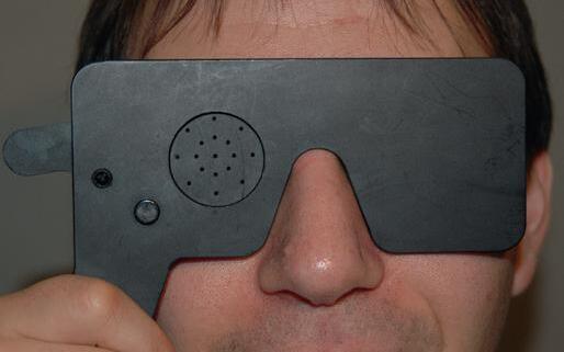
Visual acuity is a measurement of the individual’s capacity for visual discrimination of fine details of high contrast.5,6 Best corrected visual acuity should be tested for each eye separately with the other eye covered by a tissue, hand, or occluding device (Fig. 2.1A). Distance vision is most commonly evaluated with a standard Snellen chart (Fig. 2.2A) or with a computer monitor that displays the black optotypes on a white background. Near vision can be tested with a hand-held card (Fig. 2.2B). Ideally, best corrected vision should be assessed using current corrective lenses or manifest refraction. If these are unavailable, a pinhole will improve most mild to moderate refractive errors in cooperative patients (Fig. 2.1B). Patients with subnormal acuity despite best refracted correction should also be tested with pinholes, which may further resolve some refractive errors (irregular astigmatism) and media opacities (cataract). When acuity cannot be corrected by a pinhole, nonrefractive causes of visual loss (see later discussion) should be considered.
Acuity is most often recorded as a fraction (e.g., 20/40), where the numerator refers to the distance (in feet) from
which the patient sees the letters and the denominator refers to the distance from which a patient with normal vision sees the same letters. The normal eye can resolve a figure that subtends a visual angle of 5 minutes at a distance of 20 feet. At the distance at which a normal patient can see a line of letters on a Snellen eye chart, the widths of the lines on each letter subtend a visual angle of 1 minute, or one-fifth of the entire letter.6 A fraction of 20/20 2 indicates the patient saw all the letters on the 20/20 line except two, while 20/20+2 means the patient was able to see the 20/20 letters plus two letters on the next (20/15) line. Usually up to two mistakes on a line or two extra letters on the next line are allowed in this notation. Most normal adults younger than 40 years have best corrected visual acuities of 20/20 or better in each eye.
Visual acuity at distance can also be recorded using the metric or decimal systems. When the testing is at 6 meters (close to 20 feet), the normal visual acuity is recorded as 6/6. The decimal system uses the numeric equivalent of the fractional notation: 20/20 or 6/6 is a visual acuity of 1.0. A visual acuity of 20/100 would be recorded as an acuity of 0.2.
If a patient is unable to read the largest Snellen letters (20/200 or 20/400), the acuity should be recorded by moving a 200-size letter towards the patient until it is seen (Fig. 2.3). That distance is recorded as the numerator. For example, an acuity of 4/200 means the patient was able to see the 200-size letter at 4 feet. Alternatively, the degree of vision can be recorded using the phrases “count fingers” (CF) (and at what distance), “detect hand motions” (HM), and “have light perception” (LP). An eye that is blind has “no light perception” (NLP). Criteria are used by different agencies to determine a level of vision loss that qualifies for disability or benefits (i.e., “legal blindness”) based on a best corrected acuity worse than 20/200 in the better seeing eye or binocular visual field constriction to less than 20 degrees.
Unfortunately Snellen charts have several deficiencies,6,7 the most important of which is the nonlinear variation in the sizes of the letters from line to line. Thus, if one patient’s visual acuity decreases by 20/100 to 20/200 using the chart in Fig. 2.2A, and another from 20/80 to 20/100, both are considered to have a decrease in visual acuity by one line. However, in the first instance the difference in letter size is 100%, but in the second it is only 25%. Furthermore, a typical Snellen chart has a different number of letters on each line. The largest letters have the fewest, while the smallest letters have the most in each line. Therefore, more letters must be identified to complete smaller lines in contrast to larger lines. In addition, some letters, such as the “E,” are harder to identify than the “A” or “L,” for example.
2.1. A. Occluder for testing vision one eye at a time. By convention the right eye is tested first. B. Occluder with pinholes. If the visual acuity is subnormal but can be improved with pinholes, refractive error or media opacities should be suspected.
Box 2.1 Neuro-Ophthalmic Examination
Afferent Visual Function
Visual acuity
Contrast sensitivity (optional)
Color perception
Confrontation visual fields
Amsler grid testing
Higher cortical visual function (optional)
Efferent System
Pupils Size
Reactivity
Swinging flashlight test
Near (optional)
Eyelids
Facial nerve function
Ocular motility
Inspection
Ductions
Vergences
Assessment of ocular misalignment
External Examination (Including Orbit)
Slit-Lamp Examination/Applanation Tensions
Ophthalmoscopic Examination
Directed Neurologic Examination
Mental status
Cranial nerves
Motor function
Cerebellar function
Sensation
Gait
Reflexes
Directed General Examination
To eliminate these issues and when consistency is desired among testing locations, as in multicenter clinical trials, for instance, standardized Early Treatment in Diabetic Retinopathy Study (ETDRS) charts have become the gold standard (Fig. 2.2C).7 Each line contains five letters, the spacing
between the letters and lines is proportional to the letter sizes, the sizes of the letters decrease geometrically, and the recognizability of each letter is approximately the same.6 Using ETDRS charts, a linear scale for visual acuity can be created by calculating the base 10 logarithm of (1/Snellen decimal notation) in what is termed the logarithm of the minimal angle of resolution (logMAR). Each line on the ETDRS chart is therefore separated by 0.1 logMAR units. So in the previous examples, the logMAR score for the first patient would worsen from 0.7 to 1.0, while the second would worsen from 0.6 to 0.7, more accurately reflecting the greater decrease in visual acuity for the first patient.
Visual acuity with the near card is often recorded using the Snellen fraction or the standard Jaeger notation (J1, J3, etc.). When near visual acuity is tested, presbyopic patients older than 40 years of age should wear their reading glasses or bifocals. Near acuities are not as accurate as those obtained at distance, especially when the card is not held at the requisite distance specified on the card.
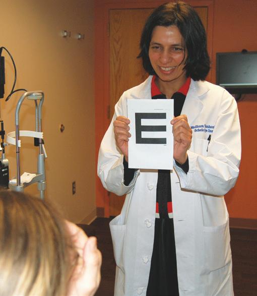
For illiterate individuals or children unable to read letters, acuity can be tested with tumbling Es8 (Fig. 2.4), Allen or Lea figures9,10 (Fig. 2.5A), or HOTV letters11 (Fig. 2.5B, C). In younger preverbal patients, assessment of fixing on and following a light or toy by each eye separately in most instances is sufficient. Caution should be used when examining small infants, since visual fixation normally may be inconsistent or absent until 8–16 weeks of age.12 When quantification of visual acuities is required in very young children (for serial examinations, for instance), preferential looking tests (Teller acuities13,14) may be used (Fig. 2.6). These tests are based on the principle that a child would rather look at objects with a pattern stimulus (alternating black and white lines of specific widths) than at a homogeneous field. The frequency of the smallest pattern that the child seems to prefer is termed the grating acuity, which can be converted to Snellen equivalents if the test distance is known. Visual acuity in a newborn is roughly 20/400 to 20/600; it improves to approximately 20/60 by 12 months of age and reaches the 20/20 level by 3–5 years of age.15
Ocular causes of reduced visual acuity include refractive error, amblyopia, macular lesion, or media opacity such as cataract, vitreous hemorrhage, vitritis, or corneal opacities or irregularities. Neuro-ophthalmic processes that can
Figure
Figure 2.2. Visual acuity charts. A. Snellen eye chart for testing visual acuity. The largest letter at the top is the 20/200 E, while the letters at the bottom represent the 20/10 line. B. Near card with pupil gauge. Note that near visual acuity is often recorded using the Jaeger notation, as in “Jaeger 2” or “J2,” to indicate 20/30 near acuity.
2.2., cont’d
C. Early Treatment in Diabetic Retinopathy Study (ETDRS)–type chart.
decrease visual acuity are those that affect the optic nerve or chiasm. Disturbances that are posterior to the chiasm (retrochiasmal, i.e., tract, optic radiations, and occipital lobe) affect visual acuity only if they are bilateral.16 Functional visual loss should always be considered when visual acuity is decreased without any obvious abnormality of the eye or visual pathways.
CONTRAST SENSITIVITY AND LOW-CONTRAST LETTER ACUITY
Contrast sensitivity testing with sine-wave or square-wave gratings may be a useful adjunct in the evaluation of vision loss.17,18 Conventional visual acuity measures spatial resolution at high contrast, while contrast sensitivity testing assesses spatial resolution when contrast varies. In one variation of the test, the patient is asked to identify in which direction the gratings, which span a spectrum of spatial and temporal frequencies and are arranged in increasing difficulty, are
Figure
Figure 2.3. Determination of visual acuity using 200-size letters in an eye with worse than 20/200 but better than hand motions vision. The distance at which the patient can see the 20/200 E is determined. If this distance is 3 feet, then the visual acuity is recorded as “3/200.”
Figure 2.4. “Tumbling Es” for assessment of visual acuity in illiterate adults or children. The patient can be asked to indicate in which direction the E points (right, left, down, up). The sizes of the Es correlate with the size of the letters on Snellen chart.