Enhance your learning with Evolve Student Resources.
These online study tools and exercises can help deepen your understanding of textbook content so you can be more prepared for class, perform better on exams, and succeed in your course.
Activate the complete learning experience that comes with each NEW textbook purchase by registering with your scratch-off access code at http://evolve.elsevier.com/Brown/
If your school uses its own Learning Management System, your resources may be delivered on that platform. Consult with your instructor.
If you rented or purchased a used book and the scratch-off code at right has already been revealed, the code may have been used and cannot be re-used for registration. To purchase a new code to access these valuable study resources, simply follow the link above.
LAVIN’S
Radiography for Veterinary Technicians
This page intentionally left blank
LAVIN’S
Radiography for Veterinary Technicians
Marg Brown, RVT, BEd Ad Ed
Formerly of Seneca College of Applied Arts and Technology King City, Ontario; Formerly of Penn Foster College, Scranton, Pennsylvania; Active Member:
Ontario Association of Veterinary Technicians
Association of Veterinary Technician Educators
National Association of Veterinary Technicians in America
No part of this publication may be reproduced or transmitted in any form or by any means, electronic or mechanical, including photocopying, recording, or any information storage and retrieval system, without permission in writing from the publisher. Details on how to seek permission, further information about the Publisher’s permissions policies and our arrangements with organizations such as the Copyright Clearance Center and the Copyright Licensing Agency, can be found at our website: www.elsevier.com/permissions
This book and the individual contributions contained in it are protected under copyright by the Publisher (other than as may be noted herein).
Notice
Practitioners and researchers must always rely on their own experience and knowledge in evaluating and using any information, methods, compounds or experiments described herein. Because of rapid advances in the medical sciences, in particular, independent verification of diagnoses and drug dosages should be made. To the fullest extent of the law, no responsibility is assumed by Elsevier, authors, editors or contributors for any injury and/or damage to persons or property as a matter of products liability, negligence or otherwise, or from any use or operation of any methods, products, instructions, or ideas contained in the material herein.
Previous editions copyrighted 2018, 2014, 2007, 2003, 1999, and 1994.
Library of Congress Control Number: 2021939951
Director, Content Development: Laurie Gower
Senior Content Strategist: Brandi Graham
Content Strategist: Melissa Rawe
Content Development Specialist: Brooke Kannady
Publishing Services Manager: Deepthi Unni
Senior Project Manager: Manchu Mohan
Design Direction: Amy Buxton
Printed in India
To my contributors and reviewers: Susan McNeal, RVT, Ashley Jenner, RVT, BSc, VTS-DI, Lorelei D’Avolio, LVT, VTS (Exotics), CVPM, Shana Lemmenes, CVT, VTS-EVN, and Julia Bitan, RVT. Thank you for your expertise and dedication in making this a valuable resource.
To all our students and the veterinary profession: may you never stop learning. To Phil, Alina, Jacquie and Tylor: love always. Marg Brown
To my contributors.
Darryl Bonder, DVM; Stephanie Holowka, MRT(R), MRT, (MR) MRSO, (MRSC TM) Bob Hylands, DVM; Lori Ponte, Animal Health Partners, Toronto Arvind Singh, HND Mech (UK) Raymax Medical Corporation; This would be a very thin book without your expertise. Thank you for your prompt and enthusiastic presentations.
Susan MacNeal, RVT, CVDT, BSc Coordinator, Veterinary Technician Georgian College Orillia, Ontario
Reviewers
Laura L. Black, LVT, BAAS
Associate Professor of Veterinary Technology
Veterinary Technology Distance Learning Program
San Juan College Farmington, New Mexico
Lorelei D’Avolio, LVT, VTS (Exotics), CVPM
Practice Manager
The Center for Avian and Exotic Medicine
New York, New York
Lynda Forgie, RVT
Professor, Veterinary Technician Georgian College Orillia, Ontario
Jennifer V. Freese, DVM
Program Director
Veterinary Technology University of Maine at Augusta Bangor, Maine
Michelle E. Goodnight, DVM, MS, DACVECC
Instructor, Veterinary Technology Gwinnett Technical College Lawrenceville, Georgia
Susan Guttschow, DVM
Program Director/Instructor
Veterinary Science Gateway Technical College Elkhorn, Wisconsin
Amanda Hackerott, RVT
Program Director, Health Sciences
WSU Tech College of Applied Sciences and Technology Wichita, Kansas
Ashley Jenner, BSc, RVT, VTS-DI
Diagnostic Imaging
Toronto Veterinary Emergency Toronto, Ontario
Shana Lemmenes, CVT, VTS-EVN
Large Animal Technician
University of Minnesota St. Paul, Minnesota
Shauna Lesick, RVT
RVT Instructor, Animal Studies
Northern Alberta Institute of Technology Edmonton, Alberta
George W. McCommon, DVM
Chair/Professor
Veterinary Science and Public Health
Fort Valley State University Fort Valley, Georgia
Sarah Meyer-Paterson, CVT, AAS
Program Director, Veterinary Technology Pensacola State College Pensacola, Florida
Gordon Smok, DVM, BA, AS
Program Chair Veterinary Technology City College Gainesville, Florida
We are excited to present the seventh edition of Lavin’ s Radiography for Veterinary Technicians. This text continues to focus on teaching the science of imaging used for veterinary medicine and veterinary technology education and referencing. The purpose of the book is to instill a working knowledge of radiologic science as it applies to producing a diagnostic quality image, to prepare radiography students in veterinary technology programs for the certification exam, to assist in the training of veterinary medicine students, and to provide a base from which practicing veterinary radiographers can make informed decisions about technical factors and diagnostic image quality in the workplace.
This text is divided into two major parts: the technical side of diagnostic imaging, and radiographic positioning and related anatomy. Part One puts theory into practice, and each of the chapters in Part Two have been expanded to include not only essential positioning information but also anatomic references to support the technician in understanding normal anatomic features so that accurate images for diagnosis are produced. This text is a mainstay for teaching radiographic anatomy and positioning of all common species.
Each chapter is unique, yet succinct, so that all important information is available and easy to understand without the need to reference other sources. This is a valuable reference for both technicians and veterinarians when they have finished their training. This seventh edition provides a thorough yet practical level of imaging and positioning coverage to equip individuals with the knowledge they need to produce high-quality images on the first attempt with a focus on radiation safety.
Photographs, radiographs, and a consistent style of color line drawings, along with tables and boxes, have been updated. Numerous images are scattered throughout the text, with many chapters ending in an image gallery for further observation. Application information and technician notes boxes continue to provide practical information to help prepare the veterinary technician for on-the-job challenges. Review questions are included in the textbook at the end of each chapter. Further review questions can be found in the accompanying Evolve website in the Student Resources. A comprehensive glossary in which key words and other terms are defined has been rewritten and is also now included in the text.
Preface
Features of Part One
The world of diagnostic imaging continues to evolve with better and clearer images in all aspects of the science. In this,
the seventh edition of the text, we have once again rewritten large sections and modified other parts of the chapters.
Information on film processing, both automatic and manual, has been included in this edition. There are still many facilities that use film/screen technology and for this reason, it is presented here.
The computed radiography and digital imaging information has been greatly expanded. As that field moves forward, the various modes of digital imaging are presented, some of which are here to stay and others which are transitional.
The intrepid contributors have, once again, presented their chapters with new images and expanded explanations of the technologies they present. It is always a pleasure to interact with colleagues who are experts in their field and to share their enthusiasm for their subjects. To that end:
Dr. Hylands has rewritten his Ultrasound chapter with new images and text.
Dr. Bonder has enlarged his chapter on Nuclear Medicine and added new images.
Horst Bruning of Animage has contributed wonderful images from technologies that are being introduced and accepted worldwide.
Stephanie Holowka has revised her chapters and added new images.
Chapters 1 and 2 once again present the science of x-ray technology.
Chapter 3 expands on how to protect your staff and the patients from the effects of radiation.
Chapters 4 to 7 present a revised and more succinct information on the modality of film/screen imaging.
Chapter 8 is the digital imaging chapter. It has been revised and expanded with a number of new sections. This field is ever evolving, and as new technology is presented, I will update the chapter on the Evolve website.
Chapter 9 presents quality control, the testing and the expected results. I have included the tests that can be completed in house if there is a problem prior to a visit from a service engineer.
Section 3 is the specialized imaging area. The experts in their fields have written their chapters, which are included to demonstrate the enhancement of anatomical parts evidenced from the use of specialized imaging.
Features of Part Two: Radiographic Positioning and Related Anatomy
This part is all about positioning and is a one-source reference for major species and procedures. Each chapter has been updated, with additional images, strategies and
techniques including digital imaging. There are separate chapters on the abdomen, thorax, forelimb, pelvis and hindlimb, spine, and skull of small animals. Small animal dental radiography, special procedures, large animal, and avian and exotic radiographic imaging have also been significantly expanded.
All information required for accurate positioning is clearly indicated. Routine and ancillary views are included for each position. All chapters include an outline, learning objectives, key terms, where to measure, the location of the central ray, borders, a step-by-step approach to positioning, further comments, and tips to ensure that the perfect image is obtained both through analog and digital imaging. Technician notes scattered throughout the chapters emphasize important points.
Positioning views are accompanied by high-quality color photographs and graphic color drawings that visibly indicate ideal radiographic imaging and anatomic features. Through the integrative application of radiography and anatomy, veterinarians will receive optimized images. Further related and essential descriptive anatomy is included in the appendixes on the Evolve website.
The emphasis in these chapters is on non-manual restraint techniques required for the safety and radiation protection of the radiographer and for the benefit of our patients. It is essential that the veterinary profession understands and applies these safety principles.
Diagnostic imaging is an exciting and changing field in which the radiographer plays an important role. With the tools and information presented here, an accurate diagnosis should be ensured.
Welcome to the seventh edition. We hope that you enjoy learning from it as much as we enjoyed writing it.
Marg Brown Lois C. Brown
Additional Learning Resources
The Evolve Student Resources include review questions, crossword puzzles, and fill-in-the-blank exercises, as well as five appendixes and a comprehensive glossary. A full complement of support materials for teaching and learning are also available and include a complete collection of all images in the text.
Acknowledgments
I express my deepest gratitude to everyone who has contributed to the production of this edition. Your dedication and constant additions keeps this resource one of the most relevant and current in the field.
A big thank you to the reviewers who gave us great input on what should be changed or continued in this edition. Your guidance has been essential to improve the content and layout. Lois, your perspective and enthusiasm continue to amaze me. It has been a pleasure to work on this with you once again.
I am forever indebted to my former colleagues of the Seneca College Veterinary Technician Program in King City, Ontario, for your constant support, generosity, enthusiasm, and dedication. Unless otherwise indicated, the positioning photographs were taken at Seneca College. Thanks to our models, Sam, Ace, and Spud, and to their owners. A big thank you to my former students. Because of your enthusiasm, curiosity and search for knowledge, I never worked a day in my life. You were a constant source of motivation and joy. Nothing gives me more pleasure than to continue learning and hearing from you.
Special thanks to Sue MacNeal, RVT, CVDT, BSc, for totally upgrading the dentistry chapter. Your knowledge and logical explanations ensure that dentistry remains a valuable chapter of this edition.
Lorelei D’Avolio, LVT, VTS (Exotics). CVPM, your expertise and years of experience significantly improved the avian and exotic medicine chapter. Shana Lemmenes, CVT, VTS-EVN, your assistance in the large-animal positioning was invaluable. Ashley Jenner, RVT, BSc, VTS-DI, your practical suggestions were indispensable for editing the small animal positioning chapters. Along with your colleague, Julia Bitan, RVT of https://handsfreexrays.com/, the images added greatly to the chapters. Jennifer White, RVT, your further images were helpful. Evelyn Kelly, RN, MRT(R), ACR, BSc, and Carolyn Bennett, AHT, your assistance with the special procedures chapter was appreciated. Heartfelt thanks to you all for your input.
I continue to express my appreciation to those who contributed to previous editions. Jeanne Robertson, your graphic images continue to be awesome.
Huge credit is extended to the editing team at Elsevier, especially Brandi Graham, Brooke Kannady and Manchu Mohan. Your inspiration, suggestions, and patience have helped make this book an essential resource.
As Albert Einstein said “once you stop learning, you start dying.” May you all learn forever.
Marg Brown
I am, once again, humbled and impressed with the knowledge, experience, and enthusiasm that I received as we ventured into a seventh edition of this work.
Robert Hylands, DVM, rewrote the ultrasound chapter, adding information and new images, and sent it and responses to my questions from an island in the North country of Canada while self-isolating from the COVID pandemic. Horst Bruning, president of Animage, sent images of his FIDEX system via California as he was quarantined in Italy, unable to travel due to COVID. Stephanie Holowka, MRT(R), MRT (MR), MRSO (MRSC TM), was especially involved with the computed tomography (CT), magnetic resonance imaging (MRI) and the new positron emission tomography section, as well as reviewing the new and revised digital imaging chapter. Her thorough enjoyment of the veterinary aspect of imaging is always refreshing. Animal Health Partners, North York, Ontario invited us to visit their MRI and computed radiography departments and then contributed enthusiastically to our seventh edition. They photographed their CT and MRI units and supplied images as requested by Stephanie as she compiled the MRI and CT chapters. Darryl Bonder, DVM has contributed to all three editions with his encyclopedic knowledge of equine nuclear medicine. Raymax Medical Corporation was invaluable in reviewing the
I Curious Tiberius—the literary—always providing assistance and voicing concerns—mainly about dinner and the lateness of the hour.
xii ACKNOWLEDGMENTS
manuscripts of Section 1. Every time I called with a request, the response or the image would show up in my inbox within hours.
The many, many contributors of images and expertise would fill several pages, and to those people, veterinarians, technicians, technical educators, and other staff members, I once again offer my special thanks.
Once again, our reviewers and the suggestions they made helped immensely to guide the content of the seventh edition. The expertise of our editors, Brooke Kannady and
Brandi Graham, and their staff certainly kept us on track and led to this new and very special seventh edition.
As before, Marg, great to work with you again… stay safe. The restrictions of COVID could have made this project very difficult, but everyone worked from home, or in isolation far from home, and with the able assistance of our pets, moved the project to completion. For me, I Curious Tiberius was once again a mainstay of editing
Lois C. Brown
Part One: Diagnostic Imaging, 1
Section One: The Technical Side of Imaging, 2
1 The Basics of Atoms and Electricity, 2
2 Diagnostic X-Ray Production, 9
3 Radiation Safety and Protection, 30
Section Two: Film Processing and Digital Imaging, 45
4 Imaging on Film, 45
5 Producing the Image, 67
6 Optimizing the Image, 83
7 Processing the Image on Film, 99
8 Computerized Radiography and Digital Imaging, 121
9 Quality Control, Testing, and Artifacts, 139
Section Three: Specialized Imaging, 170
10 Ultrasonography, 170
11 Fluoroscopy, 186
12 Computerized Tomography, 194
13 Magnetic Resonance Imaging, 210
14 Nuclear Medicine and Introduction to Positron Emission Tomography, 224
Part Two: Radiographic Positioning and Related Anatomy, 235
15 Overview of Positioning, 236
16 Small Animal Abdomen, 249
17 Small Animal Thorax, 264
18 Small Animal Forelimb, 288
19 Small Animal Pelvis and Pelvic Limb, 319
20 Small Animal Vertebral Column, 352
21 Small Animal Skull, 381
22 Dental Imaging and Radiography, 407
23 Small Animal Special Procedures, 470
24 Equine and Large Animal Radiography, 506
25 Avian and Exotic Radiography, 579
Glossary, 627
Index, 641
This page intentionally left blank
Diagnostic Imaging PART ONE
In Part One, you will be introduced to the technical side of imaging. The main emphasis is on the practical applications of rules and laws guiding the principles of radiography. You will learn about the inner workings of the radiographic unit. Film, digital, and specialized imaging are also addressed. Identifying radiographic artifacts and quality control testing techniques are also included.
The Technical Side of Imaging
CHAPTER
1
The Basics of Atoms and Electricity
Lois C. Brown, RTR (Can/USA), ACR, MSc, P.Phys
OUTLINE
The Discovery of X-Rays 3 Elements and Atomic Theory 3 Matter and Energy 4
LEARNING OBJECTIVES
The Electromagnetic Spectrum 4
The Dual Nature of X-Rays –Wave-Particle Duality 5
When you have finished this chapter, you will be able to:
1. Describe the events related to the discovery of x-rays.
2. List the properties of x-rays.
3. Understand the relationship between atoms and elements.
APPLICATIONS
Imagination is the highest form of research.
—Albert Einstein 1879–1955
Energy as Wavelengths and Frequencies 6
Energy as Particles 7
4. Describe the basics of atomic theory.
5. Understand the relationship between matter and energy.
6. Describe the basics of the table of the elements.
7. Define x-ray photons as waves and particles.
This chapter provides basic information that will be expanded on in subsequent chapters.
KEY TERMS
Key terms are defined in the Glossary on the Evolve website.
Atomic theory
Atoms
Diagnostic imaging
Electricity
Electromagnetic spectrum
Elements
Energy
Fluorescence
Frequency
Heterogeneous
Matter
Nuclear energy
Nucleus
Particles
Polyenergetic
Thermal energy
Thermionic emission
Wavelength
X-rays
Wilhelm Roentgen’s laboratory. (Photo courtesy Röntgen-Kuratorium Würzburg
e. V., Röntgenring 8, 97070 Würzburg.)
The Discovery of X-Rays
X-rays were discovered by German scientist Wilhelm Conrad Roentgen in 1895. He was working in his laboratory and setting up experiments for the next morning when he discovered a platinocyanide plate glowing a few feet from where he was working.
The fortuitous decision by Roentgen to understand what caused the plate to glow, how to prevent it from glowing (by removing the radiation source), and how to categorize the rays that caused the glow was the beginning of the profession that we know as diagnostic imaging.
Roentgen named the rays “x-rays” because of their unknown nature (“x” in science denotes an unknown quantity). He was also the first person to patent the phenomenon at a registry office.
As Roentgen developed his experiments and identified the characteristics of the new rays that he was investigating, he listed 12 unique properties (Table 1.1). He thought that these were just the beginning of a list of properties, but such was the thoroughness of his experiments and his scientific investigation that to this day no one has added to the original list. It is important that we review the contents of the list, as we will encounter these characteristics throughout the balance of the text.
Elements and Atomic Theory
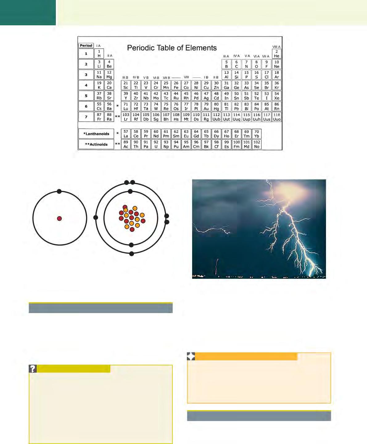
An element is the smallest particle of a substance. Elements are arranged on a table of the elements (Fig. 1.1). This table was put together to understand the nature of the atoms, which make up the elements contained within the table. Scientists realized early on that some elements were much more stable than others and some were very unstable.
PROPERTIES OF X-RAYS
Are invisible
Throughout the text, various elements will be described in relation to their position on this table.
The highest number of stable nonradioactive elements is #83 Bismuth, preceded by #82 Lead. We will meet Lead many times within the chapters of this text.
The smallest particle of an element is an atom (Fig. 1.2). An atom consists mainly of empty space, but the units within the atom are very small. An atom consists of a nucleus containing protons and neutrons. Protons have a positive electrical charge, and neutrons have no charge. Circling around the nucleus are negatively charged electrons. The electrons are held “in orbit” by the positive electrical charge of the protons and their own negative charge. They are arranged in very definite “rings,” with specific numbers of electrons in each ring matched with specific numbers of protons within the nucleus. As the numbers of protons within an atom’s nucleus increases, the number of corresponding electrons also increases. With this increase, the number of rings of electrons increases and, because of the distance from the nucleus, the electrical charge holding the electrons within the rings lessens to the point that individual electrons may leave their orbits, or they can be “boiled off” if heat is applied to the element. Boiling electrons off the filament of the cathode is the first step in creating x-rays.
APPLICATION INFORMATION
When the x-ray generator is turned on and heat is applied to the cathode of the x-ray tube, electrons are “boiled off” in an effect called thermionic emission
When the exposure switch is closed, it is the energy of the electrons (at this point called “photons” or energy packets)—drawn across from the cathode to the anode and interacting with the anode—that produces x-rays.
NOTES
One cannot hear, smell, or see x-rays.
Are electrically neutral X-rays carry neither a positive or negative charge.
Have no mass X-rays have no resistance or force when put into motion.
Travel at the speed of light in a vacuum X-rays move at 186,000 miles/second in a vacuum (3 3 108 meters/second). Cannot be focused by a lens
Form a polyenergetic (heterogeneous) beam
The beam will pass through the lens unchanged.
Multiple photon energies are contained within one exposure.
Peak kilovoltage (kVp) is the maximum energy in one exposure. One exposure equals the energy used to produce one image.
Can be produced in a range of energies (kV) The useful range for diagnostic imaging is 25–125 kV; 25–40 is used in specialized imaging.
Travel in straight lines
Cause fluorescence in certain substances
Can cause chemical changes to occur in radiographic and photographic film
Can be absorbed or scattered by tissues in the body; can produce scattered and secondary radiation
Can cause chemical and biological damage to living tissue
Individual photons travel in a divergent (straight line) beam from the x-ray tube.
Fluorescence is useful to intensify the effect of radiation on a film with the use of intensifying screens.
Depending on the energy of the photons, x-rays either penetrate matter or are absorbed by it, and therefore an image can occur on film.
Based on the energy of the x-rays and on the composition and thickness of the tissues being exposed, characteristic radiation may be produced when the photon interacts with matter.
Through excitation and ionization (removal of electrons) of atoms comprising cells, damage to cells may occur.
TABLE 1.1 Roentgen’s List of Unique Properties of X-Rays
1.1 A periodic table of the elements. (From Ziessman HA, O’Malley JP, Thrall JH. Nuclear Medicine: The Requisites in Radiobiology. 4th ed. London: Elsevier; 2014.)
FIG. 1.2 Two examples of elements. The hydrogen element has 1 proton inside the nucleus and 1 electron orbiting the nucleus. The oxygen atom has 8 protons, 8 neutrons within the nucleus, and 8 electrons orbiting the nucleus.
Matter and Energy
Every element on earth has substance, either matter (mass) or energy. It is the conversion of matter into energy that we use to create x-rays. The principal characteristic of matter is mass or weight. Weight involves gravity. The principal characteristic of energy is movement or motion (Fig. 1.3).
POINTS TO PONDER
• Dr. Einstein said it best when he developed his famous formula E 5 mc2
• E 5 energy, m 5 mass (or matter), c2 5 the speed of light, which is a constant.
• This means that everything that has mass can be changed into energy, and energy can be converted to mass. Neither can be created or destroyed.
• This matters to the production of x-rays because the electrons that are boiled off the cathode in the x-ray tube will be absorbed into whatever they encounter.
The combination of matter and energy in the universe is a constant. Matter can become energy. Energy can become matter. Neither can be created or destroyed. Each can only be changed in form from one to the other.
APPLICATION INFORMATION
The production of x-rays depends on the tungsten wire in the negatively charged cathode. As energy is infused into the wire, creating heat, electrons are emitted from the wire. These electrons are drawn across to the positively charged anode as “packets of energy” when the switch is closed and the circuit is complete.
The Electromagnetic Spectrum
There are several types of energy: mechanical, chemical, thermal, nuclear, electromagnetic, and electrical. Electrical energy and electromagnetism are the two major types of
FIG.
Hydrogen Oxygen
FIG. 1.3 Lightning is an excellent example of electric energy. The negatively charged electrons build up in the clouds until they are attracted by the powerful force of the positively charged earth.
FIG. 1.4 The entire electromagnetic spectrum is much larger than just the visible light portion. This chart shows the values of energy, frequency, and wavelength for all portions and identifies the three imaging windows. (From Bushong SC. Radiologic Science for Technologists. 11th ed. St Louis: Elsevier; 2017.)
energy used in x-ray technology. This is evident when we turn on the power to the x-ray generator at the electrical box, and the energy (both electric and electromagnetic) is directed to the production of x-rays. The electrical spectrum covers a vast number of energies. We are concerned with the portions that involve radiation and visible light. In Fig. 1.4 the portion of the spectrum that involves radiation is near the top of the scale, whereas the visible light area is below that and divides into all the colors of the rainbow (Fig. 1.5).
The Dual Nature of X-Rays –
Duality
As can be seen in Fig. 1.4, visible light and x-radiation are listed on the table of the electromagnetic spectrum. They are very similar except that x-rays have a much higher frequency and a shorter wavelength.
• Visible light photons tend to react in experiments in the manner of waves.
• X-rays tend to react in the manner of particles. (An x-ray photon is a unit of pure energy).
This is known as wave-particle duality and is useful to know when we investigate the creation of x-rays within an
FIG. 1.5 A rainbow shows all the colors in the visible light portion of the electromagnetic spectrum.
electric circuit. In the electric circuit, the x-rays can be described as waves emanating from the tungsten target in the x-ray tube where they are produced. At other times during the discussion of x-radiation, the concept is changed to x-rays as particles.
An electromagnetic wave has height (amplitude) and moves at the speed of light.
APPLICATION INFORMATION
Waves and particles of energy are important in the production of radiation. Setting the voltage and current on the x-ray generator controls the nature of the quantity and energy of the x-rays produced.
Energy as Wavelengths and Frequencies
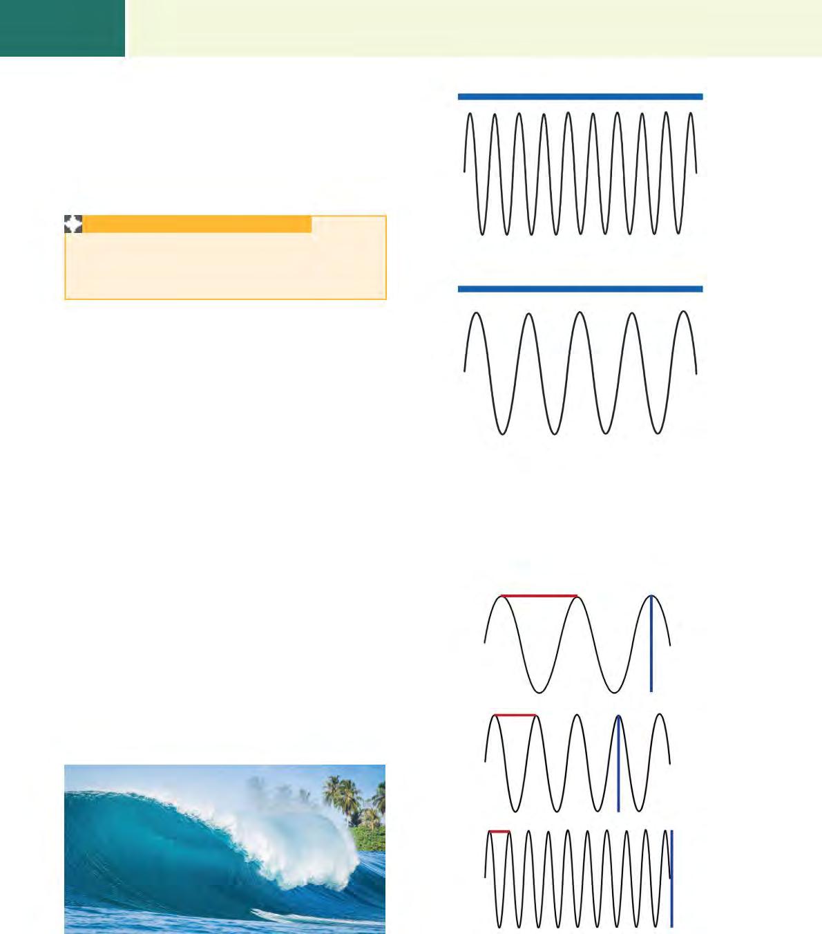
Watching waves on the seashore is an excellent example of energy transformed into waves (Fig. 1.6). There are crests and troughs, and there is a measurable distance between the crests or the troughs. There is a time factor between each crest and trough. That time factor is the frequency of the waves (Fig. 1.7). One wavelength is the measurement of one crest to the peak of the following crest (or one trough to the depth of the following trough (Fig. 1.8).
A sinusoidal (sine) wave is the tracing of the crests and troughs that the waves describe as they travel through the ocean. Sine waves have a high point and a low point, with time being the constant that runs through the middle. This is very important when we set x-ray technical factors and choose the time over which the x-rays will be produced and the kilovoltage that is used to produce them.
Frequency is the number of waves passing a given point per given unit of time. With x-ray production, frequency is connected to the kilovoltage used to penetrate the tissue of the patient and the time that is set to produce the x-rays. Shorter wavelengths with higher frequencies penetrate the tissue more effectively than long wavelengths with low frequency.
Wavelength and frequency are inversely related so that as the frequency increases, the wavelength decreases.
Time line (1 second)
Short wavelength — high-frequency wave
Time line (1 second)
Long wavelength — low-frequency wave
FIG. 1.7 If waves have a short wavelength, they will pass a fixed point frequently (short wavelength/high frequency). If the waves have a long wavelength, they will pass that given point less frequently (long wavelength/low frequency).
FIG. 1.8 These three sine waves have different wavelengths (y). The shorter the wavelength, the higher the frequency as measured over a constant time. The height of the wave from crest to trough is the amplitude (x). A, Long wavelength. B, Medium wavelength. C, Shorter wavelength.
FIG. 1.6 Energy represented by waves on the ocean. The power of the wind creates waves with frequency and wavelength. This image represents one wave with a crest and one trough. (Copyright 2015 petesphotography/Getty Images.)
APPLICATION INFORMATION
Electrical energy can be transformed from one type of energy to another. Electrical energy applied to a hair dryer is converted to heat. Electrical energy applied to an x-ray tube is converted into heat and then, by electromagnetism, into x-rays.
Wavelength and frequency are important when setting the technical factors on the x-ray unit. The choice of increasing or decreasing each of the factors is dependent on the voltage, current, and time—all of which are directly related to wavelength and frequency.
Energy as Particles
Photons are the smallest quantity of any type of electromagnetic radiation. They have neither mass nor electrical charge but interact with matter as though they are particles (Fig. 1.9). It is this energy that has been produced within the cathode to create the potential x-ray beam. A photon may be pictured as a small bundle of energy. This is particularly true when using high-frequency energies such as x-rays or gamma rays. The photon particle carries a specific energy that is dependent on frequency. The energy and the frequency are directly proportional. If the energy is doubled, then the frequency is doubled. When described as particles, it is possible to mathematically quantify the relationship between frequency and photons and the amount of energy required to perform work.
Summary
Roentgen discovered x-rays in the late 1800s and described the nature of the phenomenon completely. The electromagnetic spectrum describes all forms of energy and categorizes it on a table depending on wavelength, with x-rays near the top of the scale and light near the middle of the scale.
The table of the elements categorizes all the elements known at this time. Throughout the text, we will describe some of these elements and show how their place on the table is important.
Scientists soon realized that x-rays can act as continuous waves or as particles (photons of energy), depending on their activity. Sinusoidal (sine) waves are examples of how x-rays behave as waves. Each wave has a crest and a trough, and there is an important relationship between the crests and the troughs.
REVIEW QUESTIONS
1. X-rays are described as:
a. Invisible, but they travel in straight lines at the speed of light
b. Electrically neutral unless they are emitted from a charged source
c. Visible through a lens during x-ray exposure
d. Causing electrical changes in photographic film
2. An element is the smallest part of a substance. Which of the following statements is true?
a. The element is combined with a proton.
b. The smallest part of an element is an atom.
c. The element is composed of neutrons and particles.
d. Electrons circle the element in random order.
3. The electrons are held in place by:
a. The neutral charge of the neutrons
b. Their own attachment to the nucleus
c. The ionization of the atom
d. The positive charge of the protons
4. On the table of the elements:
a. The elements are arranged alphabetically.
b. The order of the elements depends on when they were discovered.
c. The elements are arranged in specific groups.
d. All the known substances are listed with the radioactive ones at the beginning.
5. Matter and energy are basic to every substance on earth. Which of the following statements is true?
a. A principal characteristic of matter is mass or weight.
b. The principal characteristics of energy are time and space.
c. Matter represents motion and frequency.
d. Energy cannot be created out of atoms and molecules.
6. The electromagnetic spectrum represents:
a. The color wheel and the electrical properties of light
b. The vast spectrum from x-rays to radio waves
c. The intensity of light and the darkness of radiation
d. Electricity, wavelength, and frequency
7. Energy can be represented by both:
a. Waves and particles
b. Waves and frequencies
c. Mass and matter
d. Matter and waves
8. A sinusoidal wave represents x-ray energy as waves.
Which of the following statements is true?
a. It contains the energy of the x-ray beam.
b. It is an uneven wave, so it cannot be measured.
c. It has frequency and amplitude in its definition.
d. It represents the motion of the x-ray timer.
Anode
Cathode
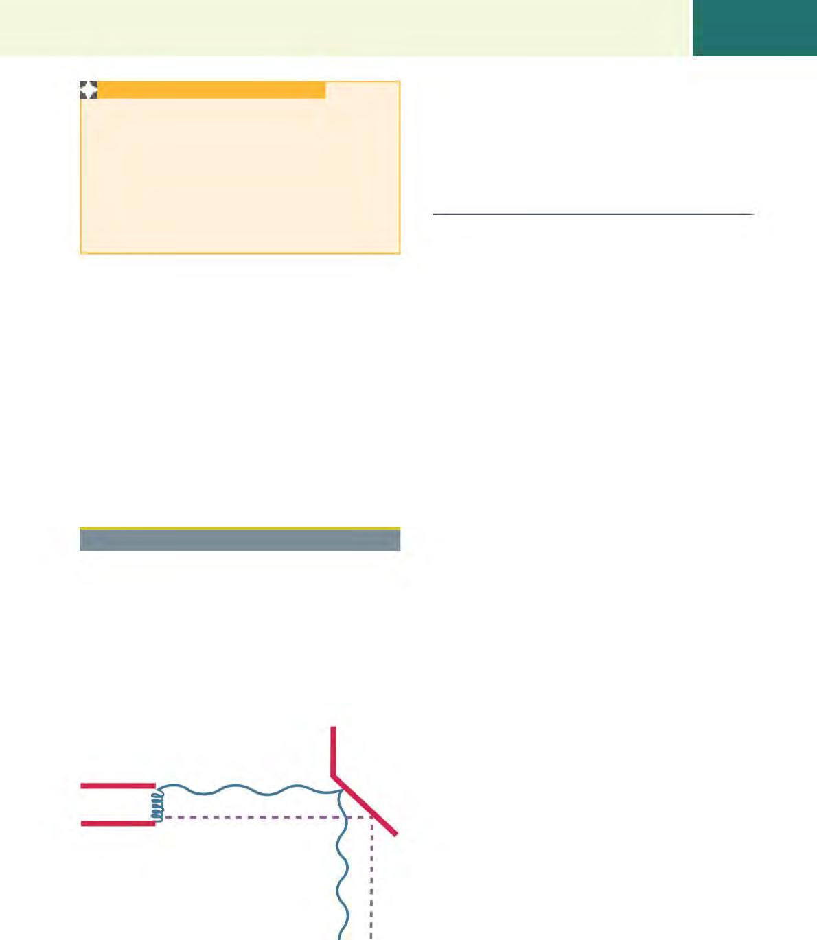
FIG. 1.9 In an x-ray tube, energy is emitted from the cathode as particles. The photons of energy are magnetically drawn to the anode when the circuit is activated.
9. Energy as particles is represented as:
a. Units of motion or as packets
b. Individual movements of atoms within a substance
c. Particles within the nucleus of the atom
d. Waves with frequencies and structure
10. Photons are described as:
a. Electromagnetic radiation that has a positive electric charge
b. Negatively charged electrons in the nucleus or the atom
c. Negatively charged protons within the nucleus of the atom
d. Electromagnetic radiation that has no electrical charge
Chapter Review Question answers are located in the Instructor Resources on Evolve and can be provided to students at the discretion of the instructor.
CHAPTER
2
Diagnostic X-Ray Production
Lois C. Brown, RTR (Can/USA), ACR, MSc, P.Phys
“The important thing is to not stop questioning. Curiosity has its own reason for existing.”
—Albert Einstein
1879–1955
OUTLINE
Electricity 10
The Wall Switch 10
The Electric Circuit 11
The Operating Console 11
Voltage 11
Current (Amperage) 12
Time 12
Watt (a Unit of Power) 12
Potential Difference 12
Line Voltage Compensator 12
Circuit Breakers: Amperage and Ground 13
Amperage 13
Fuses 13
Ground 13
Direct Current and Alternating
Current 13
LEARNING OBJECTIVES
Direct Current 14
Alternating Current 14
Transformers 14
Rectifiers 16
Single-Phase Circuits 16
Three-Phase Circuits 16
High-Frequency 16
The Complete X-ray Circuit 17
The X-Ray Unit 17
Components of the X-Ray Unit 17
The X-Ray Tube 18
The Cathode 20
The Anode 21
The Anode Heel Effect 21
The Line Focus Principle 22
When you have finished this chapter, you will be able to:
1. Describe the functions of each part of the x-ray tube.
2. List the four criteria necessary to produce x-rays.
3. Understand the construction of the x-ray tube.
4. Describe the anode heel effect and what causes it.
5. Discuss the line focus principle and anode heat bloom.
6. Distinguish between single-phase, three-phase, and high-frequency generators.
APPLICATIONS
Off-Focus Radiation and Heat Bloom 23
Large-Animal Portable X-Ray Units 23
X-Ray Production 23
The Exposure Switch 23
Exposure Switch Variations 24
X-Rays Production 24
Heat Dissipation 25
The Tube Rating Chart 26
Focal Spot Bloom 27
Minimum Power Supply Requirements 27
7. Understand potential difference.
8. Be familiar with direct and alternating current.
9. Know the difference between transformers and rectifiers.
10. Understand heat dissipation and know how to interpret the cooling charts.
11. Describe the activation of the single-stage and two-stage exposure switches.
The application of the information in this chapter is relevant to the following areas:
1. Production of radiation using electricity
2. The use of electricity in radiography and all the other imaging modalities
3. The application of switches, circuit breakers, transformers, and rectifiers in the production of radiation
4. The use of transformers to increase and decrease the voltage of the incoming power to the x-ray unit
5. The application of electricity in all aspects of work in every area of the profession
The representation of the distribution of electricity of the world. (Courtesy US National Aeronautics and Space Administration [NASA].)
KEY TERMS
Key terms are defined in the Glossary on the Evolve website.
Alternating current
Amperage
Anode
Anode heel effect
Bremsstrahlung radiation
Cathode
Characteristic radiation
Circuit
Circuit breaker
Current
Direct current
Exposure switch
Filament
Focal spot
Focal spot bloom
Generator
Ground
Heat bloom
Heat dissipation
Hertz
High frequency
Line focus principle
Electricity
All x-ray units are activated by an electric source. This source may be anything from a field battery to a large, very powerful connection in a hospital setting. So to start, we will learn about electricity and the factors that generate the power to activate the x-ray tube.
The x-ray unit consists of a closed circuit with five main criteria:
1. It must have enough power to eventually produce x-rays.
2. It must have selections where the power can be increased or decreased as necessary.
3. The electric current must travel in the same direction through the x-ray tube.
Line voltage compensator
Off-focus radiation
Photons
Potential difference
Power
Pulses (timer)
Rectifier
Resistance
Rotating anode
Rotor
Space charge effect
Stationary anode
Target
Thermionic emission
Transformer
Unsharpness
Voltage
Watt
Waveform
X-ray tube
4. There must be a way to produce free electrons with enough energy to produce x-rays.
5. There must be an efficient way to dissipate the heat that results in the interaction of the photons and the anode. (The production of x-rays is very inefficient and results in 99% heat and 1% x-rays).
The Wall Switch
Before the on/off switch for the x-ray unit itself is the wall switch (Fig. 2.1). It is very important—and in fact, the law in most countries—that the x-ray unit is installed with a separate wall switch mounted at eye level (about 5 feet) above the floor within reach of the x-ray generator. This is in place so that if there is an equipment malfunction and the x-ray unit