Physical Therapy and Rehabilitation Sciences Department
Drexel University
Philadelphia, Pennsylvania
Brian J. Eckenrode, PT, DPT, OCS
Assistant Professor
Department of Physical Therapy
Arcadia University
Glenside, Pennsylvania
Andrew R. Karduna, PhD
Associate Professor
Department of Human Physiology
University of Oregon
Eugene, Oregon
David M. Kietrys, PT, PhD, OCS
Associate Professor, Doctor of Physical Therapy Program
Rutgers, The State University of New Jersey–School of Health
Related Professions
Stratford, New Jersey
Margery A. Lockard, PT, PhD
Clinical Associate Professor
Health Sciences Department
Physical Therapy and Rehabilitation Sciences Department
Drexel University
Philadelphia, Pennsylvania
Joseph M. Mansour, PhD
Professor
Department of Mechanical and Aerospace Engineering
Case School of Engineering
Case Western Reserve University
Cleveland, Ohio
Stuart M. McGill, PhD Professor
Department of Kinesiology
University of Waterloo Waterloo, Canada
Carol A. Oatis, PT, PhD
Professor
Department of Physical Therapy
Arcadia University
Glenside, Pennsylvania
Peter G. Osmotherly, PhD
Senior Lecturer in Physiotherapy
School of Health Sciences
Faculty of Health
The University of Newcastle New South Wales, Australia
Theresa M. Spitznagle, PT, DPT, WCS
Associate Professor
Program in Physical Therapy
Washington University
St. Louis, Missouri
L. D. Timmie Topoleski, PhD
Professor
Department of Mechanical Engineering
University of Maryland, Baltimore County Baltimore, Maryland
Robert G. Wise, PT, DPT, SFMA
Good Shepherd Penn Partners
Penn Therapy and Fitness
Jenkintown, Pennsylvania
Contributors to Earlier Editions
Emily L. Christian, PT, PhD
Julie E. Donachy, PT, PhD
Z. Annette Iglarsh, PT, PhD, MBA
Thomas P. Mayhew, PT, PhD
Susan R. Mercer, PhD, BPHTY (Hon), FNZCP
Peter E. Pidcoe, PT, DPT, PhD
Neal Pratt, PT, PhD
FOREWORD
A noteworthy development in Physical Therapy was the official adoption of an identity, the Human Movement System [1]. The definition is “The human movement system comprises the anatomic structures and physiologic functions that interact to move the body or its component parts” [2]. The recognition of this system of physiological organ systems that effect and that are affected by movement further emphasizes the importance of the underlying science of kinesiology.
Now, even more than 7 years ago at the time of the last edition of Kinesiology: The Mechanics and Pathomechanics ofHumanMovement, physical inactivity has been linked to a variety of diseases including cancer, cardiovascular disease, and diabetes [3–7]. Similarly, exercise and physical activity are major treatments for these conditions and even for dementia [8–10]. But you cannot exercise if you are injured. That is where knowledge of kinesiology and pathokinesiology is so important. There is not enough recognition of how daily activities, no matter the level of intensity, alter the precision of joint motion. The general belief is that if you can move in a way that does not cause pain, that is acceptable. This belief is analogous to the belief that you can eat whatever and as much as you want without experiencing consequences.
Of course, the irony of the situation is that now that there is a great deal of evidence of the health costs of obesity, the problem continues to increase. As the health care community increases its efforts to promote exercise and physical activity, all levels of exercise providers must
augment their understanding of kinesiology to provide the highest level of guidance to prevent the development of musculoskeletal pain problems.
With the adoption of the identity of the Human Movement System, the physical therapy community is going to place even greater emphasis than in the past on movement and on understanding the factors that alter the precision of joint movement. Small alterations in movement induced by cartilage or ligament injury can lead to osteoarthritis [11,12]. Evidence is growing that, rather than strengthening exercises being the primary treatment mode movement, pattern training is being shown to be more effective [13–15]. At the same time, in the orthopedic surgery community, there is growing recognition that such things as shoulder impingement are not a pathoanatomic problem but are caused by movement patterns [16]. Similarly, there is a growing body of evidence that structural variations of the hip joint causing femoroacetabular impingement are related to excessive force from intensive sports activity [17]. Here again, knowledge of kinesiology is going to play a key role in developing safe guidelines for protecting joints while participating in sports activities as well as during daily activities.
This book provides the necessary information for the practitioner to understand normal movement as well as pathokinesiologic movement and, if used optimally, kinesiopathologic movement. That means the book can be used as the guide to help individuals avoid the development of pathology caused by movement impairments, which has as much if not more importance than recognizing how movement becomes altered by pathology in one of the contributing systems.
The scope of information in this text—from basic biomechanics to coverage of all body regions in chapters written by experts in the respective regions—provides the
insights and guidance that will serve all levels of students and practitioners well. The material is here, and the only necessary step is for the reader to take advantage of the expert information that is in this book. If they do, they will be enhancing their knowledge of the human movement system and offering the best care to their patients and clients.
American Physical Therapy Association HOD RC 14A-13, RC 15–13. American Physical Therapy Association Board Movement System Work Group 2015.
Kumar B, Robinson R, Till S: Physical activity and health in adolescence ClinMed2015;15(3):267–272
Fernandes RA, Coelho-E-Silva MJ, Spiguel Lima MC, et al.: Possible underestimation by sports medicine of the effects of early physical exercise practice on the prevention of diseases in adulthood. Curr DiabetesRev2015; 11(3): 201–205.
Physical inactivity may kill more than obesity. PerspectPublicHealth2015; 135(2): 59.
MacAnaney O, McLoughlin B, Leonard A, et al.: Inverse relationship between physical activity, adiposity and arterial stiffness in healthy middle-aged subjects. JPhysActHealth2015.
Sallis R: Exercise is medicine: a call to action for physicians to assess and prescribe exercise. PhysSportsmed2015; 43(1): 22–26.
Coombs N, Stamatakis E, Lee IM: Physical inactivity among older adults: implications for life expectancy among non-overweight and overweight or obese individuals. ObesResClinPract2015; 9(2): 175–179.
Barnes JN: Exercise, cognitive function, and aging. AdvPhysiolEduc2015; 39(2): 55–62.
Hayes SM, Alosco ML, Forman DE: The effects of aerobic exercise on cognitive and neural decline in aging and cardiovascular disease. Curr GeriatrRep2014; 3(4): 282–290.
Blalock D, Miller A, Tilley M, et al.: Joint instability and osteoarthritis Clin MedInsightsArthritisMusculoskeletDisord2015; 8: 15–23
Svoboda SJ: ACL injury and posttraumatic osteoarthritis. Clin Sports Med 2014; 33(4): 633–640
Aasa B, Berglund L, Michaelson P, et al.: Individualized low-load motor control exercises and education versus a high-load lifting exercise and education to improve activity, pain intensity, and physical performance in patients with low back pain: a randomized controlled trial. JOrthopSports PhysTher2015; 45(2): 77–85.
Graci V, Salsich GB: Trunk and lower extremity segment kinematics and their relationship to pain following movement instruction during a singleleg squat in females with dynamic knee valgus and patellofemoral pain. J SciMedSport2015; 18(3): 343–347.
Yemm B, Krause DA. Management of a patient with patellofemoral pain syndrome using neuromuscular training in decreasing medial collapse: a case report. PhysiotherTheoryPract2015; 31(3): 221–229.
Ludewig PM, Lawrence RL, Braman JP: What is in a name: using movement system diagnoses versus pathoanatomic diagnoses. J Ortho Sports Phys Ther2013; 43(5): 280–283.
17 Keogh MJ, Batt ME: A review of femoroacetabular impingement in athletes. SportsMed2008; 38(10): 863–878.
PREFACE
Movement is a core function of most living creatures. As humans, we understand that movement can be impaired for many reasons including injury or pain. We also recognize a common desire to improve or optimize a movement. Normal movement results from an exquisite interplay within the nervous, muscular, and skeletal systems and is heavily influenced by other systems including the respiratory, cardiovascular, integumentary, and immune systems. Taken as a whole, movement can itself be considered a “system” [1]. The Human Movement System integrates the functions of the other physiologic systems to produce movement. Kinesiology is the scientific study of movement of the human body or its parts and is the foundational science needed to understand the movement system. Restoration of normal movement, reduction of movement dysfunction, or optimization of movement each requires a firm foundation in kinesiology. This third edition of Kinesiology: The MechanicsandPathomechanicsofHumanMovementbuilds on the first two editions to provide the most current scientific data and clinical applications to help movement specialists understand the complex interactions within the movement system. As in the previous editions, it focuses on the musculoskeletal components of the movement system.
To evaluate and treat a movement disorder effectively, the clinician must consider two central questions: what is required to perform the movement and what effects does themovementproduceontheindividual?Consider a jogger who typically runs 2 or 3 miles three times weekly and has now decided to train for a marathon or one who has begun
to complain of lateral knee pain after a 2-mile run. To effectively guide the runner in training for a 26.2 mile (42.2 km) run or to help the runner eliminate the knee pain, the clinician must understand the requirements of running and recognize the loads applied to the musculoskeletal system while running. This textbook helps the reader develop knowledge and enhance skills that permit him or her to assist individuals to optimize movement or to reduce movement dysfunction.
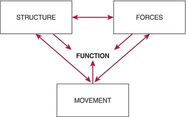
Two general factors govern the movement of a structure: the composition of the structure and the forces applied to it. A central principle in kinesiology is that the form or shape of a biological structure is directly influenced by its function. In fact, the relationship among movement, structure, and force is interdependent and multidirectional. Structure influences a body's movement. Movement affects the forces applied to the structure. The forces, in turn, influence the structure (see Figure). Function is the purposeful application of movement and, consequently, depends on the interactions of structure, force, and movement. The jogger who wants to run a marathon will be flexing and extending both knees thousands of times daily during training. The unique structure of the tibiofemoral joint produces complex threedimensional motion as the knee flexes and extends. That complex motion leads to intricate loading patterns on the tibia and femur in which the loads are distributed unevenly across the bony surfaces. These loads can lead to normal adaptation with increased bone mineralization and thickening of articular cartilage or can lead to bony edema and articular degradation.
An abnormal structure produces abnormal movement and abnormal forces on that or neighboring structures, contributing to further alterations in structure. The runner with lateral knee pain may exhibit hip muscle weakness that leads to faulty knee motion, overuse of hip musculature, and inflammation of the muscle's tendon or at the site of muscle attachment. The clinician needs to understand these interrelationships to design and direct interventions for restoring or optimizing human movement.
An understanding of the relationships among structure, force, and movement requires a detailed image of the structure of a region as well as a grasp of the fundamental laws of motion and the basic material properties of the tissues comprising the musculoskeletal system. The purposes of this textbook are to
Provide a detailed analysis of the structures of the musculoskeletal system within individual functional regions
Discuss how the structures affect function within each region
Analyze the forces sustained at the region during function
This textbook will help the clinician recognize the relationships between form and function, and between abnormal structure and dysfunction. This foundation should lead to improved evaluation and intervention approaches to movement dysfunction.
This book uses terminology that is standard within health care to describe elements of disablement based on a classification of function developed by the World Health Organization (WHO). In this classification scheme, a disease process, or pathology, alters a body structure or function, producing an impairment. The impairment may cause an individual to have difficulty executing a task or activity, producing an activity limitation or dysfunction. When the dysfunction alters the individual's ability to participate in life functions, the individual has participation restriction or a disability [2].
Although improving activity and participation is usually the primary objective in rehabilitation, the WHO model of disease provides a framework for clinicians to improve function not only by intervening directly at the level of the dysfunction but also by addressing the underlying impairments. By understanding the detailed structure and precise movement of an anatomical region, the clinician has tools to identify impairments and their influence on function and devise interventions that focus on the mechanism producing the dysfunction.
Organization of This Text
The needs of individual readers vary, and, as in the preceding editions, I have designed this book to allow readers to use it in ways that best meet their needs.
Part I of this textbook introduces the reader to the principles of biomechanics and material properties and then examines the material properties of the major component tissues of the musculoskeletal system: bone, muscle, cartilage, and dense connective tissue. These chapters lay out the biomechanical foundation for examining human movement.
Parts II through IV explore movement by anatomical region, investigating the detailed structure of the bones, joints, and muscles in that region and examining how their structures influence its movement. The ability of the region to sustain the forces generated during movements and function also is explored in Parts II through IV.
PartVconsiders more global, or whole-body, movements, specifically posture and locomotion.
Detailed discussions of forces at joints are presented in separate chapters so that readers may access that information as they need it. Although many readers will be interested in delving into the mathematical analyses used to determine forces on joint structures, others will find little need for such detail. The actual calculations are set apart in boxes that accompany the chapters. Conclusions based on the calculations are contained within the chapters' text so that readers can read the chapters and glean the essential information and return to the specific analyses as desired.
Conclusions regarding structure, function, and dysfunction in this text are based on the best available evidence, and each chapter is extensively referenced using both current and classic resources. I believe that the clinician is best equipped to evaluate current practice and to debunk long-held beliefs by having access to the classic resources that have established a concept and to the most current evidence that confirms or refutes standard impressions. Throughout this book, common clinical beliefs that are unsupported—or actually refuted—by strong evidence are explicitly identified so that the clinician hones the skill of healthy skepticism and develops the practice of demanding evidence to support a concept. The book also notes where evidence is meager or inconclusive or the conclusion is the opinion of the author. A strong, evidencebased background in kinesiology also helps develop clinician scholars who can contribute to our understanding of movement and movement dysfunction through the systematic, thoughtful observation and reporting of clinical phenomena. Despite the comment made long ago by a fellow graduate student that there was “nothing left to learn in gross anatomy,” there is much to be learned yet in functional anatomy and kinesiology.
New and Updated Features in the Third Edition
A new edition of a book allows us the opportunity to directly apply the wisdom of Maimonides:
This third edition does indeed “correct the errors of yesterday” and sheds light on emerging knowledge by including the most up-to-date science in kinesiology and biomechanics. The science in these areas continues to expand, and we have worked diligently to ensure that each chapter reflects that growth of understanding. Chapter authors have reviewed the literature and added updated references to provide the most current perspectives. Just one example of this is the inclusion of more muscle physiology in the chapter on muscle biomechanics (Chapter 4)to help readers understand muscle function in light of the growing understanding of muscle biology. In response to requests by users, we also have added a section on the basic mechanics of running to the chapter on gait (Chapter 48).
Additional Clinical Relevance boxes (with new Clinical Bottom Lines) have been added to most chapters to assist the reader in understanding the applicability of detailed kinesiological and biomechanical information. Videos of clinical cases demonstrating the use of a kinesiological
perspective to the assessment of a patient have been added to the already extensive video collection.
As in the second edition, Muscle Action tables introduce the discussion of muscle actions for each muscle. Actions of each muscle are introduced in table format and include the conclusions drawn from the evidence regarding each action. The evidence is discussed in detail after the table. This format allows the reader to identify at a glance which reported actions are supported by evidence, which are refuted, and which remain controversial.
Muscle Attachment boxes provide muscle innervation and attachment information, and also include brief descriptions of palpation strategies.
Thought problems have been added to the end of each chapter to allow the reader to practice applying the principles discussed in the chapter to clinical scenarios. Answers to these thought problems are available on (http://thePoint.lww.com), where students and instructors can also find additional practice questions and problems to solve.
A strong understanding of the effects of forces on joint function is critical to the analysis of movement. However, movement specialists are often unfamiliar with the analysis techniques needed to understand the forces generated during motion. All of the biomechanical analyses (Examining the Forces Boxes) have been revised to provide more detailed explanations of the analyses. These revisions are designed to make the analyses more understandable and accessible to those less familiar with biomechanical principles.
Finally, and perhaps most obviously, virtually all of the artwork is in full color to enhance the visual appeal of the book and to make the principles demonstrated by the
artwork more accessible to a whole new generation of readers.
I have made these changes because I firmly believe that people with musculoskeletal disorders or those who want to optimize their already normal function require the wisdom and guidance of individuals who have a clear, evidencebased understanding of musculoskeletal structure and function, a firm grasp of biomechanical principles, and the ability to observe and document movement. This third edition is designed to enhance the ability of exercise and rehabilitation specialists to serve in this role.
—CarolA.Oatis
Online Resources for Students and Instructors
Approximately 150 video clips provide dynamic illustrations of concepts discussed in the textbook and demonstrate movement disorders that can occur as a result of impairments. The clips also include demonstrations of palpations of bony landmarks for each anatomical region. A video icon is used throughout the text to identify concepts with related video material. These elements will help the reader integrate the relationships among structure, force, movement, and function and provide examples for students and teachers to analyze and discuss.
Laboratory Manuals for both students and instructors continue to offer activities for students to enhance learning and applications. The instructors' laboratory manual includes solutions and brief discussions of most activities. The laboratory manual for Chapter 1 contains additional problems for students to test their analytical skills. The solutions are provided in the Instructors' Manual.
The Instructors' Guide is a chapter-by-chapter outline to assist instructors with preparing class lectures. This ancillary has been updated to include the materials added to the revised chapters.
2. REFERENCES
1.
Sarhmann SA. The human movement system: our professional identity. PhysTher2014; 94: 1034–1042. http://www.who.int/classifications/icf/en/
ACKNOWLEDGMENTS
Completion of this third edition required the work and commitment of several individuals. First I am most thankful for the wonderful work and creativity of the contributing authors. The authors continuing from the original book include Drs. Beattie, Karduna, Lockard, Mansour, McGill, and Topoleski. They each willingly reviewed their work and listened to suggestions we received from readers. They updated information and added clinical examples to help the information be even more relevant to rehabilitation specialists. New contributors include Drs. Ebaugh, Eckenrode, Keitrys, Osmotherly, Spitznagle, and Wise. These authors also carefully updated chapters and added clinical perspectives. Together, the efforts of all of the authors to identify changes in knowledge or perspective help ensure that this textbook remains at the forefront of kinesiologic science.
The team at Lippincott Williams & Wilkins (Wolters Kluwer) has provided invaluable developmental, managerial, and technical support throughout the project. Linda Francis, Managing Editor, has been patient, persistent, and enthusiastic—frequently at the same time! She has provided the organization and help I needed to manage the complexities of revising this book. She worked with me to create a timetable that worked for both me and the publishers and then has helped me stick to it! Throughout the process, Linda has remained positive and encouraging for which I am very grateful! I also want to thank David Orzechowski, Production Project Manager at Wolters Kluwer, and Dhinakaran Arumugam, Senior Project Manager at SPi
Global, whose careful attention to detail as well as their patient tolerance of my mistakes were essential in the final production process.
I am indebted to several people who provided particular insights. Dr. David Kietrys provided wonderful feedback on how to make the Examining the Forces Boxes easier to follow by readers less familiar with biomechanical analysis. He willingly engaged in long back-and-forth conversations on ways to present the analysis, which ultimately led to a significant improvement in presentation. Dr. Robert Wise provided many wonderful clinical relevance examples and formed them into additional Clinical Relevance Boxes that will help the reader grasp the clinical importance of kinesiology. Rob also helped create many of the thought problems found at the end of each chapter to allow students to test their understanding. Rob also led the effort to create the new clinical cases presented in video for each of the book's units. Dr. Marge Tull, PT, CHT, and Wendy McCoy, PT, CHT, provided careful reviews of the chapters on the wrist and hand and offered clinical perspectives that enhance those chapters. Dr. Jamie Rosenburg provided guidance, creativity, and oversight for the new video demonstrations of EMG activity during hip exercises.
The art program underwent a complete overhaul in this third edition. These extensive revisions were expertly overseen by Jennifer Clements, Art Director. Jen kept us on schedule but also was patient and accepting as I requested “tweaks” to new and old figures needing to be fine-tuned. I am also most grateful to her for allowing me to continue my collaboration with the original artist, Kim Battista, and with the original photographer, Gene Smith. These two artists continue to create images that bring kinesiology and biomechanics to life. Kinesiology is a visual science and without the images created by these two, I would never be able to “tell the story.”
I am grateful for the invaluable support of three individuals who provided essential research and manuscript preparation assistance, without which I could never have completed this project. Rebecca Adams, DPT, provided expert research assistance and invaluable editorial insights. Elizabeth Dalrymple, DPT, and Hilary Park, SPT, were essential research assistants gathering and organizing references and citations. I am also indebted to the physical therapy students who volunteered as models for new images in this third edition: Rebecca Dobson, Nina Galleli, Stacey Gorter, Shane Harris, Emanuela Mannino, Betsy Michel, Nathalie Musey, and Junsik Yoon. Again I wish to thank Arcadia University and the Department of Physical Therapy for their support during this process.
I am particularly grateful for the support provided by Margaret M. Fenerty, Esq., who listened to my fears, tolerated my stress, and encouraged my efforts. And as many of my students know, I was lucky to have Daisy and Bruiser to keep me company as I sat at my desk day after day working on this project.
I wish to thank Arcadia University and my colleagues in the Department of Physical Therapy for their support throughout this process. I also want to thank all the students who have used the first two editions, particularly those at Arcadia University. They have provided insightful feedback and valuable suggestions that have informed this new edition. They helped identify errors, offered new ideas, and graciously told me what worked and what did not. I look forward to hearing new ideas and suggestions as students and clinicians use this new edition. Finally, I want to thank all of the students I have been lucky to work with over my career. You have inspired me with your dedication to learning and enthusiasm for excellence. You have made me a better teacher, and I know that the lives of my patients are in good hands.
Contributors
Foreword
Preface
Acknowledgments
Biomechanics of Tendons and Ligaments
10 Analysis of the Forces on the Shoulder Complex During Activity
Unit 2—Elbow Unit
11 Structure and Function of the Bones and Noncontractile Elements of the Elbow
12 Mechanics and Pathomechanics of Muscle Activity at the Elbow
13 Analysis of the Forces at the Elbow During Activity
Unit 3—Wrist and Hand Unit
14 Structure and Function of the Bones and Joints of the Wrist and Hand
15 Mechanics and Pathomechanics of the Muscles of the Forearm
16 Analysis of the Forces at the Wrist During Activity
17 Mechanics and Pathomechanics of the Special Connective Tissues in the Hand
18 Mechanics and Pathomechanics of the Intrinsic Muscles of the Hand
19 Mechanics and Pathomechanics of Pinch and Grasp