The right of Brad Bowling to be identified as author of this work has been asserted by him in accordance with the Copyright, Designs and Patents Act 1988.
No part of this publication may be reproduced or transmitted in any form or by any means, electronic or mechanical, including photocopying, recording, or any information storage and retrieval system, without permission in writing from the publisher. Details on how to seek permission, further information about the Publisher’s permissions policies and our arrangements with organizations such as the Copyright Clearance Center and the Copyright Licensing Agency, can be found at our website: www.elsevier.com/permissions.
This book and the individual contributions contained in it are protected under copyright by the Publisher (other than as may be noted herein).
Notices
Knowledge and best practice in this field are constantly changing. As new research and experience broaden our understanding, changes in research methods, professional practices, or medical treatment may become necessary.
Practitioners and researchers must always rely on their own experience and knowledge in evaluating and using any information, methods, compounds, or experiments described herein. In using such information or methods they should be mindful of their own safety and the safety of others, including parties for whom they have a professional responsibility.
With respect to any drug or pharmaceutical products identified, readers are advised to check the most current information provided (i) on procedures featured or (ii) by the manufacturer of each product to be administered, to verify the recommended dose or formula, the method and duration of administration, and contraindications. It is the responsibility of practitioners, relying on their own experience and knowledge of their patients, to make diagnoses, to determine dosages and the best treatment for each individual patient, and to take all appropriate safety precautions.
To the fullest extent of the law, neither the Publisher nor the authors, contributors, or editors, assume any liability for any injury and/or damage to persons or property as a matter of products liability, negligence or otherwise, or from any use or operation of any methods, products, instructions, or ideas contained in the material herein.
ISBN: 978-0-7020-5572-0
978-0-7020-5573-7
15
16
Preface to the Eighth Edition
I first met Jack Kanski when I rotated to The Prince Charles Eye Unit in Windsor as part of the Oxford Deanery ophthalmology residency programme. Jack had actually just retired from clinical practice, but continued to attend the unit’s weekly education meetings. As the senior registrar, I was responsible for the organization of these sessions, to which Jack brought the same qualities that have facilitated his amazing success as a medical author – his encyclopaedic knowledge of ophthalmology and unerring ability to isolate the critical issues in a topic, not to mention his incisive wit, made the meetings extraordinarily effective as well as hugely enjoyable.
Jack was aware that I had done some textbook writing previously, and after one of the teaching sessions asked me whether I would be interested in writing a basic interactive text with him for medical students and novice ophthalmologists. I was a little daunted at first – Jack had written more than thirty ophthalmology textbooks by this time – but duly proceeded; we worked together extremely well, the book was written to deadline, was critically popular and sold lots of copies.
After I left Windsor, Jack and I worked with each other again on one or two projects and kept in touch socially, and a couple of years later he raised the possibility of collaboration on the next edition of Clinical Ophthalmology. I was thrilled. I recall vividly when, just prior to my first ophthalmology post, I contacted two registrars independently to enquire about initial textbook choice, receiving a curt single-word response from both: ‘Kanski’, with the implication that there was no need to ask. Big shoes to fill.
I have striven to maintain Jack Kanski’s approach of presenting core clinical knowledge in a systematic and succinct form; the extent of subject coverage by the later editions of the book is easily underestimated, and it is intended that a thorough acquaintance with its contents will provide a comprehensive basis for general ophthalmic practice. In the present edition every attempt has been made to completely update each chapter, with inclusion of the latest practical evidence-based diagnostic and treatment approaches, and replacement and upgrading of images as appropriate, such as where novel imaging modalities offer an enhanced
perspective. The index for this edition has been written by the author to ensure its ease of use and clinical applicability.
I am incredibly indebted to Jack Kanski for the opportunity to contribute to Clinical Ophthalmology and other books, and for his ongoing mentoring and support. I have received invaluable help with the eighth edition from colleagues; Simon Chen generously furnished a large number of photographic and other images and gave his time to advise in depth on various posterior segment topics, Chris Barry also kindly provided and edited very numerous images, and many other ophthalmologists, optometrists, ophthalmic photographers and other eyecare professionals contributed one or a small number of figures and are acknowledged in individual legends. Philip Spork was good enough to review the section on macular antioxidant supplements. I am also indebted to the numerous colleagues who contacted Jack Kanski or myself with helpful comments on particular points in the seventh edition. Many individuals have helped substantially with the previous editions of Clinical Ophthalmology, the core of which has been brought forward into the present book; Ken Nischal and Andy Pearson both carried out detailed reviews of sections in the seventh edition, Jay Menon made a major contribution to the fifth edition, Anne Bolton and Irina Gout provided photographic expertise over many years and, of course, Terry Tarrant supplied a large number of amazingly authentic ocular paintings. My wife, Suzanne, and sons, Edward and Oliver, supported me unreservedly during the extended revision of the book, tolerating my absence over the course of many months without complaint. Finally, I would like to acknowledge the cheerful and expert support and commitment of the staff at Elsevier, especially Russell Gabbedy, Louise Cook, John Leonard, Anne Collett and Marcela Holmes.
It would be impossible for me to replicate Jack Kanski’s style precisely, but I have tried to retain the essence of his approach as faithfully as possible, and hope that this book will prompt in the reader at least some of the enthusiasm for the subject that the second edition of Clinical Ophthalmology engendered in me.
SFUprogressive subretinal fibrosis and uveitis syndrome
SICsolitary idiopathic choroiditis
SJSStevens–Johnson syndrome
SLKsuperior limbic keratoconjunctivitis
SLTselective laser trabeculoplasty
SRFsubretinal fluid
SSSjögren syndrome
STIRshort T1 inversion recovery
TALtotal axial length
TBtuberculosis
TENtoxic epidermal necrolysis
TGFtransforming growth factor
TIAtransient ischaemic attack
TTTtranspupillary thermotherapy
TMtrabecular meshwork
TRDtractional retinal detachment
UBMultrasonic biomicroscopy
USultrasonography
VAvisual acuity
VEGFvascular endothelial growth factor
VEPvisual(ly) evoked potential(s)
VFIvisual field index
VHLvon Hippel–Lindau syndrome
VKCvernal keratoconjunctivitis
VKHVogt–Koyanagi–Harada syndrome
VZVvaricella zoster virus
XLX-linked
INTRODUCTION 2
Anatomy 2
Terminology 3
General considerations 3
NON-NEOPLASTIC LESIONS 3
Chalazion 3
Other eyelid cysts 5
Xanthelasma 6
BENIGN EPIDERMAL TUMOURS 7
Squamous cell papilloma 7
Seborrhoeic keratosis 8
Actinic keratosis 8
BENIGN PIGMENTED LESIONS 9
Freckle 9
Congenital melanocytic naevus 9
Acquired melanocytic naevus 9
BENIGN ADNEXAL TUMOURS 10
Syringoma 10
Pilomatricoma 10
MISCELLANEOUS BENIGN TUMOURS 12
Capillary haemangioma 12
Port-wine stain 12
Pyogenic granuloma 13
Neurofibroma 13
MALIGNANT TUMOURS 13
Rare predisposing conditions 13
Basal cell carcinoma 15
Squamous cell carcinoma 17
Keratoacanthoma 18
Sebaceous gland carcinoma 19
Lentigo maligna and melanoma 19
Merkel cell carcinoma 20
Kaposi sarcoma 20
Treatment of malignant tumours 22
Eyelids 1
DISORDERS OF THE EYELASHES 25
Misdirected lashes 25
Eyelash ptosis 27
Trichomegaly 27
Madarosis 27
Poliosis 27
ALLERGIC DISORDERS 30
Acute allergic oedema 30
Contact dermatitis 30
Atopic dermatitis 30
BACTERIAL INFECTIONS 31
External hordeolum 31
Impetigo 31
Erysipelas 31
Necrotizing fasciitis 32
VIRAL INFECTIONS 32
Molluscum contagiosum 32
Herpes zoster ophthalmicus 32
Herpes simplex 33
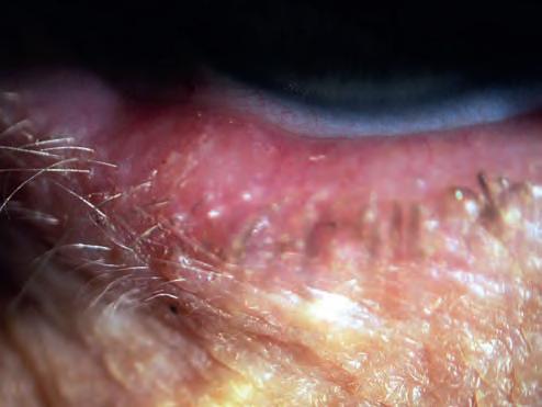
BLEPHARITIS 34
Chronic blepharitis 34
Phthiriasis palpebrarum 37
Tick infestation of the eyelid 38
Angular blepharitis 38
Childhood
blepharokeratoconjunctivitis 38
PTOSIS 38
Classification 38
Clinical evaluation 39
Simple congenital ptosis 41
Marcus Gunn jaw-winking syndrome 41
Third nerve misdirection syndromes 41
Involutional ptosis 44
Mechanical ptosis 44
Surgery 44
ECTROPION 45
Involutional ectropion 45
Cicatricial ectropion 47
Paralytic ectropion/facial nerve palsy 47
Mechanical ectropion 50
ENTROPION 50
Involutional entropion 50
Cicatricial entropion 51
MISCELLANEOUS ACQUIRED DISORDERS 51
Varix 51
Dermatochalasis 52
Floppy eyelid syndrome 52
Blepharochalasis 55
Eyelid imbrication syndrome 55
Upper lid retraction 55
Lower lid retraction 55
COSMETIC EYELID AND PERIOCULAR SURGERY 56
Involutional changes 56
Non-surgical techniques 56
Surgical techniques 56
CONGENITAL MALFORMATIONS 57
Epicanthic folds 57
Telecanthus 57
Blepharophimosis, ptosis and epicanthus inversus syndrome 59
Epiblepharon 59
Congenital entropion 59
Coloboma 59
Cryptophthalmos 60
Euryblepharon 61
Microblepharon 61
Ablepharon 61
Congenital upper lid eversion 62
Ankyloblepharon filiforme adnatum 62
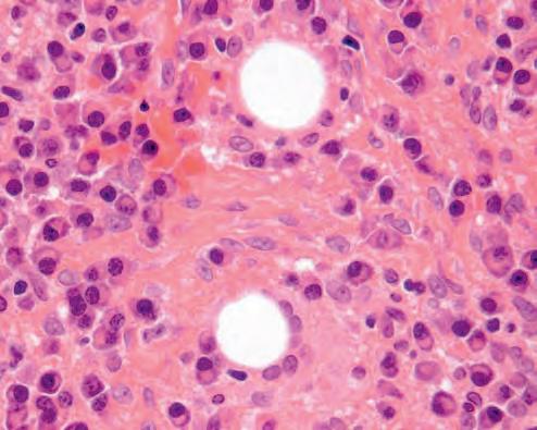
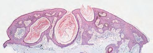
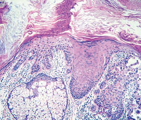
1.2 Chalazion. (A) Histopathology shows a lipogranuloma; the large pale cells are epithelioid cells and the welldemarcated empty space contained fat dissolved out during processing; (B) uninflamed chalazion; (C) acutely inflamed lesion; (D) conjunctival granuloma; (E) marginal chalazion; (F) conjunctival view of chalazion clamp in place prior to incision and curettage
Fig.
(Courtesy of J Harry and G Misson, from Clinical Ophthalmic Pathology, Butterworth-Heinemann 2001 – fig. A; J Nerad, K Carter and M Alford, from ‘Oculoplastic and Reconstructive Surgery’, in Rapid Diagnosis in Ophthalmology, Mosby 2008 – fig. F)
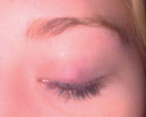
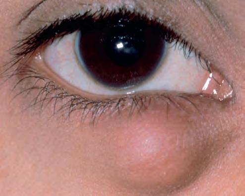
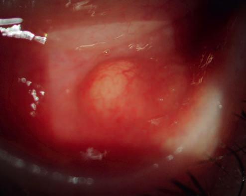
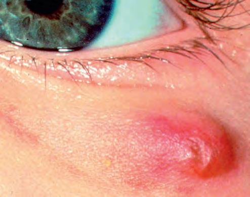
○ Acute: sterile inflammation or bacterial infection with localized cellulitis (Fig. 1.2C); differentiation may be difficult. A secondarily infected meibomian gland is referred to as an internal hordeolum.
• Signs
○ A nodule within the tarsal plate, sometimes with associated inflammation.
○ Bulging inspissated secretions may be visible at the orifice of the involved gland.
○ There may be an associated conjunctival granuloma (Fig. 1.2D).
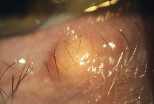
○ A lesion at the anterior lid margin – a marginal chalazion (Fig. 1.2E) – may be connected to a typical chalazion deeper in the lid or be due to isolated involvement of a gland of Zeis.
Treatment
• Oral antibiotics are required for significant bacterial infection, but not for sterile inflammation.
• Conservative. At least a third of chalazia resolve spontaneously so observation may be appropriate, especially if the lesion is showing signs of improvement, though early definitive treatment has been reported to lead to higher patient satisfaction.
• Hot compress application several times daily may aid resolution, particularly in early lesions.
• Expression. Compression between two cotton-tipped applicators is sometimes effective in expressing the contents of a fresh lesion near the lid margin.
• Steroid injection into or around the lesion has been reported to give similar resolution rates to incision and curettage (see below). It may be preferred for marginal lesions or lesions close to structures such as the lacrimal punctum because of the risk of surgical damage.
○ Reported regimens include 0.2–2 ml of triamcinolone acetonide aqueous suspension diluted with lidocaine to a concentration of 5 mg/ml, and 0.1–0.2 ml of 40 mg/ml, injected with a 27- or 30-gauge needle.
○ The success rate following one injection is about 80%; a second can be given 1–2 weeks later.
○ Local skin depigmentation and fat atrophy are potential but uncommon complications, the risk of which may be reduced by avoidance of infiltration immediately subcutaneously or by utilizing a conjunctival approach.
○ Retinal vascular occlusion has been described as a complication, probably due to intravascular injection with subsequent embolization.
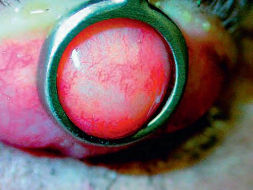
• Surgery
○ Following local anaesthesia infiltration, the eyelid is everted with a specialized clamp (Fig. 1.2F), the cyst is
incised vertically through the tarsal plate and its contents curetted.
○ Limited excision of solid inflammatory material (sent for histopathology) with fine scissors may be helpful in some cases, especially if there is no focus of secretions.
○ A suture should not be used.
○ Topical antibiotic ointment is used three times daily for 5–7 days following curettage.
• Marginal lesions can be managed by steroid injection, by curettage of an associated deeper chalazion, by shave curettage or by incision and curettage via a horizontal incision on the conjunctival surface or vertically through the grey line.
• Prophylaxis
○ Treatment of blepharitis, e.g. daily lid hygiene regimen.
○ Systemic tetracycline may be required as prophylaxis in patients with recurrent chalazia, particularly if associated with acne rosacea.
Other eyelid cysts
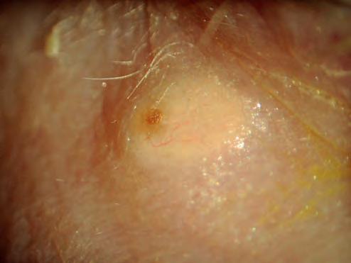
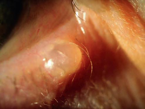
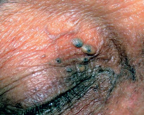
• Cyst of Zeis is a small, non-translucent cyst on the anterior lid margin arising from obstructed sebaceous glands associated with the eyelash follicle (Fig. 1.3A).
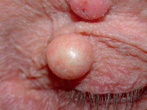
• Cyst of Moll (apocrine hidrocystoma) is a small retention cyst of the lid margin apocrine glands. It appears as a round, non-tender, translucent fluid-filled lesion on the anterior lid margin (Fig. 1.3B).
• Sebaceous (pilar) cyst is caused by a blocked pilosebaceous follicle and contains sebaceous secretions; the gland orifice will often be visible (Fig. 1.3C). It is only rarely found on the eyelid although it may occasionally occur at the inner canthus.
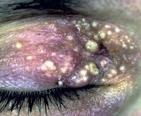
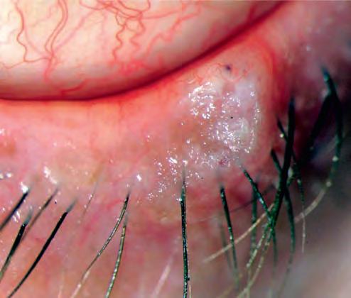
• Comedones are plugs of keratin and sebum within the dilated orifice of hair follicles that often occur in patients with acne vulgaris. They may be either open (blackheads) containing a darkened plug of oxidized material (Fig. 1.3D), or closed (whiteheads).
• Milia are caused by occlusion of pilosebaceous units resulting in retention of keratin. They are tiny, white, round, superficial papules that tend to occur in crops (Fig. 1.3E).
• Epidermal inclusion cyst is usually caused by implantation of epidermis into the dermis following trauma or surgery. It is a slow-growing, round, firm, superficial or subcutaneous lesion containing keratin (Fig. 1.3F).
• Epidermoid cyst is uncommon and usually developmental, occurring along embryonic lines of closure. It is similar in appearance to an epidermal inclusion cyst.
• Dermoid cyst is usually subcutaneous or deeper and is typically attached to the periosteum at the lateral end of the brow (Fig. 1.3G). It is caused by skin sequestered during embryonic development.
• Eccrine hidrocystoma is less common but similar in appearance to a cyst of Moll except that it is usually located along the medial or lateral aspects of the lid, and is close to but does not involve the lid margin itself (Fig. 1.3H).
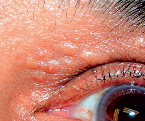
Xanthelasma
Introduction
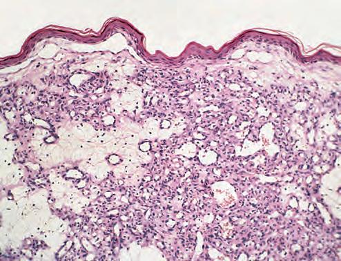
Xanthelasma (plural – xanthelasmata) is a common, frequently bilateral condition typically affecting middle-aged and elderly individuals. It is a subtype of xanthoma. Hyperlipidaemia is found in about one-third of patients, in whom corneal arcus may also be
present. In contrast to chalazion, fat in xanthelasmata is mainly intracellular, with lipid-laden histiocytes (foam cells) in the dermis (Fig. 1.4A).
Diagnosis
Xanthelasmata are yellowish subcutaneous plaques, usually in the medial aspects of the eyelids (Fig. 1.4B), commonly bilateral and are multiple (Fig. 1.4C).
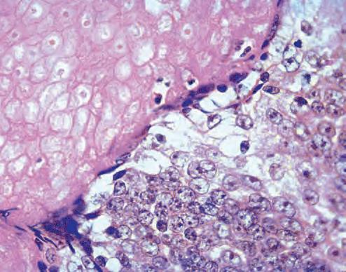
Fig. 1.6 Basal cell papilloma. (A) Typical ‘stuck-on’ appearance; (B) histopathology showing an elevated expansion of the epidermis with proliferation from basal cells – horn cysts and pseudohorn cysts are present (Courtesy of A Pearson – fig. A; J Harry – fig. B)
Acquired melanocytic naevus
Diagnosis
The clinical appearance and potential for malignant transformation of naevi are determined by their histological location within the skin.
• Junctional naevus occurs in young individuals as a uniformly brown macule or plaque (Fig. 1.10A). The naevus cells are located at the junction of the epidermis and dermis and have a low potential for malignant transformation (Fig. 1.10B).
• Compound naevus occurs in middle age as a raised papular lesion. The shade of pigment varies from light tan to dark
low, for transformation into squamous cell carcinoma. Treatment involves biopsy followed by excision or cryotherapy.
BENIGN PIGMENTED LESIONS
Freckle
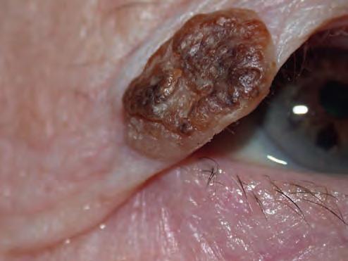
A freckle (ephelis, plural ephelides) is a small (generally 1–5 mm) brown macule due to increased melanin in the epidermal basal layer, typically in sun-exposed skin (Fig. 1.8); numbers vary with the level of sun exposure and can sometimes regress completely. Histopathology shows hyperpigmentation of the basal layer of the epidermis, with a normal melanocyte population.
Congenital melanocytic naevus
Congenital naevi are uncommon and histologically resemble their acquired counterparts (see below). They are usually small and of uniform colour. Rare variants include a ‘kissing’ or split naevus that involves the upper and lower eyelid (Fig. 1.9A) and may occasionally contain numerous hairs (Fig. 1.9B), and a very large lesion covering an extensive area of the body (‘giant hairy naevus’ – Fig. 1.9C). Large lesions have the potential for malignant transformation (up to 15%). Treatment, if necessary, involves complete surgical excision.
Fig. 1.7 Actinic keratosis. (A) Clinical appearance; (B) histopathology shows irregular dysplastic epidermis with hyperkeratosis, parakeratosis and cutaneous horn formation
(Courtesy of M Jager – fig. A; J Harry and G Misson, from Clinical Ophthalmic Pathology, Butterworth-Heinemann 2001 – fig. B)
ophthalmologists. It affects children and young adults and is more common in females. Clinically it appears as a mobile purplish dermal nodule that may have a hard consistency due to calcification (Fig. 1.12A). Histopathology shows irregular epithelial islands exhibiting viable basophilic cells at the periphery and degenerate ‘shadow’ cells more centrally (Fig. 1.12B). Calcification is frequently present and there is often a foreign body giant cell reaction. Treatment involves excision. Malignant change is rare. Other, less common, hair follicle proliferations include trichofolliculoma, trichoepithelioma and trichilemmoma.
MISCELLANEOUS BENIGN TUMOURS
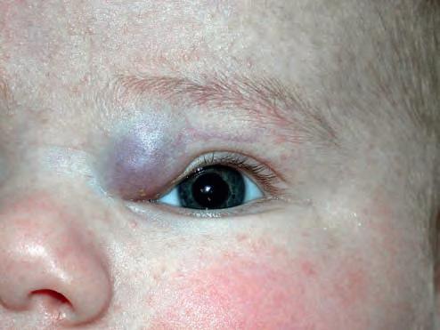
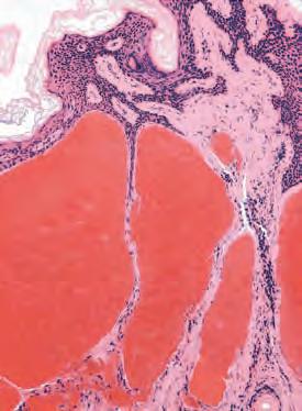
Capillary haemangioma
Capillary haemangioma (strawberry naevus) is one of the most common tumours of infancy; it is three times as common in boys as girls. It presents shortly after birth as a unilateral, raised bright red lesion (Fig. 1.13A), usually in the upper lid; a deeper lesion appears purplish (Fig. 1.13B and see also Fig. 3.31). Ptosis is frequent. The lesion blanches on pressure and may swell on crying. There may be orbital extension (see Ch. 3). Occasionally the lesion may involve the skin of the face and some patients have strawberry naevi on other parts of the body. Histopathology shows proliferation of varying-sized vascular channels in the dermis and subcutaneous tissue (Fig. 1.13C). It is important to be aware of an association between multiple cutaneous lesions and visceral haemangiomas, and to consider systemic assessment in appropriate cases. Treatment is described in Ch. 3.
Port-wine stain
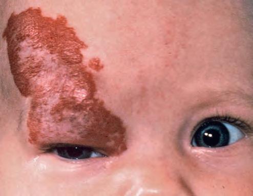
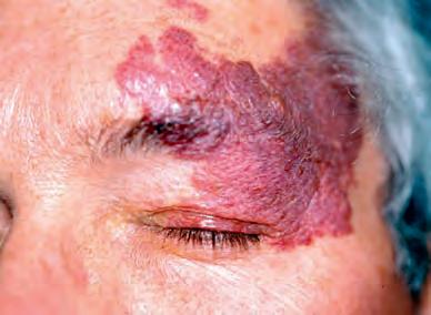
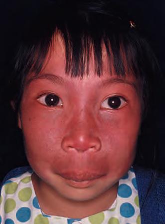
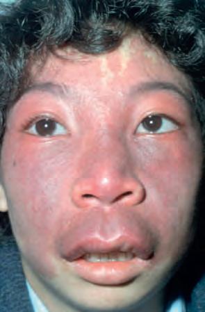
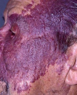
Introduction
Port-wine stain (naevus flammeus) is a congenital malformation of vessels within the superficial dermis, consisting histopathologically
of vascular spaces of varying calibre separated by thin fibrous septa (Fig. 1.14A). About 10% have associated ocular or CNS involvement, including Sturge–Weber (see below) and other defined syndromes.
Diagnosis
Port-wine stain manifests clinically as a sharply demarcated soft pink patch that does not blanch with pressure, most frequently located on the face. It is usually unilateral and tends to be aligned with the skin area supplied by one or more divisions of the trigeminal nerve (Figs 1.14B and C). Darkening to red or purple takes place with age, and there is commonly associated soft tissue hypertrophy (Figs 1.14D–F). Bleeding may occur from focal overlying lobulations (pyogenic granulomas – see below).
Treatment
Treatment with laser (e.g. pulsed-dye) is effective in decreasing skin discoloration; cosmetically superior results are usually
Fig. 1.11 Syringomas
(Courtesy of A Pearson)
Fig. 1.12 Pilomatricoma. (A) Clinical appearance; (B) histopathology shows viable basophilic cells to the right and degenerate ‘shadow’ cells to the left (Courtesy of J Krachmer, M Mannis and E Holland, from Cornea, Elsevier 2005 – fig. A; J Harry and G Misson, from Clinical Ophthalmic Pathology, Butterworth-Heinemann 2001 – fig. B)
Fig. 1.13 Capillary haemangioma. (A) Medium-sized haemangioma; (B) mechanical ptosis due to a large lesion; (C) histopathology shows vascular channels of varying size within the dermis and subcutaneous tissue (Courtesy of S Chen – fig. A; J Harry – fig. C)
achieved by early treatment. Topical preparations such as imiquimod and rapamycin, alone or with adjuvant laser, show promise. Soft tissue debulking is used in a small number of cases. Screening for glaucoma should begin in infancy. Systemic investigation is considered in some patients, particularly those with a lesion of the lumbar area.
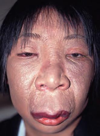
Sturge–Weber syndrome
Sturge–Weber syndrome (encephalotrigeminal angiomatosis) is a congenital, sporadic phacomatosis.
• Port-wine stain, extending over the area corresponding to the distribution of one or more branches of the trigeminal nerve.
• Leptomeningeal haemangioma involving the ipsilateral parietal or occipital region may cause contralateral focal or generalized seizures, hemiparesis or hemianopia.
• Ocular features may include ipsilateral glaucoma, episcleral haemangioma, iris heterochromia and diffuse choroidal haemangioma (see Ch. 12).
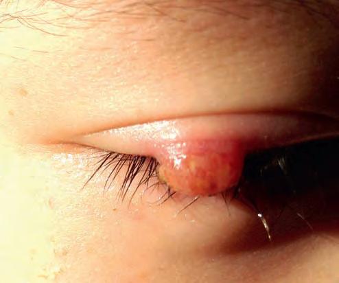
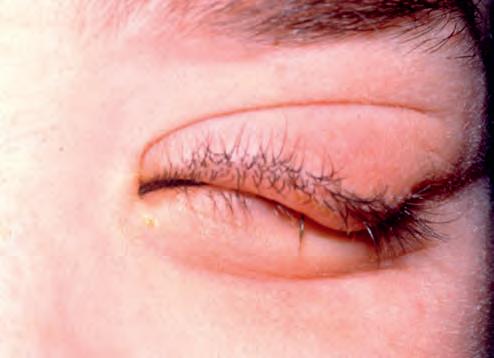
Pyogenic granuloma
Pyogenic granuloma is a rapidly growing vascularized proliferation of granulation tissue that is usually antedated by surgery, trauma or infection, although some cases are idiopathic. Clinically there is a painful, rapidly growing, vascular granulating polypoidal lesion (Fig. 1.15) that may bleed following relatively trivial trauma. Treatment of cutaneous lesions involves excision; conjunctival pyogenic granuloma is discussed in Ch. 5.
Neurofibroma
Cutaneous neurofibromas are benign nerve tumours, usually nodular or pedunculated, that can be found anywhere on the skin. Isolated neurofibromas are common in normal individuals, but if multiple lesions are present neurofibromatosis (see Ch. 19) should be excluded. Plexiform neurofibromas typically present in childhood as a manifestation of neurofibromatosis type 1 with a characteristic S-shaped deformity of the upper eyelid (Fig. 1.16). Treatment of solitary lesions involves simple excision but removal of the more diffuse plexiform lesions may be difficult.
MALIGNANT TUMOURS
The treatment of malignant eyelid tumours in general is discussed at the end of this section.
Rare predisposing conditions
Young patients who suffer from one of the following conditions may develop eyelid malignancies.
• Xeroderma pigmentosum is characterized by skin damage on exposure to sunlight, leading to progressive cutaneous abnormalities (Fig. 1.17A). It is inherited in an autosomal recessive (AR) fashion. Affected patients have a bird-like facies and a great propensity to the development of basal cell carcinoma (BCC), squamous cell carcinoma (SCC)
Fig. 1.14 Port-wine stain. (A) Histopathology shows widely dilated blood-filled spaces separated by fibrous septa; (B) and (C) clinical appearance; (D–F) progression of port-wine stain over time, with associated underlying soft tissue hypertrophy (Courtesy of L Horton – fig. A)
Fig. 1.15 Pyogenic granuloma
Fig. 1.16 Plexiform neurofibroma – characteristic S-shaped upper lid (Courtesy of J Harry)