

https://ebookmass.com/product/year-one-nora-roberts/
ebookmass.com


https://ebookmass.com/product/year-one-nora-roberts/
ebookmass.com
Edited by
C. Wayne McIlwraith, BVSc, PhD, Dr med vet (h.c. Vienna), DSc (Purdue), DSc (h.c. Massey), Laurea Dr (h.c. Turin), D vet med (h.c. London), FRCVS, Diplomate ACVS, ECVS, & ACVSMR
University Distinguished Professor
Barbara Cox Anthony University Chair in Orthopaedics
Director Orthopaedic Research Center
Gail Holmes Equine Orthopaedic Research Center
College of Veterinary Medicine and Biomedical Sciences
School of Biomedical Engineering
Colorado State University
Fort Collins, Colorado
David D. Frisbie, DVM, PhD, Diplomate ACVS & ACVSMR
Professor
Gail Holmes Equine Orthopaedic Research Center
College of Veterinary Medicine and Biomedical Sciences
School of Biomedical Engineering
Colorado State University
Fort Collins, Colorado
Christopher E. Kawcak, DVM, PhD, Diplomate ACVS & ACVSMR
Professor
Iron Rose Ranch University Chair in Musculoskeletal Research
Gail Holmes Equine Orthopaedic Research Center
College of Veterinary Medicine and Biomedical Sciences
School of Biomedical Engineering
Director of Equine Clinical Services
Colorado State University
Fort Collins, Colorado
P. René van Weeren, DVM, PhD, Diplomate ECVS
Professor of Equine Musculoskeletal Biology
Department of Equine Sciences
Faculty of Veterinary Medicine
Utrecht University
Utrecht, The Netherlands
Myra F. Barrett, DVM, MS, Diplomate ACVR
Assistant Professor of Diagnostic Imaging
Gail Holmes Equine Orthopaedic Research Center
College of Veterinary Medicine and Biomedical Sciences
Environmental and Radiological Health Sciences
Colorado State University
Fort Collins, Colorado
Pieter A. J. Brama, DVM, MBA, PhD, Diplomate ECVS
Professor of Veterinary Surgery School of Veterinary Medicine
Veterinary Science Centre University College Dublin Belfield, Dublin, Ireland
Janny C. de Grauw, DVM, PhD Resident in Veterinary Anesthesiology Faculty of Veterinary Medicine Department of Equine Sciences Utrecht University Utrecht, The Netherlands
David D. Frisbie, DVM, PhD, Diplomate ACVS & ACVSMR
Professor
Gail Holmes Equine Orthopaedic Research Center College of Veterinary Medicine and Biomedical Sciences School of Biomedical Engineering
Colorado State University
Fort Collins, Colorado
Laurie R. Goodrich, DVM, PhD, Diplomate ACVS
Associate Professor in Equine Surgery and Lameness
Gail Holmes Equine Orthopaedic Research Center College of Veterinary Medicine and Biomedical Sciences School of Biomedical Engineering
Colorado State University
Fort Collins, Colorado
Kevin K. Haussler, DVM, DC, PhD, Diplomate ACVSMR
Associate Professor
Gail Holmes Equine Orthopaedic Research Center
College of Veterinary Medicine and Biomedical Sciences School of Biomedical Engineering
Colorado State University
Fort Collins, Colorado
Christopher E. Kawcak, DVM, PhD, Diplomate ACVS & ACVSMR
Professor
Iron Rose Ranch University Chair in Musculoskeletal Research
Gail Holmes Equine Orthopaedic Research Center College of Veterinary Medicine and Biomedical Sciences School of Biomedical Engineering
Director of Equine Clinical Services
Colorado State University
Fort Collins, Colorado
Melissa R. King, DVM, PhD, Diplomate ACVSMR
Assistant Professor
Equine Sports Medicine and Rehabilitation
Gail Holmes Equine Orthopaedic Research Center College of Veterinary Medicine and Biomedical Sciences School of Biomedical Engineering
Colorado State University
Fort Collins, Colorado
C. Wayne McIlwraith, BVSc, PhD, Dr med vet (h.c. Vienna), DSc (Purdue), DSc (h.c. Massey), Laurea Dr (h.c. Turin), D vet med (h.c. London), FRCVS, Diplomate ACVS, ECVS, & ACVSMR
University Distinguished Professor
Barbara Cox Anthony University Chair in Orthopaedics Director Orthopaedic Research Center
Gail Holmes Equine Orthopaedic Research Center College of Veterinary Medicine and Biomedical Sciences School of Biomedical Engineering
Colorado State University
Fort Collins, Colorado
Kurt Selberg, MS, DVM, MS, Diplomate ACVR
Assistant Professor
Department of Veterinary Biosciences and Diagnostic Imaging
College of Veterinary Medicine University of Georgia Athens, Georgia
P. René van Weeren, DVM, PhD, Diplomate ECVS
Professor of Equine Musculoskeletal Biology
Department of Equine Sciences
Faculty of Veterinary Medicine
Utrecht University
Utrecht, The Netherlands
Natasha M. Werpy, DVM, Diplomate ACVR Associate Professor
Department of Large Animal Clinical Sciences
College of Veterinary Medicine
University of Florida
Gainesville, Florida
P. René van Weeren
The horse has always taken a special position among the species that have been domesticated by humankind. The horse was domesticated rather late, around 3500 BC,1 millennia after such species as goat, sheep, and cattle. Unlike these other species, the main purpose of the horse’s domestication was not the provision of edible products or products that could be somehow transformed into clothing, such as meat, milk, fur, or skin, but for a less tangible commodity: the combination of physical power and athletic capacity.
Horses have been the major power source for all Eurasian and Northern African civilizations since their introduction from roughly 3500 to 500 BC until the invention of the steam engine that started the Industrial Revolution in the late 1700s. The ultimate personification of the role of the horse in society is perhaps Bucephalus, the legendary horse of Alexander the Great, who conquered the vastest land empire the world has ever known. Bucephalus served Alexander who, according to legend, was the only person able to mount the stallion, from a young age to its death at the age of 30 after the battle of Hydaspes in what is now Pakistan, 2900 miles from its native Macedonia. There, Alexander named the city of Bucephala (present-day Jhelum) after him. After the Industrial Revolution horses still remained essential for many sectors of human society until after World War II, when the combustion engine definitively took over all traditional roles of the horse in warfare, transport, and agriculture. Some have predicted that the loss of its classic duties would make the horse into a zoo species,2 but they were proven entirely wrong by the rapidly increasing popularity of the horse as a sports and leisure animal from the mid-1960s onwards. Over the millennia, humans and horses appeared to have bonded in a way that goes far beyond economic value or utility and is more profound than with any other domesticated species, with the exception of the dog. Though admittedly the equine industry is susceptible to the fluctuations of economic prosperity, this fascination for the equine species is not likely to disappear soon, if ever. This obviously guarantees the horse its
privileged place in the big family of animal species with which humankind has surrounded itself.
Where the role of the horse in society has changed profoundly in the past century, the underlying reasons of its use and popularity have not changed at all. It is still the stamina of the animal and athletic capacity of its locomotor system that form the basis for almost all present-day use. The most critical body systems for athletic performance are the cardiorespiratory system and the musculoskeletal system. Within the latter system, joints are literally pivotal elements. It may not be surprising that orthopedic malfunctioning or other musculoskeletal disorders account for the vast majority of reasons to consult an equine vet.3 Of the specific elements of the musculoskeletal system, joint disorders invariably rank first or second in importance (together with tendinopathies, depending on discipline). Most figures come from the racing industry,4,5 but the relatively scarce data for sport horses also point in the same direction.6,7 In a survey of U.S. horse owners in 1998 it was estimated that 60% of all lameness was related to osteoarthritis (OA) and approximately $145 million was spent on veterinary bills relating to the problem.8 In this respect, the clinical importance of joint disorders in the equine species is very comparable to the situation in humans where musculoskeletal disorders in general and articular pathologies in particular represent an enormous burden to society in terms of loss of quality of life and costs of healthcare with 151 million sufferers of OA worldwide.9 For this and a number of important biologic reasons the horse is increasingly recognized as a suitable, if not the best, model for human joint disease.10 This translational aspect of equine joint disease will be dealt with in more detail in Chapter 27, which discusses arthritis research and future directions in joint disease.
This first chapter gives a general introduction into the anatomy and physiology of the (equine) joint, as a basis for the understanding of the following chapters that address in detail specific disorders, diagnostic possibilities, and therapeutic interventions.
Whereas the necessary stability of the equine musculoskeletal system is provided by the rigid bony components, joints permit motion of these bony components in relation to each other and, indirectly, the displacement of the entire individual with respect to the environment, that is, locomotion. To accomplish this, joints have to meet several requirements. They have to be as robust as the bony elements of the musculoskeletal system, as the forces generated by locomotion and other (athletic) activities are transmitted through joints as they are through bones. They also have to allow for smooth and as frictionless as possible motion of the bony ends that articulate with respect to each other. Lastly, they have a role, together with other structures, such as the digital cushion in the foot, to mitigate and dampen the accelerations and associated vibrations that are generated during the impact peak of the stride cycle at hoof landing. This latter aspect has been relatively well studied in the equine literature.11,12
All the aforementioned requirements that are at least partially contradictory (strength comparable to bone, smooth surfaces for supple gliding, and resilience for shock absorption) have to be accommodated in a single structure, which is a challenging task. As will be explained, nature deals with these challenges in an ingenious way, however, at the cost of flexibility and repair capacity. For reasons of clarity the components that make up a joint will be dealt with separately, but it is important to stress that a joint is more than a collection of tissues with separate characteristics and functions. There is common agreement nowadays that the joint should be seen as a complex multicomposite organ not unlike structures such as the liver, kidney or heart.13,14 Within this organ the constituting elements act together to ensure proper joint function. There is a strong interplay of all these components in health and disease and mutual influencing of physiologic functioning; malfunctioning of the components will also inevitably affect the other constituents and hence performance of the entire joint at a shorter or longer term.
Joints can be classified in several ways. A gross division can be made between classification according to structural characteristics, that is, the type of tissue(s) that form the interface between the articulating bony parts of the skeleton, and classification according to function, or the degree and type of movement joints allow.
The currently used basic classification is three major categories, which are fibrous joints with the bone connected by dense connective tissue, cartilaginous joints where cartilage is the interface, and synovial joints in which there is a fluid-filled cavity.15 In the horse, the articulations between the bodies of the vertebrae that make up the axial skeleton are fibrous joints, with the exception of the articulation between the first and second cervical vertebrae (C1-C2), which is a synovial joint. A cartilaginous joint has an interface consisting of hyaline or fibrous cartilage; examples are the human intervertebral
disk and the symphysis of the pubic bones in both humans and horses. In synovial joints there is no structural connection between the bony parts of the skeleton, but both ends are capped with hyaline cartilage and articulate by gliding over each other although contained in a joint capsule that is filled with synovial fluid, a viscous liquid. A sliding bearing in mechanical engineering basically functions according to the same principle.
In a functional sense, there are several other ways to classify joints. A common way is according to the degree of motion they permit. Although the following nomenclature is currently seen as obsolete,15 it is still widely used and will hence be mentioned here. A synarthrosis is a joint permitting little mobility. Most of these joints are of fibrous nature, such as the sutures that connect the bony components that make up the skull. Amphiarthroses are joints that permit more, but still very limited, mobility. They are generally of either fibrous or cartilaginous nature, with the intervertebral joints (again with the exception of C1-C2) as the best examples. Finally, diarthrodial joints permit maximal motion. These are always synovial joints and their motion is limited by periarticular or intraarticular structures such as capsules or ligaments, but not by the nature of the joint. Virtually all joints of the appendicular skeleton of the horse are diarthrodial joints.
Other functional classifications are based on the degrees of freedom a joint has. Any three-dimensional body in space has six potential degrees of freedom within the global coordinate system: three translations along the x, y, and z axes of the coordinate system, and three rotations around these axes. In aeronautical terms these rotations are indicated as pitch, yaw, and roll. In joints, translations of bony parts with respect to each other are limited (but may occur, for instance in the middle carpal joint), but rotations can be substantial and may comprise rotations around more than one axis, as is the case in the hip joint. The horse has evolved as a flight-and-fright animal specialized in fast motion, for which reason most of the joints of the appendicular skeleton are largely monoaxial, permitting excursions that are basically restricted to flexionextension in the sagittal plane.
This chapter is limited to the general anatomy and physiology of diarthrodial joints only, as the other joint types in the horse hardly, if ever, give rise to clinical problems.
The axial and limb skeleton is derived from the embryonic paraxial and lateral plate mesoderm, which is the precursor tissue of, among other tissues, the hyaline cartilage that is found in diarthrodial joints. The mesenchymal progenitor cells, originating from the lateral plate mesoderm, differentiate into chondrocytes that form a cartilaginous skeletal anlagen as precursor for the later bony skeleton and connecting diarthrodial joints.16 Joint formation occurs when cells at the future joint site start to flatten and form a region that is distinct from the adjacent cartilaginous areas.17 This zone, once morphologically distinct, is called the interzone. The cells in this zone lose their chondrogenic phenotype and cease the
expression of collagen type II. The interzone is further characterized by the expression of growth/differentiation factor 5 (Gdf5), Wnt9a, double cortin, and versican, whereas matrilin-1 is not expressed anymore.16,18 The importance of the interzone for joint formation has been demonstrated unequivocally by the experimental removal of the interzone from the elbow joint in chicken embryos, which led to the fusion of the humerus with the radius and the ulna in the absence of joint formation.19
The moment when interzone development starts during embryonic development varies per species. Recently, equine embryonic development has been mapped in detail using magnetic resonance imaging.20 When taking the day in which ovulation was first detected as day 0 of pregnancy and hence of embryonic life (E0), it has been shown that at E40 the interzone is fully formed and consists of three distinct layers: the inner interzone (II) that will develop into the joint cavity, intraarticular structures, and articular cartilage, and two adjacent outer interzones, which are precursors to the epiphyseal growth cartilage and will eventually turn into bone.21 Using laser capture microdissection to harvest tissue samples from outer and inner interzones, respectively, it was shown that the mRNA expression patterns of both tissue types varied markedly for genes related to chondrogenesis. Further, several genes involved in cell adhesion, transcription regulation, and various signaling pathways were expressed differentially. The top 25 genes expressed more in the outer than in the intermediate interzone were mostly associated with endochondral ossification, cartilage, and growth plate matrix composition. Examples are genes for matrilin-1 and 3, BMP5, and Col2al. They also partake in Wnt/b-catenin signaling, bone morphogenetic protein (BMP) signaling, and sonic hedgehog signaling, which are essential regulatory pathways for chondrogenesis and osteogenesis.21 This information is important for the further development of regenerative techniques for articular lesions in which the full recovery of the original structure and function is still a major challenge (see also Chapter 27).
Figure 1-1 presents a semischematic and simplified drawing of a diarthrodial joint. The basic structures common to all synovial joints are layers of articular cartilage covering the ends of the articulating bones that together constitute the joint, subchondral bone beneath this cartilage, synovial fluid that surrounds the articulating bone ends, and some structure that restrains the synovial fluid within the joint. This latter structure will often be a joint capsule, but other structures may serve this purpose as well, as is the case of the proximal interphalangeal joint that has no capsule but in which the synovial fluid is retained by the ligamentous and tendinous structures that surround the joint. Additional structures that serve principally to stabilize the joint and to restrict motion in unwanted directions are collateral or other periarticular ligaments, intraarticular ligaments, such as the cruciate ligaments in the femorotibial joint, and menisci, as in the femorotibial
Joint capsule
Synovial membrane
Joint cavity containing synovial fluid
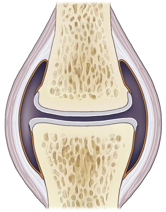
Trabecular bone
Subchondral bone plate
Articular cartilage
FIGURE 1-1 Schematic representation of a diarthrodial joint. (Adapted from: De Grauw J.C. (2010). Molecular monitoring of equine joint homeostasis. Thesis, Utrecht University.)
and temporomandibular joints. These constituting structures of the joint will be discussed separately in the following paragraphs.
The functional characteristics of a construction are largely determined by the interplay of the material properties of the building blocks or components that construction is made of and the way these components are arranged and interconnected, that is, the architecture of the construction. For structured tissues, such as articular cartilage or bone, this is similar. However, as in all living tissues, the situation is more complex as cellular action, driven by a wide variety of cues and effected through various signaling pathways, determines tissue homeostasis and the response to external stimuli. If that response can somehow not cope with the demands made by these stimuli, pathology may ensue.
The major components of the extracellular matrix (ECM) of articular cartilage are collagen, proteoglycans (PGs), and water. Water content varies from 70% to 80%, depending on age. The other components account for approximately 50% (collagen) and 35% (PGs) on a dry weight basis. The remaining 15% consists of about two thirds (10% of total dry weight) of glycoproteins (substances such as proteinases and inhibitors of these, growth factors, specific molecules such as fibronectin, lubricin, cartilage oligomeric protein [COMP], etc.) Minor fractions are minerals (3%), lipids (1%), and miscellaneous components (1%).22 The cellular component of articular cartilage is relatively small and accounts for approximately 1% to 12% volume percentage, depending on the location within the joint and the depth in relation to the surface.22
Whereas cartilage macroscopically is seemingly a homogeneous tissue, there are large differences in structure and composition from the surface down to the transition to the subchondral bone. Classically, four layers or zones are discerned, although the transitions between these layers are
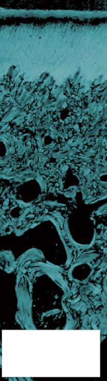

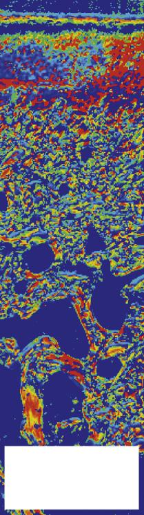

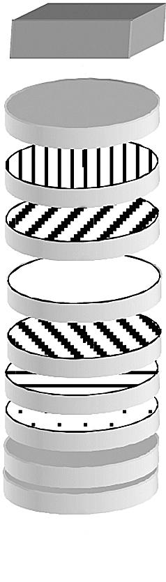
FIGURE 1-6 The use of polarized light microscopy for the determination of orientation angle and parallelism index. Collagen fibrils run parallel to each other in the deep layer of the cartilage and in the superficial layer, indicated by a white color. In the transitional zone they arch and hence lose their parallel arrangement (dark color). The orientation is perpendicular to the calcified layer (90°) in the deep zone (red color), changing to tangential (0°) in the superficial layer (blue color), with angles in between in the transitional zone. The pictures are from samples from the dorsoproximal margin (site I) and the central fovea (site II) of the proximal articular surface of the equine proximal phalanx. (From: Holopainen J.T., Halmesmäki E., Harjula T., et al. (2008). Changes in subchgoondral bone mineral density and collagen matrix organization in growing horses. Bone 43(6), 1108-1114.)
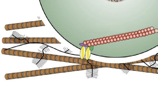
Cross-link formation is one of the so-called posttranslational modifications of collagen and is the last chemical modification occurring during the formation of the primary collagen structure. There are various types of cross-links. Common covalent cross-links are the pyridinoline cross-links that form between lysyl and hydroxylysyl residues in the collagen network in a largely irreversible process (lysylpyridinoline [LP], cross-links and hydroxylysylpyridinoline [HP] cross-links, respectively, the last being more abundant in articular cartilage). They have a major influence on the structural and hence on the biomechanical characteristics of the collagen network.32 A special category of cross-links is formed through the process of nonenzymatic glycation. Collagen molecules have an exceptionally long lifetime once incorporated into the ECM of cartilage, which makes them susceptible to the accumulation of advanced nonenzymatic glycation end products (AGEs) via the Maillard reaction.33 This process results in increased cross-linking, such as pentosidine formation from lysine, sugar, and arginine moieties. Pentosidine is one of the few Maillard cross-links of which the structure has been elucidated and can be used as a sensitive marker for the process of nonenzymatic glycation.34 As the accumulation of AGEs depends on the turnover rate of a protein or tissue, it can be used as a measure for the metabolic rate of that structure.35 Apart from collagen type II, many other collagens can be found in the ECM of articular cartilage. Some, but not all, of these are fibrillar and some have a structural role. The exact role is not known of all collagens. Minor collagens that form,
together with collagen type II as a copolymer, the fibril network of developing cartilage, are collagens IX and XI. Other minor collagen species that can be found in extracts of articular cartilage are types III, VI, XIII, and XIV. Collagen type X is restricted to the hypertrophic zone of cartilage actively undergoing the process of endochondral ossification.
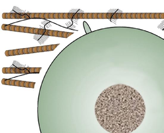
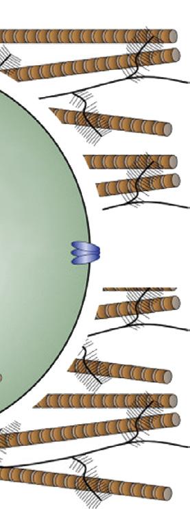
The nonfibrillar collagen IX molecules are attached to the surface of the collagen type II fibrils. They are more abundant in juvenile articular cartilage (approximately 10% of total collagen), which is characterized by collagen type II fibrils that are on average smaller in diameter than in mature cartilage, in which the concentration of collagen IX is about 1% of total collagen.36 Seven cross-linking sites have been described on the collagen IX molecule that interact with collagen II and other collagen IX molecules.37 Collagen XI is a fibrillar collagen that is located in the core of the collagen II fibrils and of which the molecules are primarily cross-linked to each other in a head-to-tail manner (Figure 1-7). They are believed to form a template that constrains the lateral growth of collagen II fibrils.38 Also collagen XI is more prominently present in juvenile tissue (approximately 10%) than in mature cartilage (approximately 3%).36 Both collagens IX and XI are critical to the correct functioning of the collagen network in articular cartilage.
Collagen type III is a fibrillar collagen consisting of a homotrimer of α1(III) chains that functions as a copolymer of collagen I in many tissues and is known to be prominent at sites of healing and repair of many tissues, including tendons.39
Collagenases
Collagen II and IX “telopeptidases”
Collagen XI
Collagen XI “telopeptidase”
In articular cartilage a small, but significant amount of collagen III can be found, mostly in the matrix surrounding the chondrocytes. Recent studies indicate that the molecule may function as a covalently bound (to collagen II) modifier of the fibril network and may, as in other tissues, have a role in the response of the tissue to damage.39 Collagen type VI is a nonfibrillar collagen that can be found in the matrix of most tissues, including articular cartilage, at low concentrations (<1%). It is predominantly localized within the pericellular matrix and is a structural component of the chondron. Chondron is the name for the ensemble of the chondrocyte and its pericellular microenvironment, which is generally considered the primary structural, functional, and metabolic unit of hyaline cartilages.40 Collagen VI can self-assembly into a filamentous network and is more concentrated in fibrocartilage than in hyaline cartilage.41 Collagens type XII and XIV are members of the so-called FACET (fibril-associated collagen with interrupted triple-helix) collagen subfamily. They are not covalently bound to the large collagen II network and their function is largely unknown.41
Collagen X has a special position in articular cartilage. The molecule is specific for cartilage of juvenile individuals in which an active process of endochondral ossification is ongoing. It is synthesized by hypertrophic chondrocytes and may make up to 18% of the total amount of collagen in the hypertrophic region of the epiphyseal growth cartilage in growing animals. It has a role in the regulation of matrix mineralization and the compartmentalization of matrix components.42 Collagen X is expressed by clinically affected cartilage in mature patients with, for example, OA, where it is associated with enhanced mineralization, or other cartilage disorders,43 where it may interfere with the normal interaction between collagens, which may result in ECM dysregulation.44
Proteoglycans
The other main component of cartilage ECM, apart from water, is formed by PG aggregates, which are interspersed between the collagen fibrils and connected to them either directly or via hyaluronan (hyaluronic acid) molecules. The PGs form a group of composite molecules featuring a protein
FIGURE 1-7
Known and speculated sites of cleavage peptide bonds of collagens, necessary for either degradation or lateral growth, within the collagen heteropolymer that forms the backbone of the collagen network. (From: Eyre D.R., Weis M.A., Wu J.J. (2006). Articular cartilage collagen: an irreplaceable framework? Euro Cells Mater, 12, 57-63.)
(hence: proteo) and a sugar (hence: glycol) component. In the case of cartilage the PGs feature some number of chondroitin sulfate side chains, which are part of the so-called lectican family.45 The family includes aggrecan, versican, neurocan, and brevican. The best researched and by far most abundant and emblematic member of this family in articular cartilage is aggrecan. Various forms exist and the aggrecan molecule may lose several parts when the animal ages or when pathology occurs, but the typical aggrecan monomer consists of a large core protein to which several hundreds of glycosaminoglycan side chains are attached (see Figure 1-8). The core protein has an amino (N-) terminal side and a carboxy (C-) terminal of which the former is normally connected via a link protein to another component of the ECM, mostly hyaluronan. Three distinct globular domains have been identified. The N-terminal G1 domain anchors the aggrecan molecule to hyaluronan via the link protein. The G2 domain is unique to aggrecan and is known to be highly conserved, but it has (as yet) no known function. The C-terminal G3 domain links the PG aggregates to the ECM.46 This domain is not without importance, as in man hereditary disorders have been described, among which autosomal dominant familial osteochondritis dissecans, because of missense mutations in the aggrecan C-type lectin repeat in this domain.46
Between the G2 and G3 domains the glycosaminoglycan side chains attach to the aggrecan core protein. Keratan sulfate (KS) is a glycosaminoglycan consisting of repeats of disaccharides of galactose and sulfated or nonsulfated N-acetylglucosamine. Chondroitin sulfate (CS) is composed of disaccharide repeats of glucuronic acid and sulfated or nonsulfated N-acetylgalactosamine. The KS side chains are attached closest to the G2 domain, and the CS chains are situated more towards the terminal G3 domain (see Figure 1-8). Up to 30 KS and 100 CS side chains may be bound to a single core protein.47 The sulfate groups in both side chains are highly hydrophilic because they are negatively charged. These groups account for the high viscosity of the molecules the CS and KS chains form part of, such as the aggrecan monomers. The molecular configuration of the aggrecan monomer is neither uniform nor stable over time. The length of both the core protein and the side chains tends to decrease with advancing age,
Chondroitin sulfate chain (n = 100)
Keratan sulfate chain (n = 30
N-linked oligosaccharides
0-linked oligosaccharide (n = 42)
Primary site of cleavage of stromelysin
FIGURE 1-8 Schematic presentation of an aggrecan molecule, consisting of a core protein that is bound to hyaluronic acid (HA) by a link protein. To the core protein a large number of side chains consisting of sulfated glycosaminoglycans is attached. These are keratin sulfate (KS) and chondroitin sulfate (CS) side chains. Globular domains 1 (hyaluronic acid binding region [HABR]), 2 and 3 are indicated. (From: Ray C.S., Pool A.R., McIlwraith C.W. (1996). Use of synovial fluid and serum markers in articular disease. In: McIlwraith C.W., Trotter G.W. (eds.) Joint disease in the horse (1st ed.) (pp. 203-216). Philadelphia, PA: Saunders.)
which affects both nanostructure of the cartilage ECM and the nanomechanical properties of the tissue48 (Figure 1-9).
The aggrecan monomers bind with their G1 domains via a link protein with hyaluronan or hyaluronic acid (HA). Hyaluronan is also a glycosaminoglycan, but it is not sulfated. The molecule consists of repeats of the disaccharides glucuronic acid and N-acetyl glucosamine. Hyaluronan is a major component of the ECM of articular cartilage (Figure 1-10), but it can also be found in free form in synovial fluid. As the aggrecan monomers are strongly negatively charged through the sulfate groups of their CS and KS side chains and
hence attract water, they will repel each other. Consequently, strongly water-binding PG aggregates will form that may contain over 100 aggrecan monomers and may be about 2.107 Dalton in size.49 Within these large PG aggregates the aggrecan monomers assume a fan-shaped position with respect to the central HA molecule.
Apart from aggrecan, there are several minor PGs (minor both in size and in fraction of the total) present in the ECM of articular cartilage. The family of small leucine-rich proteins/ PGs (SLRPs) has an important role in the creation and maintenance of the structure and function of articular cartilage. They are characterized by a number of repeats of around 25 amino acids with leucine residues at conserved locations.50 Some of these molecules have CS or dermatan sulfate side chains, making them into proper PGs. Others have not and are actually proteins, hence the double meaning of the “p” in the abbreviation SLRP. The most well-known SLRPs in articular cartilage are decorin, biglycan, fibromodulin, lumican, and chondroadherin. An important function of several SLRPs is the regulation of fibrillogenesis, as they bind to collagens during fibril formation via their leucine-rich repeat domain.51,52 Biglycan and decorin are known to bind via their core protein to filaments of collagen type VI.53 In combination with members of another group of noncollagenous proteins called matrilins, which bind to triple helical collagen, links are provided that interconnect the entire ECM network and thus provide structural coherence. Fibromodulin and lumican also affect fibrillogenesis and seem to share many functions.50 Chondroadherin is abundant in cartilage but also present in bone where it decreases the production of cytokines that activate osteoclasts, such as the interleukins IL-1 and IL-6.50
The other SLRPs are also involved in many signaling pathways. An example is the role of biglycan in the regulation of inflammation through its function as an endogenous ligand of the innate immunity toll-like receptors TLR-4 and TLR-2 in macrophages.52
Apart from the major components (collagen and PGs), the ECM of cartilage contains numerous other components, including a number of noncollagenous proteins. The matrilins have already been mentioned. Another well-known constituent of the ECM is COMP, a member of the thrombospondin family, also called trombospondin 5. The COMP molecule has five identical subunits that can all bind to (different) collagen molecules. It is thought that COMP can bring five collagen molecules together, thus facilitating assembly of the matrix. It does not bind to established collagen fibrils but seems to act predominantly at an early stage of fibril formation. In mature ECM it has a more stabilizing role through its binding with collagen type IX and matrilins.50
Mature articular cartilage has long been considered a tissue containing a single cell type only: the chondrocyte. Relatively recently it has become clear, however, that, as in many tissues, there is a population of progenitor cells.54 The presence
can be supposed to exist, as there are distinct and quantitatively substantial topographical differences in loading over a given joint surface. This indeed is the case. Branch et al.63 showed a repeatable pattern of subchondral bone thickness in the distal tarsal bones of horses without hind limb lameness, reflecting similar loading patterns across joints. The effect may be joint-dependent and is most probably not seen until a certain threshold is passed. In a study on the effect of treadmill exercise on subchondral bone density, exercised horses showed a higher subchondral bone density in the metacarpal bones, but not in the carpal bones.64 In a developmental sense it has been shown that different exercise regimens can induce substantial differences in the biochemical composition of the subchondral bone of juvenile horses.65-68
Biomechanically, the subchondral bone plays an important role in the attenuation of the forces generated by locomotion and athletic activity. Although much less deformable than the overlying cartilage, the subchondral plate has been shown to be approximately 10 times more deformable than the cortical shaft of long bones.69 It follows that changes in the rigidity of the subchondral bone will have repercussions for the mechanical loading of the articular cartilage. This consideration, together with the fact that sclerosis of the subchondral bone (and hence a decrease of elasticity of the subchondral plate) is a hallmark of OA, has led to the theory that subchondral bone sclerosis may be an initiating factor rather than a secondary sign of OA.70,71 Whereas current opinion sees articular cartilage as the tissue where OA initiates, it is clear that the degenerative and reactive processes in the cartilage layer and in the subchondral bone are intricately linked and influence each other from the very early stages of the disease onwards.72
An important difference between the cartilage layer and the subchondral bone is the avascular and aneural character of the former versus the very rich vascularization and innervation of the latter. The abundant vascularization of subchondral bone allows for an extensive response of the tissue to both physiologic and pathologic stimuli. In the case of the latter this will translate as the formation of sclerosis, osteophytes, and fibrocartilaginous repair tissue in those cases where subchondral bone becomes exposed in the joint cavity because of deterioration of the cartilage layer. The nerve supply of the subchondral bone is one of the main vehicles of pain perception in the case of joint disease, together with the nerves that connect to nociceptors in the joint capsule and intraarticular and extraarticular ligaments (see the section on innervation of articular tissues).
The capsules of most joints grossly consist of two distinct layers. The outer layer of the joint capsule is made up of relatively stiff fibrous tissue. It is often tightly connected to extraarticular structures such as collateral ligaments and mainly serves mechanical stability. Apart from this, it hosts a great number of proprioceptive nerve endings that provide information on actual joint position to the brain. The inner layer of the capsule, which is the actual lining of the joint cavity, often called
Joint space
Type A synoviocyte
Hyaluronan molecules
Type B synoviocyte
Blood vessel
Erythrocyte
FIGURE 1-12 Semischematic drawing of the synovial membrane. The subintimal layer is highly vascularized and the lack of a basement membrane facilitates passage of small molecules to the synovial cavity. The synoviocytes have both secretory and phagocytic properties. The main molecule that is secreted is hyaluronan. (From: Frisbie D.D. (2012). Synovial joint biology and pathobiology. In: Auer J.A., Stick J.A. (Ed). Equine surgery (4th ed.) (pp. 1096-1114). St. Louis, MO: Elsevier-Saunders.)
the synovium or synovial membrane, consists itself of two layers: the intimal and the subintimal layers (Figure 1-12). The subintimal layer is made up of loose connective tissue and is very well vascularized (the source of hemorrhage during arthroscopic surgery) and innervated (see the paragraph on the innervation of articular tissues). The intimal layer is the layer directly lining the joint cavity. This is a very thin layer, one to four cells thick without a basement membrane; the cells are located within a loose and porous bed of collagen fibrils and other matrix proteins. This lack of basement membrane and the immediate presence of a large number of blood vessels facilitate the passage of plasma components from the blood to the synovial cavity. In fact, synovial fluid is often described as an ultrafiltrate of blood plasma, in the sense that all but the larger molecules (the estimated size limit is 10 kDa22) can move freely from the blood to the synovial cavity (and vice versa). This does not mean that synovial fluid is formed by a largely passive process. There is a large excretory activity of the cells that constitute the intima, the so-called synoviocytes. Classically, they have been divided into two major categories. Type A are macrophage-like synoviocytes that are mostly involved in phagocytic actions. Type B are fibroblastlike synoviocytes that are mainly responsible for production and excretion into the synovial fluid of proteins and other molecules such as hyaluronan, the nonsulfated glycosaminoglycan macromolecule that is both a major component of the cartilage ECM and a principal component of synovial fluid, determining to a large extent the viscosity of this fluid.73 An intermediate type C exists that is currently thought to be a
through the action of certain cytokines. Lastly, active MMPs may form clusters with so-called TIMPs (tissue inhibitors of MMPs), leading to their inactivation.92 As this bond is reversible, these MMPs may rapidly regain an active status once the bond is dissolved under the influence of certain stimuli. The last two mechanisms explain why MMP activity can increase very rapidly after an insult to the joint. In pathologic conditions there is an increase in the activity of many MMPs, which makes these enzymes potential targets for molecular therapies. However, it should be realized that they play an important role in the maintenance of joint homeostasis in healthy joints. In human articular cartilage there is a slight excess of TIMPs in relation to MMPs in healthy joints, and this equilibrium is disturbed in case of pathology.93 Therefore, therapeutic targeting of MMPs certainly has potential, but, given the widespread effects that MMPs exert on natural processes in many parts of the body, including the joint, it seems that total inhibition is not the way to go and that targeted MMP modulation is more promising.92
The role of the chondrocyte as the sole source of the major ECM components in articular cartilage is undisputed, but not all the exact mechanisms through which the maintenance of tissue homeostasis is realized have been elucidated. The balance between anabolic and catabolic growth factors and cytokines is important in this respect, but also mechanical loading plays a role and most probably both mechanisms are mutually influencing each other. In studies on the regulation of chondrogenesis it has been shown that the effects of dynamic compressive loading alone (i.e., in the absence of exogenous growth factors) were relatively minimal and much less than the effects of application of growth factors alone.94 This may implicate that the right growth factor/cytokine environment is primary to the effects of mechanical loading (which are substantial once this condition has been met). However, this refers to research on cartilage formation, which is different from maintenance of homeostasis in existing cartilage. It may well be that in a physiologic situation where an appropriate cytokine/growth factor balance is present, it is the mechanical loading that produces the primary cues for the maintenance of tissue homeostasis.
Growth factors with a known anabolic effect on articular cartilage include transforming growth factor-β (TGF-β) and insulin-like growth factor-1 (IGF-1). TGF-β stimulates PG synthesis by chondrocytes and expression of collagen type II; possibly it downregulates matrix degrading enzymes.95,96 It may therefore counteract the effects of the major catabolic cytokine IL-1.97 However, not all effects of TGF-β are seen as positive, as it may also stimulate osteophyte formation when used over a longer period.98 IGF-1 is important for the maintenance of ECM homeostasis in articular cartilage through the stimulation of matrix production and inhibition of degradation.96 Chronic deficiency of IGF-1 and growth hormone (which induces its production) leads to OA-like lesions in rats.99
Mechanical loading
Hydrostatic pressure
Cell deformation
Fluid flow, shear stress
Primary cilium
Nuclear deformation
Cytoskeleton
Ca2
Osmotic pressure
Mechano- or osmosensitive ion channels
FIGURE 1-13 Schematic drawing depicting the mechanisms of mechanically induced chondrogenesis. Joint loading results in both direct cellular and nuclear deformation and in change of biophysical factors, such as osmotic and hydrostatic pressure and fluid flow. (From: O’Conor C.J., Case N., Guilak F. (2013). Mechanical regulation of chondrogenesis. Stem Cell Res Ther, 4(4), 61.)
Bone has long been known to respond to mechanical loading,62 but all musculoskeletal tissues are highly sensitive to their mechanical environment and all respond to some degree or another. Articular cartilage is no exception. This is most evident in the juvenile phase when growth and development prevail100 (see also Chapter 8), but seems less obvious in mature individuals where the ECM of cartilage is characterized by very long turnover times and extensive remodeling appears no longer possible. Still, there is compelling evidence that the chondrocyte is very sensitive to mechanical stimuli and these may well be the principal cues for tissue homeostasis and maintenance under physiologic conditions, as suggested above. The regular loading of articular cartilage provokes important changes in the biomechanical environment of the chondrocyte, both directly and indirectly. Directly, loading of the cartilage will lead to deformation of both nucleus and cell, eliciting a cellular response. Indirectly, there are changes in hydrostatic pressure, osmolality and, related to these, flow of interstitial fluid. This is schematically depicted in Figure 1-13 These physical events may have direct and indirect effects. Changes in flow of interstitial fluid will have effects on nutrient supply and waste removal, but may also be a stimulus for the activation of metabolically active substances such as TGFβ. 101 The chondrocyte possesses a so-called primary cilium (Figure 1-13). This organelle was described long ago,102 but it has more recently become clear that it may have an important role in chondrocyte mechanotransduction.103 The molecular
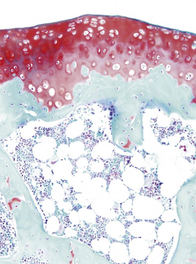
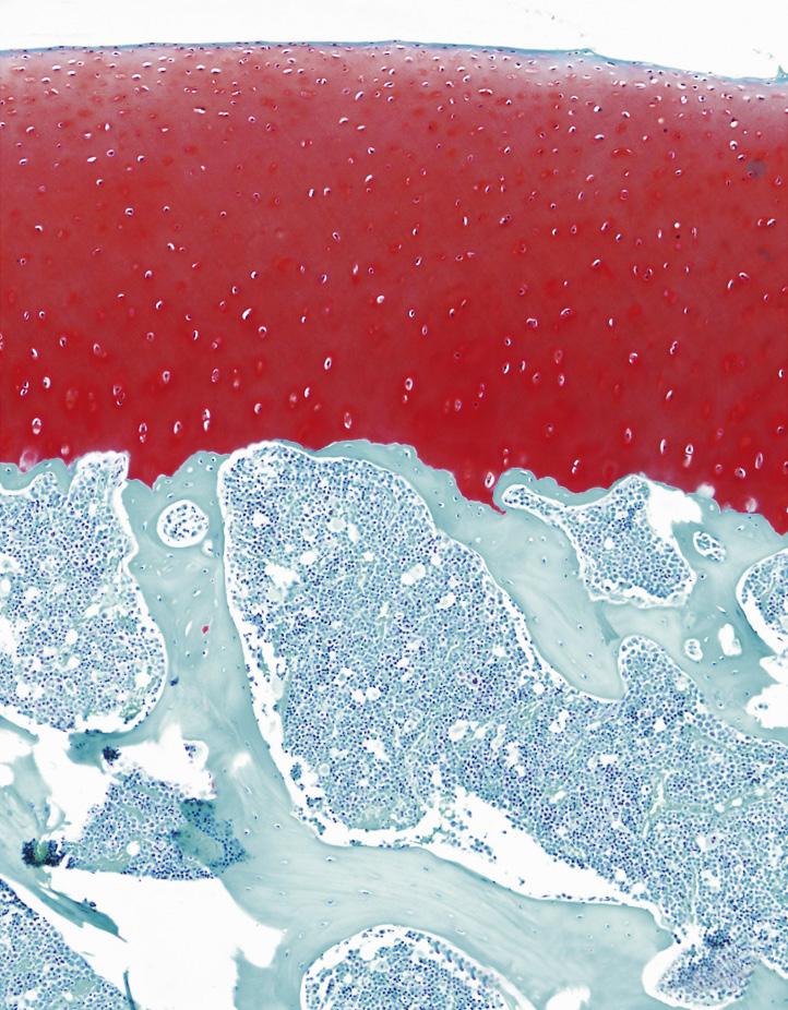
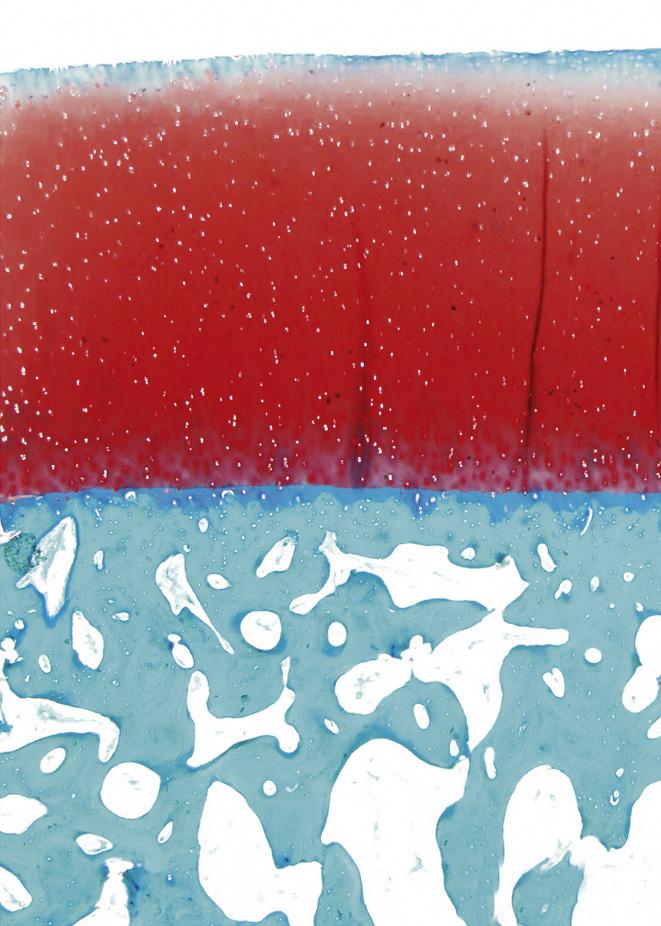
FIGURE 1-16 Safranin-O staining (stains glycosaminoglycans red) of osteochondral tissue of the (A) rat, (B) Barbary macaque, and (C) white rhinoceros. Scale bars indicate (A) 200 μm, (B) 400 μm, and (C) 1000 μm. Cartilage of heavier species is thicker, but the increase is not isometrical. Cartilage of heavier species is similar in cell density, but lighter species have more cell-dense cartilage.
(From: Malda J., de Grauw J.C., Benders K.E.M., et al. (2013). Of mice, men and elephants: the relation between articular cartilage thickness and body mass. PLoS ONE 8(2), e57683.)
margin and the central part of the fovea of the proximal articular surface of the proximal phalanx (Figure 1-17). These are areas that are subjected to entirely different loading patterns. The central fovea is loaded at every instance the horse is on its feet, independent of athletic activity, whereas the dorsal joint margin is nonweight-bearing under most circumstances, but is quite heavily loaded when there is severe hyperextension of the metacarpophalangeal joint, as occurs during strenuous exercises such as galloping and jumping. The relationship between the loading pattern and the biomechanical composition became clear in another study of the same groups where, in an ex vivo setting, limb loading as occurring during athletic activity was mimicked and intraarticular pressure measured using pressure-sensitive films.114 Results showed that high intermittent loading was related to high collagen content with a high level of cross-linking; less intense but more consistent loading was associated with higher PG content (Figure 1-18). Later biomechanical studies confirmed that this topographic heterogeneity in a biochemical sense indeed represented differences in biomechanical properties of the tissue.115
The topographic variation in biochemical and biomechanical properties is not genetically determined, but develops for the larger part in the early phase of life, that is, during the first year after birth in the horse, with most changes in the first 5 months of life. Biomechanical loading is an important cue in this development100; this issue is discussed in more detail in Chapter 8.
Pain transmission through afferent fibers is more complex than it may seem with peripheral sensory neurons functioning
as afferent conductors, but at the same time also exerting important efferent functions mediated by neuropeptides.116 Neuropeptides are small molecules that are synthesized in the dorsal root and autonomic ganglion neurons and then transported via the axon to peripheral nerve terminals. They can induce the release of other mediators, such as cytokines, prostaglandins, and nitric oxide. They are relatively potent, but usually have a limited range of action, both in space and time, as they are chemically labile. In healthy or regenerating tissue, they may have growth factor-like functions,117 meaning that they also play a role in maintenance of joint homeostasis. Well-known neuropeptides include substance P (SP), calcitonin gene-related peptide (CGRP), vasoactive intestinal polypeptide (VIP), neuropeptide Y (NPY), and somatostatin (SOM).116 Neuropeptides play an important role in the complex mechanisms that modulate and mitigate nociceptive input. An example is VIP, which is thought to be important for the augmentation of responsiveness to mechanical stimuli in case of joint inflammation.118
In joint disease, numerous pathologic processes can contribute to the joint pain experienced by affected subjects; the precise tissue origin of pain is rarely identified in the individual patient (Figure 1-19). The sensory innervation of the subchondral bone, marginal periosteum, synovial membrane, and joint capsule will contribute to a variable extent to pain perception and associated loss of function. Within the joint tissues several pathologic processes may be at the origin of pain, such as subchondral bone exposure, remodeling, marrow edema (causing a rise in intraosseous pressure), and marginal periosteal activation related to osteophyte formation.78 Synovitis is an important factor generating pain through joint
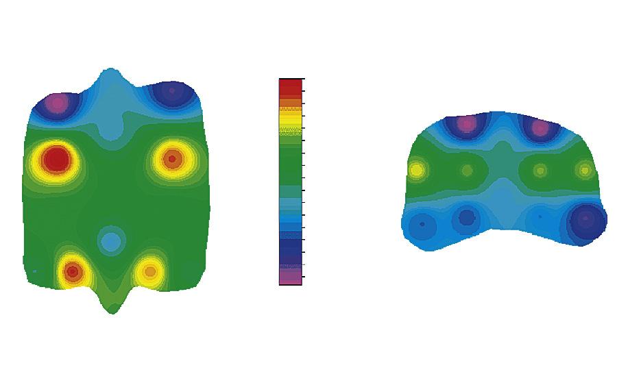
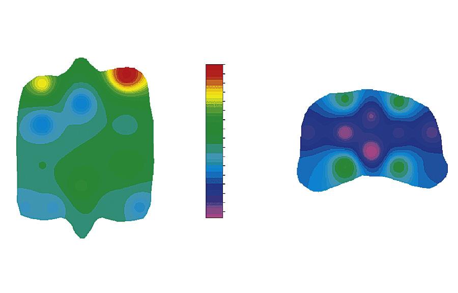
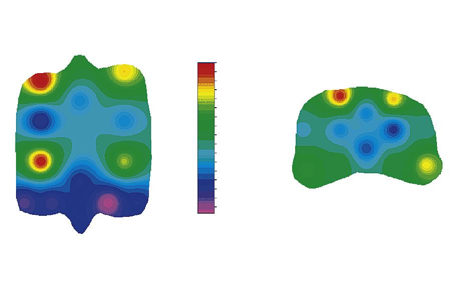
FIGURE 1-17 Distribution of glycosaminoglycans (GAG), collagen, and HP-cross-links over the proximal articular surface of the proximal phalanx and the distal articular surface of the third metacarpal bone. Dors., Dorsal; GAG, glycosaminoglycan; HP, hydroxylysyl pyridinoline; lat, lateral; MC, distal third metacarpal bone; med., media; palm., palmar; P1, proximal first phalanx. (From: Brama P.A.J., TeKoppele J.M., Bank R.A., et al. (2000). Topographical mapping of biochemical properties of articular cartilage in the equine fetlock joint. Equine Vet J, 32(1), 19-26.)
effusion, swelling, and/or fibrosis that will activate mechanoreceptors in the joint capsule, and through direct chemical stimulation of nociceptors.
Pathways and Mediators of Pain in the Joint Nociception will in most cases be stimulated or enhanced by inflammation. In this context, the somewhat complex and mutually influencing interrelationship between mechanoreceptors and nociceptors needs to be pointed out: mechanoreceptors can become sensitized by chemical stimuli released
during inflammatory processes, but mechanical stimulation may also, through tissue damage, lead to an inflammatory response with release of pro-nociceptive mediators. In inflammation, pain originates from the chemical stimulation of nerve afferents by a variety of endogenous mediators. Interleukin-1β and TNF-α are the two major cytokine players in the pathogenesis of OA.119 Both cytokines increase the synthesis of inflammatory mediators that are involved in pain perception, such as prostaglandin E2 (PGE2), by stimulating cyclo-oxygenase (COX)-2, microsomal PGE synthase-1 (MPES-1), and soluble phospholipase A2 (SPLA2). They further upregulate the production of nitric oxide via inducible nitric oxide synthetase (iNOS).120 Relatively little work has been done on the exact identification of pain-related mediators in joint disease. Prostaglandins have always been seen as the major pain mediators in arthritis,121 but their role is not as clear-cut as it may seem. In a horse study comparing PGE2 levels in synovial fluid between lame horses that did or did not respond to an intraarticular block, there was no significant difference. However, when comparing the levels of these lame horses (blocking to a low 4-point or 6-point perineural nerve block) to those of sound horses, lame horses were found to have significantly higher and more variable PGE2 levels than the sound control horses.122 The other branch of the arachidonic acid cascade, the pathway that leads via 5-lipoxygenase (LOX) to the formation of leukotrienes, may play a role in nociception in joints, too. Leukotriene B4 (LTB4) has been shown to be implicated in hyperalgesia in joints of mice.123 In the horse, LTB4 levels were increased in animals suffering from osteochondrosis (OC) and may be involved in the extensive joint distension that is often seen in OC.124 However, lameness in these patients is uncommon and thus far no link between LTB4 and joint pain in the horse has been established. Various neuropeptides have been identified as direct pain mediators in joint disease in humans. These include NPY, serotonin, and CGRP.125,126 In the horse, substance P was the only mediator that could be directly linked to outcome of intraarticular analgesia,122 but it could not be related to radiographic OA status of a joint in another study.127 As radiographic OA status is more a measure of cumulative damage to the joint than of actual pain, this was perhaps not to be expected. More recently, other families of cytokines, such as kinins and chemokines, have been implicated in the generation and maintenance of chronic pain in joints (for an overview see Miller et al.128). None of these possible mediators have as yet been investigated in the horse except for bradykinin, which showed a strong correlation with lameness and joint hyperalgesia in chemically induced synovitis.129 The concentration, however, was not related to the outcome of intraarticular analgesia.122
Whereas the joint should be seen as an organ that is composed of the variety of structures and tissues outlined above, this organ itself is a constituting element of the entire body. Joints are integral parts of the musculoskeletal system and their functioning is partly determined by the other