PREFACE
Agriscience programs vary nationwide and most have undergone extensive curricular changes within the past decade. Many include advanced placement-type coursework, such as veterinary science. While teaching agricultural education at Greenwood High School in Millerstown, Pennsylvania, Dr. Baker searched for materials to be used in a new veterinary science course. After a futile hunt, and hearing similar concerns from other instructors, Dr. Baker teamed with Dr. Lawhead, a practicing veterinarian who served the local area where she taught, in an effort to author a veterinary science text that was both student and teacher friendly.
The authors believe that two of the most useful features in this book are the “A Day in the Life” of a veterinarian, coupled with the “Clinical Practice” chapter features. These two elements tie the real-life work of a veterinarian, which can have less than desired outcomes, with the technical and, sometimes, dry and dif cult text material. Therefore, the next time a student says, “I want to be a veterinarian,” a venture into Introduction to Veterinary Science will provide the learner with a realistic preview of both veterinary work and the academic rigor needed to achieve success in the profession.
Simply put, the goals of this text are to afford learners a base knowledge of veterinary science by moving through topics ranging from the cell to surgery, and to provide a view of the practice of veterinary medicine through the eyes of an experienced practitioner. Chapters 1 and 2 begin the text with a comprehensive investigation of cells and tissues. Following chapters examine the musculoskeletal, circulatory, respiratory, renal, digestive, reproductive, nervous, endocrine, and immune systems. The basic physiology learned in the beginning of the text is then applied in concluding chapters covering nutrition, species differentiation in nutrition, principles and prevention of infectious disease, disease classi cation, zoonotic diseases, disease diagnosis, and surgery.
NEW TO THIS EDITION
• Additional hands-on activities that use easyto- nd materials have been added to the chapters.
These new activities will help instructors reinforce student learning using a variety of applications.
• The new safety chapter provides guidelines to help teachers ensure student safety in the classroom and eld laboratories, while another new chapter further explores veterinary careers.
• Technical material has been further explained by the author, Dr. James Lawhead. These expanded and updated explanations will help students grasp more advanced material.
• Additional photos and gures bring the veterinary practice into the classroom, helping to keep students engaged.
• The new edition discusses the most current technology used in veterinary practice, providing a look into recent advances in the eld of veterinary medicine.
EXTENSION TEACHING/LEARNING MATERIALS
Instructor’s Companion Website
The Instructor Resources are now available on the companion website. Updated for the third edition, this robust suite of teaching resources includes the following components to help minimize instructor prep time and engage students:
• Instructor’s Guide to Text—The Instructor’s Guide provides answers to the end-of-chapter questions and additional material to assist the instructor in the preparation of lesson plans.
• PowerPoint—Chapter outlines with images for each textbook chapter.
• Computerized Test Bank in Cognero—Hundreds of modi able questions for exams, quizzes, inclass work, or homework assignments, in an online platform.
• Image Gallery—Hundreds of images from the textbook that can be used to easily customize the PowerPoint outlines.
Each chapter in the textbook begins with clear educational objectives to be learned by the student in the reading, a list of important key terms, and an introduction overview of the chapter content.
Basic Cell Biology
Objectives
Upon completion of this chapter, you should be able to:
■ Explain the molecular makeup of cells.
■ Identify the basic structures of the cell and their corresponding functions.
■ Review the basic function of the cell.
■ Describe the process of protein synthesis.
Key Terms
■ Discuss mitosis and its clinical signi cance in diseases such as cancer.
■ Detail meiosis in mammalian reproduction.
■ Connect cellular parts and function to clinical veterinary practice.
Introduction
The cell is the basic structure of animal life. However, the cell contains other structures and molecules. Cells conduct many functions and are also able to reproduce. Animals not only have millions of cells that comprise
A Day in the Life ADR—Ain’t Doin’ Right…
I remember the day in veterinary school when our stethoscopes arrived. The air lled with excitement as we listened to our own heartbeats. This instrument became a necessary tool in everyday life as I began to examine animals. I must admit I felt cool walking around the hospital in a white lab coat with a stethoscope draped around my neck! It seems like yesterday, even though more than a few years have passed.
Several months ago I examined a cow that was ADR—ain’t doin’ right. As I walked into the pen, I could see she obviously wasn’t feeling well at all. She appeared quite droopy, had lost a lot of weight, and had developed a swelling under her jaw. During the physical, I listened to her heart. It sounded like the noise from a washing machine in midcycle. The heart made a sloshing sound with every beat. Using the stethoscope, I diagnosed hardware disease . The cow had eaten a piece of metal that migrated from the stomach and lodged close to the heart. The location and structure of the heart provided me with the information necessary to interpret the symptoms of this disease. Hardware disease is often found during my appointed rounds. The next diagnosis is not.
This week, Dr. Deppen and I were both doing evening small animal appointments at the office. It was snowing heavily and we were hoping to nish at a reasonable hour. Dr. Deppen was seeing Lucky, a 12-yearold Schnauzer mix that had a history of having what the owners thought was a seizure. She detected that the dog’s heart rate was too slow and the rhythm was very irregular. I had a chance to listen to the dog’s heart as well and agreed that we should do more tests to detect the underlying problem.
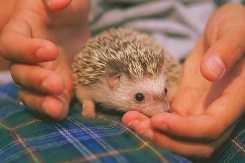
The author James Herriot portrayed veterinary work in his best-selling collection of stories, All Creatures Great and Small. Times have changed considerably since Herriot practiced. Much more information and sophisticated medicines and techniques are now readily available. Still, I cannot possibly be an expert on all animals. Last year our of ce received a call from a local school. The sixth grade class mascot, Sonic the hedgehog, had a sore foot. In this case, my experience with hedgehogs was limited to reading just one obscure
journal article. I had never even met one in real life. Therefore, I advised the teacher of my lack of experience but agreed to examine Sonic. Sonic arrived at the of ce in a cage (Figure 4–1). He looked just like a miniature porcupine. Because hedgehogs are nocturnal animals, Sonic was apparently taking his afternoon nap when he arrived at the of ce.
I disturbed him as I tried to examine his leg. Sonic jumped and snorted in an attempt to scare me. To be honest, it worked! His prickly quills were quite sharp.
My assistant and I then put on thick leather gloves and proceeded with the examination. Sonic countered with another protective measure. He rolled himself into a tight ball, so tight his legs were completely hidden. I referred to the journal article for help.
Following the recommendations, I anesthetized Sonic with an inhalant anesthetic. We placed him in the large clear mask. The anesthetic was slowly delivered with every breath. Finally Sonic relaxed enough so I was able to have a more thorough look. Once Sonic’s leg was exposed, the problem was quite obvious. The rags that Sonic used as a nest had tattered edges with loose strings. One of these strings had wrapped tightly around his foot and stopped the circulation. The foot had turned dark and was oozing. All mammals rely on circulation to maintain their bodies. What happened to Sonic’s foot when the blood supply was stopped?
the body but also many different cell types. The combination of these cell types makes an animal function. This chapter will discuss the structure of cells, and how they work.
Each chapter features “A Day in the Life” of a veterinarian vignette that relays James Herriot–type stories with relevance to clinical practice and the real-life work of a veterinarian.
55 Chapter 4 The Circulatory System
FIGURE 4–1 A hedgehog.
attach to these vertebrae, forming a sling that supports internal organs.
The sacrum, a group of three sacral vertebrae, fuses to support the pelvis (Figure 3–14). In addition, the sacrum articulates with the last lumbar vertebra and the rst caudal vertebra. The sacrum then joins with the pelvis, allowing the hind limbs to support the weight of the body. This connection can be damaged. The pelvis may split away from the sacrum when dogs and cats are hit by cars (HBC). During this type of accident, fracture of the pelvis itself is also common. Very painful lameness often results from a split pelvis or pelvic fracture. Many of these fractures heal if the animal’s activities are restricted. In severe cases, surgeries may be required.
The nal group of vertebrae is called caudal. These small vertebrae comprise the tail. As mentioned, the numbers of vertebrae vary among species and within
41 Chapter 3 The Musculoskeletal
System
a species. The typical dog has 20 caudal vertebrae, but this can range from 6 to 23. The appendicular skeleton includes the bones of the forelimbs and hind limbs. A study of this part of the skeleton provides a clear examination of comparative anatomy. Although the same anatomic terms are used for all mammals, great differences exist in the numbers and sizes of bones in the mammalian appendicular skeleton. For instance, a dog has four or ve toes, whereas a horse has only one. The forelimb, or thoracic limb, does not have a bony connection to the axial skeleton. The scapula, or shoulder blade, lies at against the rib cage (Figure 3–15). The scapula connects to the axial skeleton with a group of muscles. This attachment allows the scapula to move over the rib cage. This rotation ranges as high as 25 degrees in animals such as cats while running. This exibility is also useful in cats as they land after a jump. As the cat falls, it extends its front legs fully at both the scapula and the elbow. As the front feet hit the ground, the elbow exes and the scapula rotates. The cat makes this very coordinated act look quite graceful. Clinically, this is of signi cance when cats fall from extreme heights. In large cities, this happens often as cats tumble from balconies or windows of tall buildings. In high-rise syndrome, the falling cat rarely breaks a leg; however, it will often break its lower jaw. The high speed of the falling cat forces the jaw to contact the ground.
Each chapter contains combinations of charts, illustrations, photographs, radiographs, and the like that help to illustrate and enhance the concepts presented.
5 Chapter 1 Basic Cell Biology
allowing veterinarians to diagnose what speci c organism is causing the sickness.
Nucleic acids provide plans for the differing construction of proteins. Nucleic acids are fabricated with a series of nucleotides. The nucleotides are made up of a ve-carbon sugar, a phosphate group, and a nitrogen-containing base (Figure 1–5). Ribonucleic acid (RNA) claims ribose as its sugar, whereas deoxyribonucleic acid (DNA) has deoxyribose as its sugar. There are four different bases for RNA and DNA (Table 1–1).
Notice that the bases are the same except for thymine and uracil. The order of base combination determines what amino acids are used to make proteins. This information is stored in the cell’s genetic material. Both DNA and RNA have a backbone of sugar alternating with phosphate. The nitrogenous bases are attached to this backbone. In DNA, a double-stranded molecule is formed as the bases are loosely bonded together. The molecule has a twisted structure, which is described as a double helix (Figure 1–6). The bases join, speci cally, thymine to adenine and cytosine to guanine. Later in the chapter, a process of transcription will be described, in which the sequence of DNA nitrogenous bases is converted to a molecule of RNA. In this situation, adenine in the DNA molecule bonds to a uracil base of RNA. The sequence of nitrogenous bases is used to de ne the amino acids used in protein synthesis. A group of three nitrogenous bases is the code for a speci c amino acid. The order of the nitrogenous bases makes up the genetic code of the animal. Each gene provides the code for one peptide chain.
FIGURE 1–5 Chemical structure of a nucleotide.
Table 1–1 RNA and DNA Bases
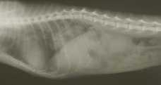
Thoracic Vertebrae
Rib Sternum Lumbar Vertebrae
FIGURE 3–13 Radiograph of a cat, showing the thoracic and lumbar spine. Ribs and sternum are also visible.
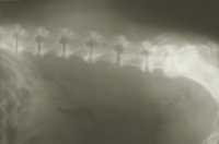
Pelvis
Lumbar Vertebrae
FIGURE 3–14 Radiograph of the lumbar spine of a dog. A portion of the pelvis is also visible. This dog is showing an age-related change called spondylosis. In spondylosis, bone spurs are formed that can eventually bridge between vertebrae.
FIGURE 3–15 The scapula. Spine
Basic Cell Biology
Objectives
Upon completion of this chapter, you should be able to:
■ Explain the molecular makeup of cells.
■ Identify the basic structures of the cell and their corresponding functions.
■ Review the basic function of the cell.
■ Describe the process of protein synthesis.
Key Terms
■ Discuss mitosis and its clinical signi cance in diseases such as cancer.
■ Detail meiosis in mammalian reproduction.
■ Connect cellular parts and function to clinical veterinary practice.
anesthetize antibiotics cancer lipid hydrophilic hydrophobic glucose diabetes glycogen enzymes antibodies exocytosis metabolism anabolism catabolism homeostasis diffusion osmosis active transport endocytosis benign malignant pathologists
Introduction
The cell is the basic structure of animal life. However, the cell contains other structures and molecules. Cells conduct many functions and are also able to reproduce. Animals not only have millions of cells that comprise the body but also many different cell types. The combination of these cell types makes an animal function. This chapter will discuss the structure of cells, and how they work.
allowing veterinarians to diagnose what speci c organism is causing the sickness.
Nucleic acids provide plans for the differing construction of proteins. Nucleic acids are fabricated with a series of nucleotides. The nucleotides are made up of a ve-carbon sugar, a phosphate group, and a nitrogen-containing base (Figure 1–5). Ribonucleic acid (RNA) claims ribose as its sugar, whereas deoxyribonucleic acid (DNA) has deoxyribose as its sugar. There are four different bases for RNA and DNA (Table 1–1).
Notice that the bases are the same except for thymine and uracil. The order of base combination determines what amino acids are used to make proteins. This information is stored in the cell’s genetic material.
Both DNA and RNA have a backbone of sugar alternating with phosphate. The nitrogenous bases are attached to this backbone. In DNA, a double-stranded molecule is formed as the bases are loosely bonded together. The molecule has a twisted structure, which is described as a double helix (Figure 1–6). The bases join, speci cally, thymine to adenine and cytosine to guanine. Later in the chapter, a process of transcription will be described, in which the sequence of DNA nitrogenous bases is converted to a molecule of RNA. In this situation, adenine in the DNA molecule bonds to a uracil base of RNA. The sequence of nitrogenous bases is used to de ne the amino acids used in protein synthesis. A group of three nitrogenous bases is the code for a speci c amino acid. The order of the nitrogenous bases makes up the genetic code of the animal. Each gene provides the code for one peptide chain.
or Pyrimidine)
FIGURE 1–5 Chemical structure of a nucleotide.
Table 1–1 RNA and DNA Bases
the inside of the cell contained. The cell membrane is so ne that it cannot be seen with a normal light microscope. The cell membrane is about half protein and half lipid (phospholipid type). One end of phospholipids is attracted to water, whereas the other end is repelled by water. The cell membrane, which is surrounded by water on both sides, has two layers of lipid in its wall (Figure 1–7). The ends of the lipid that are attracted to water face outward. Protein is also included in the membrane, both between the lipid molecules and on the surface. The position of the protein molecules is not rmly established; rather, the molecules are mobile within the membrane. Cholesterol, another molecule in the cell membrane, provides stabilization of the membrane.
Cell membranes are semipermeable, meaning they allow certain substances but not others to pass. Some molecules, such as water, are able to pass through easily. The specialized proteins in the cell membrane in uence which molecules are able to pass readily. In addition, the intrinsic membrane proteins can act as receptors. These receptors can process a signal from the extracellular uid to in uence the cell’s interior (e.g., a hormone can trigger a reaction within the cell). Other molecules, such as proteins, starches, and some ions, are unable to pass.
CELL STRUCTURE
Objective
■ Identify the Basic Structures of the Cell and Their Corresponding Functions
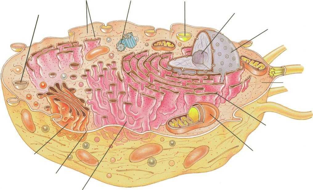
Many cell types exist. These cells not only look different but function differently as well. Nevertheless, many features are common among cells. Specialized structures within the cells are called organelles. These organelles are present in most but not all cells. Red blood cells, for example, lack a nucleus.
The cell membrane (or plasma membrane) is common to all cells. It serves as the boundary that keeps
Many of the organelles within the cell are also surrounded by a membrane. The basic structure remains the same for all the membranes. The speci cs of the makeup differ, depending on function.
Cell contents are divided into the nucleus and the cytoplasm. Cytoplasm generally describes the organelles and uid in the cell. A nucleus comes as a standard part of most cells (with a few exceptions such as the red blood cell; Figure 1–8). The nucleus contains the genetic material (i.e., DNA) of the cell, which controls cellular activities by coding for protein synthesis. The DNA in the nucleus is called chromatin. As the cell divides, the chromatin clumps into chromosomes. Identical DNA is passed to all daughter cells. All the cells in the body have the same chromatin. However, cells take on different roles by using certain areas of the chromatin more than others.
FIGURE 1–6 DNA structure: The structure is described as a double helix. Phosphate and sugar groups make up the two strands. The strands are joined by hydrogen bonds between two nitrogenous bases.
FIGURE 1–7 Illustration of cell membrane. The cell membrane has a double layer of phospholipid. In addition, protein molecules are present on and within the phospholipid layers.
A membrane made of two lipid bilayers surrounds the nucleus. This membrane is often joined to other organelles, such as the endoplasmic reticulum and ribosomes. Such a close association helps the nucleus control cell function.
In cells not dividing, a nucleolus is often seen in the nucleus. The nucleolus produces RNA that forms the ribosomes, which in turn produce protein. Cells with large nucleoli actively produce protein.
Ribosomes are small granular-like structures that can be found in the cytoplasm. They contain roughly 60% RNA and 40% other protein. Ribosomes manufacture the protein used in the cell. Growing cells require large amounts of protein and, therefore, have a greater number of ribosomes. The speci c proteins produced by a cell are governed by the nucleus.
The endoplasmic reticulum (ER) is a collection of folded membrane. This membrane attaches to the membrane of the nucleus. The ribosomes often line this membrane, giving it a bumpy appearance and therefore its name, rough endoplasmic reticulum (RER). Protein produced by the ribosomes is then deposited into the RER. These proteins can be further changed in the RER. This protein may be used by the cell or moved to the surface of the cell for secretion. The protein is moved through the membrane in a process called exocytosis, which will be discussed later in the chapter.
Smooth endoplasmic reticulum (SER) has no ribosomes attached. This form is not as common. Some liver cells contain a large amount of SER. The SER in these cells produces glycogen and lipids, and removes toxins.
The Golgi apparatus is formed with large amounts of folded membrane that looks similar to SER. The Golgi apparatus produces polysaccharides and special protein sacs called lysosomes. Protein produced in the RER is moved to the Golgi apparatus. The Golgi apparatus then changes the protein and collects it in the lysosomes. These sacs are pinched from the Golgi apparatus and then moved to the surface of the cell and released.
The proteins contained in the lysosomes are enzymes (remember, enzymes are molecules that help speed chemical reactions in the body). Lysosomes contain enzymes that help to break down other molecules. Varying enzymes match differing molecules. The membrane surrounding lysosome prevents the enzymes from attacking other parts of the cell.
Lysosomes are used to digest food taken in by the cell and to destroy cell structures no longer needed. In Millie, the dog with the abscess, her white blood cells were using lysosomes to destroy bacteria. Cells that die in the body are eliminated when enzymes within lysosomes are released into the cytoplasm. This process of autolysis makes room for replacement cells.
Vacuole
Ribosomes
Cytoplasm
Rough Endoplasmic Reticulum
Mitochondrion
Smooth Endoplasmic Reticulum
Plasma Membrane
Golgi Apparatus (Complex)
© 2017 Cengage Learning ®
FIGURE 1–8 Illustration of cell structure.
Mitochondria are small rod-shaped organelles found in varying numbers in cells. The more active the cell, the more mitochondria are present. Mitochondria have a double membrane, similar to the cell membrane. The outer membrane is smooth and forms the shape of the mitochondria. The inner membrane is highly folded. These shel ike infolded ridges are called cristae.
The role of mitochondria is well de ned. The mitochondria convert food substances into energy that can be used by the cell. Mitochondria contain the enzymes necessary for this process. Because of this role, mitochondria are called the powerhouses of the cell. The mitochondria are found within cells at their areas of highest activity.
CELL FUNCTION
Objective
■ Review the Basic Function of the Cell
The cell constantly reacts to its environment. Metabolism describes all the reactions going on in cells. Metabolism can be categorized into two main types. Anabolism describes reactions in which smaller molecules are combined into larger ones. The joining of amino acids to form proteins serves as an example. Catabolism, the opposite, occurs when large molecules are broken down into smaller ones. The breaking down of glycogen to release energy is an example of catabolism.
A liquid called extracellular uid (ECF) surrounds living cells. The ECF supplies cells with all the products necessary for their functions. ECF is derived from blood. The outermost skin cells are not covered in liquid; however, they are no longer living.
Other cells exposed to the surface, such as those of the eye, need moisture. In the eye, tears produced by glands act as the source of moisture and nutrients. The eyelids help to sweep the tears across the surface of the eye. Certain breeds of dogs, such as the pug, have eyes that bulge from the eye socket. The bulging can be so severe that the eyelids cannot keep the surface of the eye moist with tears. This results in a disease condition on the surface of the eye. Arti cial tears are often used to keep the surface moist.
Table 1–2 summarizes the makeup of ECF. Water is the major component of ECF. Oxygen passes to the cells through the ECF. Conversely, carbon dioxide passes from the cells through it. There are many inorganic ions in the ECF. Some ions, macrominerals, are present in large amounts. Trace minerals are present in much smaller amounts. Both macrominerals and trace minerals are essential for cellular function. Many of the trace minerals are needed for enzymes to function. Organic compounds, including the lipids, proteins, and carbohydrates, are also delivered by the ECF. Metabolism produces waste products, which must be removed from the cells. These waste products are
Table 1–2 Components of the Extracellular Fluid
1. Water
2. Dissolved gases: oxygen, carbon dioxide
3. Inorganic ions
Macrominerals: sodium, potassium, chloride, phosphate, calcium, bicarbonate
Trace minerals: copper, zinc, manganese, cobalt, selenium, uoride, iron
4. Organic compounds (carbon-containing compounds): proteins, amino acids, lipids, carbohydrates, vitamins
5. Hormones: compounds produced by glands to in uence metabolism of cells
6. Waste products
eliminated by the ECF. Without elimination, the waste products actually become toxic to the cell.
Many of the products in ECF must be maintained at constant normal concentrations. Cells will be unable to function properly if there is too much or too little of certain products. Glucose provides an excellent example. Small puppies can become low in blood sugar if they have too many parasites robbing them of nutrients. When the sugar in ECF becomes too low, the cells do not have adequate energy. The puppy can become weak or, in severe cases, develop a seizure. Homeostasis is the maintenance of ECF. Homeostasis allows maintenance of normal concentrations of molecules in spite of a wide variety of external conditions.
Cells must be able to obtain products from the ECF. It is not enough that the chemicals just exist in the ECF; there must be means for their exchange with the cell. Table 1–3 summarizes the mechanisms by which materials are exchanged across the cell membrane. The rst mechanism is a process called diffusion (Figure 1–9), in which molecules move from higher to lower concentrations. Because molecules are always moving, there is a greater chance that they will move toward areas of lower concentration. This movement continues until the concentrations are equalized.
The cell membrane does not allow totally free diffusion. Diffusion is in uenced by the size of the molecule, its charge, and its ability to dissolve in lipid. In general, the smaller the molecule, the more easily
Table 1–3 Mechanisms of Cellular Exchange
1. Diffusion
2. Osmosis
3. Active transport
4. Endocytosis
5. Exocytosis
Molecule
Equilibrium
1–9 Diffusion: Random movement of molecules allows equalization of concentrations across a membrane.
the diffusion occurs. Some large molecules such as proteins are unable to diffuse through the membrane and must be transported in other ways.
As previously learned, the property of allowing only certain molecules to diffuse through the membrane is called semipermeability. This characteristic sets the stage for a special type of diffusion, called osmosis
A solvent (in the following case, water) moves across the membrane to equalize the concentration; however, the molecules dissolved in the water (called solutes) cannot pass through the membrane (Figure 1–10). This process can be observed in red blood cells when they are placed in a concentrated solution. The water from the cell moves outward into the solution. Microscopically, the red blood cells can be seen to shrink.
1–10 Osmosis: The semipermeable membrane prevents the passage of large molecules. In this situation, water moves across the membrane to equalize the concentration.
In certain situations, a cell may require a higher concentration of a molecule than is found in the ECF. For example, red blood cells have higher levels of potassium than the surrounding uid. Diffusion constantly attempts to equalize the concentrations (e.g., potassium continually diffuses from the cell). In this case, the potassium is pumped back into the cell, and the higher concentration is maintained. This process is referred to as active transport (Figure 1–11). Active transport requires the cell to burn energy and use enzymes to aid the process. Many different cell types perform the function. Another example occurs in intestinal cells, which transport glucose into the bloodstream, where it is present at higher levels.
Large molecules, such as proteins, must be moved through the membrane in a process called endocytosis
1–11
FIGURE
FIGURE
(Figure 1–12). During endocytosis, the cell membrane wraps around the particle, pinches off, and moves into the cytoplasm as a vacuole. Lysosomes then join with the vacuole, providing the enzymes necessary to break down the particle. The smaller fragments produced are then released into the cell.
In cells producing protein, the opposite process occurs. In exocytosis, a membrane-bound sac containing the protein joins with the cell membrane and releases it into the ECF (Figure 1–13). These sacs are produced within the Golgi apparatus. In intestinal cells, fat droplets can be taken into the cell through endocytosis. The vacuole is transported across the cell and released into the bloodstream by exocytosis.
PROTEIN SYNTHESIS
Objective
■ Describe the Process of Protein Synthesis
As mentioned previously, every cell contains all the genetic material of the animal. The expression of certain genes produces speci c proteins that allow cell specialization. Protein synthesis begins within the nucleus on the basis of the DNA structure. During transcription, information within the DNA is transferred to a strand of messenger RNA (mRNA) that moves into the cytoplasm. An enzyme called RNA polymerase binds to DNA, causing a separation of the double-helix strands (Figure 1–14). This pulling apart exposes a gene. The
enzyme begins at a speci c series of bases (thymine, adenine, cytosine) called a promoter. The RNA polymerase moves along the length of the DNA molecule, creating a complementary strand of RNA. The RNA bases are added in the speci c order that bonds to the bases of the DNA. The corresponding bases were discussed earlier in the chapter. This process continues until the polymerase reaches a terminator series of bases (adenine, thymine, thymine). The mRNA is released and the DNA helix reconnects.
1–14 Transcription of mRNA: RNA polymerase separates the strands of DNA and creates a strand of mRNA coded by the nucleotides of the DNA molecule.
FIGURE 1–12 Endocytosis: A large particle is engulfed by the cell membrane and brought into the cytoplasm within a vacuole.
FIGURE 1–13 Exocytosis: A membrane-bound sac joins with the cell membrane to release the particle.
FIGURE