Impacted Third Molars
Edited by John Wayland
San Francisco Bay Area, California, USA
Second Edition
To my wife and best friend, Betty Yee
List of Contributors
Jamieson Brady, DDS
Faculty, Bay Area IV Wisdom
Private Practice, Chicago, IL.
SUNY Buffalo School of Dental Medicine, 2016 University at Buffalo summa cum laude Dental Degree in 2016
Oral & Maxillofacial Surgery internship at Chicago Cook County Hospital
Member of the American Dental Association
Matthew Diercks, DDS
Faculty, Bay Area IV Wisdom
Private Practice, University of the Pacific School of Dentistry
Private Practice, Los Gatos, CA
Faculty, Bay Area IV Wisdom
University of the Pacific Dental Degree in 1998 Member of the American Dental Association
Andy Le, DDS
Faculty, Bay Area IV Wisdom
Private Practice, San Leandro, CA
Faculty, Bay Area IV Wisdom
Marquette University Dental Degree in 2001
Diplomat of the American Board of Oral Implantology and Implant Dentistry
Fellow of the International Congress of Oral Implantology
Member of the American Dental Association.
Prophylatic Removal of Third Molars Controversy
There is no debate about the removal of third molars when pain or pathology is present. However, the prophylactic removal of third molars is controversial. There are many studies published to support either side of this controversy. However, the author believes common sense would support prophylactic removal.
Most patients with retained third molars will develop pathology. Third molars are difficult to keep clean. Every hygienist routinely records deep pockets near retained third molars. Caries are commonly found on third molars or the distal of second molars.
It is well documented that early removal of wisdom teeth results in fewer surgical complications. The incidence of postoperative infections and dry socket is also reduced.
Intended Audience
This book is intended for general dentists who would like to predictably, safely, and efficiently remove impacted third molars. It can be read cover to cover or by selected areas of interest. Emphasis has been placed on practical and useful information that can be readily applied in the general dentistry office.
1
Anatomy
Third molar surgical complications can be minimized or eliminated with proper case selection, surgical protocol, and a thorough knowledge of oral anatomy. Removal of third molars, including impactions, can become routine. A brief review of oral anatomy related to third molars is the first step in your journey to become proficient in the safe removal of impacted third molars. The structures relevant in the safe removal of third molars are:
1) Nerves
2) Blood vessels
3) Buccal fat pad
4) Submandibular fossa
5) Maxillary sinus
6) Infratemporal fossa
Nerves
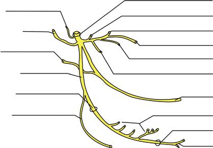
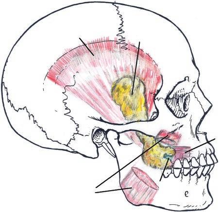
In classical anatomy there are 12 paired cranial nerves (I–XII) providing sensory and motor innervation to the head and neck. (See Figure 1.1.)
The trigeminal nerve (V), the fifth cranial nerve, is responsible for sensations of the face and motor functions of the muscles of mastication. This cranial nerves derives its name from the fact that each trigeminal nerve (one on each side of the pons) has three major branches: the ophthalmic nerve (V1), the maxillary nerve (V2), and the mandibular nerve (V3). (See Figure 1.2.) The ophthalmic and maxillary nerves are purely sensory, while the mandibular nerve has sensory and motor functions. (See Figure 1.3.)
The mandibular nerve (V3) is the largest of the three branches or divisions of the trigeminal nerve, the fifth (V) cranial nerve. (See Figure 1.4.) It is made up of a large sensory root and a small motor root. The mandibular nerve exits the cranium through the foramen ovale and divides into an anterior and posterior trunk in the infratemporal fossa. The mandibular nerve divides further into 9 main branches, 5 sensory and 4 motor.
The infratemporal fossa also contains many blood vessels in addition to nerves. This becomes important during the removal of high, palatally placed, maxillary third molars. These teeth may be displaced into the infratemporal fossa which contains these vital structures.
The five sensory branches of the mandibular nerve control sensation to teeth, tongue, mucosa, skin, and dura.
● Average diameter of the main trunk of the lingual nerve is 3.5 mm.3
3) Auriculotemporal – innervation to the skin on the side of the head.
4) Buccal or long buccal – innervation to the cheek and second and third molar mucosa.
5) Meningeal – innervation to dura mater.
The four motor branches of the mandibular nerve control the movement of eight muscles, including the four muscles of mastication: the masseter, the temporal, and the medial and lateral pterygoids. The other four muscles are the tensor veli palatini, tensor tympani, mylohyoid, and the anterior belly of the digastric. Nerves to the tensor veli tympani and tensor palatini are branches of the medial pterygoid nerve. Nerves to the mylohyoid and anterior belly of the digastric muscles are branches of the inferior alveolar nerve.
6) Masseteric
7) Deep temporal
8) Lateral pterygoid
9) Medial pterygoid
Figure 1.3 Sensory innervation of the three branches of the trigeminal nerve. (by Madhero88 / Wikimedia commons / CC BY 3.0).
Nerve Complications Following the Removal of Impacted Third Molars
Injury to the inferior alveolar, lingual, mylohyoid, and buccal nerves may cause altered or complete loss of sensation of the lower 1/3 of the face on the affected side.
The majority of serious nerve complications result from inferior alveolar or lingual nerve injuries. Most surgical injuries to the inferior alveolar nerve and lingual nerve cause temporary sensory change, but in some cases they can be permanent. Injury to these nerves can cause anesthesia (loss of sensation), paresthesia (abnormal sensation), hypoesthesia (reduced sensation), or dysesthesia (unpleasant abnormal sensation). Injury to the lingual nerve and associated chorda tympani nerve can also cause loss of taste of the anterior 2/3 of the tongue.
Damage to the mylohyoid nerve has been reported to be as high as 1.5% following lower third molar removal, but this is probably due to the use of lingual retraction.4 Most third molars can be
5 Meningeal Branch
3 Auriculotemporal nerve
Chorda Tympani
Joins Lingual Ner ve
1 Inferior Alveolar Ner ve
Mandibular Foramen
Ner ve to Mylohyoid And Digastric
Foramen Ovale
Masseteric Branch 6
Deep Temporal Ner ve 7
Buccal Ner ve 4
Ner ve to Lateral Pter ygoid 8
Ner ve to Medial Pter ygoid, Tensor Tympani, & Tensor Palatini 9
Lingual Ner ve 2
Inferior Dental Ner ves
Mental Foramen
Mental Ner ve
Figure 1.4 Mandibular nerve branches from the main trunk; anterior, and posterior divisions. (Drawing by Michael Brooks).
V1
V3
V2
removed by utilizing a purely buccal technique. Utilizing this technique, it is not necessary to encroach on the lingual tissues or to remove distal, distolingual, or lingual bone.5
A search of the literature found no specific reports of long buccal nerve involvement (AAOMS white paper, March 2007), although one article did note long buccal involvement when the anatomical position was aberrant. In this case, the long buccal nerve was coming off the inferior alveolar nerve once it was already in the canal and coming out through a separate foramen on the buccal side of the mandible.6 Long buccal nerve branches are probably frequently cut during the incision process, but the effects are generally not noted.7
Blood Vessels
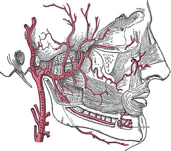
Life-threatening hemorrhage resulting from the surgical removal of third molars is rare. However, copious bleeding from soft tissue is relatively common. One source of bleeding during the surgical removal of third molars is the inferior alveolar artery and/or vein. These central vessels can be cut during sectioning of third molars leading to profuse bleeding. The path of vessels leading to the inferior alveolar neurovascular bundle begins with the common carotid arteries and the heart.
The common carotid arteries originate close to the heart and divide to form the internal and external carotid arteries. The left and right external carotid arteries provide oxygenated blood to the areas of the head and neck outside the cranium. These arteries divide within the parotid gland into the superficial temporal artery and the maxillary artery. The maxillary artery has three portions; maxillary, pterygoid, and pterygomaxillary. (See Figures 1.5a and 1.5b.)
The first portion of the maxillary artery divides into five branches. The inferior alveolar artery is one of the five branches of the first part of the maxillary artery. The inferior alveolar artery joins the inferior alveolar nerve and vein to form the inferior alveolar neurovascular bundle within the mandible. Three studies confirm that the inferior alveolar vein lies superior to the nerve and that there are often multiple veins. The artery appears to be solitary and lies on the lingual side of the nerve, slightly above the horizontal position.8
Bleeding during and after third molar impaction surgery is expected. Local factors resulting from soft-tissue and vessel injury represent the most common cause of postoperative bleeding.9 Systemic causes of bleeding are not common, and routine preoperative blood testing of patients, without a relevant medical history, is not recommended.10
Hemorrhage from mandibular molars is more common than bleeding from maxillary molars (80% and 20%, respectively),11 because the floor of the mouth is highly vascular. The distal lingual aspect of mandibular third molars is especially vascular and an accessory artery in this area can be cut leading to profuse bleeding.12,13 The most immediate danger for a healthy patient with severe post-extraction hemorrhage is airway compromise.14
Most bleeding following third molar impaction surgery can be controlled with pressure. Methods for hemostasis will be discussed further in Chapter 3.
Buccal Fat Pad
The buccal fat pad is a structure that may be encountered when removing impacted third molars. It is most often seen when flap incisions are made too far distal to maxillary second molars. It is a deep fat pad located on either side of the face and is surrounded by the following structures. (See Figure 1.6.)
● Anterior – angle of the mouth
● Posterior – masseter muscle
(a)
Incisor branch
Figure 1.5a The Maxillary Artery. (Henry Gray / Wikimedia commons / Public Domain).
(b)
Mid. meningeal
Ant. t ympanic
Super cial te almpor
Deep auric
Ext er nal ca r otid
Post. deep temporal
Access. meningeal
1stpart
Art of Pterygoid canal
Pharyngeal
Desc. pal
Ant. deep temp.
Plerygoid
2ndpart
Sphenopalatine
Infraorbital
3rd part
Post. sup. alveolar
Buccinator
Masseteric
Pterygoid
Inferior alveolar
Mylohyoid
Figure 1.5b Branches of the maxillary artery depicting maxillary, pterygoid, and pterygomaxillary portions. (by Henry Vandyke Carter / Wikimedia commons / Public Domain).
● Medial – buccinator muscle
● Lateral – platysma muscle, subcutaneous tissue, and skin
● Superior – zygomaticus muscles
● Inferior – depressor anguli oris muscle and the attachment of the deep fascia to the mandible Zhang, Yan, Wi, Wang, and Liu reviewed the anatomical structures of the buccal fat pad in 11 head specimens (i.e., 22 sides of the face).
The enveloping, fixed tissues and the source of the nutritional vessels to the buccal fat pad and its relationship with surrounding structures were observed in detail. Dissections showed that the buccal fat pad can be divided into three lobes – anterior, intermediate, and posterior, according to the structure of the lobar envelopes, the formation of the ligaments, and the source of the nutritional vessels. Buccal, pterygoid, pterygopalatine, and temporal extensions are derived from the posterior lobe. The buccal fat pad is fixed by six ligaments to the maxilla, posterior zygoma, and inner and outer rim of the infraorbital fissure, temporalis tendon, or buccinator membrane. Several nutritional vessels exist in each lobe and in the subcapsular vascular plexus forms. The buccal fat pads function to fill the deep tissue spaces, to act as gliding pads when masticatory and mimetic muscles contract, and to cushion important structures from the extrusion of muscle contraction or outer force impulsion. The volume of the buccal fat pad may change throughout a person’s life.15
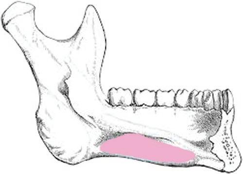
Submandibular Fossa
The submandibular fossa is a bilateral space located in the mandible, medial to the body of the mandible, and below the mylohyoid line. (See Figure 1.7.) It contains the submandibular salivary gland which produces 65–70% of our saliva.
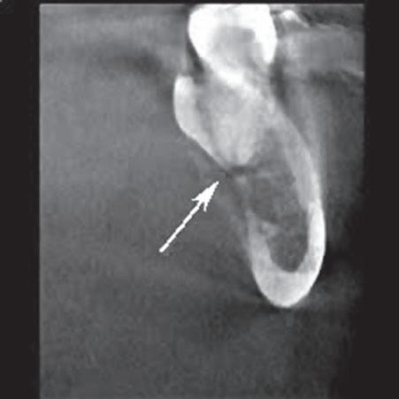
Third molar roots are often located in close proximity to the submandibular space. (See Figure 1.8.) The lingual cortex in this area may be thin or missing entirely. Therefore, excessive or
M. Temporalis Buccal Fat
M. Buccinator
Parotid Duct
M. Masseter
Figure 1.6 Buccal fat pad. (By Otto Placik / Wikimedia commons / CC BY 3.0).
1.7 Submandibular fossa. (Adapted from Henry Vandyke Carter, via Wikimedia Commons).
misplaced force can dislodge root fragments or even an entire tooth into the adjacent submandibular space.16
Patients presenting with partially impacted third molars can develop pericoronitis. This localized infection can spread to the submandibular, sublingual, and submental spaces. Bilateral infection of these spaces is known as Ludwig’s angina.17 Prior to the advent of antibiotics this infection was often fatal due to concomitant swelling and compromised airway.
Maxillary Sinus
The maxillary sinus is a bilateral empty space located within the maxilla, above the maxillary posterior teeth. It is pyramidal in shape consisting of an apex, base, and four walls. (See Figure 1.9 and Box 1.1.)
1.8
roots
fossa. (Reproduced with permission of Dr. Jason J. Hales, DDS).
Figure
Figure
Third molar
near submandibular
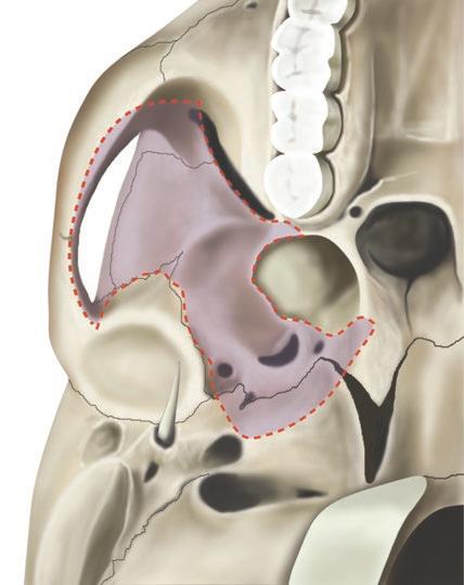
Box 1.2 Boundaries of the infratemporal fossa.
Anterior: posterior maxilla
Posterior: tympanic plate and temporal bone
Medial: lateral pterygoid plate
Lateral: ramus of the mandible
Superior: greater wing of the sphenoid bone
Inferior: medial pterygoid muscle
Although rare, there are documented cases of maxillary third molars displaced into the infratemporal fossa. Unlike the maxillary sinus, the infratemporal fossa is not an empty space. It contains many vital structures including nerves, arteries, and veins. A third molar displaced into the infratemporal fossa is considered a major complication. Dentists removing impacted maxillary third molars should understand the anatomy of the infratemporal fossa.
This chapter is not intended to be a comprehensive review of oral anatomy, but instead is a review of structures relevant to third molars. This knowledge is essential to avoid surgical complications. Although no surgical procedure is without risk, most impacted third molars can be removed safely and predictably.
An important key to avoid complications is deciding when to refer to an oral surgeon. This will be different for each dentist depending on experience and training. “When to refer” may be the most important factor to consider prior to treating your patients. Case selection, including surgical risk and difficulty, is discussed in the next chapter.
Figure 1.10 Boundaries of the infratemporal fossa. (Reproduced with permission from Joanna Culley BA(hons) IMI, MMAA, RMIP).
References
1 Svane TJ. Vascular Characteristics of the Human Inferior Alveolar Nerve, J Oral Maxillofacl Surg. 1986;44:431.
2 Graff-Radford SB, Evans RW. Disclosures. Headache. 2003;43(9): 9.
3 Zur KB, Mu L, Sanders I. Distribution pattern of the human lingual nerve. Clin Anat. 2004 Mar;17(2):88–92.
4 Carmichael FA, McGowan DA. Incidence of nerve damage following third molar removal: a West of Scotland Surgery Research Group study. Brit J Oral Maxillofac Surg. 1992;30:78.
5 Gargallo-Albiol J, Buenechea-Imaz R, Gay-Escoda C. Lingual nerve protection during surgical removal of lower third molars. J Oral Maxillofac Surg. 2000;29:268.
6 Singh S. Aberrant buccal nerve encountered at third molar surgery. Oral Surg Oral Med Oral Pathol. 1981;52:142.
7 Merrill RG. Prevention, treatment, and prognosis for nerve injury related to the difficult impaction. Dent Clin North Am. 1979;23:471.
8 Pogrel MA, Dorfman D, Fallah H. The anatomic structure of the inferior alveolar neurovascular bundle in the third molar region. J Oral Maxillofac Surg. 2009 Nov;67(11):2452–4.
9 Allen FJ. Postextraction hemorrhage. A study of 50 consecutive cases. Br Dent J 1967;122(4):139–43.
10 Suchman AL, Mushlin AI. How well does activated partial thromboplastin time predict postoperative hemorrhage? JAMA. 1986;256(6):750–3.
11 Jensen S. Hemorrhage after oral surgery. An analysis of 103 cases. Oral Surg Oral Med Oral Pathol. 1974;37(1):2–16.
12 Funayama M, Kumagai T, Saito K, Watanabe T. Asphyxial death caused by post extraction hematoma. Am J Forensic Med Pathol. 1994;15(1):87–90.
13 Goldstein BH. Acute dissecting hematoma: a complication of an oral and maxillofacial surgery. J Oral Surg. 1981;39(1):40–3.
14 Moghadam HG, Caminiti MF. Life-threatening hemorrhage after extraction of third molars: case report and management protocol. J Can Dent Assoc. 2002;68(11):670–5.
15 Zhang HM, Yan YP, Qi KM, Wang JQ, Liu ZF. Anatomical structure of the buccal fat pad and its clinical adaptations. Plast Reconst Surg. 2002;109(7):2509–18; discussion 2519–20.
16 Aznar-Arasa L, Figueiredo R, Gay-Escoda C. Iatrogenic displacement of lower third molar roots into the sublingual space: report of 6 cases. Int J Oral Maxillofac Surg. 2012;70:e107–15.
17 Vijayan A, et al. Ludwigs angina: report of a case with extensive discussion on its management. URJD. 2015;5(2):82–86.
18 Kim JH. A review of the maxillary sinus. Sinus, OK: Perio Implant Advisory.
19 Hargreaves KM, Cohen S, web editor, Berman LH, editor. Cohen’s pathways of the pulp, 10th edition. St. Louis, MO: Mosby Elsevier, 2010, 590,592.
2
Case Selection
The best way to avoid complications when removing impacted third molars is to select patients and surgeries that are commensurate with your level of training and experience. Will you treat medically compromised patients? Or, will you only remove impacted third molars for healthy teens? Have you removed thousands of impactions? Or, are you about to remove your first maxillary soft tissue impaction? This chapter will help you decide which third molar surgery patients should be referred to a maxillofacial surgeon or kept in your office. It will also help you know when you are ready to move to the next level of difficulty.
Medical Evaluation
The medical evaluation includes a complete health history/patient interview, physical assessment, clinical exam, and psychological evaluation of the patient. The removal of impacted third molars is an invasive surgical procedure with risk of complications higher than most dental procedures. Furthermore, patients are often apprehensive and have anxiety about the procedure.
Health History and Patient Interview
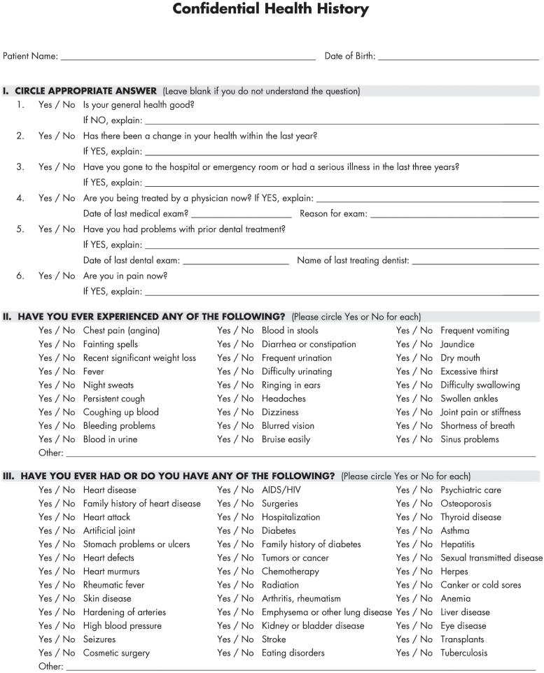
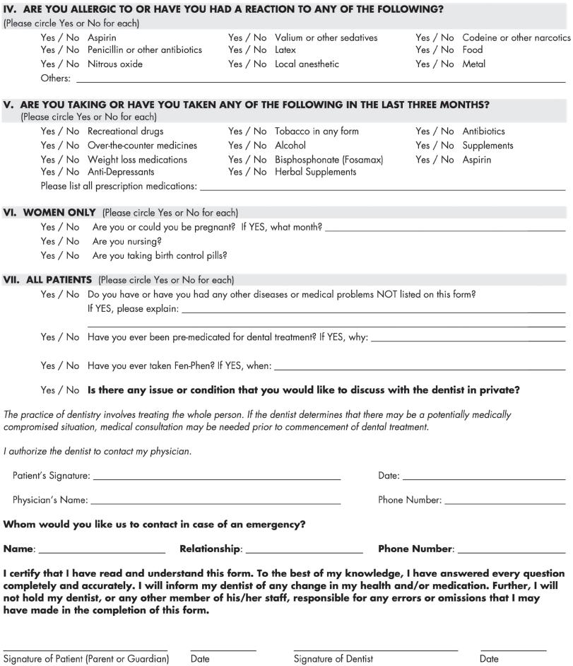
A thorough health history and patient interview should be completed prior to treatment. The primary purpose of a patient’s health history is to attempt to find out as much about each patient as possible, so that the patient can be treated safely and knowledgeably. A health history form, completed by the patient, should be reviewed before interviewing the patient. The American Dental Association’s 2014 Health History form is provided as an example. (See Figures 2.1a, 2.1b, and 2.1c.)
The patient’s health history can be subpoenaed in court cases, such as a malpractice suit, or when disciplinary action is taken against a dental professional by a regulatory board. Medical evaluation documents can be used as legal evidence and must be thorough and comprehensive.
The patient interview is an essential part of a medical evaluation. It’s not uncommon to have an unremarkable health history, only to learn during the interview that the patient has a history of health issues and medication. Good interview technique requires open-ended questions and active listening. Open-ended questions always begin with What, How, When, or Where. These questions cannot be answered with a simple yes or no answer. Yes or no questions should be limited to the health history form.
CAMP is a useful mnemonic to remember key interview questions.
Chief complaint – What brings you to the office?
Allergies – What are you allergic to? What else?
Medications – What medications do you take? What medications have you taken previously?
Past Medical History – What medical problems have you had in the past and when did you have them?
2.1a Health History (Reproduced by permission of ADA).
Figure
2.1b Health History Update (Reproduced by permission of ADA).
Figure