Enhance your learning with Evolve Student Resources.
These online study tools and exercises can help deepen your understanding of textbook content so you can be more prepared for class, perform better on exams, and succeed in your course.
Activate the complete learning experience that comes with each
http://evolve.elsevier.com/Turgeon/immunology/
If your school uses its own Learning Management System, your resources may be delivered on that platform. Consult with your instructor.
has already been revealed, the code may have been used and cannot be re-used for registration. To purchase a new code to access these valuable study resources, simply follow the link above.
Quick Reference
Chapter
1 Highlights of Innate and Adaptive Immune Systems
2 Soluble Mediators of the Immune System
3 Antigens and Antibodies
4 Cellular Activities and Clinical Disorders of Innate and Adaptive Immunity
5 Basic Safety in the Immunology-Serology Laboratory
6 Basic Quality Control and Quality Assurance Practices
7 Basic Serologic Laboratory: Techniques and Clinical Applications
8 Precipitation and Particle Agglutination Methods
9 Electrophoresis Techniques and Chromatography
10 Labeling Techniques in Immunoassay
11 Flow Cytometry
12 Molecular Laboratory Techniques
13 Infectious Diseases: Overview and TORCH Diseases
• Identification of Leukocytes Related to Immune Function
• Screening Test for Phagocytic Engulfment
• C-Reactive Protein Rapid Latex Agglutination Test
• ABO Blood Grouping (Forward Antigen Typing)
• Screening Test for Phagocytic Engulfment
• Assessment of Cellular Immune Status
• Test Your Safety Knowledge
• Validation of a New Procedure Write-Up
• Card Pregnancy Testing
• ABO Blood Grouping (Reverse Grouping)
• Immunofixation Electrophoresis
• Pregnancy Testing
• Direct Fluorescent Antibody Test for Neisseria gonorrhoeae
• Laboratory Activities
• Molecular testing – Group A Streptococcus Direct Test
• Rapid TORCH Testing
• Passive Latex Rubella Agglutination Test
• Passive Latex Agglutination for Detection of Antibodies to Cytomegalovirus
• Quantitative Determination of Antibodies to Cytomegalovirus
14 Streptococcal Infections
• Antistreptolysin O Latex Test Kit
• OSOM Ultra Streptococcus A Test
• Group A Streptococcus Direct Test
• Antistreptolysin O Classic Procedure
• Classic Venereal Disease
CASE 1.1 A 1-month-old female infant born 6 weeks prematurely was admitted to the hospital because she had a high fever and was crying all of the time
CASE 2.1 A 39-year-old woman was admitted for a cholecystectomy. The patient became febrile 1 day after surgery.
CASE 3.1 A 38-year-old white woman presented to the emergency department of her local hospital with increased difficulty in breathing and chronic diarrhea.
CASE 4.1 A family had a son who died age 2 weeks because of overwhelming bacterial infection. When their newborn daughter began developing recurrent infections, she was immediately taken to a pediatrician.
CASE 4.2 A 6-year-old white male patient was taken to a pediatrician because of recurring abscesses since the age of 1 month.
CASE 4.3 A 33-year-old man, the child of unrelated parents of Mexican descent, was examined because of a history of frequent sore throats and sinus headaches.
CASE 5.1 When a new employee in a rural laboratory started to work, she wiped down the work bench with 5% bleach and donned latex gloves that she had rinsed off the night before.
CASE 6.1 A new employee was asked to examine a CLISI procedural protocol worksheet and rate the write-up.
CASE 7.1 A 9-year-old boy was taken to the emergency department with a sore throat.
CASE 7.2 A 28-year-old woman has been trying to get pregnant for the past 6 months. Although she has no health problems, conceiving a child is proving to be difficult.
CASE 8.1 An 85-year-old man had a discrepancy between his forward grouping (ABO antigens) and reverse grouping (ABO antibodies).
CASE 9.1 A 40-year-old woman with a long-term history of alcohol abuse comes to the emergency department complaining of difficulty breathing.
CASE 10.1 A 25-year-old woman had a missed menstrual period 3 weeks earlier.
CASE 11.1 The parents of a 6-year-old boy brought him to the hospital complaining of back pain and refusal to walk since falling a week earlier.
CASE 12.1 A 38-year-old man drove himself to the emergency department because of a worsening condition of shortness of breath. He had a sore throat, felt tired, and had a fever, unproductive cough, and mild chest pain.
CASE 13.1 A 34-year-old African-American male was a local delivery truck driver until 2 weeks ago when he was laid off because of the SARS-CoV-2 pandemic. He has been an organist at his local church for the past 5 years. Two weeks ago, he began to feel very tired but had no other medical complaints.
CASE 13.2 A 24-year-old woman with a history of acquired immunodeficiency syndrome (AIDS) comes to the clinic for evaluation of left-sided weakness. She has been experiencing headaches and seizures, and others had observed an alteration in her mental status.
CASE 13.3 A 20-year-old college junior comes to the student health office because she had been exposed to rubella during a recent outbreak at the college. She had been immunized as a child.
CASE 13.4 A 35-year-old man recently received a kidney transplant. He had been feeling well until 2 weeks before, when he experienced a sore throat, fever, chills, profound malaise, and myalgia.
CASE 14.1 A 19-year-old woman visited the emergency department (ED) with swelling and redness of her right leg.
15 Syphilis
Research Laboratory Test –Venereal Disease Research
Laboratory Qualitative Slide Test
• Rapid Plasma Reagin Card Test
• Fluorescent Treponemal Antibody Absorption Test
CASE 15.1 A 25-year-old woman comes to an ambulatory center with pain in the right side of her pelvis and a slightly elevated temperature.
aDigital enrichment files for animated content, virtual labs, web-based videos, and additional chapter-specific outline web resources are available on the Elsevier Evolve website for approved textbook adopters (instructors).
bFully developed case studies and associated questions are published in associated chapters. A full, narrative discussion of the questions for each case study is posted on the Elsevier Evolve website for approved textbook adopters (instructors).
cThe principles and clinical applications of these procedures are explained in the textbook. Procedural protocols and other technical details are posted and explained on the Elsevier Evolve website for approved textbook adopters (instructors).
CASE 16.1 A 42.year-old executive lives in New York City. Her company annually sponsors a Memorial Day weekend golf outing at a Long Island club. In early June, she noticed a solid bright red spot on her left thigh.
CASE 16.2 A 25-year-old graduate student visits his local family physician because of episodic arthromyalgia, sporadic global headaches, fatigue, irritability, and depression. Over the last several months, he had become seriously dysfunctional at work and home.
CASE 16.3 A 45-year-old man from upstate New York visits his physician because of a worsening headache, myalgia, arthralgia, and generalized weakness. He had been in good health until about 1 week before the appointment.
CASE 16.4 A 73-year-old previously healthy man had spent the previous summer on Martha’s Vineyard. On returning to his home in Boston after Labor Day, he began to feel unusually tired and had difficulty breathing.
CASE 16.5 A 35-year-old field biologist from central Missouri was positive for human immunodeficiency virus (HIV). Her work required that she spend a great deal of time in the woods in the surrounding areas. Although she was in good health despite the HIV positivity, she began having back pain, fever, chills, sweats, productive cough, and extreme tiredness before her visit to the emergency department.
17 Infectious Mononucleosis
18 Viral Hepatitis
19 Primary and Acquired Immunodeficiency Syndromes
• Paul-Bunnell Screening Test
• Davidsohn Differential Test
• MonoSlide Test
CASE 17.1 A female college freshman visits the infirmary complaining of extreme fatigue, frequent headaches, and a sore throat.
• Rapid Hepatitis C Virus Testing CASE 18.1 Several workers at a local fast food restaurant called in sick and reported to the local ambulatory clinic for treatment. They all complained of extreme fatigue. All of them complain of extreme fatigue. In addition, another 26-year-old food handler, who returned from visiting his relatives in Costa Rica a month ago, is sick.
CASE 18.2 A 30-year-old phlebotomist presents with fever, persistent fatigue, and joint pain. She reports that a needle in a plastic garbage bag nicked her finger about 2 months ago.
CASE 18.3 A 75-year-old woman had an 18-month history of right-sided abdominal pain and progressive fatigue. Her other medical problems include insulin-dependent diabetes mellitus and hypertension.
CASE 18.4 A 45-year-old previously healthy medical technologist visits her primary care physician because of increasing fatigue and loss of appetite.
20 Hypersensitivity Reactions
• Rapid HIV Antibody Test
• GS HIV Combo Ag/Ab EIA
• Simulation of HIV-1 Detection
• Rapid Test for Food Allergy Direct Antiglobulin Test
21 Immunoproliferative Disorders • Bence Jones Protein Screening Procedure
22 Tolerance, Autoimmunity, and Autoimmune Disorders
23 Systemic Lupus Erythematosus
• Rapid Slide Test for Antinucleoprotein
• Antinuclear Antibody Visible Method
• Rapid Slide Test for Antinucleoprotein
• Autoimmune Enzyme Immunoassay ANA Screening Test
24 Rheumatoid Arthritis • Rapid Agglutination
25 Transplantation: Human Leukocyte Antigens, Solid Organ, and Hematopoietic Stem Cells
26 Tumor Immunology and Applications of Massive Parallel Sequencing/ Next-Generation Sequencing
• Longitudinal Assessment of Posttransplant Immune Status
• Prostate-Specific Antigen Rapid Test of Seminal Fluid (Seratec)
CASE 19.1 Mary is a freshman in college. She began to feel ill with diarrhea and a cough. She tried to ignore the symptoms but woke up the next morning with a severe headache and a painful stiff neck.
CASE 19.2 Mr. J.J. Smith, aged 68 years, had retired to Florida and was meeting with his new primary care provider for the first time. His medical history indicated that he had suffered from recurrent upper and lower respiratory and gastrointestinal infections throughout his life.
CASE 19.3 A 40-year-old man with a history of IV drug use came to the emergency department because of a rash and fever. In addition, the patient complained of a several-day history of malaise, fatigue, fever, headache, and sore throat.
CASE 20.1 A 60-year-old man was stung by a bee while gardening.
CASE 20.2 A 35-year-old gravida 4 para 1 + 2 was seen by her gynecologist when she was 8 weeks pregnant. Her first pregnancy 4 years ago was unremarkable. The patient reported that her second and third pregnancies had resulted in a stillbirth at 36 weeks and a spontaneous abortion at 10 weeks of gestation.
CASE 20.3 A patient had a medical history that included frequent sore throats as a child. He had been treated with antibiotics, particularly penicillin. Eventually, he developed a rash. He was told that he had developed an allergy to penicillin and should not have it again.
CASE 20.4 A 19-year-old college student went to the Student Health Services because she had a slowly developing rash on both earlobes, her hands and her wrists, and around her neck.
CASE 20.5 A 35-year-old woman reported that she had experienced three bouts of urticaria of unknown origin about 10 years ago.
CASE 21.1 A 58-year-old nuclear power plant worker saw his family physician because of increasing fatigue and weakness.
CASE 22.1 A 50-year-old white woman visited her primary care provider because of extreme fatigue. She also reported experiencing mild pain in her abdominal region.
CASE 22.2 A right-handed 25-year-old woman had no significant medical history. She came to the emergency department because of a sudden onset of slurred speech.
CASE 23.1 A 39-year-old African-American woman with SLE was diagnosed with the illness 20 years ago.
CASE 23.2 A 27-year-old white woman sought medical attention because of persisting pain in her wrists and ankles and an unexplained skin irritation on her face.
CASE 24.1 A 62-year-old woman experienced pain in her left knee unrelated to trauma. The pain occurred primarily with weight bearing. She is currently being treated for hypertension but is otherwise healthy.
CASE 24.2 A 31-year-old patient was referred to a rheumatologist because of pain and stiffness in her fingers and wrists. Before her last pregnancy 3 years earlier, she had experienced similar symptoms, but these had gone away.
CASE 25.1 A 40-year-old was seen by her family physician after several episodes of painless hematuria. On direct questioning, she complained of worsening malaise and swelling of her legs and hands over the previous 2 weeks.
CASE 26.1 A 59-year-old white man visited his primary care provider because of his need to urinate frequently and urgently. Over the past several years, his urine output had been in small volumes, with a decreasing flow rate.
CASE 26.2 A 65-year-old African-American woman visited her primary care provider for an annual examination, including a routine pelvic examination. Although she had gained some weight since her last examination, she reported that her general health was good, but that she had been experiencing some gastrointestinal problems over the last 6 weeks.
27 Primer on Vaccines • Tetanus Antibodies (IgG) CASE 27.1 A 25-year-old female medical student came to the emergency department because of a fever, cough, and shortness of breath.
aDigital enrichment files for animated content, virtual labs, web-based videos, and additional chapter-specific outline web resources are available on the Elsevier Evolve website for approved textbook adopters (instructors).
bFully developed case studies and associated questions are published in associated chapters. A full, narrative discussion of the questions for each case study is posted on the Elsevier Evolve website for approved textbook adopters (instructors).
cThe principles and clinical applications of these procedures are explained in the textbook. Procedural protocols and other technical details are posted and explained on the Elsevier Evolve website for approved textbook adopters (instructors).
IMMUNOLOGY & SEROLOGY in
Laboratory Medicine
Mary Louise Turgeon, EdD, MLS(ASCP)CM
Associate Professor (Adjunct), University of Texas Medical Branch, Galveston, Texas
Clinical Laboratory Education Consultant, Mary L. Turgeon and Associates
Boston, Massachusetts; St. Petersburg, Florida
Elsevier
3251 Riverport Lane
St. Louis, Missouri 63043
IMMUNOLOGY & SEROLOGY IN LABORATORY MEDICINE, SEVENTH EDITION ISBN: 978-0-323-71193-7
No part of this publication may be reproduced or transmitted in any form or by any means, electronic or mechanical, including photocopying, recording, or any information storage and retrieval system, without permission in writing from the publisher. Details on how to seek permission, further information about the Publisher’s permissions policies and our arrangements with organizations such as the Copyright Clearance Center and the Copyright Licensing Agency, can be found at our website: www.elsevier.com/permissions
This book and the individual contributions contained in it are protected under copyright by the Publisher (other than as may be noted herein).
Notice
Practitioners and researchers must always rely on their own experience and knowledge in evaluating and using any information, methods, compounds or experiments described herein. Because of rapid advances in the medical sciences, in particular, independent verification of diagnoses and drug dosages should be made. To the fullest extent of the law, no responsibility is assumed by Elsevier, authors, editors or contributors for any injury and/or damage to persons or property as a matter of products liability, negligence or otherwise, or from any use or operation of any methods, products, instructions, or ideas contained in the material herein.
International Standard Book Number: 978-0-323-71193-7
Senior Content Strategist: Tamara Myers
Senior Content Development Specialist: Heather Bays
Publishing Services Manager: Julie Eddy
Senior Project Manager: Richard Barber
Design Direction: Bridget Hoette
Printed in India
To the continuing sense of curiosity and learning adventures shared with Dick and Murphy.
REVIEWERS
Shauna N. Hay, MT(ASCP), MPH VP of Clinical Programs Beacon LBS Laboratory Corporation of America (LabCorp) Burlington, North Carolina
Deborah Josko, PhD. MLT(ASCP)M, SM Director Medical Laboratory Science Program; Associate Professor Clinical Laboratory and Medical Imaging Sciences Rutgers, The State University of New Jersey Newark, New Jersey
Steven R. Schwarze, PhD, MLS(ASCP) SBB Associate Professor College of Health Sciences University of Kentucky Lexington, Kentucky
MOLECULAR LABORATORY TECHNIQUES
Kyle P. Miller, BS, MLS(ASCP)CM, MS, MB(ASCP)CM Chemistry Supervisor, Chemistry Molecular Biology Technical Specialist Cumberland Medical Center Crossville, Tennessee
The goal of the 7th edition of Immunology & Serology in Laboratory Medicine is to facilitate problem-based learning needed by medical laboratory technician (MLT) and medical laboratory science (MLS) students to achieve high scores on board certification or licensure examinations upon graduation and master entry-level professional competencies for career success.
Since the 1st edition of Immunology & Serology in Laboratory Medicine, the goals continue to be to capture and share the latest knowledge and emerging practices of an ever-changing field. Immunology & Serology in Laboratory Medicine continues to strive to present relevant content in sufficient detail and clarity to clinical laboratory science students. Sharp focusing on the most important principles and practices of clinical immunology at the undergraduate level has restrained any overexpansion of the length of the book.
The 7th edition of Immunology & Serology in Laboratory Medicine has been reviewed and revised to continue to include the most recent advances in concepts and practices related to the study of immunology applied to the clinical laboratory. Immunology & Serology in Laboratory Medicine continues to be a unique textbook because of the recognition of the importance of the professional knowledge and practice guidelines in the current ASCLS Entry Level Curriculum and ASCLS Professional Body of Knowledge competencies in immunology and applicable molecular applications. In addition, the latest ASCP Board of Certification Immunology Examination Content Guideline & Outline for the Medical Laboratory Technician, MLT(ASCP), and Medical Laboratory Scientist, MLS(ASCP), categories of certification are used as reference guidelines.
ORGANIZATION
This 7th edition of Immunology & Serology in Laboratory Medicine continues to use the original, classic platform to present and integrate new content and practices. Each chapter has an integration of narrative content and visual reinforcements with related fully developed case studies and student laboratory procedures. Because students in the digital age are more visual learners, the inclusion of new and highly acclaimed illustrations, tables, and boxes has increased again in this edition. Additional up-to-date, professionally developed laboratory assay algorithms are included in the 7th edition. These algorithms are a unique feature of Immunology & Serology in Laboratory Medicine and are included because of the value of high impact visuals that accommodate learning preferences of today’s students. Outstanding features of this textbook are the inclusion of 50 fully developed clinical case studies with associated review and critical thinking questions and more than 650 end-of-chapter, multiple choice review questions.
Immunology & Serology in Laboratory Medicine clearly addresses changes in the clinical laboratory and challenges for students to learn— and instructors to teach—more information in a fixed time frame. The organization of the book allows for tremendous flexibility in instructional design and delivery. The book is well suited for traditional on-campus instruction, hybrid, or blended modes of teaching, and online delivery of courses The 7th edition provides students with a basic foundation in the theory and practice of clinical immunology and practical serology in a one- or two-term course at MLT or MLS levels of instruction.
Each chapter in this edition capitalizes on the strengths of previous editions based on up-to-date information presented at annual meetings and conferences; publications in the professional literature; and comments received from students, teaching faculty, faculty book
reviewers, and working professionals from around the globe. Some traditional content has been retained in the book or transferred to the web-based Evolve platform, because some questions related to classic content may appear on certification or licensure examinations, and under-resourced locations in the United States and worldwide may use classic manual techniques.
The procedural protocol, including specimen collection, the required materials, actual procedure, and expected reference results, is published on the Evolve websites for students and instructors who wish to select a particular laboratory exercise in their curriculum. Instructors can easily select procedures and create a customized laboratory manual that students can print, as needed. This reduces the risk of soiling or contaminating their textbook in a wet laboratory. By reducing the number of pages devoted to laboratory procedures in the text, which may not be desired in a course, the planet gets a little greener in addition to the associated savings in the production cost.
Content correlation between lecture and reading, procedures, and case studies is featured in the inside cover of the book for easy reference. Suggestions for web-based videos and virtual laboratories have been compiled by chapter and are presented on the Evolve site.
DISTINCTIVE FEATURES AND LEARNING AIDS
Immunology & Serology in Laboratory Medicine underscores the importance of clarity, conciseness, and continuity of information for an entry-level student. Integration of fundamental narrative content, clinical case studies, and laboratory procedures occurs in every chapter. Sole authorship of this textbook ensures a smooth transition from chapter to chapter without unnecessary redundancy or changes in writing style.
This edition of Immunology & Serology in Laboratory Medicine continues to strengthen the robust pedagogy that has set the quality benchmark for clinical laboratory science textbooks since the 1st edition. Critical thinking is essential and has a renewed emphasis in this edition, with more clinical case studies. Every chapter has applicable cases. A total of fifty clinical cases are fully developed and presented in ed. 7. These cases have extensively developed content, case-related multiple-choice questions, and critical analysis group discussion questions. These cases not only promote critical thinking and stimulate an overall interest in medicine but also highlight the essential role of the laboratory in patient diagnosis and treatment.
HIGHLIGHTS OF THE 7TH EDITION
The content in Immunology & Serology in Laboratory Medicine is organized into six parts. All of the content has been reviewed and refreshed as needed. New features include Key Concepts interwoven throughout each chapter to highlight important facts. Use of these Key Concepts enables more focused learning of the content narrative as it unfolds in a chapter.
Part I—Basic Immunologic Mechanisms (Chapters 1-4)
In this section, content has been newly rearranged into chapters 1-4 The rearrangement focuses on examining the immune system in a holistic manner rather than as independent entities.
Chapter 1, Highlights of Innate and Adaptive Immunity, organizes the cells and tissues of both systems to enable students to relate the new immunology content to various blood cells that may have been studied in previous courses. A comparison of the three lines of
immunologic defense introduces student to normal immunologic body defense activities.
Chapter 2, Soluble Mediators, is introduced earlier than in past editions. The early introduction of information related to soluble mediator substances, e.g. complement and cytokines, allows students to link the activities of these constituents to innate body defenses discussed in chapter 1. The “cytokine storm” related to the pathophysiology of COVID-19 has raised the interest level in cytokines activity. The list of cytokines has been extensively expanded in Appendix C.
To assist with the understanding of the importance of testing complement, new testing algorithms and tables summarizing complement testing are included in Chapter 2
Chapter 3, Antigens and Antibodies, the hallmarks of immunologic activities, has been placed in this position in the flow of information to facilitate association of content with the previous chapters.
Chapter 4, Innate and Adaptive Immunity: Cellular Functions and Related Clinical Disorders, integrates the content from the first three chapters into the functional characteristics of body defenses and associated clinical disorders.
Part II—The Theory of Immunologic and Serologic Procedures (Chapters 5-12)
Chapter 6, Basic Serologic Laboratory: Techniques & Clinical Applications, now features integrated laboratory techniques supported by examples of non-instrument, rapid testing techniques, including malaria, HIV, and pregnancy testing.
Chapter 12, Molecular Laboratory Techniques, has been expanded to include the latest innovations in testing. The chapter continues to begin with genetic information that bridges classic information with exciting state-of-the-art molecular techniques and applications. Molecular diagnostic testing and treatment strategies have assumed a more visible presence in the clinical laboratory. The content of this chapter as well as throughout the book reflects the importance of molecular diagnostics in today’s medical laboratories.
Single nucleotide polymorphisms (SNPs) content is introduced early in the chapter because they are the most abundant source of genetic variation in the human genome. This chapter has expanded content on molecular amplification methods and the molecular analysis of amplification products. The alternatives to electrophoresis-based techniques of pyrosequencing, mass spectrophotometry, and high performance liquid chromatography continue to be important topics in this chapter. Other important topics are microarrays and Next Generation Sequencing Technology/Massively Parallel Sequencing.
Part III—Immunologic Manifestations of Infectious Diseases (Chapters
13-18)
Chapter 13, Infectious Diseases: Overview & TORCH Diseases is a newly organized chapter that continues to present a traditional overview of laboratory testing in infectious diseases. However, in this edition TORCH (Toxoplasmosis, Other Viruses, Rubella, and Cytomegalovirus) infectious diseases are combined into a single chapter to emphasize their similarities. The use of infectious diseases immunohistochemistry (IHC), advantages of polymer-based immunohistochemistry methods, and newer molecular testing approaches are carried over into the new chapter. All of the statistics for chapters 13-18 have been reviewed and updated as needed. All of the numerous testing algorithms have been reviewed and updated as needed.
Part IV—Immune Disorders (Chapters
19-24)
Chapter 19, Primary and Acquired (Secondary) Immunodeficiencies, has been expanded to include additional content related to innate
immunity and two new associated clinical case studies. Laboratory testing algorithms are emphasized in order to simplify the diagnostic process. Therapeutic drugs in an extensive number of viral mechanistic categories for the treatment of human immunodeficiency virus are described in terms of viral impact.
Part V—Transplantation and Tumor Immunology
(Chapters 25 and 26)
Chapter 26, Tumor Immunology and Up-to-Date Applications of Next-Generation Sequencing/Massive Parallel Sequencing, has an extensively expanded table of diagnostic tumor markers currently in common use. Content on Next-Generation Sequencing, now called Massive Parallel Sequencing, is re-introduced as an important aspect of tumor immunology. In addition, continuous field-flow–assisted dielectropheresis, a new method to isolate and characterize rare circulating tumor cells (CTCs) is presented in this chapter. In addition, examples of currently approved monoclonal antibodies and mechanisms of action have been updated.
Part VI—Vaccines (Chapter 27)
Knowledge of vaccines has never been more important to healthcare professionals. A new vaccine initiative directed at prevention of COVID-19 has raised awareness of the importance of vaccines. Continuing research on human immunodeficiency virus (HIV), malaria, and cancer vaccines is discussed. Immunization schedules have been reviewed and updated as needed.
COVID-19: AN IMMUNOLOGY TEACHING AND LEARNING OPPORTUNITY
Information related to COVID-19 has exploded in the scientific literature, social media, news conferences on television, and newspapers since the declaration of the SARS-CoV-2 virus. At every turn, medical terms and concepts in immunology are being articulated on a global scale. Ten of the 27 chapters in this new edition of Immunology and Serology in Laboratory Medicine integrate the latest COVID-19 research to concepts and laboratory techniques. Here are the applicable chapters and an example of the kind of COVID-19 questions related to a chapter’s content.
Chapter 1—Highlights of Innate and Adaptive Immune Systems
What are immune responses to SARS-CoV-2 virus?
Chapter 2—Soluble Mediators of the Immune System
What’s a cytokine storm?
Chapter 3—Antigens and Antibodies
What are neutralizing antibodies?
Chapter 4—Cellular Activities and Clinical Disorders of Innate and Adaptive Immunity
What are cytokine storm syndromes?
Chapter 5—Basic Safety in the Immunology-Serology Laboratory
Is there new guidance for safety in the laboratory?
Chapter 6—Quality Assurance and Quality Control
What are the major quality assurance topics related to the use of unapproved laboratory diagnostic testing?
Chapter 9—Electrophoresis Techniques and Chromatography
What three laboratory methods of direct or indirect testing for COVID19 are available?
Chapter 12—Molecular Laboratory Techniques
What is the value of Next Generation Sequencing testing?
Chapter 13—Infectious Diseases
What kind of molecular testing and serological (antibody) assays are needed to assess the extent of virus spread and to determine the infection-related fatality rate?
Chapter 27—Primer on Vaccines
What is the difference among the eight research platforms being used to develop a SARS-CoV-2 vaccine?
GENERAL, OVERALL IN-TEXT FEATURES
• More Algorithms and other student-desired visual learning formats (figures, tables, and boxes) to meet the preferences of today’s student.
• Key Concepts interwoven in every chapter to assist students to focus on important topics as narrative content unfold in each chapter. .
• Numerous Fully Developed Case Studies with etiology, pathophysiology, laboratory findings, and critical thinking group discussion questions link key concepts and procedures with a disorder, disease, or condition.
• Applicable Procedures help students solidify concepts and gain practical skills.
• More Full-Color Images offer student-desired visual guides to concepts and procedures.
• Extensive Number (total of more than 650) End-of-Chapter Review Questions assist students in self-evaluation of content upon successful completion of each chapter.
ANCILLARIES
Immunology & Serology in Laboratory Medicine includes additional resources for both instructors and students that are available on the book’s companion website, Evolve.
For the Instructor
Evolve
The companion Evolve website offers several features to aid instructors:
• Critical Analysis Group Discussion Questions: Complete explanations are on the instructor’s side of Evolve for the open-ended, case-related discussion questions.
• Test Bank: This test bank of more than 990 multiple choice questions features answers, explanations, and cognitive levels. The test bank can be used for review in class or for test development. More than 330 of the questions in the instructor test bank are available for student use.
• PowerPoint Presentations: One PowerPoint presentation is given per chapter; this feature can be used as is or as a template to prepare lectures.
• Image Collection: All the images from the book can be downloaded into PowerPoint presentations. The figures can be used during lectures to illustrate important concepts.
• Procedures: This feature presents the principles and application of procedures in every chapter.
• Sample Syllabi for MLT and MLS Students: One- and two-semester courses are available.
• Answers to Additional Review Questions: Students have access to more than 330 questions that test their knowledge on the concepts presented in the text. The questions and answers are available to instructors.
• Chapter-Linked Digital Enrichment References: References to videos, animations, and virtual laboratories are available.
For the Student Evolve
The student resources on Evolve include the following:
• Additional Review Questions: A set of more than 330 multiple choice questions provides extra review and practice.
• Case Studies: Case studies provide additional opportunities for student application of chapter content in real-life scenarios.
ACKNOWLEDGMENTS
My objective in writing Immunology & Serology in Laboratory Medicine, 7th edition, continues to be to share basic scientific concepts, procedural theory, and clinical applications with colleagues and students. Because the knowledge base and technology in immunology and serology continues to expand, writing and revising a book that addresses the need of teachers and students continues to be a challenge. In addition, this book continues to provide me with the opportunity to learn and share my laboratory and teaching experience, and insight as an educator, with others.
Thank you to Ellen Wurm-Cutter and Heather Bays for guiding this project. Also, thank you to Rich Barber for managing the production of the book. Feedback from faculty reviewers and students has been extremely helpful.
Comments from instructors and students are always welcome at Turgeonbooks@gmail.com.
Mary L. Turgeon St. Petersburg, Florida
ABOUT THE AUTHOR
Mary Louise Turgeon, EdD, MLS(ASCP)CM, is an educator, author, and consultant in medical laboratory science education. Her career as an educator includes 15 years as a community college professor and Medical Laboratory Technology program director and 14 years as an undergraduate and graduate university professor, Medical Laboratory Science program director, and departmental chairperson. Other university teaching has included teaching graduate Physician Assistant students at South University, Tampa, Florida and Northeastern University, Boston, Massachusetts. Most recently, Dr. Turgeon taught a graduate immunology course to students in the Doctorate in Clinical Laboratory Science program at the University of Texas Medical Branch, Galveston, Texas.
Dr. Turgeon is currently an ad hoc educational content specialist for the College of Professional Studies, Northeastern University, Boston, and maintains an active clinical laboratory science consulting practice. Her practice, Mary L. Turgeon and Associates, focuses on new program development, curriculum revision, and increasing teaching effectiveness through the use of technology and interactive teaching strategies.
The presentation of numerous professional workshops and lectures complements Dr. Turgeon’s extensive teaching and writing activities. Her career in medical laboratory science has spanned the globe with active participation in a wide variety of professional meetings and workshops. Enthusiastic professional involvement has offered her the opportunity to share leading-edge knowledge with students and to meet and collaborate with medical laboratory science colleagues in the United States and worldwide, including China, Italy, Japan, Qatar, Saudi Arabia, and the United Arab Emirates. Professional volunteer activities have taken her to distant locations such as Cambodia and Lesotho, Africa.
She is the author of medical laboratory science books (sold in more than 45 countries):
• Immunology & Serology in Laboratory Medicine, 6th edition (2018)
Louis Pasteur is generally considered to be the “father of immunology.” Some historic benchmarks in immunology are listed in Table 1.1
KEY CONCEPTS: Characteristics of the Immune System
• The function of the immune system is to recognize self from nonself and to defend the body against nonself.
• An effective immune response requires cooperative interaction between specific cells of the immune system’s cellular elements, cell products, and nonlymphoid elements for optimal functioning.
• Desirable consequences of immunity include natural resistance, recovery, and acquired resistance to infectious diseases.
• Undesirable consequences of immunity include allergy, rejection of a transplanted organ, or an autoimmune disorder, in which the body’s own tissues are attacked as nonself.
• The immune system is usually conceptualized as having two divisions: the innate immune system, the immediate protection component, and the adaptive immune system, the slower but more focused defense component.
The term immunity has historically referred to resistance to pathogens based on the ability of the immune system to recognize and dispose of foreign (nonself) material. Today it is recognized that the immune system plays a critical role in both health and disease (Table 1.2). Desirable consequences of immunity include natural resistance, recovery, and acquired resistance to infectious diseases. Another advantage is the scientific ability to manipulate the immune system to protect against or treat a wide variety of clinical conditions. A deficiency or dysfunction of the immune system can cause various disorders. Undesirable consequences of immunity include allergy, rejection of a transplanted organ, or an autoimmune disorder, in which the body’s own tissues are attacked as if they were foreign.
The immune system is usually conceptualized as having two divisions: the innate immune system (also called natural immunity or native immunity), the immediate protection component, and the adaptive immune system, the slower but more focused defense component (Fig. 1.1).
Innate immunity reflects a person’s ability to resist infections with first and second lines of defense that are nonspecifically directed at all pathogens or foreign particles without memory of prior exposure. Cells
macrophages major histocompatibility complex (MHC)
in the innate division have receptors to recognize microbial products. The innate division can respond rapidly, but rather indiscriminately, to a danger signal.
In contrast, adaptive immunity is specific and exhibits memory of prior exposure to individual pathogens or foreign particles. Antibody formation is a significant characteristic of adaptive immunity. In addition, the adaptive division can rearrange genes, such as antibody or antigen receptors, and achieve a highly targeted, precise response. This process takes time, but it is much more selective than innate immunity.
Various specific cells and nonspecific constituents of the immune system, including soluble mediators (e.g., complement, the proteins that are the major [fluid] component of natural immunity; see Chapter 2, Soluble Mediators), in addition to antibodies, participate in body defenses. The entire leukocytic cell system is designed to defend the body against disease. Each cell type has a unique function and behaves independently, or, in many cases, in cooperation with other cell types.
This chapter presents the general features of cornerstone concepts that support modern immunology. These themes are woven into subsequent chapters throughout this book.
KEY CONCEPTS: Overview of Cells of the Innate and Adaptive Immune Systems
• The major blood and tissue cell components of the innate immune system include surface epithelial barriers.
• Tissue sentinel cells include macrophages, dendritic cells, and mast cells.
• Leukocytes associated with the innate immune system include neutrophils, eosinophils, and basophils, monocytes and macrophages derived from monocytes, natural killer (NK) cells, and innate lymphoid cells.
• Lymphocytes are the key mediators of the adaptive immune system.
• Tissue resident cells (dendritic cells, macrophages, and mast cells) act as sentinels to detect the presence of microbes in tissues and initiate immune responses.
CELLS OF THE INNATE AND ADAPTIVE IMMUNE SYSTEMS
The cells of the immune system are located in the peripheral blood and different tissues. These cells function differently in host defense. Most of the cells derived from bone marrow precursors circulating in the
TABLE 1.1 Significant Milestones in
Immunology
Date Scientists Discovery
1798 Jenner Smallpox vaccination
1862 Haeckel Phagocytosis
1880–1881 Pasteur Live attenuated chicken cholera and anthrax vaccines
1883–1905 Metchnikoff Cellular theory of immunity through phagocytosis
1885 Pasteur Therapeutic vaccination
First report of live “attenuated” vaccine for rabies
1890 Von Behring, Kitasatoa Humoral theory of immunity proposed
1891 Koch Demonstration of cutaneous (delayed-type) hypersensitivity
1903 Arthus Arthus reaction of intermediate hypersensitivity
1938 Marrack Hypothesis of antigen–antibody binding
1944 Hypothesis of allograft rejection
1949 Salk, Sabin Development of polio vaccine
1951 Reed Vaccine against yellow fever
1953 Graft-versus-host reaction
1957 Burnet Clonal selection theory
1957 Interferon
1958–1962 Human leukocyte antigens (HLAs)
1964–1968 T cell and B cell cooperation in immune response
1972 Identification of antibody molecule
1975 Köhler First monoclonal antibodies
1985–1987 Identification of genes for T cell receptor
1986 Monoclonal hepatitis B vaccine
1986 Mosmann Th1 versus Th2 model of T helper cell function
1996–1998 Identification of toll-like receptors
2001 FOXP3, the gene directing regulatory T cell development
2005 Frazer Development of human papillomavirus vaccine
peripheral blood are leukocytic white blood cells. Other immunologically active cells reside in tissues. Some cells function mainly in innate immunity, others in adaptive immunity, and some in both innate and adaptive immune responses.
Leukocytes can be functionally divided into the general categories of granulocyte, monocyte-macrophage, lymphocyte, or plasma cell. The primary phagocytic cells are the polymorphonuclear neutrophil (PMN) leukocytes and the mononuclear monocytes-macrophages. The response of the body to pathogens involves cross-talk among many immune cells, including macrophages, dendritic cells, and T lymphocytes.
The role of the innate system in tissue injury, particularly neutrophils and macrophages, is that these types of cells are recruited to sites of ischemic injury. Macrophages have a complex program in which they first release proinflammatory mediators to fight pathogens but may then initiate programs to help in the clearance of dead tissue and
TABLE 1.2 Importance of the Immune System in Health and Disease
Role of Immune System Health Implications
Defense against infections Deficient immunity results in increased susceptibility to infections
Vaccination boosts immune defenses and protects against infections
Defense against tumors Potential for immunotherapy to treat malignancies
Control of tissue regeneration and scarring
Injury to cells and induction of pathologic inflammation
Recognition and response to tissue grafts and newly introduced proteins
Repair of damaged tissues
Immune responses cause allergic, autoimmune, and other inflammatory diseases
Immune responses are barriers to transplantation and gene therapy
Adapted from Abbas AK, Lichtman AH, Pillae S: Basic immunology: functions and disorders of the immune system, Philadelphia, 2020, Elsevier.
tissue repair. Although the latter response is beneficial if short lived, continued activation of the repair program may be detrimental if it becomes chronic.
In contrast, lymphocytes participate in adaptive body defenses primarily through the recognition of foreign antigens and production of antibodies. Plasma cells are antibody-synthesizing cells.
ORIGIN AND DEVELOPMENT OF BLOOD CELLS
Blood cell production, or hematopoiesis, begins in embryonic development. Embryonic blood cells, excluding the lymphocyte type of white blood cell (WBC), originate from the mesenchymal tissue that arises from the embryonic germ layer, the mesoderm (Fig. 1.2). The sites of hematopoiesis follow a definite sequence in the embryo and fetus: 1. In the embryo, self-renewing hematopoietic stem cells develop initially in the primitive yolk sac. The first blood cells are primitive red blood cells (RBCs, or erythroblasts), which are formed in the islets of the yolk sac during the first 2 to 8 weeks of life. Advances in molecular genetics have concluded that a large proportion of tissue-resident macrophage populations, including those in the pancreas, liver, brain, and skin, are derived from embryonic yolk sac progenitors. 2. Gradually the liver and spleen replace the yolk sac as the sites of blood cell development. By the second month of gestation, the liver becomes the major site of hematopoiesis. Beginning in the fetal liver and later in bone marrow, hematopoietic stem cells give rise to the earliest myeloid and lymphoid progenitors. Pancreatic Langerhans cells in the epidermis were discovered to originate from embryonic tissue, predominantly from the fetal liver. The liver and spleen predominate from about 2 to 5 months of fetal life.
3. In the fourth month of gestation, bone marrow begins to produce blood cells. After the fifth fetal month, bone marrow begins to assume its ultimate role as the primary site of hematopoiesis. The cellular elements of the blood are produced from a common CD34+ multipotential hematopoietic cell, the stem cell. Cluster of differentiation (CD) nomenclature for cell surface markers is a common system. The CD system can identify and discriminate between, or “mark,” different cell populations, or it can differentiate maturational stages of various leukocytes, particularly lymphocytes (Table 1.3; also see Fig. 4.5 and Table 4.3).
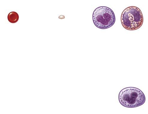
After stem cell differentiation, blast cells arise for each of the major categories of cell types. Subsequent maturation of these blast cells produces the major cellular elements of the circulating blood, the erythrocytes (RBCs), thrombocytes (platelets), and specific types of leukocytes (WBCs). In normal peripheral or circulating blood, the following types of leukocytes can be found, in order of frequency: neutrophils, lymphocytes, monocytes, eosinophils, and basophils.
BLOOD AND TISSUE CELLS ASSOCIATED WITH INNATE AND ADAPTIVE IMMUNE SYSTEMS
Cooperation is required for optimal function of the immune system. This cooperative interaction involves specific cells of the immune system, cell products, and soluble mediators. Cells of the immune system consist of specialized cells that capture and display microbial antigens, effector cells that eliminate microbes, and various subsets of lymphocytes.
The principal functions of the major cell types involved in the immune response are:
1. Specific recognition of antigens or foreign particles
2. Capture and processing of antigens or foreign particles for display to lymphocytes
3. Elimination of offending antigens or foreign particles
The major blood and tissue cell components of the innate immune system are surface epithelial barriers; tissue sentinel cells, including macrophages, dendritic cells, and mast cells; leukocytes, including granulocytes (neutrophils, eosinophils, and basophils), monocytes and macrophages derived from monocytes, and NK cells; and innate lymphoid cells. Lymphocytes are the key mediators of the adaptive immune system. Some cells, such as tissue-resident cells (dendritic cells, macrophages, and mast cells), act as sentinels to detect the presence of microbes in tissues and initiate immune responses. Formed elements of the circulating blood go through a series of developmental stages.
GRANULOCYTES
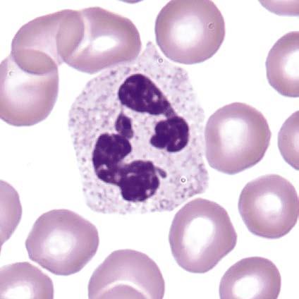
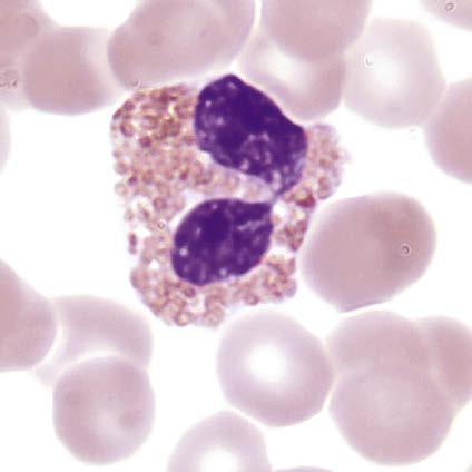
Neutrophils
Fig. 1.1 Principal Mechanisms of Innate and Adaptive Immunity. The mechanisms of innate immunity provide the initial defense against infections. Some mechanisms (e.g., epithelial barriers) prevent infections, and other mechanisms (e.g., phagocytes, natural killer [NK] cells, and other innate lymphoid cells [ILCs] and the complement system) eliminate microbes. Adaptive immune responses develop later and are mediated by lymphocytes and their products. Antibodies block infections and eliminate microbes, and T lymphocytes eradicate intracellular microbes. The kinetics of the innate and adaptive immune responses are approximations and may vary in different infections. (From Abbas AK, Lichtman, AH, Pillai S: Basic immunology: functions and disorders of the immune system, ed 5, Philadelphia, 2016, Elsevier.)
Normally only mature cells are seen in the peripheral blood circulation, but immature cells may appear in the peripheral blood in certain disease states. Each mature cell type has an identifiable appearance, a unique function, independent behavior, or cooperative interaction with other cell types. Each cell type has a normal life span and function.
Granulocytic leukocytes can be further subdivided on the basis of morphology into neutrophils, eosinophils, and basophils. Granulocytes develop in the bone marrow. The maturation stages are similar for all granulocytes; each begins as a multipotential stem cell in the bone marrow.
The most numerous of the granulocytes are PMN leukocytes, or segmented neutrophil leukocytes. Neutrophils make up about 59% of the leukocytes in the peripheral blood of adults, with a range of 35% to 71%. Infants and children have fewer neutrophils and more lymphocytes. Cells of the neutrophil series are generally round with smooth margins or edges. As the cells mature, they become progressively smaller. Most immature cells have cytoplasm that stains dark blue and becomes light pink as the cells mature. As the cells mature, nonspecific granules that stain blue to reddish purple appear in the cytoplasm. Eventually, these nonspecific granules are replaced by specific neutrophilic granules. Nuclear changes also occur as the cells mature. As the cell matures, the nucleus decreases in relative size and begins to contort or form lobes. At the same time the nuclear chromatin changes from a fine, delicate pattern to the more clumped pattern characteristic of the mature cell. The staining of the nucleus also changes from reddish purple to bluish purple as the cell matures. Nucleoli may be apparent in the early forms but gradually disappear as the chromatin thickens and the cell matures.
CD80 Dendritic cells, activated B lymphocytes, macrophages
MHC, Major histocompatibility complex.
Adapted from Abbas AK, Lichtman AH, Pillae S: Basic immunology: functions and disorders of the immune system, ed 6, St. Louis, 2020, Elsevier.
Neutrophils (Fig. 1.3) are recognized as an essential part of the innate immune response. Previously neutrophils were believed to be directly eliminated in the marrow, liver, and spleen after circulating for less than 1 day. It has now been established that neutrophils redistribute into multiple tissues. Neutrophils exist in the peripheral blood for about 10 hours after they are released from the marrow. During this
Agranulocytes
Fig. 1.2 Diagram of Hematopoiesis. Diagram shows derivation of cells from the pluripotential stem cell. (From Waugh A, Grant A: Ross and Wilson anatomy and physiology in health and illness, ed 13, Philadelphia, 2018, Elsevier. Photographic inserts from Telser AG, Young JK, Baldwin KM: Elsevier’s integrated histology, Edinburgh, 2007, Mosby; and Young B, Lowe JS, Stevens A, et al: Wheater’s functional histology: a text and colour atlas, Edinburgh, 2006, Churchill Livingstone. Reproduced with permission.)
Fig. 1.3 Segmented Neutrophil. (From Rodak BF, Carr JH: Clinical hematology atlas, ed 4, St. Louis, 2013, Elsevier.)
time they move back and forth between the general blood circulation and the walls of the blood vessels, where they accumulate. They also leave the blood and enter the tissues, where they carry out their primary functions. In the tissues neutrophils fight bacterial infections and are then destroyed or eliminated from the body by the excretory system (intestinal tract, urine, lungs, or saliva).
Neutrophilic leukocytes, particularly the PMN type, provide an effective host defense against bacterial and fungal infections. The antimicrobial function of PMNs is essential in the innate immune response. Although other granulocytes are also phagocytic cells, the PMNs and macrophages are the principal cells associated with phagocytosis (i.e., the capture and destruction of invading microorganisms; see Chapter 4) and a localized inflammatory response. Inflammatory exudate (pus), which develops rapidly in an inflammatory response, is composed primarily of neutrophils and monocytes.
PMNs can prolong inflammation by the release of soluble substances, such as cytokines and chemokines. The role of neutrophils in influencing the adaptive immune response is believed to include shuttling pathogens to draining lymph nodes, antigen presentation, and modulation of T lymphocyte responses.
Mature neutrophils are found in two evenly divided pools: the circulating and the marginating pools. The marginating granulocytes adhere to the vascular endothelium. In the peripheral blood these cells are only in transit to their potential sites of action in the tissues. Movement of granulocytes from the circulating pool to the peripheral tissues occurs by movement through the vessel wall. Once in the peripheral tissues the neutrophils are able to carry out their function of capture and destruction of invading pathogens or foreign particles.
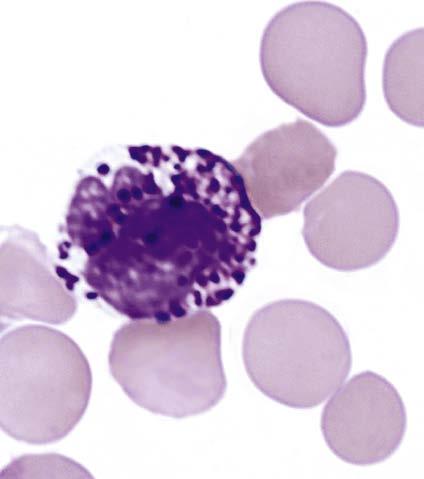
Eosinophils
Although capable of participating in phagocytosis, eosinophils and basophils have less phagocytic activity. The ineffectiveness of these cells results from the small number of cells in the circulating blood and a lack of powerful digestive enzymes. However, both eosinophils and basophils are functionally important in the body’s defense.
Eosinophils (Fig. 1.4) are granulocytes and generally make up about 3% of the circulating leukocytes. They are slightly larger than neutrophils. The nucleus occupies a relatively small part of the cell. The nucleus is usually bilobed, and occasionally three lobes are seen. The nuclear structure is similar to that of the neutrophil, but the lobes are
plumper and the chromatin often stains lighter purple than in the neutrophil. The nuclear membrane is distinct, and no nucleoli are visible. The cytoplasm is usually colorless, but it may be faintly basophilic. It is crowded with spheric acidophilic granules, which stain red-orange with eosin and are larger and more distinct than neutrophilic granules. The granules are evenly distributed throughout the cytoplasm but are rarely seen overlying the nucleus. A second population of eosinophilic granules is highly refractive, a feature that is often a valuable distinguishing characteristic.
The eosinophil is considered to be a regulator of inflammation. Functionally this means that the eosinophil attempts to suppress an inflammatory reaction to prevent the excessive spread of the inflammation. The eosinophil may also play a role in the host defense mechanism because of its ability to kill certain parasites. Although capable of participating in phagocytosis, eosinophils and basophils are less effective in phagocytosis than neutrophils. Eosinophils exist in the peripheral blood for less than 8 hours after release from the marrow and have a short survival time in the tissues.
An increase in eosinophils is associated with a wide variety of conditions, but especially with allergic reactions, drug reactions, certain skin disorders, parasitic infestations, collagen vascular diseases, Hodgkin disease, and myeloproliferative diseases.
Basophils
Basophils (Fig. 1.5) are granulocytes that normally constitute an average of 0.6% of the total circulating leukocytes. They are about the same size as neutrophils, but their nuclei usually occupy a relatively greater portion of the cell. The nucleus is often extremely irregular in shape, varying from a lobular form to a form showing indentations that are not deep enough to divide it into definite lobes. The nuclear pattern is indistinct and stains purple or blue. The nuclear membrane is fairly distinct, and no nucleoli are visible. The cytoplasm is usually colorless; it contains a variable number of deeply stained, coarse, round, or angular basophilic granules. The granules (metachromatic) stain deep purple or black; occasionally a few smaller, brownish granules may be present. They may overlie and obscure the nucleus. Because the granules are soluble in water, occasionally a few or even most of them may be dissolved during the staining procedure. When this occurs, the cell
Fig. 1.4 Eosinophil. (From Rodak BF, Carr JH: Clinical hematology atlas, ed 4, St. Louis, 2013, Elsevier.)
Fig. 1.5 Basophil. (From Rodak BF, Carr JH: Clinical hematology atlas, ed 4, St. Louis, 2013, Elsevier.)
contains vacuoles in place of granules, and the cytoplasm may appear grayish or brownish in their vicinity. The cytoplasm of a mature basophil is colorless.
Basophils have high concentrations of heparin and histamine in their granules. If events are triggered by antigens from pollen, food, drugs, or insect venom, the result is an immediate hypersensitivity reaction.
Tissue Basophils (Mast Cells)
Tissue basophils are also called mast cells Mast cells are bone marrow–derived cells found in the skin and mucosal barriers. These cells resemble basophils with abundant basophilic, cytoplasmic granules.
Tissue basophils are activated by microbial binding and complement components as part of innate immunity or by an antibody-dependent mechanism of adaptive immunity. Degranulation occurs when an antigen such as pollen binds to two adjacent immunoglobulin E (IgE) antibody molecules located on the surface of mast cells. The events resulting from the release of the contents of these basophilic granules include increased vascular permeability, smooth muscle spasm, and vasodilation. If severe, this reaction can result in anaphylactic shock (see Chapter 20).
Tissue basophils are an important defense against parasitic helminths and other pathogens, in addition to snake and insect venom. Some subsets of basophils are involved in normal tissue homeostasis (e.g., neurogenic and endocrine responses).
MONONUCLEAR PHAGOCYTE SYSTEM
In the past the mononuclear phagocyte system (MPS) was known only as a scavenger cell system. Recently the role of the MPS as a complex component of the immune system in the host defense against infection has been recognized. The MPS is considered a cellular system because of the common origin, similar morphology, and shared functions, including rapid phagocytosis, mediated by receptors and a major fragment of complement (C1).
Cells of the MPS system originate in the bone marrow from the multipotential stem cell. This common committed progenitor cell can differentiate into the granulocyte or monocyte-macrophage pathway, depending on the microenvironment and chemical regulators. Maturation and differentiation of these cells may occur in various directions. Circulating monocytes may continue to be multipotential and give rise to different types of macrophages.
The MPS consists of monocytes, dendritic cells, and macrophages. The system, comprised of promonocytes and their precursors in the bone marrow, monocytes in circulation, and terminally differentiated macrophages at peripheral tissue sites, represents a continuum that is dynamically controlled through proliferation, differentiation, maturation, and precise movement of cells (trafficking). Macrophages and monocytes resident in the tissues of the body are already strategically placed to recognize and deal with an intruding agent. They migrate freely into the tissues from the blood to replenish and reinforce the macrophage population.
The MPS contributes to various functions that are essential for maintaining homeostasis, activation of innate immunity, and bridging it with the adaptive immunity. The MPS is highly significant in bolstering immunity against pathogens.
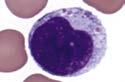
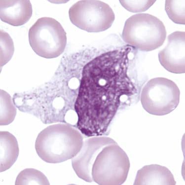
MONOCYTES
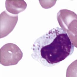
Monocytes (Fig. 1.6), as with granulocytes, are produced mainly in the bone marrow. Monocytes, like granulocytes, are derived from the myeloid cell line. They make up about 4% to 6% of normal circulating
leukocytes, ranging from 2% to 10%, depending on the laboratory or author. Monocytes are the largest of the normal leukocytes.
The nucleus is fairly large; it may be round, oval, indented, lobular, notched, or rarely even segmented, but most frequently it is indented or horseshoe shaped. The nuclear chromatin stains light purple and is delicate or lacy. Chromatin and parachromatin are sharply segregated, and the chromatin is distributed in a linear arrangement of delicate strands, which gives the nucleus a stringy appearance. The nuclear membrane is delicate but not distinct, and nucleoli usually are not seen.
The cytoplasm is abundant and stains gray or gray-blue. It may contain numerous small, poorly defined granules, resulting in a “ground-glass” appearance, and is often vacuolated. Extremely fine and abundant azurophilic granules are present; this granulation is called azure dust and is seen only in monocytes. The granules vary in color from light pink to bright purplish red. In addition, phagocytized particles may be seen in the cytoplasm.
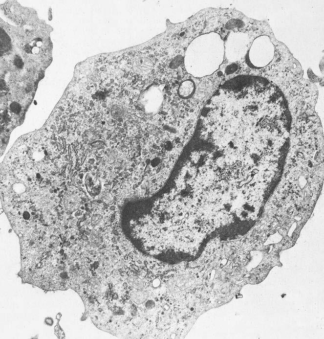
Macrophages
The macrophage (Fig. 1.7) and its precursors are widely distributed throughout the body. Macrophages have long been viewed as major components of the innate immune system as a result of their ability to coordinate directional movement with the detection and elimination of foreign entities. Macrophages also facilitate tissue repair and remodeling, resolution of inflammation, maintenance of homeostasis, and disease progression.
Although distinct macrophage subsets populate the developing embryo and fetus in three distinct waves, little is known about the functional differences between in utero macrophage populations and how they might contribute to fetal and neonatal immunity. Macrophage populations may share common functional properties in the moments after their differentiation from myeloid progenitors during fetal hematopoiesis. Upon trafficking to the circulation and into various tissues, the local microenvironment then shapes macrophage biology and promotes specialized, tissue-specific functions.
An initial concept was that tissue macrophages were derived from multiple organs in early-stage monocyte differentiation, the kinetics of tissue macrophage repopulation by circulating monocytes, and macrophage proliferation in tissues. Now it is recognized that a steady-state maintenance of tissue macrophage populations is not the result of the recruitment of circulating macrophages, but rather of proliferation of tissue-resident cells. There is a fundamental distinction in macrophage
Fig. 1.6 Monocyte. (From Rodak BF, Carr JH: Clinical hematology atlas, ed 4, St. Louis, 2013, Elsevier.)
populations and function. Tissue homeostasis is maintained by resident macrophages established during development; macrophages generated from adult hematopoiesis participate predominantly in response to tissue insult and disease resolution. Resident macrophages can be distinguished from macrophages that enter tissues from the circulation because they tend to express higher levels of the G-protein–coupled receptor F4/80.
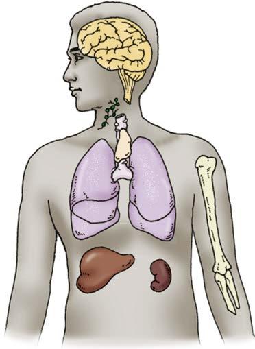
Specialized macrophages, such as the pulmonary alveolar macrophages, are the so-called dust phagocytes of the lung that function as the first line of defense against inhaled foreign particles and bacteria. Macrophages line the endothelium of capillaries and the sinuses of organs such as the bone marrow, spleen, and lymph nodes. Kupffer cells, also known as stellate macrophages, are specialized macrophages located in the liver, lining the walls of the sinusoids that form part of the MPS (Fig. 1.8).
Functionally the most important step in the maturation of macrophages is the mediator (cytokine)–driven conversion of the normal resting macrophage to the activated macrophage. Macrophages can be activated during infection by the release of macrophage-activating cytokines, such as interferon-gamma (IFN-γ) and granulocyte colony-stimulating factor (G-CSF), from T lymphocytes specifically sensitized to antigens from the infecting microorganisms. This interaction constitutes the basis of cell-mediated immunity. In addition, macrophages exposed to an endotoxin release a hormone, tumor necrosis factor α (TNF-α), or cachectin, which can activate macrophages itself under certain in vitro conditions.
The terminal stage of development in the mononuclear phagocyte cell line is the multinucleated giant cell, which characterizes
granulomatous inflammatory diseases, such as tuberculosis. Both monocytes and macrophages can be shown in the lesions in these diseases before the formation of giant cells, thought to be precursors of the multinucleated cells.
Macrophages and neutrophils have different characteristics, but they are the most effective phagocytic cells (Table 1.4).
Dendritic Cells
Dendritic cells (DCs) (also known as accessory cells ) resemble the microscopic appearance of nerve cells (i.e., dendrites) with long, stringlike extensions. DCs act as sentinels in tissues that respond to microbes by producing soluble substances, cytokines. They act as messengers between the innate and the adaptive immune systems.
The common portals of entry for microbes are the skin and the gastrointestinal, respiratory, and urogenital tracts. These tissue locations contain the classic, or conventional, DCs (Table 1.5). DCs or macrophages residing in tissues capture microbes or antigens and present processed antigen to lymphocytes; these are the first steps in the development of an adaptive immune response.
The main function of tissue DCs is to process antigens and present them on the cell surface to the T lymphocytes of the immune system. Other antigen-presenting cells (macrophages and B lymphocytes) present antigens to differentiated effector T lymphocytes in various immune responses.
Follicular dendritic cells reside in the germinal centers of lymphoid follicles in peripheral lymphoid organs and display antigens that stimulate the differentiation of B lymphocytes in the follicles.
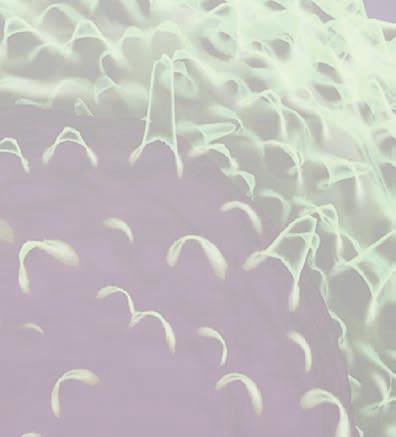
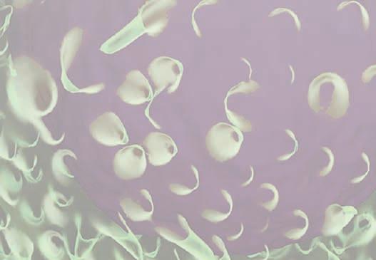
Fig. 1.7 Electron Micrograph of a Macrophage. (From Barrett JT: Textbook of immunology, ed 5, St. Louis, 1988, Mosby.)
Lungs (pulmonary alveolar macrophages)
Liver (Kupffer cells)
Kidney (glomerular mesangial cells)
Connective tissue (tissue macrophages) or histiocytes
Nervous tissue (microglial cells)
Lymph nodes (macrophages)
Bone (osteoclasts)
Spleen (macrophages)
Fig. 1.8 Mononuclear Phagocyte System. (Adapted from Roitt IM: Essential immunology, ed 5, Oxford, 1984, Blackwell Scientific.)
TABLE 1.4 Comparison of Characteristics of Neutrophils and Macrophages
Neutrophils
Life cycle Highly variable concentration in peripheral circulating blood, but react with increased concentration in infection
Only migrate to inflamed tissue injury
Die a few hours after migrating into tissue and encountering pathogen
Killing of pathogens
Interaction with other immune components
Phagocytosis
Kill pathogens by release of toxic molecules and enzymes
Produce neutrophil cellular trapsa
Short-lived secretion of soluble mediators, chemokines, that recruits more neutrophils to the site of inflammation/infection
Respond to soluble mediator interleukin (IL-17) from the adaptive immune system
Do not provide many signals to interact with adaptive immune system
Monocytes-Macrophages
Consistent concentration produced at a steady state
Migrate into tissues without the presence of inflammation
Can survive for many years after encountering pathogens
Phagocytosis
Kill pathogens by release of toxic molecules and enzymes
Recruit neutrophils to site of inflammation/infection by secreting soluble mediators, cytokines
Respond to soluble mediator, interferon, from adaptive immune system
Stimulate adaptive immune system by presenting processed antigen and secreting soluble mediators, cytokines
aNeutrophil extracellular traps (NETs) are networks of extracellular fibers, primarily composed of DNA from neutrophils, which bind pathogens. NETs are a first line of defense against infection because NETs kill invading pathogens by engulfing microbes and secreting antimicrobial mediators.
Adapted from Helbert M: Immunology for medical students, ed 3, St. Louis, 2017, Elsevier.
TABLE 1.5 Classic Human Dendritic Cell Subsets
Major
Major proposed functions Innate immunity: Source of inflammatory cytokines
Adaptive immunity: Capture and presentation of antigens mostly to CD4+ T lymphocytes
Cross Presentinga
Adaptive immunity: Capture and cross presentation of antigens to CD8+ T lymphocytes
Plasmacytoid Dendritic Cellsb
Antiviral immunity: Early innate response; priming of antiviral T lymphocytes
aPresentation of exogenous antigens on major histocompatibility complex (MHC) class I molecules, which is essential for the initiation of CD8+ T cell responses.
bA rare type of immune cell that circulates in the blood and is found in peripheral lymphoid organs. These cells secrete large quantities of type 1 interferon (IFN) in response to a viral infection.
Adapted from Abbas AK, Lichtman AH, Pillai S: Cellular and molecular immunology, ed 9, Philadelphia, 2018, Elsevier.
LYMPHOCYTE VARIATIONS
The adaptive immune system is composed of the cellular and humoral systems. Each of the two arms of the adaptive immune system has fundamental mechanisms allowing the body to attack an invading pathogen. The immunologically specific cellular component of the immune system is organized around two classes of specialized cells, T lymphocytes and B lymphocytes. Lymphocytes recognize foreign antigens, directly destroy some cells, or produce antibodies as plasma cells.
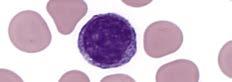
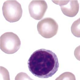
Virgin, or naïve, lymphocytes (Fig. 1.9) are mature T or B lymphocytes that have not encountered or been stimulated by a foreign antigen. Naïve and memory lymphocytes are both called resting lymphocytes because they are not actively dividing or performing immune functions.
A newly discovered lymphocyte, the innate lymphoid cell, straddles the innate and adaptive immune systems. Unlike lymphocytes associated with the adaptive immune system, innate lymphoid cells cannot perform gene rearrangement of their antigen receptors. Although conventional classes of lymphocytes greatly outnumber innate lymphoid cells, innate cells have been implicated in a variety of diseases, especially ones associated with mucosal surfaces such as the skin, lung, and gut.
KEY CONCEPTS: Facts About Lymphocyte Development
• In mammalian immunologic development the precursors of lymphocytes arise from progenitor cells of the yolk sac and liver in the embryo.
• Later in fetal development and throughout the life cycle, the bone marrow becomes the sole provider of undifferentiated progenitor cells, which can further develop into lymphoblasts.
• Continued cellular development and proliferation of lymphoid precursors occur as the cells travel to the primary and secondary lymphoid tissues.
• Mature lymphocytes differentiate into the main categories of T lymphocytes or B lymphocytes and include different subsets of highly specialized lymphocytes.
• T cells arise in the thymus from fetal liver or bone marrow precursors that seed the thymus during embryonic development.
• B lymphocytes most likely mature in the bone marrow and function primarily in antibody production or the formation of antibodies.
• Plasma cells arise as the end stage of B cell differentiation into a large, activated plasma cell.
• An increase in the number of lymphocytes is associated with viral infections.
• NK lymphocytes are very important in host defense and are recognized as a unique and important part of the immune system with roles in both infectious disease defense and tumor surveillance.
• A newly discovered type of lymphocyte, the innate lymphoid cell, straddles the innate and adaptive immune systems.
Lymphocyte Development
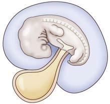
In mammalian immunologic development the precursors of lymphocytes arise from progenitor cells of the yolk sac and liver in the embryo (Fig. 1.10). Later in fetal development and throughout the life cycle the bone marrow becomes the sole provider of undifferentiated progenitor cells, which can further develop into lymphoblasts. Continued cellular development and proliferation of lymphoid precursors occur as the cells travel to the primary and secondary lymphoid tissues. Mature lymphocytes differentiate into the main categories of T lymphocytes or B lymphocytes (Table 1.6) and include different subsets of highly specialized lymphocytes (discussed in Chapter 4).
Naïve lymphocytes must see proteins displayed by dendritic cells to initiate clonal expansion and differentiation of T lymphocytes into effector and memory cells. Naïve lymphocytes produce cytokines similar to helper T cells but do not express T cell antigen receptors.
Lymphocytes make up about 34% of the leukocytes in the normal adult. Infants and children normally have more lymphocytes and fewer neutrophils than adults, a reversed differential. Lymphocytes fall into two general groups, small and large. Most normal lymphocytes are small. When observed microscopically, lymphocytes are described based on their size and cytoplasmic granularity. Small lymphocytes are found in the greatest numbers. The small lymphocyte is composed chiefly of nucleus and is the type of lymphocyte predominating in normal adult blood. It is about the same size as a normocytic (nucleated) RBC and is a useful size marker during examination of the peripheral blood film, especially in cases of megaloblastic anemia, in which all cell forms other than lymphocytes are increased in diameter.
The nucleus is round or slightly notched, and the nuclear chromatin is in the form of coarse, dense, deeply staining blocks. There is relatively little parachromatin, and it is not very distinct. Almost the entire nucleus stains deep purple. The nuclear membrane is heavy and distinct, and nucleoli are not usually seen. The cytoplasm appears in the form of a narrow band that stains pale blue with few, if any, red (azure) granules.
The large lymphocyte shows a further increase in the size of the nucleus and an increase in the relative amount of cytoplasm. The nucleus contains more parachromatin and thus stains more lightly than the nuclei of the smaller forms. The chromatin is still present in clumps, without distinct outlines because of the blending of chromatin and parachromatin. The nuclear membrane is distinct, and nucleoli usually are not seen. The cytoplasm in this form can be abundant, and azure granules are frequently seen. The cytoplasm color varies from colorless to a clear light or medium blue. The cytoplasm of the large granular lymphocyte can be deeply basophilic. Morphologically T and B lymphocytes appear identical on a Wright-stained blood film, but
Fig. 1.9 Lymphocytes. (From Rodak BF, Carr JH: Clinical hematology atlas, ed 4, St. Louis, 2013, Elsevier.)
Yolk sac Phar yngeal pouches
Fig. 1.10 Development of Immunologic Organs. The anatomy of the human fetus illustrates the development of the mammalian immune system. Cells of the pharyngeal pouches migrate into the chest and form the thymus. Precursors of lymphocytes originate early in embryonic life in the yolk sac and eventually migrate to the bone marrow via the spleen and liver.
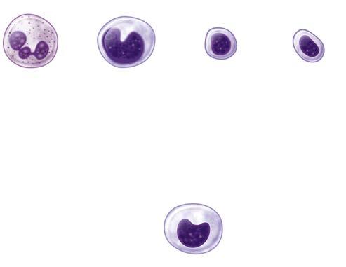
TABLE 1.6 Stages of Maturation of T and B Lymphocytes
Stages of Maturation Stem Cell Immature Cell Mature Cell
T Lymphocyte
Anatomic location Bone marrow Thymus Peripheral blood
Response to antigen
B Lymphocyte
None
Positive and negative selection
Activation (and differentiation)
Anatomic location Bone marrow Bone marrow/ peripheral blood Peripheral blood
Adapted from Turgeon ML: Clinical hematology, ed 6, Philadelphia, 2018, Lippincott.
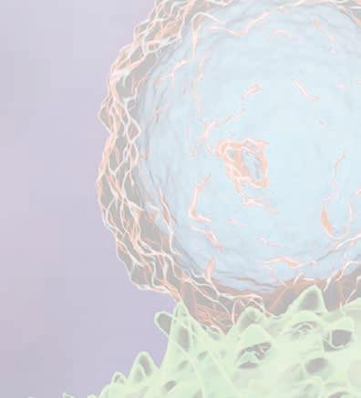
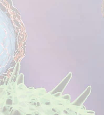
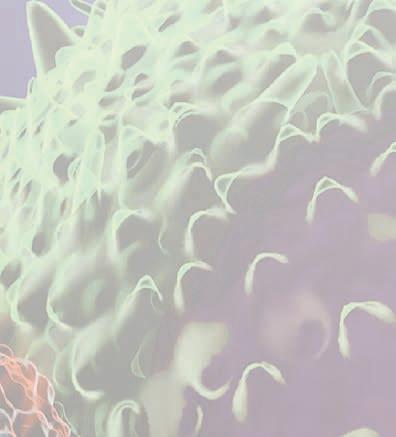
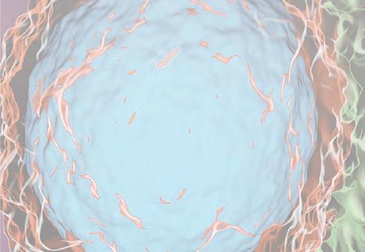
their appearance is very different with scanning electron microscopy (Fig. 1.11)
After antigenic stimulation, small lymphocytes can undergo transformation. These transformed cells appear large on Wright-stained films, with a relatively large amount of deep blue cytoplasm, and are called large granular lymphocytes. The large nucleus has a reticular appearance, with uniform chromatin and prominent nucleoli. Such cells have various names, including reactive, atypical, variant, and reticular lymphocytes.
Nucleoli can be observed in reactive or neoplastic lymphocyte nucleoli but are not routinely observed in normal resting lymphocytes.
An increase in the number of lymphocytes is associated with viral infections. It is characteristic of certain acute infections, such as infectious mononucleosis; pertussis, mumps, and rubella; and German measles, and of chronic infections, such as tuberculosis, brucellosis, and infectious hepatitis. The changes seen in these diseases have been referred to as reactive or atypical changes and are particularly associated with infectious mononucleosis. The cells are called reactive lymphocytes because the increased amount and apparent activity of the cytoplasm indicate that it may be reacting to some sort of stimulus. These cells can be referred to as variant forms.
Lymphocytes act to direct the immune response system of the body. Maturation of lymphocytes in the bone marrow or thymus results in cells that are immunocompetent. The cells are able to respond to antigenic challenges by directing the immune responses of the host defense.
They migrate to various sites in the body to await antigenic stimulus and activation. Only when immunologic studies are performed can these cells be identified as belonging to specific subsets of lymphocytes. As lymphocytes mature, their identity and function are specified by the antigenic structures on their external membrane surface.
T Cells
T cells arise in the thymus from fetal liver or bone marrow precursors that seed the thymus during embryonic development. These CD34+ progenitor cells develop in the thymic cortex. B lymphocytes are derived from hematopoietic stem cells by a complex series of differentiation events. T lymphocytes mature in the thymus and function in cell-mediated immune responses, such as delayed hypersensitivity, graft-versus-host reactions, and allograft rejection. T cells make up the majority of the lymphocytes circulating in the peripheral blood. In the periphery of the thymus they further differentiate into multiple different T cell subpopulations with different functions, including cytotoxicity and the secretion of soluble factors (cytokines). Many different cytokines have been identified, including 25 interleukin molecules and more than 40 chemokines; their functions include growth promotion, differentiation, chemotaxis, and cell stimulation.
B Cells
B lymphocytes most likely mature in the bone marrow and function primarily in antibody production or the formation of immunoglobulins. B cells constitute about 10% to 30% of the blood lymphocytes. Some activated B cells differentiate into memory B cells, long-lived cells that circulate in the blood. Memory B cells may live for years, but mature B cells that are not activated live only for days. B lymphocytes undergo blast transformation into plasma cells with appropriate antigen stimulation.
Mammalian B cell development encompasses a continuum of stages that begins in primary lymphoid tissue, such as human fetal liver and fetal or adult marrow, with subsequent functional maturation in secondary lymphoid tissue, such as human lymph nodes and spleen. The functional or protective endpoint is antibody production by terminally differentiated plasma cells. At least 10 distinct transcription factors regulate the early stages of B cell development.
B cells are derived from progenitor cells through an antigenindependent maturation process occurring in the bone marrow and gut-associated lymphoid tissue (GALT). Plasma cells or antibody-forming cells are terminally differentiated B cells. These cells are entirely devoted to antibody production, a primary host defense against microorganisms. Thymus