Imaging anatomy. ultrasound 2nd edition paula j. woodward - Download the ebook today and own the com

Page 1


https://ebookmass.com/product/imaging-anatomy-

Instant digital products (PDF, ePub, MOBI) ready for you

Download now and discover formats that fit your needs...

Diagnostic Imaging: Obstetrics 4th Edition Paula J. Woodward Md

https://ebookmass.com/product/diagnostic-imaging-obstetrics-4thedition-paula-j-woodward-md/

ebookmass.com

Chest, abdomen, pelvis 2nd Edition Federle

https://ebookmass.com/product/chest-abdomen-pelvis-2nd-editionfederle/

ebookmass.com

Fundamentals of Sectional Anatomy: An Imaging Approach 2nd Edition

https://ebookmass.com/product/fundamentals-of-sectional-anatomy-animaging-approach-2nd-edition/

ebookmass.com

Marketing:

The Core 9TH Edition (International Edition) Roger A. Kerin

https://ebookmass.com/product/marketing-the-core-9th-editioninternational-edition-roger-a-kerin/

ebookmass.com

Artistic and Cultural Dialogues in the Late Medieval

Mediterranean 1st ed. Edition María Marcos Cobaleda

https://ebookmass.com/product/artistic-and-cultural-dialogues-in-thelate-medieval-mediterranean-1st-ed-edition-maria-marcos-cobaleda/

ebookmass.com

Empowerment Series: Social Work with Groups: Comprehensive Practice and Self-Care 10th Edition Charles Zastrow

https://ebookmass.com/product/empowerment-series-social-work-withgroups-comprehensive-practice-and-self-care-10th-edition-charleszastrow/

ebookmass.com

Pleading Innocence Carmen Black

https://ebookmass.com/product/pleading-innocence-carmen-black/

ebookmass.com

The New Politics Of Numbers: Utopia, Evidence And Democracy 1st Edition Andrea Mennicken

https://ebookmass.com/product/the-new-politics-of-numbers-utopiaevidence-and-democracy-1st-edition-andrea-mennicken/

ebookmass.com

Understanding Nutrition - Standalone Book 15th Edition

https://ebookmass.com/product/understanding-nutrition-standalonebook-15th-edition-ellie-whitney/

ebookmass.com

https://ebookmass.com/product/etextbook-978-0132871693-engineeringvibration/

ebookmass.com

WOODWARD

GRIFFITH | ANTONIO | AHUJA
WONG | KAMAYA | WONG-YOU-CHEONG

SECOND EDITION

Paula J. Woodward, MD

Professor

of Radiology

David G. Bragg, MD and Marcia R. Bragg Presidential Endowed Chair in Oncologic Imaging

Adjunct Professor of Obstetrics and Gynecology

University of Utah School of Medicine

Salt Lake City, Utah

James F. Griffith, MD, MRCP, FRCR

Professor

Department of Imaging and Interventional Radiology

The Chinese University of Hong Kong

Hong Kong (SAR), China

Gregory E. Antonio, MD, DRANZCR, FHKCR

Honorary Professor

Department of Imaging and Interventional Radiology

The Chinese University of Hong Kong

Consultant Radiologist

Scanning Department

St. Teresa’s Hospital

Hong Kong (SAR), China

Anil T. Ahuja, MBBS (Bom), MD (Bom), FRCR, FHKCR, FHKAM (Radiology)

Professor of Diagnostic Radiology & Organ Imaging

Faculty of Medicine

The Chinese University of Hong Kong

Prince of Wales Hospital

Hong Kong (SAR), China

K. T. Wong, MBChB, FRCR, FHKCR, FHKAM (Radiology)

Consultant & Clinical Associate Professor (Honorary) Department of Imaging and Interventional Radiology

Prince of Wales Hospital

Faculty of Medicine

The Chinese University of Hong Kong

Hong Kong (SAR), China

Aya Kamaya, MD, FSRU, FSAR

Associate Professor of Radiology

Director, Stanford Body Imaging Fellowship

Stanford University School of Medicine

Stanford, California

Jade Wong-You-Cheong, MBChB, MRCP, FRCR

Professor

Department of Diagnostic Radiology and Nuclear Medicine

University of Maryland School of Medicine

Director of Ultrasound

University of Maryland Medical Center

Baltimore, Maryland

1600 John F. Kennedy Blvd.

Ste 1800 Philadelphia, PA 19103-2899

IMAGING ANATOMY: ULTRASOUND, SECOND EDITION

Copyright © 2018 by Elsevier. All rights reserved.

ISBN: 978-0-323-54800-7

No part of this publication may be reproduced or transmitted in any form or by any means, electronic or mechanical, including photocopying, recording, or any information storage and retrieval system, without permission in writing from the publisher. Details on how to seek permission, further information about the Publisher’s permissions policies and our arrangements with organizations such as the Copyright Clearance Center and the Copyright Licensing Agency, can be found at our website: www.elsevier.com/permissions.

This book and the individual contributions contained in it are protected under copyright by the Publisher (other than as may be noted herein).

Notices

Knowledge and best practice in this field are constantly changing. As new research and experience broaden our understanding, changes in research methods, professional practices, or medical treatment may become necessary.

Practitioners and researchers must always rely on their own experience and knowledge in evaluating and using any information, methods, compounds, or experiments described herein. In using such information or methods they should be mindful of their own safety and the safety of others, including parties for whom they have a professional responsibility.

With respect to any drug or pharmaceutical products identified, readers are advised to check the most current information provided (i) on procedures featured or (ii) by the manufacturer of each product to be administered, to verify the recommended dose or formula, the method and duration of administration, and contraindications. It is the responsibility of practitioners, relying on their own experience and knowledge of their patients, to make diagnoses, to determine dosages and the best treatment for each individual patient, and to take all appropriate safety precautions.

To the fullest extent of the law, neither the Publisher nor the authors, contributors, or editors, assume any liability for any injury and/or damage to persons or property as a matter of products liability, negligence or otherwise, or from any use or operation of any methods, products, instructions, or ideas contained in the material herein.

Publisher Cataloging-in-Publication Data

Names: Woodward, Paula J.

Title: Imaging anatomy. Ultrasound / [edited by] Paula J. Woodward. Other titles: Ultrasound.

Description: Second edition. | Salt Lake City, UT : Elsevier, Inc., [2017] | Includes bibliographical references and index.

Identifiers: ISBN 978-0-323-54800-7

Subjects: LCSH: Human anatomy--Handbooks, manuals, etc. | Ultrasonic imaging--Handbooks, manuals, etc. | MESH: Ultrasonography--methods--Atlases. | Anatomy, Cross-Sectional--Atlases.

Classification: LCC QM25.I43 2017 | NLM WN 17 | DDC 616.07’543--dc23

International Standard Book Number: 978-0-323-54800-7

Cover Designer: Tom M. Olson, BA

Cover Art: Richard Coombs, MS

Printed in Canada by Friesens, Altona, Manitoba, Canada

Last digit is the print number: 9 8 7 6 5 4 3 2 1

Dedication

To Anthony

Why? You know why!

But may you keep asking why (and why not) throughout your life.

In those questions, you’ll find a marvelous adventure.

Love, Lala

Contributing Authors

Jill M. Abrigo, MD, DPBR

Clinical Tutor

Department of Diagnostic Radiology and Organ Imaging

The Chinese University of Hong Kong Hong Kong (SAR), China

Shweta Bhatt, MD

Associate Professor

Department of Imaging Sciences

University of Rochester Medical Center Rochester, New York

Winnie C. W. Chu, MBChB, FRCR

Professor

Department of Diagnostic Radiology and Organ Imaging

The Chinese University of Hong Kong Hong Kong (SAR), China

Richard E. Fan, PhD

Engineering Research Associate Department of Urology

Stanford University School of Medicine Stanford, California

Bryan R. Foster, MD

Assistant Professor

Department of Radiology

Oregon Health & Science University Portland, Oregon

Simon S. M. Ho, MBBS, FRCR

Assistant Professor

Department of Diagnostic Radiology and Organ Imaging

The Chinese University of Hong Kong Hong Kong (SAR), China

Stella Sin Yee Ho, RDMS, RVT, PhD

Adjunct Associate Professor

Department of Imaging & Interventional Radiology

Prince of Wales Hospital

Faculty of Medicine

The Chinese University of Hong Kong Hong Kong (SAR), China

Anne Kennedy, MD

Professor of Radiology

Adjunct Professor of Obstetrics and Gynecology

Executive Vice Chair of Radiology

Codirector of Maternal Fetal Diagnostic Center

University of Utah School of Medicine

Salt Lake City, Utah

Barton F. Lane, MD

Assistant Professor

Clinical Director of CT

Department of Diagnostic Radiology and Nuclear Medicine

University of Maryland School of Medicine

Baltimore, Maryland

Ryan K. L. Lee, MBChB, FRCR, FHKAM (Radiology)

Associate Consultant and Clinical Assistant

Professor (Honorary)

Department of Imaging and Interventional Radiology

Prince of Wales Hospital Faculty of Medicine

The Chinese University of Hong Kong

Hong Kong (SAR), China

Yolanda Y. P. Lee, MBChB, FRCR, FHKCR, FHKAM (Radiology)

Associate Consultant and Clinical Associate Professor (Honorary)

Department of Imaging and Interventional Radiology

Prince of Wales Hospital

Faculty of Medicine

The Chinese University of Hong Kong

Hong Kong (SAR), China

Vivian Y. F. Leung, PhD, RDMS

Adjunct Assistant Professor

Department of Diagnostic Radiology and Organ Imaging

The Chinese University of Hong Kong

Hong Kong (SAR), China

Eric K. H. Liu, PhD, RDMS

Adjunct Associate Professor

Department of Imaging and Interventional Radiology

The Chinese University of Hong Kong

Hong Kong (SAR), China

Chander Lulla, MD, DMRD

Consultant Sonologist

RIA Clinic

Mumbai, India

Thomas A. Miller, DO

Assistant Professor of Pediatrics

Division of Pediatric Cardiology

University of Utah Salt Lake City, Utah

L. Nayeli Morimoto, MD

Clinical Instructor

Department of Radiology

Stanford University School of Medicine Stanford, California

Alex W. H. Ng, MBChB, FRCR, FHKCR, FHKAM (Radiology)

Consultant and Clinical Associate Professor (Honorary) Department of Imaging and Interventional Radiology

Prince of Wales Hospital

Faculty of Medicine

The Chinese University of Hong Kong Hong Kong (SAR), China

Bhawan K. Paunipagar, MBBS, MD, DNB

Senior Consultant Radiologist, Head of MRI/CT Division

Department of Radiology

Wockhardt Hospitals, South Mumbai Mumbai, Maharashtra, India

Michael D. Puchalski, MD

Professor of Pediatrics

Adjunct Professor of Radiology

Associate Director of Pediatric Cardiology Director of Non-Invasive Imaging

University of Utah/Primary Children’s Hospital Salt Lake City, Utah

Deyond Y. W. Siu, MBChB, FRCR

Honorary Clinical Tutor

Department of Diagnostic Radiology and Organ Imaging

The Chinese University of Hong Kong Hong Kong (SAR), China

Roya Sohaey, MD

Professor of Radiology

Adjunct Professor of Obstetrics and Gynecology

Director of Fetal Imaging

Oregon Health & Science University

Portland, Oregon

Sathi A. Sukumar, MBBS, FRCP (UK), FRCR

Consultant Radiologist

University Hospital of South Manchester Manchester, United Kingdom

Ali M. Tahvildari, MD

Staff Radiologist

VA Palo Alto Healthcare System

Palo Alto, California

Clinical Instructor (Affiliated) Department of Radiology

Stanford University School of Medicine

Stanford, California

Katherine To’o, MD

Staff Radiologist

Veterans Affairs Palo Alto Health Care System

Palo Alto, California

Ashish P. Wasnik, MD

Assistant Professor Department of Radiology Division of Abdominal Imaging University of Michigan Health System

Ann Arbor, Michigan

Nicole S. Winkler, MD

Assistant Professor of Radiology University of Utah Salt Lake City, Utah

Preface

Anatomy is the fundamental infrastructure upon which all comprehension of the human body builds, in both health and disease. It is essential to everyone who practices medicine but is critical to those of us who perform and interpret ultrasound. You cannot understand what is abnormal without a thorough understanding of what is normal. That is why we wrote this book. This second edition of Imaging Anatomy: Ultrasound is the single most detailed and inclusive ultrasound anatomy text available on the market.

I have always found studying anatomy a bit like reading the dictionary—there is a lot of fantastic information, but there isn’t much of a plot. Such a necessary topic is often difficult to approach. We have taken it as our mission, however, to break down those barriers and create an accessible anatomy text. Here is our story:

• The Characters: Each anatomic area (Brain & Spine, Head & Neck, Thorax, Abdomen, Pelvis, Extremities, and Developmental Anatomy) has its own complete cast of fascinating characters (organs). There is no hero in this book though; each is as important as the next, from the Parotid Gland to the Pelvic Floor to the Metatarsals and Toes. They all have their vital role to play.

• The Story Line: Every chapter begins with Gross Anatomy, followed by Imaging Anatomy, which includes best imaging techniques, helpful tips, and potential pitfalls. The tale is presented in an engaging, reader-friendly style. Convoluted descriptions are abandoned as key anatomic principles are outlined in a succinct, bulleted format for quick reference.

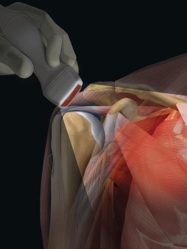
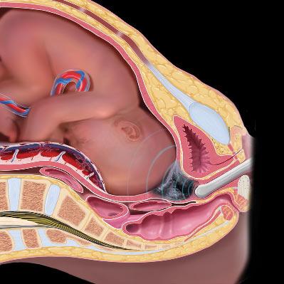
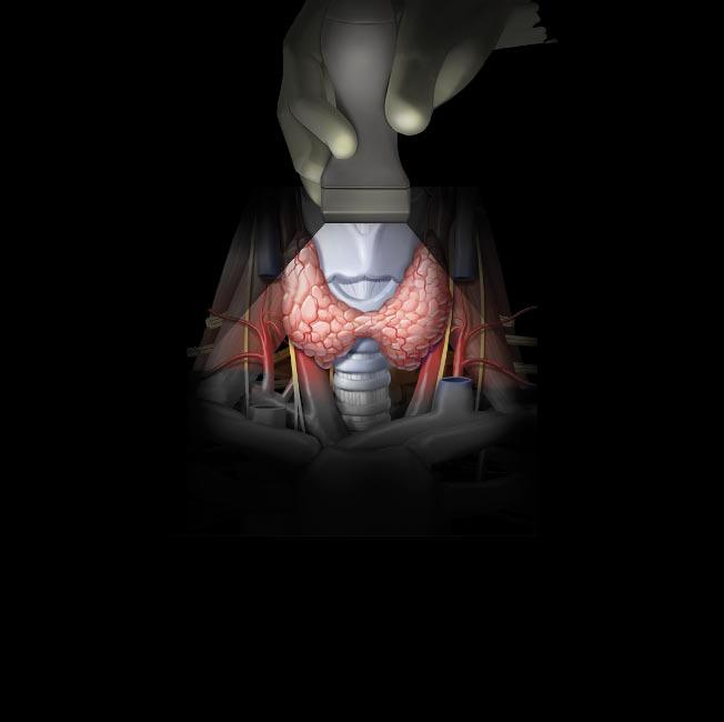
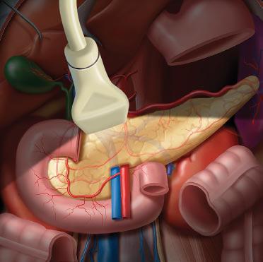
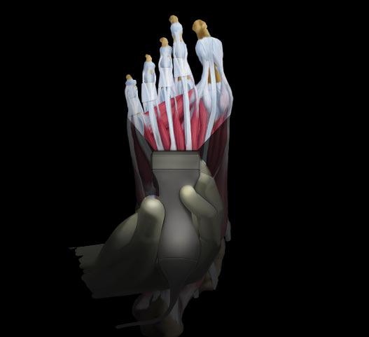
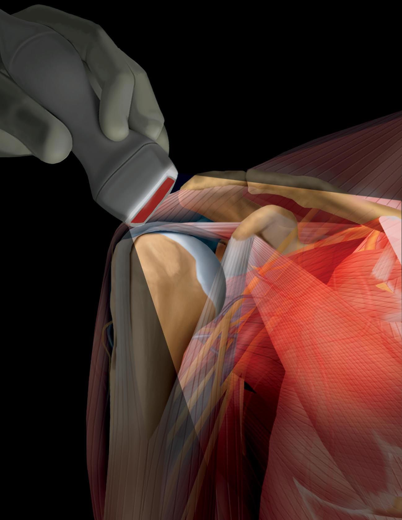
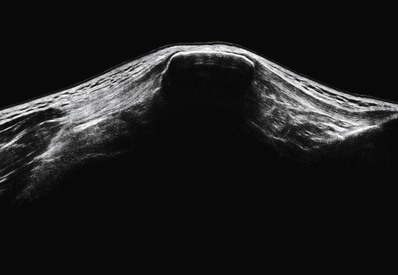
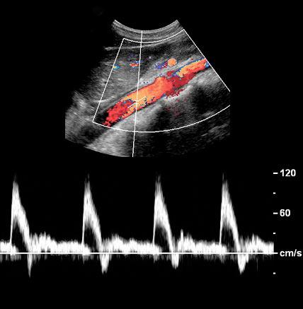
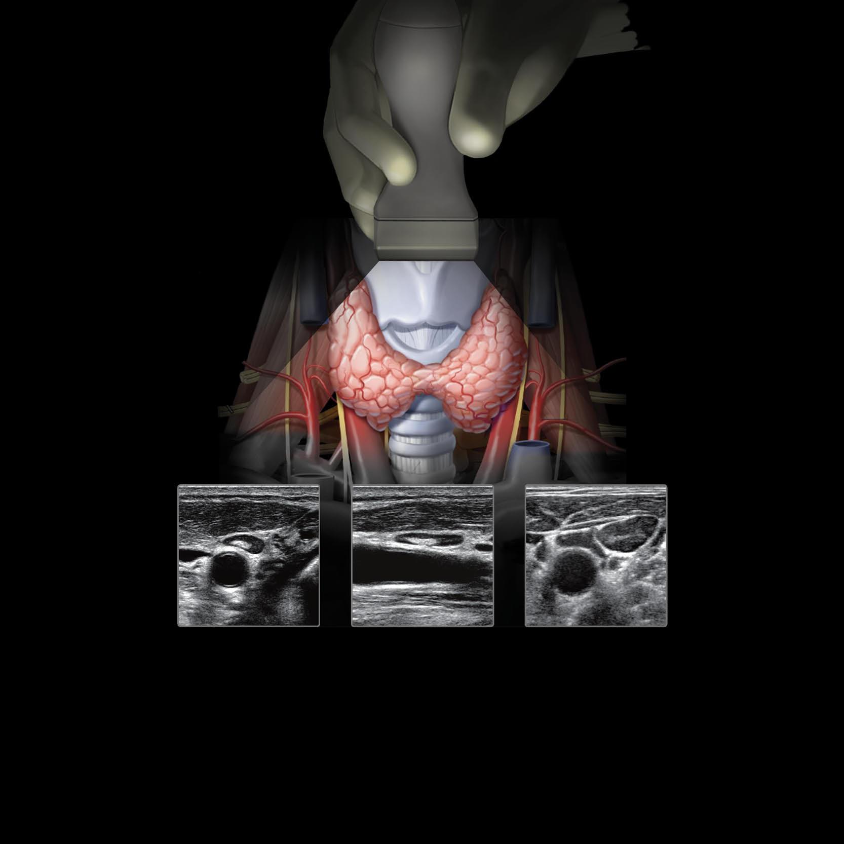
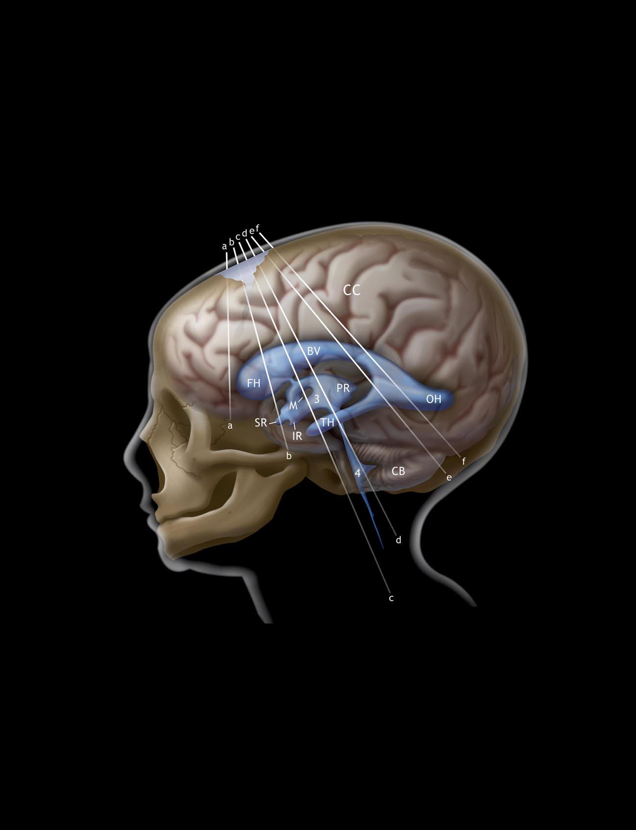
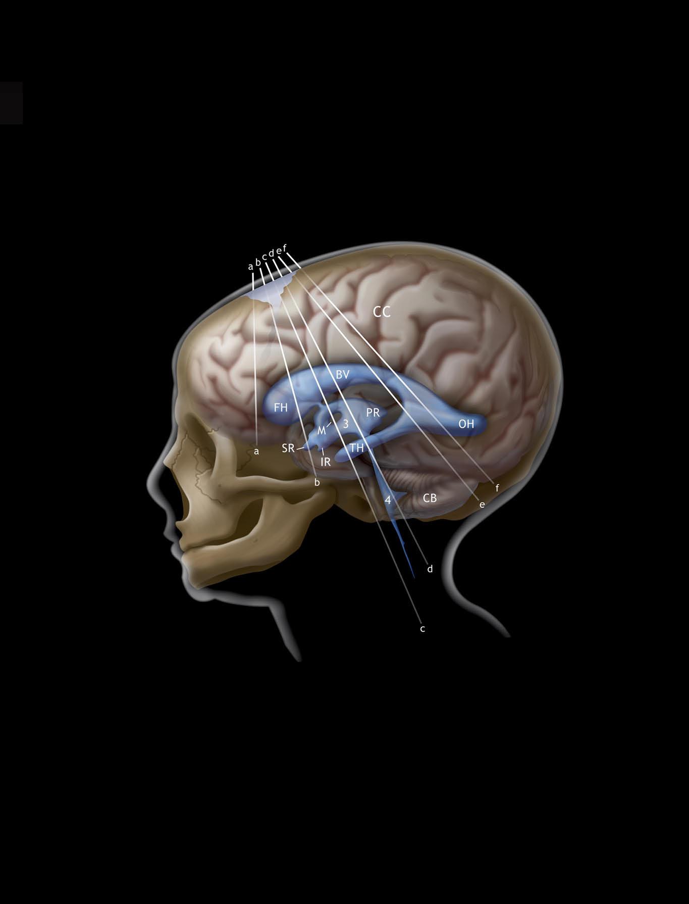
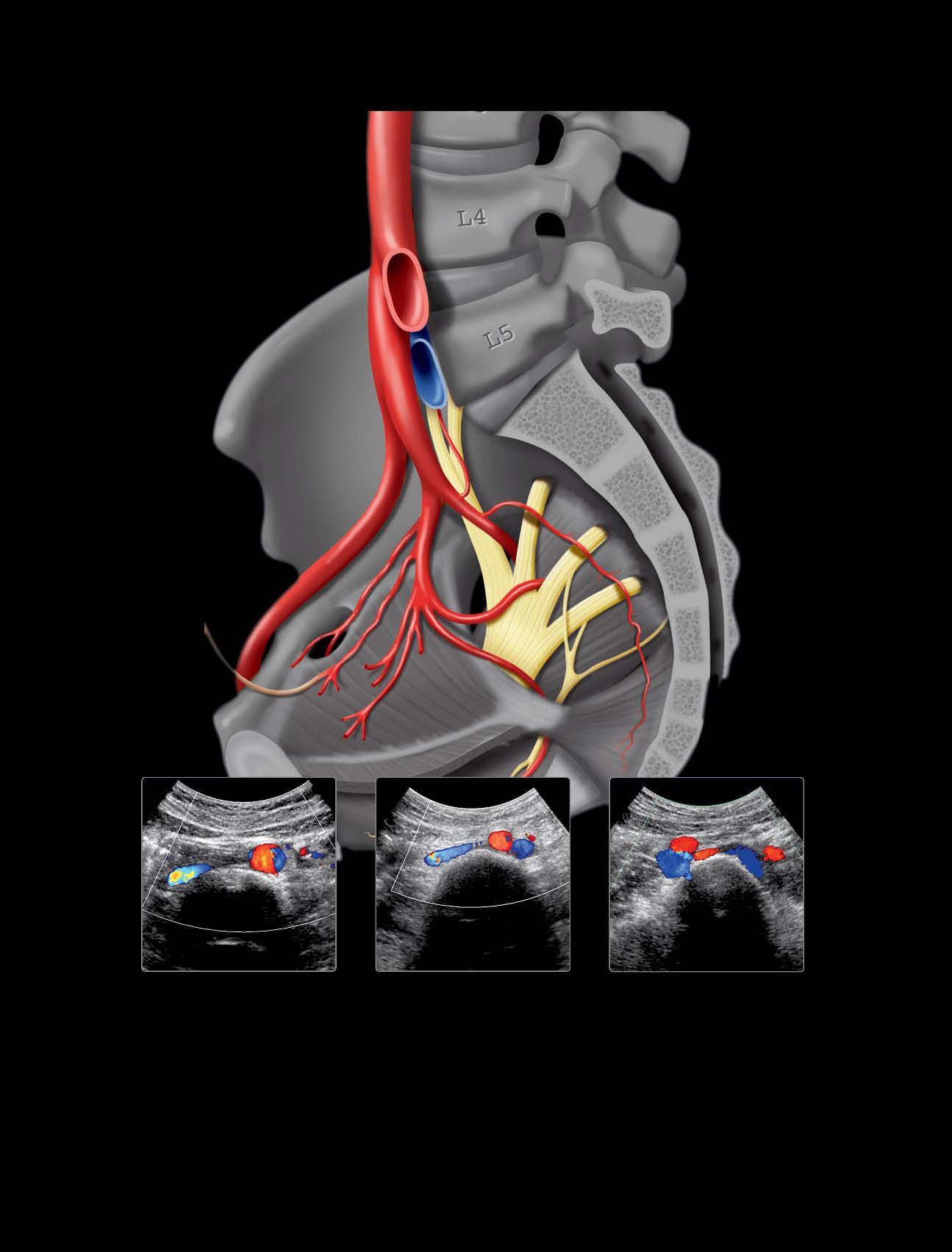
• The Illustrations: Never before has there been such a beautifully illustrated ultrasound anatomy text. The graphics, created by our own very talented group of medical illustrators, are of extraordinary quality. Those alone would make this book worth the read. But then following the graphics are extended galleries of detailed, extensively labeled, high-quality ultrasound images. A page turner for certain.

• The Authors: Given the expansive scope of this book, it required experts in all the various anatomic regions. I am quite fortunate to have some brilliant sonologists leading and editing their areas: Drs. James Griffith (Musculoskeletal), Anil Ahuja (Head & Neck), and Aya Kamaya & Jade WongYou-Cheong (Abdomen & Pelvis). In addition to the physicians, I must acknowledge the talented sonographers whose fine work is highlighted throughout this book.

• The Editorial Staff: To publish any book (especially one of this complexity) takes an incredible group of individuals working behind the scenes to make it happen. I would like to thank the wonderful Elsevier Salt Lake City editorial and production staff, medical illustrators, and image editors—with a special shout out to Matt Hoecherl, who helped me immeasurably. I’m extremely lucky to work with you guys.

It is with a great deal of pride that we present to you the second edition of Imaging Anatomy: Ultrasound. While it might not be an epic thriller, it does have a compelling narrative to keep the reader engaged and informed throughout.

David G. Bragg, MD and Marcia R. Bragg Presidential Endowed Chair in Oncologic Imaging

Adjunct Professor of Obstetrics and Gynecology

University of Utah School of Medicine

Salt Lake City, Utah

Acknowledgments

Lead Editor

Matt W. Hoecherl, BS

Text Editors

Arthur G. Gelsinger, MA

Nina I. Bennett, BA

Terry W. Ferrell, MS

Lisa A. Gervais, BS

Karen E. Concannon, MA, PhD

Megg Morin, BA

Image Editors

Jeffrey J. Marmorstone, BS

Lisa A. M. Steadman, BS

Illustrations

Richard Coombs, MS

Lane R. Bennion, MS

Laura C. Wissler, MA

Art Direction and Design

Tom M. Olson, BA

Laura C. Wissler, MA

Production Coordinators

Rebecca L. Bluth, BA

Angela M. G. Terry, BA

Emily C. Fassett, BA

Sections

SECTION 1: Brain and Spine

SECTION 2: Head and Neck

SECTION 3: Thorax

SECTION 4: Abdomen

SECTION 5: Pelvis

SECTION 6: Upper Extremity

SECTION 7: Lower Extremity

SECTION 8: Obstetrics and Developmental Anatomy

TABLEOFCONTENTS

SECTION1:BRAINANDSPINE

4 ScalpandCalvarialVault

WinnieC.W.Chu,MBChB,FRCRandVivianY.F.Leung, PhD,RDMS

8 Brain

PaulaJ.Woodward,MD

38 Orbit

PaulaJ.Woodward,MD,StellaSinYeeHo,RDMS,RVT, PhD,andDeyondY.W.Siu,MBChB,FRCR

50 TranscranialDoppler

StellaSinYeeHo,RDMS,RVT,PhD,DeyondY.W.Siu, MBChB,FRCR,andPaulaJ.Woodward,MD

74 VertebralColumnandSpinalCord

PaulaJ.Woodward,MD

SECTION2:HEADANDNECK

86 NeckOverview

K.T.Wong,MBChB,FRCR,FHKCR,FHKAM(Radiology), YolandaY.P.Lee,MBChB,FRCR,FHKCR,FHKAM (Radiology),andAnilT.Ahuja,MBBS(Bom),MD(Bom), FRCR,FHKCR,FHKAM(Radiology)

92 Sublingual/SubmentalRegion

K.T.Wong,MBChB,FRCR,FHKCR,FHKAM(Radiology), YolandaY.P.Lee,MBChB,FRCR,FHKCR,FHKAM (Radiology),andAnilT.Ahuja,MBBS(Bom),MD(Bom), FRCR,FHKCR,FHKAM(Radiology)

98 SubmandibularRegion

K.T.Wong,MBChB,FRCR,FHKCR,FHKAM(Radiology), YolandaY.P.Lee,MBChB,FRCR,FHKCR,FHKAM (Radiology),andAnilT.Ahuja,MBBS(Bom),MD(Bom), FRCR,FHKCR,FHKAM(Radiology)

104 ParotidRegion

K.T.Wong,MBChB,FRCR,FHKCR,FHKAM(Radiology), YolandaY.P.Lee,MBChB,FRCR,FHKCR,FHKAM (Radiology),andAnilT.Ahuja,MBBS(Bom),MD(Bom), FRCR,FHKCR,FHKAM(Radiology)

112 UpperCervicalLevel

K.T.Wong,MBChB,FRCR,FHKCR,FHKAM(Radiology), YolandaY.P.Lee,MBChB,FRCR,FHKCR,FHKAM (Radiology),andAnilT.Ahuja,MBBS(Bom),MD(Bom), FRCR,FHKCR,FHKAM(Radiology)

118 MidcervicalLevel

K.T.Wong,MBChB,FRCR,FHKCR,FHKAM(Radiology), YolandaY.P.Lee,MBChB,FRCR,FHKCR,FHKAM (Radiology),andAnilT.Ahuja,MBBS(Bom),MD(Bom), FRCR,FHKCR,FHKAM(Radiology)

124 LowerCervicalLevelandSupraclavicularFossa

K.T.Wong,MBChB,FRCR,FHKCR,FHKAM(Radiology), YolandaY.P.Lee,MBChB,FRCR,FHKCR,FHKAM (Radiology),andAnilT.Ahuja,MBBS(Bom),MD(Bom), FRCR,FHKCR,FHKAM(Radiology)

130 PosteriorTriangle

K.T.Wong,MBChB,FRCR,FHKCR,FHKAM(Radiology), YolandaY.P.Lee,MBChB,FRCR,FHKCR,FHKAM (Radiology),andAnilT.Ahuja,MBBS(Bom),MD(Bom), FRCR,FHKCR,FHKAM(Radiology)

136 ThyroidGland

PaulaJ.Woodward,MD,K.T.Wong,MBChB,FRCR, FHKCR,FHKAM(Radiology),andAnilT.Ahuja,MBBS (Bom),MD(Bom),FRCR,FHKCR,FHKAM(Radiology)

144 ParathyroidGlands

PaulaJ.Woodward,MD,K.T.Wong,MBChB,FRCR, FHKCR,FHKAM(Radiology),andAnilT.Ahuja,MBBS (Bom),MD(Bom),FRCR,FHKCR,FHKAM(Radiology)

150 LarynxandHypopharynx

K.T.Wong,MBChB,FRCR,FHKCR,FHKAM(Radiology), YolandaY.P.Lee,MBChB,FRCR,FHKCR,FHKAM (Radiology),andAnilT.Ahuja,MBBS(Bom),MD(Bom), FRCR,FHKCR,FHKAM(Radiology)

158 TracheaandEsophagus

K.T.Wong,MBChB,FRCR,FHKCR,FHKAM(Radiology), AnilT.Ahuja,MBBS(Bom),MD(Bom),FRCR,FHKCR, FHKAM(Radiology),andPaulaJ.Woodward,MD

164 VagusNerve

K.T.Wong,MBChB,FRCR,FHKCR,FHKAM(Radiology), YolandaY.P.Lee,MBChB,FRCR,FHKCR,FHKAM (Radiology),andAnilT.Ahuja,MBBS(Bom),MD(Bom), FRCR,FHKCR,FHKAM(Radiology)

170 CarotidArteries

K.T.Wong,MBChB,FRCR,FHKCR,FHKAM(Radiology), YolandaY.P.Lee,MBChB,FRCR,FHKCR,FHKAM (Radiology),andAnilT.Ahuja,MBBS(Bom),MD(Bom), FRCR,FHKCR,FHKAM(Radiology)

184 VertebralArteries

K.T.Wong,MBChB,FRCR,FHKCR,FHKAM(Radiology), YolandaY.P.Lee,MBChB,FRCR,FHKCR,FHKAM (Radiology),andAnilT.Ahuja,MBBS(Bom),MD(Bom), FRCR,FHKCR,FHKAM(Radiology)

190 NeckVeins

K.T.Wong,MBChB,FRCR,FHKCR,FHKAM(Radiology), YolandaY.P.Lee,MBChB,FRCR,FHKCR,FHKAM (Radiology),andAnilT.Ahuja,MBBS(Bom),MD(Bom), FRCR,FHKCR,FHKAM(Radiology)

198 CervicalLymphNodes

TABLEOFCONTENTS

K.T.Wong,MBChB,FRCR,FHKCR,FHKAM(Radiology), YolandaY.P.Lee,MBChB,FRCR,FHKCR,FHKAM (Radiology),andAnilT.Ahuja,MBBS(Bom),MD(Bom), FRCR,FHKCR,FHKAM(Radiology)

SECTION3:THORAX

208 ThoracicOutlet

GregoryE.Antonio,MD,DRANZCR,FHKCR,EricK.H.Liu, PhD,RDMS,andPaulaJ.Woodward,MD

218 Pleura

PaulaJ.Woodward,MD,GregoryE.Antonio,MD, DRANZCR,FHKCR,andEricK.H.Liu,PhD,RDMS

224 Diaphragm

GregoryE.Antonio,MD,DRANZCR,FHKCR,EricK.H.Liu, PhD,RDMS,andPaulaJ.Woodward,MD

228 ChestWall

GregoryE.Antonio,MD,DRANZCR,FHKCR,EricK.H.Liu, PhD,RDMS,andPaulaJ.Woodward,MD

234 Breast

NicoleS.Winkler,MD

SECTION4:ABDOMEN

248 Liver

AyaKamaya,MD,FSRU,FSAR

272 BiliarySystem

L.NayeliMorimoto,MD

284 Spleen

AliM.Tahvildari,MDandPaulaJ.Woodward,MD

292 Pancreas

BartonF.Lane,MD

302 Kidneys

JadeWong-You-Cheong,MBChB,MRCP,FRCR

330 AdrenalGlands

PaulaJ.Woodward,MD

336 Bowel

SathiA.Sukumar,MBBS,FRCP(UK),FRCR

352 AbdominalLymphNodes

JadeWong-You-Cheong,MBChB,MRCP,FRCR

356 AortaandInferiorVenaCava

SimonS.M.Ho,MBBS,FRCR,JillM.Abrigo,MD,DPBR, andChanderLulla,MD,DMRD

386 PeritonealCavity

JadeWong-You-Cheong,MBChB,MRCP,FRCR

394 AbdominalWall

JadeWong-You-Cheong,MBChB,MRCP,FRCR

SECTION5:PELVIS

408 IliacArteriesandVeins

SimonS.M.Ho,MBBS,FRCR,JillM.Abrigo,MD,DPBR, andChanderLulla,MD,DMRD

424 UretersandBladder

AshishP.Wasnik,MDandPaulaJ.Woodward,MD

434 ProstateandSeminalVesicles

KatherineTo'o,MD,RichardE.Fan,PhD,andPaulaJ. Woodward,MD

446 TestesandScrotum

ShwetaBhatt,MDandPaulaJ.Woodward,MD

458 PenisandUrethra

PaulaJ.Woodward,MD

468 Uterus

BartonF.Lane,MDandPaulaJ.Woodward,MD

482 Cervix

BartonF.Lane,MD

488 Vagina

BartonF.Lane,MD

494 Ovaries

BryanR.Foster,MD

504 PelvicFloor

StellaSinYeeHo,RDMS,RVT,PhD,DeyondY.W.Siu, MBChB,FRCR,andPaulaJ.Woodward,MD

SECTION6:UPPEREXTREMITY

530 SternoclavicularandAcromioclavicularJoints

JamesF.Griffith,MD,MRCP,FRCRandBhawanK. Paunipagar,MBBS,MD,DNB

536 Shoulder

JamesF.Griffith,MD,MRCP,FRCRandBhawanK. Paunipagar,MBBS,MD,DNB

554 Axilla

JamesF.Griffith,MD,MRCP,FRCRandBhawanK. Paunipagar,MBBS,MD,DNB

562 Arm

JamesF.Griffith,MD,MRCP,FRCRandBhawanK. Paunipagar,MBBS,MD,DNB

570 ArmVessels

JamesF.Griffith,MD,MRCP,FRCRandBhawanK. Paunipagar,MBBS,MD,DNB

578 Elbow

JamesF.Griffith,MD,MRCP,FRCRandBhawanK. Paunipagar,MBBS,MD,DNB

598 Forearm

JamesF.Griffith,MD,MRCP,FRCRandBhawanK. Paunipagar,MBBS,MD,DNB

606 ForearmVessels

JamesF.Griffith,MD,MRCP,FRCRandBhawanK. Paunipagar,MBBS,MD,DNB 614 Wrist

JamesF.Griffith,MD,MRCP,FRCRandBhawanK. Paunipagar,MBBS,MD,DNB 628 Hand

JamesF.Griffith,MD,MRCP,FRCRandBhawanK. Paunipagar,MBBS,MD,DNB 640 HandVessels

JamesF.Griffith,MD,MRCP,FRCRandBhawanK. Paunipagar,MBBS,MD,DNB

646 Thumb

JamesF.Griffith,MD,MRCP,FRCRandBhawanK. Paunipagar,MBBS,MD,DNB

656 Fingers

JamesF.Griffith,MD,MRCP,FRCRandBhawanK. Paunipagar,MBBS,MD,DNB

668 BrachialPlexus

JamesF.Griffith,MD,MRCP,FRCR,K.T.Wong,MBChB, FRCR,FHKCR,FHKAM(Radiology),andPaulaJ. Woodward,MD

676 RadialNerve

TABLEOFCONTENTS

JamesF.Griffith,MD,MRCP,FRCRandBhawanK. Paunipagar,MBBS,MD,DNB

684 MedianNerve

JamesF.Griffith,MD,MRCP,FRCRandBhawanK. Paunipagar,MBBS,MD,DNB

694 UlnarNerve

JamesF.Griffith,MD,MRCP,FRCRandBhawanK. Paunipagar,MBBS,MD,DNB

SECTION7:LOWEREXTREMITY

706 GlutealMuscles

RyanK.L.Lee,MBChB,FRCR,FHKAM(Radiology),

GregoryE.Antonio,MD,DRANZCR,FHKCR,andEricK.H. Liu,PhD,RDMS

716 Groin

AlexW.H.Ng,MBChB,FRCR,FHKCR,FHKAM(Radiology),

GregoryE.Antonio,MD,DRANZCR,FHKCR,andEricK.H. Liu,PhD,RDMS

726 Hip

GregoryE.Antonio,MD,DRANZCR,FHKCRandEricK.H. Liu,PhD,RDMS

736 ThighMuscles

GregoryE.Antonio,MD,DRANZCR,FHKCRandEricK.H. Liu,PhD,RDMS

748 FemoralVesselsandNerves

GregoryE.Antonio,MD,DRANZCR,FHKCRandEricK.H. Liu,PhD,RDMS

762 Knee

GregoryE.Antonio,MD,DRANZCR,FHKCRandEricK.H. Liu,PhD,RDMS

780 LegMuscles

GregoryE.Antonio,MD,DRANZCR,FHKCRandEricK.H. Liu,PhD,RDMS

792 LegVessels

GregoryE.Antonio,MD,DRANZCR,FHKCR,EricK.H.Liu, PhD,RDMS,andPaulaJ.Woodward,MD

810 LegNerves

GregoryE.Antonio,MD,DRANZCR,FHKCRandEricK.H. Liu,PhD,RDMS

814 Ankle

GregoryE.Antonio,MD,DRANZCR,FHKCRandEricK.H. Liu,PhD,RDMS

832 Tarsus

GregoryE.Antonio,MD,DRANZCR,FHKCRandEricK.H. Liu,PhD,RDMS

846 FootVessels

GregoryE.Antonio,MD,DRANZCR,FHKCRandEricK.H. Liu,PhD,RDMS

852 MetatarsalsandToes

GregoryE.Antonio,MD,DRANZCR,FHKCRandEricK.H. Liu,PhD,RDMS

SECTION8:OBSTETRICSAND DEVELOPMENTALANATOMY

860 EmbryologyandAnatomyof1stTrimester

AnneKennedy,MD

872

EmbryologyandAnatomyofBrain

AnneKennedy,MD

888 EmbryologyandAnatomyofSpine

PaulaJ.Woodward,MD

894 EmbryologyandAnatomyofFaceandNeck

RoyaSohaey,MD

906 EmbryologyandAnatomyofChest

PaulaJ.Woodward,MD

914 EmbryologyandAnatomyofCardiovascularSystem

ThomasA.Miller,DOandMichaelD.Puchalski,MD

924 EmbryologyandAnatomyofAbdominalWalland GastrointestinalTract

PaulaJ.Woodward,MD

934 EmbryologyandAnatomyofGenitourinaryTract

PaulaJ.Woodward,MD

WOODWARD

GRIFFITH | ANTONIO | AHUJA
WONG | KAMAYA | WONG-YOU-CHEONG

Brain and Spine

ScalpandCalvarialVault

TERMINOLOGY

Definitions

•Fontanelle:Broadareasofconnectivetissueatjunctionof majorsutures

GROSSANATOMY

Overview

• Scalp

○Scalphas5layers

–Skin(epidermis,dermis,hair,sebaceousglands)

–Subcutaneoustissue(veryvascularfibroadipose tissue)

–Epicranialtissue(scalpmuscles,galeaaponeurotica)

–Subaponeurotictissue(looseareolarconnective tissue)

–Pericranium(periosteumofskull)

•Skull(28separatebones,mostlyconnectedbyfibrous sutures)

○Craniumhasseveralparts

–Calvarialvault

–Cranialbase

–Facialskeleton

○Calvarialvaultcomposedofseveralbones

–2frontalbonesseparatedbymetopicsuture

–Pairedparietalbones

–Squamousoccipitalbone

–Pairedsquamoustemporalbones

○3majorserratedfibrousjoints(sutures)connectbones ofvault

–Coronalsuture

–Sagittalsuture

–Lambdoidsuture

○Outer,innertables

–2thinplatesofcompactcorticalbone

–Separatedbydiploicspace(cancellousbone containingmarrow)

○Endocranialsurface

–Linedbyouter(periosteal)layerofdura

–Groovedbyvascularfurrows

–Mayhaveareasoffocalthinning(arachnoid granulations),foramina(emissaryveins)

IMAGINGANATOMY

Overview

•Scalp:Hypoechoic,5layerscannotbefurtherseparately resolved

•Calvariumechogenicouter/innertables;diploicspacefilled withfattymarrowandappearshypoechoic,sutureappears asgapbetweenechogeniccalvarium

•Frontalbones

○Frontalsinusesshowwidevariationinaeration

○Frontalbonesoftenappearthickened,hyperostotic (especiallyinolderfemales)

•Parietalbones

○Areasofparietalthinning,granularfoveolae(for arachnoidgranulations)commonadjacenttosagittal suture

○Innertablesoftenslightlyirregular(convolutional markingscausedbygyri),groovedbypairedmiddle meningealarteries+vein

•Occipitalbone

○Deeplygroovedbysuperiorsagittal,transversesinuses

○Internaloccipitalprotuberancemarkssinusconfluence (torcularHerophili)

•Temporalbones

○Thin,innersurfacegroovedbymiddlemeningealvessels

○Outersurfacegroovedbysuperficialtemporalartery

•Fontanelle:ProvideacousticwindowforUSexaminationof underlyingbrainparenchyma

○ Anteriorfontanelle

○Between2frontaland2parietalbones,usually disappearsbyage2

○Whenfused,correspondstobregma:Meetingof sagittal,coronalsutures

○ Posteriorfontanelle

○Small,usuallyclosesbetween3-6monthsofage

○Whenfused,correspondstolambda:Meetingofsagittal, lambdoidsutures

○Pterion

–Anterolateralfontanelle;closesbetween3-6months ofage

–H-shapedjunctionbetweenfrontal,parietalbones+ greatersphenoidwing,squamoustemporalbone

○Asterion

–Posterolateralfontanelle,persistsuntil2yearsofage

○ Mastoidfontanelle

○Locatedatjunctionoftemporosquamousandlambdoid sutures

○Persistsuntil2years

ANATOMYIMAGINGISSUES

ImagingRecommendations

•High-frequencylineararraytransducersprovidesuperb resolutionofnear-fieldstructures

•Goodskin-to-transducercouplingachievedbycopioususe ofacousticcouplinggel

•Superficialstandoffpadcanbeusedtoincreasedepthof focalzone

•UScanbeusedtoevaluatecranialsuturesandassists diagnosisofcraniosynostosis(prematurefusionofsutures)

EMBRYOLOGY

EmbryologicEvents

•Skullbaseformedfromendochondralossification

•Calvarialvaultformsviamembranousossification

○Curvedmesenchymalplatesappearatday30

○Extendtowardeachother,skullbase

○Aspairedbonesmeetinmidline,metopicandsagittal suturesareinduced(coronalsutureispresentfrom onsetofossification)

○Unossifiedcentersatedgesofparietalboneform fontanelles

○Vaultgrowsrapidlyin1stpostnatalyear

Sagittalsuture

Coronalsuture

ScalpandCalvarialVault

SCALPANDCALVARIALVAULT

Frontonasalsuture

Metopicsuture Anteriorfontanelle

Coronalsuture

Anterolateralfontanelle(pterion)

Posterolateral/mastoidfontanelle (asterion)

Anteriorfontanelle

Temporosquamosalsuture

Posteriorfontanelle

Lambdoidsuture

Mendosalsuture

Sweatglandandduct

Sebaceousgland

Hairfollicle

Subcutaneousfibroadiposetissue

Pericranium

Diploicspace

Epidermis

Dermis

Superficial,deepvascularplexi

Epicranialaponeurosis

Subaponeuroticareolartissue

Outertable,calvarium

Innertable,calvarium

Venous"lake"

(Top)Graphicdepictionofaninfantcranium,frontalview,isshown.Theanteriorfontanelleispresentbetween2frontaland2parietal bones,whichusuallycloseby2yearsofage.Whenfused,thissitecorrespondstobregma:Themeetingpointofsagittalandcoronal sutures.(Middle)Lateralviewofaninfantcalvarialvaultisshown.Theposteriorfontanelleissmallandusuallyclosesby3-6monthsof age.Whenfused,thiscorrespondstolambda:Themeetingofsagittalandlambdoidsutures.Theanterolateralfontanelle(pterion) closesat~3monthsofage.Theposterolateralfontanelle(asterion)oftenpersistsuntil2yearsofage.(Bottom)Scalpandcalvarium aredepictedincrosssection.The5scalplayersaredepicted.Skinconsistsofepidermisanddermis.Hairfolliclesandasebaceousgland, thesubcutaneousfibroadiposetissue,andsweatglandsandductsaswellassuperficialanddeepcutaneousvascularplexiareshown.

ScalpandCalvarialVault

Corticalv.insubarachnoidspace

Subarachnoidspace

USgel

Scalp

Outertableofcalvarium

Periosteumcoveringcalvarium

Innertableofcalvarium

Superiorsagittalsinus

Cerebralcortex

Subarachnoidspace

Cerebralsulcus

USgel

Scalp

Outertableofcalvarium

Innertableofcalvarium

Corticalv.insubarachnoidspace

Cerebralcortex

Scalp

Superiorsagittalsinus

Outertableofcalvarium

Diploicspace

Innertableofcalvarium

Subarachnoidspace

Cerebralhemisphere

(Top)AnteriorcoronalUSscanthroughtheanteriorfontanelleshowsthescalpcoveringthefrontalboneofthecalvarialvault.The5 layersofthescalpareasfollows:Skin,connectivetissueconsistingoflobulesoffat,arteryandemissaryvein,aponeurosis,loose connectivetissue,whichaccountsformobilityofthescalpontheunderlyingboneandperiosteumadheringtotheoutertableofskull. These5layers,however,cannotberesolvedbyUS.(Middle)Midsagittalscanthroughtheanteriorfontanelleshowsthescalpcovering thefrontalboneofthecalvarialvault.Beneaththeinnertableofthecalvarialvaultistheanechoicsubarachnoidspace.(Bottom) CoronalT2MRshowsthehypointenseouterandinnertableofcalvarium.Thescalpanddiploicspacearehyperintense.Thesuperior sagittalsinusappearsassignalvoidstructurebelowtheinnertableofcalvarium.

Suture

ScalpandCalvarialVault

Suture

(Top)CoronalUSthroughtheanteriorfontanelleshowsthescalpandcalvarium.Thereisdiscontinuityinahypoechoicbandextending fromtheoutertoinnertableofthecalvarialvault.Thisrepresentsanormalsuture.Thescalpappearshypoechoiccomparedtothe echogenicouterandinnertablesofthecalvarium.The5layersofthescalpcannotberesolvedbyUS.(Middle)CoronalUSthroughthe anteriorfontanelleshowsanothersutureofthecalvarialvault.Thewidthandcurvatureofsuturesisvariableandshouldnotbe mistakenforabonyfracture.(Bottom)SagittalT2MRofthescalpandcalvariumisshown.Thesuturelineappearswiththesame signalintensityastheouterandinnertableofcalvarium.Corticalveinscanbeseenwithinthehyperintensesubarachnoidspace,which isimmediatelyundertheinnertable.

Scalp
Calvarium
Calvarium
Scalp
Suture
Scalp
Corticalvv.insubarachnoidspace
Cerebralcortex
Outertableofcalvarium
Innertableofcalvarium
USANDT2WIMRCALVARIUM

Turn static files into dynamic content formats.

Create a flipbook
Issuu converts static files into: digital portfolios, online yearbooks, online catalogs, digital photo albums and more. Sign up and create your flipbook.