Artistic and Cultural Dialogues in the Late Medieval
Mediterranean 1st ed. Edition María Marcos Cobaleda
https://ebookmass.com/product/artistic-and-cultural-dialogues-in-thelate-medieval-mediterranean-1st-ed-edition-maria-marcos-cobaleda/
ebookmass.com
Empowerment Series: Social Work with Groups: Comprehensive Practice and Self-Care 10th Edition Charles Zastrow
https://ebookmass.com/product/empowerment-series-social-work-withgroups-comprehensive-practice-and-self-care-10th-edition-charleszastrow/
ebookmass.com
Pleading Innocence Carmen Black
https://ebookmass.com/product/pleading-innocence-carmen-black/
ebookmass.com
The New Politics Of Numbers: Utopia, Evidence And Democracy 1st Edition Andrea Mennicken
https://ebookmass.com/product/the-new-politics-of-numbers-utopiaevidence-and-democracy-1st-edition-andrea-mennicken/
ebookmass.com
Understanding Nutrition - Standalone Book 15th Edition
Ellie Whitney
https://ebookmass.com/product/understanding-nutrition-standalonebook-15th-edition-ellie-whitney/
ebookmass.com
https://ebookmass.com/product/etextbook-978-0132871693-engineeringvibration/
ebookmass.com
SECOND EDITION Paula J. Woodward, MD Professor
of Radiology David G. Bragg, MD and Marcia R. Bragg Presidential Endowed Chair in Oncologic Imaging
Adjunct Professor of Obstetrics and Gynecology
University of Utah School of Medicine
Salt Lake City, Utah
James F. Griffith, MD, MRCP, FRCR
Professor
Department of Imaging and Interventional Radiology
The Chinese University of Hong Kong
Hong Kong (SAR), China
Gregory E. Antonio, MD, DRANZCR, FHKCR
Honorary Professor
Department of Imaging and Interventional Radiology
The Chinese University of Hong Kong
Consultant Radiologist
Scanning Department
St. Teresa’s Hospital
Hong Kong (SAR), China
Anil T. Ahuja, MBBS (Bom), MD (Bom), FRCR, FHKCR, FHKAM (Radiology)
Professor of Diagnostic Radiology & Organ Imaging
Faculty of Medicine
The Chinese University of Hong Kong
Prince of Wales Hospital
Hong Kong (SAR), China
K. T. Wong, MBChB, FRCR, FHKCR, FHKAM (Radiology)
Consultant & Clinical Associate Professor (Honorary) Department of Imaging and Interventional Radiology
Prince of Wales Hospital
Faculty of Medicine
The Chinese University of Hong Kong
Hong Kong (SAR), China
Aya Kamaya, MD, FSRU, FSAR
Associate Professor of Radiology
Director, Stanford Body Imaging Fellowship
Stanford University School of Medicine
Stanford, California
Jade Wong-You-Cheong, MBChB, MRCP, FRCR
Professor
Department of Diagnostic Radiology and Nuclear Medicine
University of Maryland School of Medicine
Director of Ultrasound
University of Maryland Medical Center
Baltimore, Maryland
1600 John F. Kennedy Blvd.
Ste 1800 Philadelphia, PA 19103-2899
IMAGING ANATOMY: ULTRASOUND, SECOND EDITION
Copyright © 2018 by Elsevier. All rights reserved.
ISBN: 978-0-323-54800-7
No part of this publication may be reproduced or transmitted in any form or by any means, electronic or mechanical, including photocopying, recording, or any information storage and retrieval system, without permission in writing from the publisher. Details on how to seek permission, further information about the Publisher’s permissions policies and our arrangements with organizations such as the Copyright Clearance Center and the Copyright Licensing Agency, can be found at our website: www.elsevier.com/permissions.
This book and the individual contributions contained in it are protected under copyright by the Publisher (other than as may be noted herein).
Notices Knowledge and best practice in this field are constantly changing. As new research and experience broaden our understanding, changes in research methods, professional practices, or medical treatment may become necessary.
Practitioners and researchers must always rely on their own experience and knowledge in evaluating and using any information, methods, compounds, or experiments described herein. In using such information or methods they should be mindful of their own safety and the safety of others, including parties for whom they have a professional responsibility.
With respect to any drug or pharmaceutical products identified, readers are advised to check the most current information provided (i) on procedures featured or (ii) by the manufacturer of each product to be administered, to verify the recommended dose or formula, the method and duration of administration, and contraindications. It is the responsibility of practitioners, relying on their own experience and knowledge of their patients, to make diagnoses, to determine dosages and the best treatment for each individual patient, and to take all appropriate safety precautions.
To the fullest extent of the law, neither the Publisher nor the authors, contributors, or editors, assume any liability for any injury and/or damage to persons or property as a matter of products liability, negligence or otherwise, or from any use or operation of any methods, products, instructions, or ideas contained in the material herein.
Publisher Cataloging-in-Publication Data
Names: Woodward, Paula J.
Title: Imaging anatomy. Ultrasound / [edited by] Paula J. Woodward. Other titles: Ultrasound.
Description: Second edition. | Salt Lake City, UT : Elsevier, Inc., [2017] | Includes bibliographical references and index.
Identifiers: ISBN 978-0-323-54800-7
Subjects: LCSH: Human anatomy--Handbooks, manuals, etc. | Ultrasonic imaging--Handbooks, manuals, etc. | MESH: Ultrasonography--methods--Atlases. | Anatomy, Cross-Sectional--Atlases.
Classification: LCC QM25.I43 2017 | NLM WN 17 | DDC 616.07’543--dc23
International Standard Book Number: 978-0-323-54800-7
Cover Designer: Tom M. Olson, BA
Cover Art: Richard Coombs, MS
Printed in Canada by Friesens, Altona, Manitoba, Canada
Last digit is the print number: 9 8 7 6 5 4 3 2 1
Contributing Authors Jill M. Abrigo, MD, DPBR
Clinical Tutor
Department of Diagnostic Radiology and Organ Imaging
The Chinese University of Hong Kong Hong Kong (SAR), China
Shweta Bhatt, MD
Associate Professor
Department of Imaging Sciences
University of Rochester Medical Center Rochester, New York
Winnie C. W. Chu, MBChB, FRCR
Professor
Department of Diagnostic Radiology and Organ Imaging
The Chinese University of Hong Kong Hong Kong (SAR), China
Richard E. Fan, PhD
Engineering Research Associate Department of Urology
Stanford University School of Medicine Stanford, California
Bryan R. Foster, MD
Assistant Professor
Department of Radiology
Oregon Health & Science University Portland, Oregon
Simon S. M. Ho, MBBS, FRCR
Assistant Professor
Department of Diagnostic Radiology and Organ Imaging
The Chinese University of Hong Kong Hong Kong (SAR), China
Stella Sin Yee Ho, RDMS, RVT, PhD
Adjunct Associate Professor
Department of Imaging & Interventional Radiology
Prince of Wales Hospital
Faculty of Medicine
The Chinese University of Hong Kong Hong Kong (SAR), China
Anne Kennedy, MD
Professor of Radiology
Adjunct Professor of Obstetrics and Gynecology
Executive Vice Chair of Radiology
Codirector of Maternal Fetal Diagnostic Center
University of Utah School of Medicine
Salt Lake City, Utah
Barton F. Lane, MD
Assistant Professor
Clinical Director of CT
Department of Diagnostic Radiology and Nuclear Medicine
University of Maryland School of Medicine
Baltimore, Maryland
Ryan K. L. Lee, MBChB, FRCR, FHKAM (Radiology)
Associate Consultant and Clinical Assistant
Professor (Honorary)
Department of Imaging and Interventional Radiology
Prince of Wales Hospital Faculty of Medicine
The Chinese University of Hong Kong
Hong Kong (SAR), China
Yolanda Y. P. Lee, MBChB, FRCR, FHKCR, FHKAM (Radiology)
Associate Consultant and Clinical Associate Professor (Honorary)
Department of Imaging and Interventional Radiology
Prince of Wales Hospital
Faculty of Medicine
The Chinese University of Hong Kong
Hong Kong (SAR), China
Vivian Y. F. Leung, PhD, RDMS
Adjunct Assistant Professor
Department of Diagnostic Radiology and Organ Imaging
The Chinese University of Hong Kong
Hong Kong (SAR), China
Eric K. H. Liu, PhD, RDMS
Adjunct Associate Professor
Department of Imaging and Interventional Radiology
The Chinese University of Hong Kong
Hong Kong (SAR), China
Chander Lulla, MD, DMRD
Consultant Sonologist
RIA Clinic
Mumbai, India
Thomas A. Miller, DO
Assistant Professor of Pediatrics
Division of Pediatric Cardiology
University of Utah Salt Lake City, Utah
L. Nayeli Morimoto, MD
Clinical Instructor
Department of Radiology
Stanford University School of Medicine Stanford, California
Alex W. H. Ng, MBChB, FRCR, FHKCR, FHKAM (Radiology)
Consultant and Clinical Associate Professor (Honorary) Department of Imaging and Interventional Radiology
Prince of Wales Hospital
Faculty of Medicine
The Chinese University of Hong Kong Hong Kong (SAR), China
Bhawan K. Paunipagar, MBBS, MD, DNB
Senior Consultant Radiologist, Head of MRI/CT Division
Department of Radiology
Wockhardt Hospitals, South Mumbai Mumbai, Maharashtra, India
Michael D. Puchalski, MD
Professor of Pediatrics
Adjunct Professor of Radiology
Associate Director of Pediatric Cardiology Director of Non-Invasive Imaging
University of Utah/Primary Children’s Hospital Salt Lake City, Utah
Deyond Y. W. Siu, MBChB, FRCR
Honorary Clinical Tutor
Department of Diagnostic Radiology and Organ Imaging
The Chinese University of Hong Kong Hong Kong (SAR), China
Roya Sohaey, MD
Professor of Radiology
Adjunct Professor of Obstetrics and Gynecology
Director of Fetal Imaging
Oregon Health & Science University
Portland, Oregon
Sathi A. Sukumar, MBBS, FRCP (UK), FRCR
Consultant Radiologist
University Hospital of South Manchester Manchester, United Kingdom
Ali M. Tahvildari, MD
Staff Radiologist
VA Palo Alto Healthcare System
Palo Alto, California
Clinical Instructor (Affiliated) Department of Radiology
Stanford University School of Medicine
Stanford, California
Katherine To’o, MD
Staff Radiologist
Veterans Affairs Palo Alto Health Care System
Palo Alto, California
Ashish P. Wasnik, MD
Assistant Professor Department of Radiology Division of Abdominal Imaging University of Michigan Health System
Ann Arbor, Michigan
Nicole S. Winkler, MD
Assistant Professor of Radiology University of Utah Salt Lake City, Utah
Acknowledgments Lead Editor Matt W. Hoecherl, BS
Text Editors Arthur G. Gelsinger, MA
Nina I. Bennett, BA
Terry W. Ferrell, MS
Lisa A. Gervais, BS
Karen E. Concannon, MA, PhD
Megg Morin, BA
Image Editors Jeffrey J. Marmorstone, BS
Lisa A. M. Steadman, BS
Illustrations Richard Coombs, MS
Lane R. Bennion, MS
Laura C. Wissler, MA
Art Direction and Design Tom M. Olson, BA
Laura C. Wissler, MA
Production Coordinators Rebecca L. Bluth, BA
Angela M. G. Terry, BA
Emily C. Fassett, BA
TABLEOFCONTENTS SECTION1:BRAINANDSPINE
4 ScalpandCalvarialVault
WinnieC.W.Chu,MBChB,FRCRandVivianY.F.Leung, PhD,RDMS
8 Brain
PaulaJ.Woodward,MD
38 Orbit
PaulaJ.Woodward,MD,StellaSinYeeHo,RDMS,RVT, PhD,andDeyondY.W.Siu,MBChB,FRCR
50 TranscranialDoppler
StellaSinYeeHo,RDMS,RVT,PhD,DeyondY.W.Siu, MBChB,FRCR,andPaulaJ.Woodward,MD
74 VertebralColumnandSpinalCord
PaulaJ.Woodward,MD
SECTION2:HEADANDNECK
86 NeckOverview
K.T.Wong,MBChB,FRCR,FHKCR,FHKAM(Radiology), YolandaY.P.Lee,MBChB,FRCR,FHKCR,FHKAM (Radiology),andAnilT.Ahuja,MBBS(Bom),MD(Bom), FRCR,FHKCR,FHKAM(Radiology)
92 Sublingual/SubmentalRegion
K.T.Wong,MBChB,FRCR,FHKCR,FHKAM(Radiology), YolandaY.P.Lee,MBChB,FRCR,FHKCR,FHKAM (Radiology),andAnilT.Ahuja,MBBS(Bom),MD(Bom), FRCR,FHKCR,FHKAM(Radiology)
98 SubmandibularRegion
K.T.Wong,MBChB,FRCR,FHKCR,FHKAM(Radiology), YolandaY.P.Lee,MBChB,FRCR,FHKCR,FHKAM (Radiology),andAnilT.Ahuja,MBBS(Bom),MD(Bom), FRCR,FHKCR,FHKAM(Radiology)
104 ParotidRegion
K.T.Wong,MBChB,FRCR,FHKCR,FHKAM(Radiology), YolandaY.P.Lee,MBChB,FRCR,FHKCR,FHKAM (Radiology),andAnilT.Ahuja,MBBS(Bom),MD(Bom), FRCR,FHKCR,FHKAM(Radiology)
112 UpperCervicalLevel
K.T.Wong,MBChB,FRCR,FHKCR,FHKAM(Radiology), YolandaY.P.Lee,MBChB,FRCR,FHKCR,FHKAM (Radiology),andAnilT.Ahuja,MBBS(Bom),MD(Bom), FRCR,FHKCR,FHKAM(Radiology)
118 MidcervicalLevel
K.T.Wong,MBChB,FRCR,FHKCR,FHKAM(Radiology), YolandaY.P.Lee,MBChB,FRCR,FHKCR,FHKAM (Radiology),andAnilT.Ahuja,MBBS(Bom),MD(Bom), FRCR,FHKCR,FHKAM(Radiology)
124 LowerCervicalLevelandSupraclavicularFossa
K.T.Wong,MBChB,FRCR,FHKCR,FHKAM(Radiology), YolandaY.P.Lee,MBChB,FRCR,FHKCR,FHKAM (Radiology),andAnilT.Ahuja,MBBS(Bom),MD(Bom), FRCR,FHKCR,FHKAM(Radiology)
130 PosteriorTriangle
K.T.Wong,MBChB,FRCR,FHKCR,FHKAM(Radiology), YolandaY.P.Lee,MBChB,FRCR,FHKCR,FHKAM (Radiology),andAnilT.Ahuja,MBBS(Bom),MD(Bom), FRCR,FHKCR,FHKAM(Radiology)
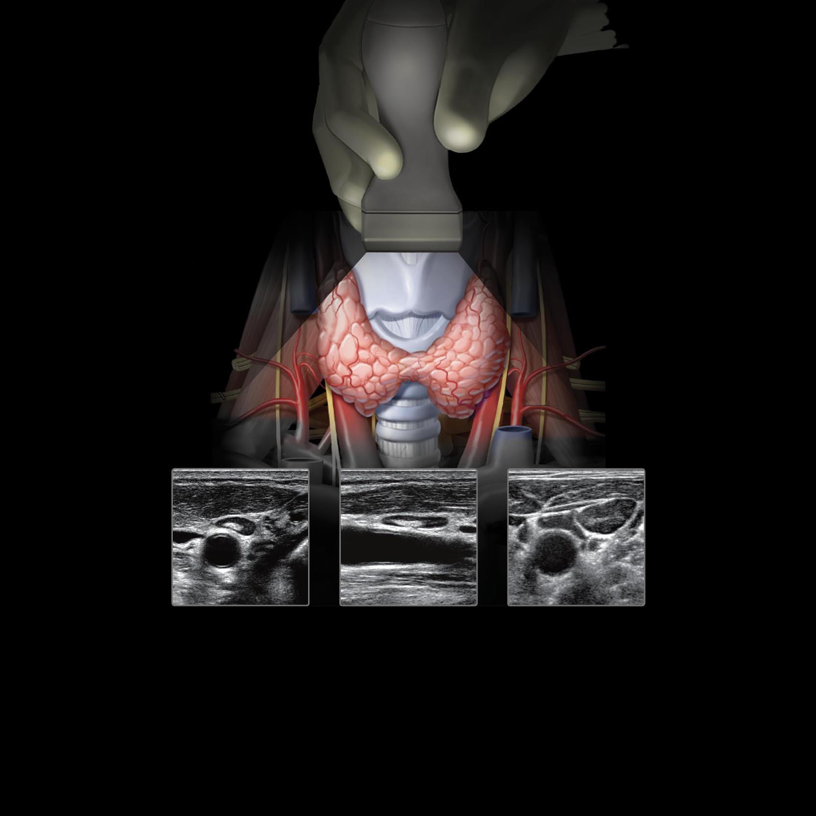
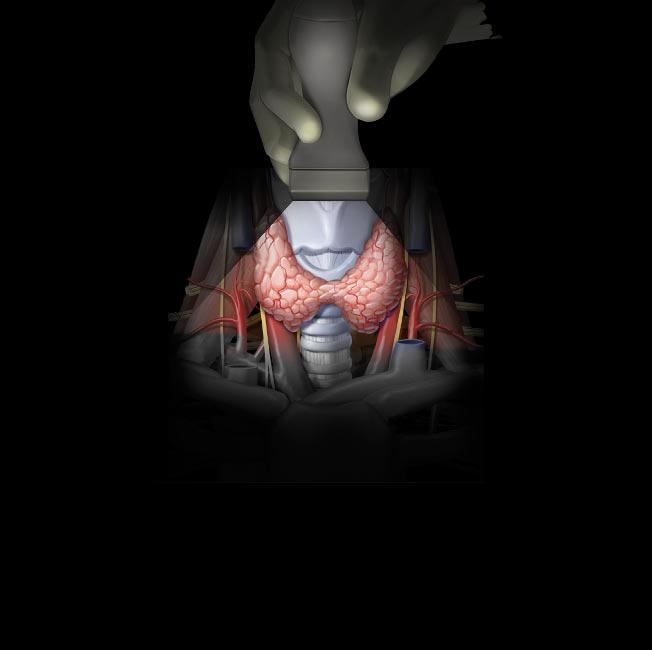
136 ThyroidGland
PaulaJ.Woodward,MD,K.T.Wong,MBChB,FRCR, FHKCR,FHKAM(Radiology),andAnilT.Ahuja,MBBS (Bom),MD(Bom),FRCR,FHKCR,FHKAM(Radiology)
144 ParathyroidGlands
PaulaJ.Woodward,MD,K.T.Wong,MBChB,FRCR, FHKCR,FHKAM(Radiology),andAnilT.Ahuja,MBBS (Bom),MD(Bom),FRCR,FHKCR,FHKAM(Radiology)
150 LarynxandHypopharynx
K.T.Wong,MBChB,FRCR,FHKCR,FHKAM(Radiology), YolandaY.P.Lee,MBChB,FRCR,FHKCR,FHKAM (Radiology),andAnilT.Ahuja,MBBS(Bom),MD(Bom), FRCR,FHKCR,FHKAM(Radiology)
158 TracheaandEsophagus
K.T.Wong,MBChB,FRCR,FHKCR,FHKAM(Radiology), AnilT.Ahuja,MBBS(Bom),MD(Bom),FRCR,FHKCR, FHKAM(Radiology),andPaulaJ.Woodward,MD
164 VagusNerve
K.T.Wong,MBChB,FRCR,FHKCR,FHKAM(Radiology), YolandaY.P.Lee,MBChB,FRCR,FHKCR,FHKAM (Radiology),andAnilT.Ahuja,MBBS(Bom),MD(Bom), FRCR,FHKCR,FHKAM(Radiology)
170 CarotidArteries
K.T.Wong,MBChB,FRCR,FHKCR,FHKAM(Radiology), YolandaY.P.Lee,MBChB,FRCR,FHKCR,FHKAM (Radiology),andAnilT.Ahuja,MBBS(Bom),MD(Bom), FRCR,FHKCR,FHKAM(Radiology)
184 VertebralArteries
K.T.Wong,MBChB,FRCR,FHKCR,FHKAM(Radiology), YolandaY.P.Lee,MBChB,FRCR,FHKCR,FHKAM (Radiology),andAnilT.Ahuja,MBBS(Bom),MD(Bom), FRCR,FHKCR,FHKAM(Radiology)
190 NeckVeins
K.T.Wong,MBChB,FRCR,FHKCR,FHKAM(Radiology), YolandaY.P.Lee,MBChB,FRCR,FHKCR,FHKAM (Radiology),andAnilT.Ahuja,MBBS(Bom),MD(Bom), FRCR,FHKCR,FHKAM(Radiology)
198 CervicalLymphNodes
TABLEOFCONTENTS K.T.Wong,MBChB,FRCR,FHKCR,FHKAM(Radiology), YolandaY.P.Lee,MBChB,FRCR,FHKCR,FHKAM (Radiology),andAnilT.Ahuja,MBBS(Bom),MD(Bom), FRCR,FHKCR,FHKAM(Radiology)
SECTION3:THORAX
208 ThoracicOutlet
GregoryE.Antonio,MD,DRANZCR,FHKCR,EricK.H.Liu, PhD,RDMS,andPaulaJ.Woodward,MD
218 Pleura
PaulaJ.Woodward,MD,GregoryE.Antonio,MD, DRANZCR,FHKCR,andEricK.H.Liu,PhD,RDMS
224 Diaphragm
GregoryE.Antonio,MD,DRANZCR,FHKCR,EricK.H.Liu, PhD,RDMS,andPaulaJ.Woodward,MD
228 ChestWall
GregoryE.Antonio,MD,DRANZCR,FHKCR,EricK.H.Liu, PhD,RDMS,andPaulaJ.Woodward,MD
234 Breast
NicoleS.Winkler,MD
SECTION4:ABDOMEN
248 Liver
AyaKamaya,MD,FSRU,FSAR
272 BiliarySystem
L.NayeliMorimoto,MD
284 Spleen
AliM.Tahvildari,MDandPaulaJ.Woodward,MD
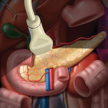
292 Pancreas
BartonF.Lane,MD
302 Kidneys
JadeWong-You-Cheong,MBChB,MRCP,FRCR
330 AdrenalGlands
PaulaJ.Woodward,MD
336 Bowel
SathiA.Sukumar,MBBS,FRCP(UK),FRCR
352 AbdominalLymphNodes
JadeWong-You-Cheong,MBChB,MRCP,FRCR
356 AortaandInferiorVenaCava
SimonS.M.Ho,MBBS,FRCR,JillM.Abrigo,MD,DPBR, andChanderLulla,MD,DMRD
386 PeritonealCavity
JadeWong-You-Cheong,MBChB,MRCP,FRCR
394 AbdominalWall
JadeWong-You-Cheong,MBChB,MRCP,FRCR
SECTION5:PELVIS
408 IliacArteriesandVeins
SimonS.M.Ho,MBBS,FRCR,JillM.Abrigo,MD,DPBR, andChanderLulla,MD,DMRD
424 UretersandBladder
AshishP.Wasnik,MDandPaulaJ.Woodward,MD
434 ProstateandSeminalVesicles
KatherineTo'o,MD,RichardE.Fan,PhD,andPaulaJ. Woodward,MD
446 TestesandScrotum
ShwetaBhatt,MDandPaulaJ.Woodward,MD
458 PenisandUrethra
PaulaJ.Woodward,MD
468 Uterus
BartonF.Lane,MDandPaulaJ.Woodward,MD
482 Cervix
BartonF.Lane,MD
488 Vagina
BartonF.Lane,MD
494 Ovaries
BryanR.Foster,MD
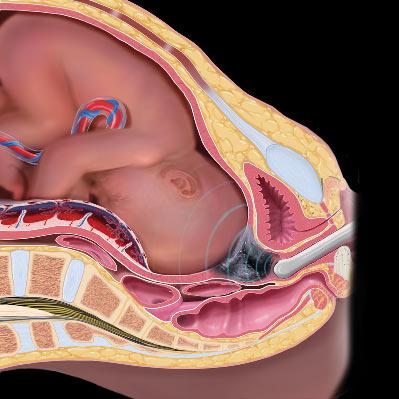
504 PelvicFloor
StellaSinYeeHo,RDMS,RVT,PhD,DeyondY.W.Siu, MBChB,FRCR,andPaulaJ.Woodward,MD
SECTION6:UPPEREXTREMITY
530 SternoclavicularandAcromioclavicularJoints
JamesF.Griffith,MD,MRCP,FRCRandBhawanK. Paunipagar,MBBS,MD,DNB
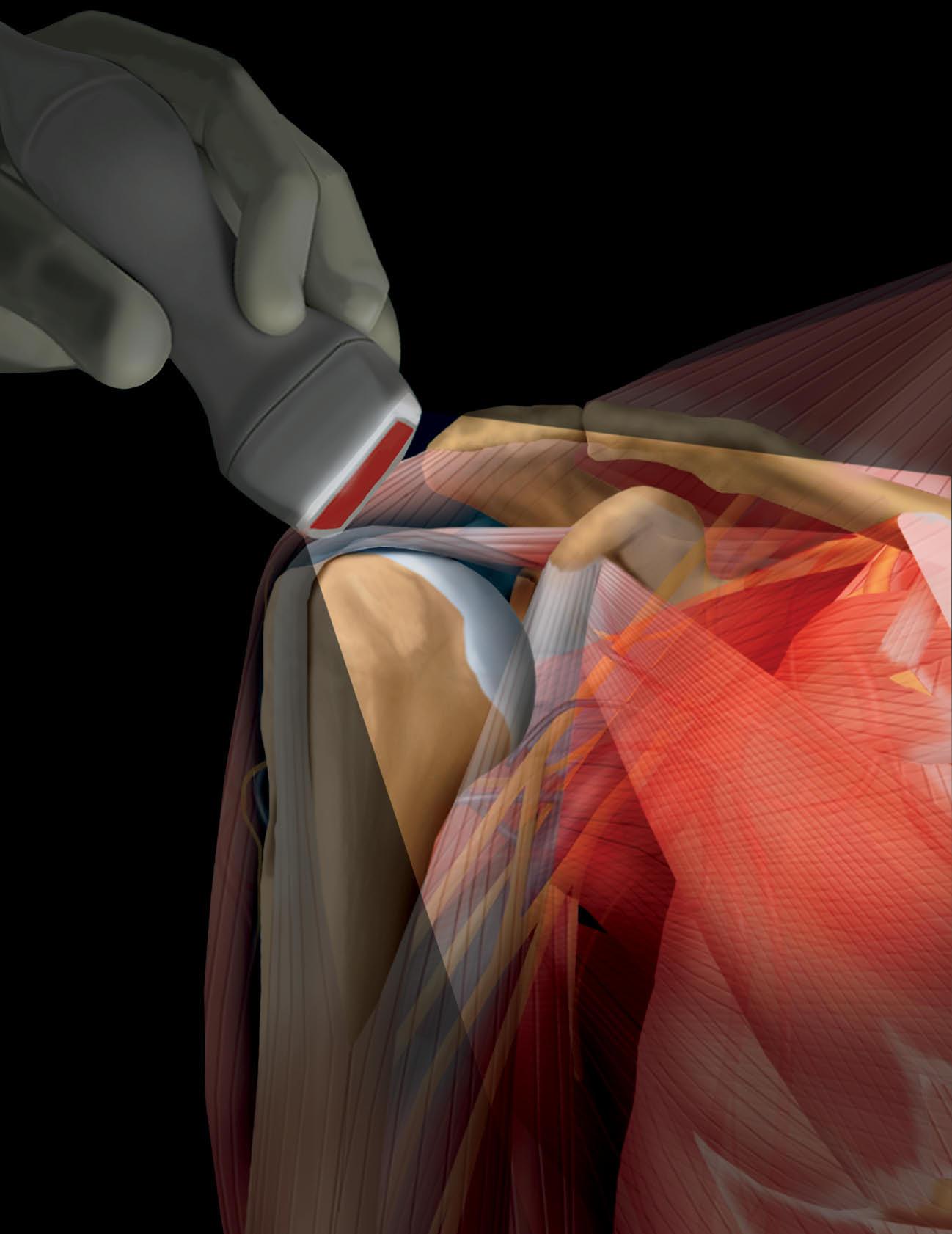
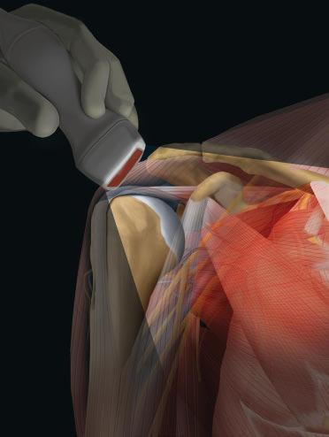
536 Shoulder
JamesF.Griffith,MD,MRCP,FRCRandBhawanK. Paunipagar,MBBS,MD,DNB
554 Axilla
JamesF.Griffith,MD,MRCP,FRCRandBhawanK. Paunipagar,MBBS,MD,DNB
562 Arm
JamesF.Griffith,MD,MRCP,FRCRandBhawanK. Paunipagar,MBBS,MD,DNB
570 ArmVessels
JamesF.Griffith,MD,MRCP,FRCRandBhawanK. Paunipagar,MBBS,MD,DNB
578 Elbow
JamesF.Griffith,MD,MRCP,FRCRandBhawanK. Paunipagar,MBBS,MD,DNB
598 Forearm
JamesF.Griffith,MD,MRCP,FRCRandBhawanK. Paunipagar,MBBS,MD,DNB
606 ForearmVessels
JamesF.Griffith,MD,MRCP,FRCRandBhawanK. Paunipagar,MBBS,MD,DNB 614 Wrist
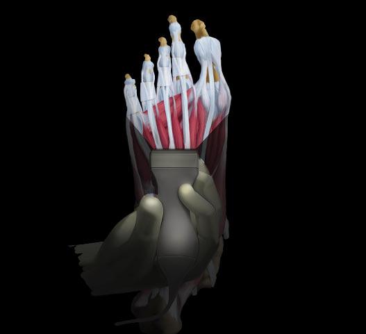
JamesF.Griffith,MD,MRCP,FRCRandBhawanK. Paunipagar,MBBS,MD,DNB 628 Hand
JamesF.Griffith,MD,MRCP,FRCRandBhawanK. Paunipagar,MBBS,MD,DNB 640 HandVessels
JamesF.Griffith,MD,MRCP,FRCRandBhawanK. Paunipagar,MBBS,MD,DNB
646 Thumb
JamesF.Griffith,MD,MRCP,FRCRandBhawanK. Paunipagar,MBBS,MD,DNB
656 Fingers
JamesF.Griffith,MD,MRCP,FRCRandBhawanK. Paunipagar,MBBS,MD,DNB
668 BrachialPlexus
JamesF.Griffith,MD,MRCP,FRCR,K.T.Wong,MBChB, FRCR,FHKCR,FHKAM(Radiology),andPaulaJ. Woodward,MD
676 RadialNerve
TABLEOFCONTENTS JamesF.Griffith,MD,MRCP,FRCRandBhawanK. Paunipagar,MBBS,MD,DNB
684 MedianNerve
JamesF.Griffith,MD,MRCP,FRCRandBhawanK. Paunipagar,MBBS,MD,DNB
694 UlnarNerve
JamesF.Griffith,MD,MRCP,FRCRandBhawanK. Paunipagar,MBBS,MD,DNB
SECTION7:LOWEREXTREMITY
706 GlutealMuscles
RyanK.L.Lee,MBChB,FRCR,FHKAM(Radiology),
GregoryE.Antonio,MD,DRANZCR,FHKCR,andEricK.H. Liu,PhD,RDMS
716 Groin
AlexW.H.Ng,MBChB,FRCR,FHKCR,FHKAM(Radiology),
GregoryE.Antonio,MD,DRANZCR,FHKCR,andEricK.H. Liu,PhD,RDMS
726 Hip
GregoryE.Antonio,MD,DRANZCR,FHKCRandEricK.H. Liu,PhD,RDMS
736 ThighMuscles
GregoryE.Antonio,MD,DRANZCR,FHKCRandEricK.H. Liu,PhD,RDMS
748 FemoralVesselsandNerves
GregoryE.Antonio,MD,DRANZCR,FHKCRandEricK.H. Liu,PhD,RDMS
762 Knee
GregoryE.Antonio,MD,DRANZCR,FHKCRandEricK.H. Liu,PhD,RDMS
780 LegMuscles
GregoryE.Antonio,MD,DRANZCR,FHKCRandEricK.H. Liu,PhD,RDMS
792 LegVessels
GregoryE.Antonio,MD,DRANZCR,FHKCR,EricK.H.Liu, PhD,RDMS,andPaulaJ.Woodward,MD
810 LegNerves
GregoryE.Antonio,MD,DRANZCR,FHKCRandEricK.H. Liu,PhD,RDMS
814 Ankle
GregoryE.Antonio,MD,DRANZCR,FHKCRandEricK.H. Liu,PhD,RDMS
832 Tarsus
GregoryE.Antonio,MD,DRANZCR,FHKCRandEricK.H. Liu,PhD,RDMS
846 FootVessels
GregoryE.Antonio,MD,DRANZCR,FHKCRandEricK.H. Liu,PhD,RDMS
852 MetatarsalsandToes
GregoryE.Antonio,MD,DRANZCR,FHKCRandEricK.H. Liu,PhD,RDMS
SECTION8:OBSTETRICSAND DEVELOPMENTALANATOMY
860 EmbryologyandAnatomyof1stTrimester
AnneKennedy,MD
872
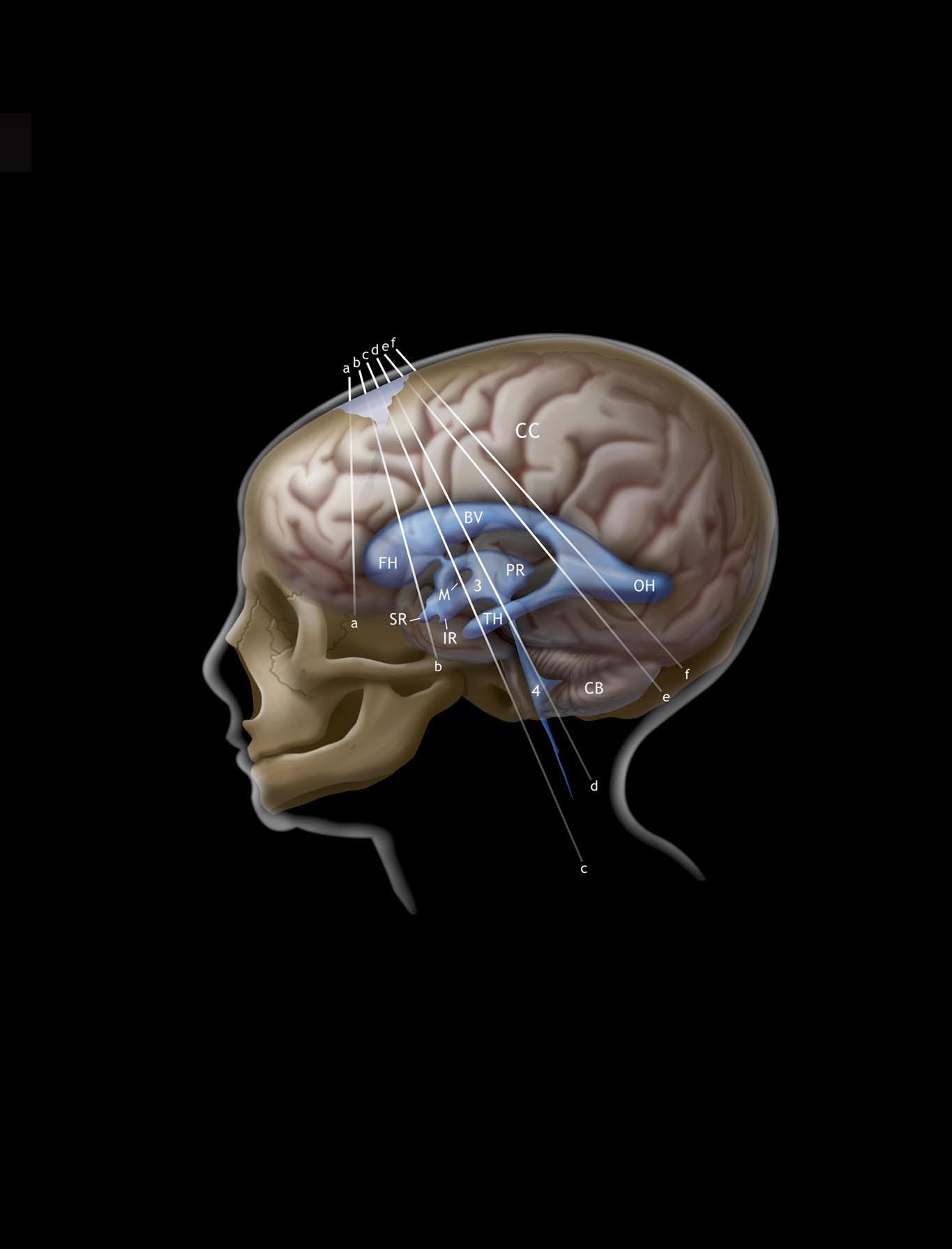
EmbryologyandAnatomyofBrain
AnneKennedy,MD
888 EmbryologyandAnatomyofSpine
PaulaJ.Woodward,MD
894 EmbryologyandAnatomyofFaceandNeck
RoyaSohaey,MD
906 EmbryologyandAnatomyofChest
PaulaJ.Woodward,MD
914 EmbryologyandAnatomyofCardiovascularSystem
ThomasA.Miller,DOandMichaelD.Puchalski,MD
924 EmbryologyandAnatomyofAbdominalWalland GastrointestinalTract
PaulaJ.Woodward,MD
934 EmbryologyandAnatomyofGenitourinaryTract
PaulaJ.Woodward,MD
WOODWARD GRIFFITH | ANTONIO | AHUJA
WONG | KAMAYA | WONG-YOU-CHEONG
ScalpandCalvarialVault TERMINOLOGY Definitions
•Fontanelle:Broadareasofconnectivetissueatjunctionof majorsutures
GROSSANATOMY Overview
• Scalp
○Scalphas5layers
–Skin(epidermis,dermis,hair,sebaceousglands)
–Subcutaneoustissue(veryvascularfibroadipose tissue)
–Epicranialtissue(scalpmuscles,galeaaponeurotica)
–Subaponeurotictissue(looseareolarconnective tissue)
–Pericranium(periosteumofskull)
•Skull(28separatebones,mostlyconnectedbyfibrous sutures)
○Craniumhasseveralparts
–Calvarialvault
–Cranialbase
–Facialskeleton
○Calvarialvaultcomposedofseveralbones
–2frontalbonesseparatedbymetopicsuture
–Pairedparietalbones
–Squamousoccipitalbone
–Pairedsquamoustemporalbones
○3majorserratedfibrousjoints(sutures)connectbones ofvault
–Coronalsuture
–Sagittalsuture
–Lambdoidsuture
○Outer,innertables
–2thinplatesofcompactcorticalbone
–Separatedbydiploicspace(cancellousbone containingmarrow)
○Endocranialsurface
–Linedbyouter(periosteal)layerofdura
–Groovedbyvascularfurrows
–Mayhaveareasoffocalthinning(arachnoid granulations),foramina(emissaryveins)
IMAGINGANATOMY Overview
•Scalp:Hypoechoic,5layerscannotbefurtherseparately resolved
•Calvariumechogenicouter/innertables;diploicspacefilled withfattymarrowandappearshypoechoic,sutureappears asgapbetweenechogeniccalvarium
•Frontalbones
○Frontalsinusesshowwidevariationinaeration
○Frontalbonesoftenappearthickened,hyperostotic (especiallyinolderfemales)
•Parietalbones
○Areasofparietalthinning,granularfoveolae(for arachnoidgranulations)commonadjacenttosagittal suture
○Innertablesoftenslightlyirregular(convolutional markingscausedbygyri),groovedbypairedmiddle meningealarteries+vein
•Occipitalbone
○Deeplygroovedbysuperiorsagittal,transversesinuses
○Internaloccipitalprotuberancemarkssinusconfluence (torcularHerophili)
•Temporalbones
○Thin,innersurfacegroovedbymiddlemeningealvessels
○Outersurfacegroovedbysuperficialtemporalartery
•Fontanelle:ProvideacousticwindowforUSexaminationof underlyingbrainparenchyma
○ Anteriorfontanelle
○Between2frontaland2parietalbones,usually disappearsbyage2
○Whenfused,correspondstobregma:Meetingof sagittal,coronalsutures
○ Posteriorfontanelle
○Small,usuallyclosesbetween3-6monthsofage
○Whenfused,correspondstolambda:Meetingofsagittal, lambdoidsutures
○Pterion
–Anterolateralfontanelle;closesbetween3-6months ofage
–H-shapedjunctionbetweenfrontal,parietalbones+ greatersphenoidwing,squamoustemporalbone
○Asterion
–Posterolateralfontanelle,persistsuntil2yearsofage
○ Mastoidfontanelle
○Locatedatjunctionoftemporosquamousandlambdoid sutures
○Persistsuntil2years
ANATOMYIMAGINGISSUES
ImagingRecommendations
•High-frequencylineararraytransducersprovidesuperb resolutionofnear-fieldstructures
•Goodskin-to-transducercouplingachievedbycopioususe ofacousticcouplinggel
•Superficialstandoffpadcanbeusedtoincreasedepthof focalzone
•UScanbeusedtoevaluatecranialsuturesandassists diagnosisofcraniosynostosis(prematurefusionofsutures)
EMBRYOLOGY
EmbryologicEvents
•Skullbaseformedfromendochondralossification
•Calvarialvaultformsviamembranousossification
○Curvedmesenchymalplatesappearatday30
○Extendtowardeachother,skullbase
○Aspairedbonesmeetinmidline,metopicandsagittal suturesareinduced(coronalsutureispresentfrom onsetofossification)
○Unossifiedcentersatedgesofparietalboneform fontanelles
○Vaultgrowsrapidlyin1stpostnatalyear
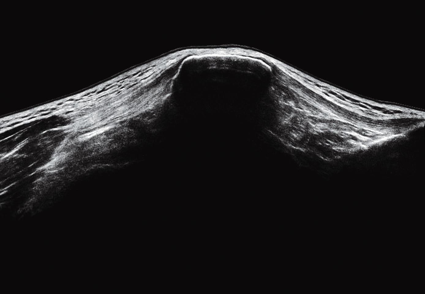
Suture
ScalpandCalvarialVault Suture
(Top)CoronalUSthroughtheanteriorfontanelleshowsthescalpandcalvarium.Thereisdiscontinuityinahypoechoicbandextending fromtheoutertoinnertableofthecalvarialvault.Thisrepresentsanormalsuture.Thescalpappearshypoechoiccomparedtothe echogenicouterandinnertablesofthecalvarium.The5layersofthescalpcannotberesolvedbyUS.(Middle)CoronalUSthroughthe anteriorfontanelleshowsanothersutureofthecalvarialvault.Thewidthandcurvatureofsuturesisvariableandshouldnotbe mistakenforabonyfracture.(Bottom)SagittalT2MRofthescalpandcalvariumisshown.Thesuturelineappearswiththesame signalintensityastheouterandinnertableofcalvarium.Corticalveinscanbeseenwithinthehyperintensesubarachnoidspace,which isimmediatelyundertheinnertable.
Scalp
Calvarium
Calvarium
Scalp
Suture
Scalp
Corticalvv.insubarachnoidspace
Cerebralcortex
Outertableofcalvarium
Innertableofcalvarium
USANDT2WIMRCALVARIUM