Equipping You with 21st-Century Skills to Succeed in A&P and Beyond…
The 12th Edition of Elaine Marieb and Katja Hoehn’s best-selling A&P text and media program motivates and supports both novice learners and expert students, more than ever before. Each carefully-paced chapter guides you in advancing from mastering terminology to applying knowledge in clinical scenarios, to practicing the critical thinking and problem-solving skills that are required for entry to nursing, allied health, and exercise science programs.
GLOBAL EDITION
Human Anatomy & Physiology
TWELFTH GLOBAL EDITION
Elaine N. Marieb Katja Hoehn
Marieb Hoehn
Identify “Big Picture” Concepts Before Exploring Details
Before you look up details and information within a chapter, read the ChapterOpening Roadmap, which visually groups and organizes “big picture” concepts and shows how they are related. To focus your studying, review the numbered Key Concept Headings, Learning Outcomes, and summaries.
UNIQUE! Chapter Roadmaps provide a visual overview of the key concepts in the chapter and show how they relate to each other. Each key concept “brick” in the roadmap corresponds to a numbered section within the chapter.
Joints
Each numbered section within the chapter begins with a Key Concept Heading that helps you quickly grasp the “big idea” of the discussion that follows.
Career Connection Videos feature a health care professional who describes how the chapter content relates to their everyday work. You can access all of the Career Connections videos through an open access web page at https:// bit.ly/3P8hiZa.
In this chapter, you will learn that
Joints determine how bones move relative to each other
The graceful movements of ballet dancers and the roughand-tumble grapplings of football players demonstrate the great variety of motion allowed by joints, or articulations the sites where two or more bones meet. Our joints have two fundamental functions: They give our skeleton mobility, and they hold it together, sometimes playing a protective role in the process.
8.1 Joints are classified into three structural and three functional categories
Learning Outcomes
✔ Define joint or articulation.
✔ Classify joints by structure and by function.
Joints are classified by structure and by function. The structural classification focuses on the material binding the bones together and whether or not a joint cavity is present. Structurally, there are fibrous, cartilaginous, and synovial joints (Table 8.1 on p. 285). Only synovial joints have a joint cavity.
The functional classification is based on the amount of movement allowed at the joint. On this basis, there are synarthroses (sin0ar-thro9sēz; syn 5 together, arthro 5 joint), which are immovable joints; amphiarthroses (am0fe-ar-thro9sēz; amphi 5 on both sides), slightly movable joints; and diarthroses (di0ar-thro9sēz; dia through, apart), or freely movable joints. Freely movable joints predominate in the appendicular skeleton (limbs). Immovable and slightly movable joints are largely restricted to the axial skeleton. This localization of functional joint types makes sense because the less movable the joint, the more stable it is likely to be.
In general, fibrous joints are immovable, and synovial joints are freely movable. However, cartilaginous joints have both rigid
Learning Outcomes are presented at the beginning of each chapter section to give you a preview of essential information to study.
Pace Yourself: Learn & Review the Basics
Summary Tables present key information and serve as “one-stop shopping” study tools.
Summary of Cutaneous Glands
Functions Temperature control
Some antibacterial properties
Type of Secretion
Method of Secretion
Secretion
Exits Duct At
May act as sexual scent glands
external ear canal. Their secretion mixes with sebum produced by nearby sebaceous glands to form a sticky, bitter substance called cerumen, or earwax, that is thought to deter insects and block entry of foreign material.
Hypotonic filtrate of blood plasma Filtrate of blood plasma with added proteins and fatty substances
Merocrine (exocytosis)
Skin surface
Body Location Everywhere, but especially palms, soles, forehead
See p. 192
Merocrine (exocytosis)
Usually upper part of hair follicle; rarely, skin surface
Lubricate skin and hair
Help prevent water loss
Chapter 5 The Integumentary System 193
Antibacterial properties
Sebum (an oily secretion)
Check Your Understanding
Holocrine
17. Which cutaneous glands are associated with hair follicles?
Usually upper part of hair follicle; sometimes, skin surface
18. When Anthony returned home from a run in 30°C weather, his face was dripping with sweat. Why?
Mostly axillary and anogenital regions
so-called pores of a person’s complexion, which are openings of hair follicles.)
Mammary glands. Mammary glands, another type of specialized sweat gland, secrete milk. Although they are properly part of the integumentary system, we will consider the mammary glands in Chapter 27 with female reproductive organs.
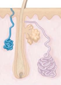
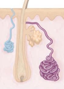
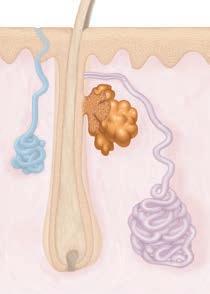
Sebaceous Glands
Eccrine gland secretion, commonly called sweat, is a hypotonic filtrate of the blood that passes through the secretory cells of the sweat glands and is released by exocytosis. It is 99% water, with some salts (mostly sodium chloride), traces of metabolic wastes (urea, uric acid, and ammonia), and a microbe-killing peptide called dermcidin. Normally, sweat is acidic with a pH between 4 and 6.
Sweating’s major role is to prevent the body from overheating. Heat-induced sweating begins on the forehead and spreads inferiorly over the remainder of the body. Emotionally induced sweating—the so-called “cold sweat” brought on by fright or nervousness—begins on the palms, soles, and axillae (armpits) and then spreads to other body areas.
Apocrine Sweat Glands
See p. 193
The approximately 2000 apocrine sweat glands (ap9o-krin) are largely confined to the axillary and anogenital areas. In spite of their name, they are merocrine glands, which release their product by exocytosis like the eccrine sweat glands. Larger than
Everywhere except palms and soles
19. What is the difference between heat-induced sweating and a “cold sweat,” and which variety of sweat gland is involved?
eccrine glands, they lie deeper in the dermis or even in the subcutaneous tissue, and their ducts empty into hair follicles.
20. APPLY Sebaceous glands are not found in thick skin. Why is their absence in those body regions desirable? For answers, see Answers Appendix.
Apocrine secretion contains the same basic components as true sweat, plus fatty substances and proteins. Consequently, it is viscous and sometimes has a milky or yellowish color. The secretion is odorless, but when bacteria on the skin decompose its organic molecules, it takes on a musky and generally unpleasant odor, the basis of body odor.
The sebaceous glands (se-ba 9shus; “greasy”), or oil glands (Figure 5.9a), are simple branched alveolar glands that are found all over the body except in the thick skin of the palms and soles. They are small on the body trunk and limbs, but quite large on the face, neck, and upper chest. These glands secrete an oily substance called sebum (se9bum). The central cells of the alveoli accumulate oily lipids until they become so engorged that they burst, so functionally these glands are holocrine glands ( p. 156). The accumulated lipids and cell fragments constitute sebum. Most, but not all, sebaceous glands develop as outgrowths of hair follicles and secrete sebum into a hair follicle, or occasionally to a pore on the skin surface. Arrector pili contractions force sebum out of the hair follicles to the skin surface. Sebum softens and lubricates the hair and skin, prevents hair from becoming brittle, and slows water loss from the skin. Perhaps even more important is its bactericidal (bacterium-killing) action. Sebaceous glands increase their activity during puberty under the influence of male sex hormones.
Apocrine glands begin functioning at puberty under the influence of the male sex hormones (androgens) and play little role in maintaining a constant body temperature. Their precise function is not yet known. Three lines of evidence suggest that they may be the human equivalent of other animals’ sexual scent glands: (1) Sexual foreplay increases their activity; (2) they enlarge and recede with the phases of a woman’s menstrual cycle; and (3) behavioral studies show that their secretions may act as pheromones (chemical messengers released by one individual that trigger a response in other members of the same species).
5.8 First and foremost, the skin is a barrier
Learning Outcome
✔ Describe how the skin accomplishes at least five different functions.
Text Recall icons guide you to review specific pages where a concept was first introduced.
Like the skin of a grape, our skin keeps its contents juicy and whole. The skin and its appendages perform a variety of functions, including protection, body temperature regulation, cutaneous sensation, metabolic functions, blood reservoir, and excretion.
Two important types of modified apocrine glands are: Ceruminous glands. Ceruminous glands (sĕ-roo9mĭ-nus; cera 5 wax) are modified apocrine glands found in the lining of the
Protection
Building Vocabulary Coaching Activities in Mastering A&P® are a fun way to learn word roots and A&P terminology while building and practicing important language skills.
Given its superficial location, the skin is our most vulnerable organ system, exposed to microorganisms, abrasion, temperature extremes, harmful chemicals, and UV radiation. The skin constitutes at least three types of barriers: chemical, physical, and biological.
Table 5.1
ECCRINE SWEAT GLANDS
APOCRINE SWEAT GLANDS
SEBACEOUS GLANDS
Study the Figures as You Read the Text
Anatomy and Physiology is a visual science. To succeed, you need to practice and develop visual literacy skills for understanding and interpreting information. To help you achieve this goal, the text and associated figures are tightly integrated so that you do not have to flip pages back and forth to connect visuals with words.
26 Focus Figures walk you through complex processes using exceptionally clear, easy-to-follow illustrations with integrated text explanations.
See pp. 840–841
Focus Figure
“Mini-Animation” Coaching Activities bring some of the Focus Figures to life using short video segments.
Activation and Differentiation of B Cells
Most cells of the clone differentiate into plasma cells, the antibody-secreting effector cells of the humoral response. Plasma cells develop the elaborate internal machinery (largely target and in and lymphocyte
pattern
An immunocompetent but naive B lymphocyte is activated when matching antigens bind to its surface receptors and cross-link adjacent receptors together. Antigen binding is quickly followed by receptor-mediated endocytosis of the cross-linked antigen-receptor complexes. As we described previously, this is called clonal selection and is followed by proliferation and differentiation into effector cells (Figure 21.11). (As we will see shortly, interactions with T cells are usually required to help B cells achieve full activation.)
Blue text represents the voice of an A&P instructor, highlighting important points to remember.
31 unique In-Line Figures are strategically placed within the text to visually reinforce the text discussion.
See p. 828
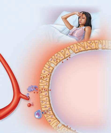
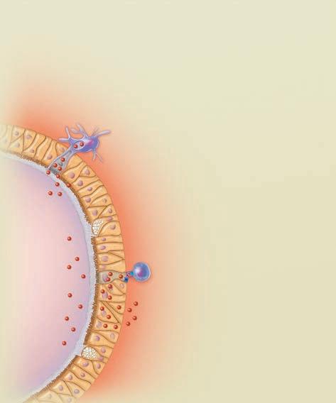
FOCUS FIGURE 21.1 An Example of a Primary Immune Response
Apply Your Knowledge to a Range & Variety of Questions
858 UNIT 4 Maintenance of the Body
As you build your knowledge and confidence in A&P, practice responding to the more challenging questions—you are likely to encounter similar questions on a test or licensing exam. Your extra effort will pay off at exam time!
always ready to mount the adaptive barriers and internal and cellular immu(the skin and Opsonization is the phagocytosis by decorating Antibodies and comple4. Our own cells viruses or when they are the cardinal caused by vasodilation of body core) to the histamine and other capillary permeability. interstitial fluid (IF), out of blood vessels to two things: prostaglandins) on nerve endings. make Julian particuadaptive immunity systemic, and it has reactivity, whereas Self-antigens, particularly immunocompetence of specific and unique is a membranereceptor.) 11. Dendritic cells are most antigen does the T cells that has cell that would surThe secondary because the immune are specific for (You can tell pentamer.) IgG antibody
a membrane attack complex (MAC)]. Specifically, in complement-mediated cell lysis, MAC complexes cause a target cell (usually a bacterium) to become leaky. This allows water to enter and cytoplasmic contents to exit the cell, killing the cell. In contrast, cytotoxic T cells and NK cells kill their targets (usually virus-infected cells) by releasing perforins and granzymes onto the identified target cell. Perforins form a pore in the target cell membrane, and granzymes enter through this pore, activating enzymes that trigger apoptosis (cell suicide).
A greater variety and range of self-assessment questions have been added to the Check Your Understanding sections within each chapter and include Apply, Predict, What If?, Draw, and Make Connections. Dozens of new visual questions ask you to label structures or interpret visual information.
Table
22.1 The Upper Respiratory System
Chapter 11 Fundamentals of the Nervous
STRUCTURE DESCRIPTION, GENERAL AND DISTINCTIVE FEATURES
Nose (external nose and nasal cavity)
Check Your Understanding
Jutting external portion is supported by bone and cartilage. Internal nasal cavity is divided by midline nasal septum and lined with mucosa. Roof of nasal cavity contains olfactory epithelium.
5. How does a nucleus within the brain differ from a nucleus within a neuron?
Paranasal sinuses
6. How is a myelin sheath formed in the CNS, and what is its function?
Some Definitions:
Voltage,
Voltage, the measure of electrical charges, is measured
Mucosa-lined, air-filled cavities in cranial bones surrounding nasal cavity ( p. 245). Lighten incoming
23. The cell being activated is a CD4 cell and so could become either a helper T cell or a regulatory T cell. 24. HIV is particularly hard for the immune system to defeat because (1) it destroys helper T cells, which are key players in adaptive immunity and (2) it has a high mutation rate and so it can avoid detection by the immune system by changing its surface antigens. 25. Binding of an allergen onto specific IgE antibodies attached to mast cells triggers the mast cells to release histamine.
Pharynx Passageway connecting nasal cavity to larynx and oral cavity to esophagus. Three subdivisions: nasopharynx, oropharynx, and laryngopharynx.
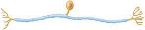
7. What is the structural classification of the neuron shown below? What is its usual functional classification? Name the parts labeled a–d.
Houses tonsils (lymphoid tissue masses involved in protection against pathogens).
8. APPLY Which structural and functional type of neuron is involved in sensing the smell of your perfume? Which type is needed to transfer the impulses to the brain for integration?
Central process
directly posterior to the larynx, where the respiratory and digestive pathways diverge, and extends to the inferior edge of the cricoid cartilage. The laryngopharynx is continuous with the esophagus posteriorly.
Review Questions 1. d; 2. b; 3. d; 4. d; 5. a; 6. a; 7. c; 8. d; 9. d; 10. (1)b, g; (2)d, i; (3)a, e; (4)a, e, f, h; (5)e, h; (6)c, f, g; 11. d; 12. The antibody classes and their likely locations in the body are: IgM—attached to B cell plasma membrane as a monomer and in plasma as a pentamer; IgA—dimers found in secretions such as saliva, tears, intestinal juice, and milk; IgD—attached to B cell plasma membrane as a monomer; IgG—in plasma; IgE—attached to mast cells in skin, mucosae of the gastrointestinal and respiratory tracts, and tonsils.
Chapter 22
“Draw” questions ask you to create visuals that reinforce important concepts by drawing a structure, annotating a figure, or creating a summary table.
The esophagus conducts food and fluids to the stomach; air enters the larynx anteriorly. During swallowing, food has the “right of way,” and air passage temporarily stops.
9. MAKE CONNECTIONS Which part of the neuron is its fiber? How do nerve fibers differ from the fibers of connective tissue (see Chapter 4) and the fibers in muscle (see Chapter 9)?
Check Your Understanding
For answers, see Answers Appendix.
(1 mV = 0.001 V) . Voltage points and is called the potential between the charge between two points, The flow of electrical current, and it can be a flashlight. The amount points depends on two tance is the hindrance through which the current electrical resistance are tance are conductors
All of the End-of-Chapter Review questions are now organized into 3 levels of difficulty based on Bloom’s Taxonomy categories:
Level 1: Remember/Understand
Level 2: Apply/Analyze
Level 3: Evaluate/Synthesize
See p. 431
1. Air moving from the nose to the larynx passes by a number of structures. List (in order) as many of these structures as you can.
2. Name the two types of mucous membrane found in the nasal cavity.
11.4 The resting membrane potential depends on differences in ion concentration and permeability
Learning Outcomes
3. DRAW Create a summary table to help you study the pharynx by comparing and contrasting its three parts. For each part, identify what it conducts (air, food, or both), the type of epithelium found there, and the associated tonsils. For answers, see Answers Appendix.
✔ Describe the relationship between current, voltage, and resistance.
Nasopharynx
Ohm’s law gives the and resistance:
Current
The conducting sageways from the provide fairly rigid sites. The conducting and warm incoming has fewer irritants the body, and it
The Larynx Basic Anatomy
Ohm’s law tells us
Current (I) is directly voltage (potential difference), There is no net current same potential. Current is inversely resistance, the smaller
Epithelium
Tonsils
✔ Identify different types of membrane ion channels.
✔ Define resting membrane potential and describe its electrochemical basis.
Oropharynx
Learning Outcomes
Pseudostratified ciliated columnar
Tubal
22.2 The lower respiratory system consists of conducting and respiratory zone structures
Stratified squamous
Laryngopharynx
Conducts Air Air and food Air and food
Stratified squamous
Pharyngeal
Palatine
✔ Distinguish between conducting and respiratory zone structures.
The larynx (lar 9 5 cm from the level Superiorly it attaches laryngopharynx. Inferiorly (Figure 22.5b). The larynx has Provide a patent Act as a switching proper channels Voice production cords)]
In the body, electrical cellular membranes. (Unlike house wiring, there are a living system.) Recall numbers of positive and lar plasma membranes tial across those membranes. the resistance to current
Lingual (none)
✔ Describe the structure, function, and location of the larynx, trachea, and bronchi.
See p. 858 and Answers Appendix
Basic Principles of Electricity
Like all cells, neurons have a resting membrane potential However, unlike most other cells, neurons can rapidly change their membrane potential. This ability underlies the function of neurons throughout the nervous system. In order to understand how neurons work, let’s first explore some basic priciples of electricity and revisit the resting membrane potential.
✔ Describe the makeup of the respiratory membrane, and relate structure to function.
✔ Identify the organs forming the respiratory passageway(s) in descending order until you reach the alveoli.
The human body is electrically neutral—it has the same number of positive and negative charges. However, there are regions where one type of charge predominates, making those regions positively or negatively charged. Because opposite charges
Check Your Understanding 1. The structures that air passes by are the nasal cavity (nares, nasal vestibule, nasal conchae), nasopharynx, oropharynx, and laryngopharynx. 2. The nasal cavity contains both respiratory mucosa and olfactory mucosa. 3. ❯ 4. The epiglottis seals the larynx when we swallow. 5. The incomplete, C-shaped cartilage rings of the trachea allow it to expand and contract and yet keep it from collapsing. 6. The many tiny alveoli together have a large surface area. This and the thinness of their respiratory membranes make them ideal for gas exchange. 7. The peanut was most likely in the right main bronchus because it is wider and more vertical than the left. 8. The two circulations of the lungs are the pulmonary circulation, which delivers deoxygenated blood to the lungs for oxygenation and returns oxygenated blood to the heart,
Anatomically, the lower respiratory system consists of the lar-
Role of Membrane Ion
The framework of nine cartilages ( Figure 22.6). Except lages are hyaline cartilages. The large, shield-shaped fusion of two cartilage upright open book, rior midline of the prominence (lah-rin Adam’s apple (Figure larger in males than stimulate its growth tilage is the ring-shaped
Recall that plasma membranes membrane proteins that of these channels is selective allows to pass. For example, only potassium ions to Membrane channels subunits. Some channels, always open. Other channels forms a molecular “gate” the channel in response types of gated channels:
Ground Substance
Ground substance is the unstructured material that fills the space between the cells and contains the fibers. It has three components:
Prepare for Your Future Career & Practice Solving Real-World Problems
Interstitial fluid . The ground substance consists of large amounts of fluid and functions as a molecular sieve through which nutrients and other dissolved substances can diffuse between the blood capillaries and the cells. The fibers embedded in the ground substance make it less pliable and hinder diffusion somewhat.
Cell adhesion proteins. These proteins serve mainly as a connective tissue glue that allows connective tissue cells to attach to the extracellular matrix.
fibrous protein collagen. Collagen molecules are secreted into the extracellular space, where they assemble spontaneously into cross-linked fibrils, which in turn are bundled together into the thick collagen fibers seen with a microscope. Because their fibrils cross-link, collagen fibers are extremely tough and provide high tensile strength (the ability to resist being pulled apart) to the matrix. Indeed, stress tests show that collagen fibers are stronger than steel fibers of the same size!
The authors of this text, Elaine Marieb and Katja Hoehn, share insights from their own clinical experience to help you prepare for your future career in health care. All clinical examples and applications are signaled with an easy-to-find “Clinical” label.
Proteoglycans. The proteoglycans consist of a protein core to which large polysaccharides called glycosaminoglycans (GAGs) (gli0kos-ah-me0no-gli9kanz) are attached. The strandlike GAGs [e.g., chondroitin sulfate and hyaluronic acid (hi0ah-lu-ron9ik)] stick out from the protein core like the fibers of a bottle brush. The proteoglycans tend to form huge aggregates in which the GAGs intertwine and trap water, forming a substance that varies from a fluid to a viscous gel. The higher the GAG content, the more viscous the ground substance.
Homeostatic Imbalance discussions alert you to the consequences of body systems not functioning optimally. Relevant photos have been added to selected discussions for visual reinforcement.
Connective Tissue Fibers
NEW! Discussions have been added on Marfan syndrome, brittle bone disease, tetanus, and anxiety disorders.
The fibers of connective tissue are proteins that provide support. Three types of fibers are found in connective tissue matrix:
Elastic Fibers Long, thin, elastic fibers form branching networks in the extracellular matrix. These fibers contain a rubberlike protein, elastin, that allows them to stretch and recoil like rubber bands. Connective tissue can stretch only so much before its thick, ropelike collagen fibers become taut. Then, when the tension lets up, elastic fibers snap the connective tissue back to its normal length and shape. Elastic fibers are found where greater elasticity is needed, for example, in the skin, lungs, and blood vessel walls.
HOMEOSTATIC IMBALANCE 4.2
CLINICAL
Marfan syndrome is an inherited disorder that causes a change in the types of proteins that comprise elastic fibers. As a result of this change, the elasticity in tissues is reduced, leading to the overgrowth (aortic enlargement and long arms, legs, and fingers) and instability (lung collapse and eye problems) of tissues. Although people suffering from Marfan syndrome are born with the condition, not all of them show symptoms at birth or during childhood; some only develop symptoms as adults.
See p. 158
Connective tissue is composed of (1) fibers, (2) ground substance, and (3) cells.
Extracellular matrix
well, complains of repeated “colds,” and is extremely “puffy” (edematous). Explain the reason for these symptoms.
Level 3 Evaluate/Synthesize
Clinical Case Studies are provided at the end of Chapters 5–29 and challenge you to apply your knowledge to realistic clinical scenarios.
• Collagen fiber
• Elastic fiber
• Reticular fiber
CLINICAL CASE STUDY
Cells Ground substance
One-Year-Old Girl with Retarded Growth
20. Which type of hormone receptor—plasma membrane bound or intracellular—would be expected to provide the most long-lived response to hormone binding and why?
21. Name two endocrine glands (or regions) that are important in the stress response, and explain why they are important.
22. How are the hyperglycemia and lipidemia of insulin deficiency linked?
23. List some problems that elderly people might have as a result of decreasing hormone production.
24. Mary Morgan has just been brought into the emergency room of City General Hospital. She is perspiring profusely and is breathing rapidly and irregularly. Her breath smells like acetone (sweet and fruity), and her blood glucose tests out at 650 mg/100 ml of blood. She is in acidosis. Which hormone drug should be administered, and why?
almost six million deaths per year. While immunization programs to prevent the outbreak of life-threatening infectious diseases tend to focus on children, these vaccines can also be effective in adults. However, access to vaccines remains regrettably unequal both for children and adults globally.
25. Kyle, a 5-year-old boy, has been growing by leaps and bounds; his height is 100% above normal for his age. He has been complaining of headaches and vision problems. A CT scan reveals a large pituitary tumor. (a) Which hormone is being secreted in excess? (b) What condition will Kyle exhibit if corrective measures are not taken? (c) What is the probable cause of his headaches and visual problems?
Conventional vaccines have shortcomings. The biggest shortcoming is that they are not always as effective or long-lasting as we would like. In some individuals, contaminating proteins (for example, egg albumin) cause allergic responses to the vaccine.
NEW! Boxes on scientists feature details about the lives and works of eminent scientists. These will show you the human side of science.
Passive humoral immunity differs from active immunity, both in the antibody source and in the degree of protection it provides (Figure 21.13). Instead of being made by your plasma cells, ready-made antibodies are introduced into your body. As a result, your B cells are not challenged by antigens, immunological memory does not occur, and the protection provided by the “borrowed” antibodies ends when they naturally degrade in the body.
Passive immunity is conferred naturally on a fetus or infant when the mother’s antibodies cross the placenta or are ingested with the mother’s milk. For several months after birth, the baby is protected from all the antigens to which the mother has been exposed.
Passive immunity can also be conferred artificially by administering exogenous antibodies (from outside your own body) as gamma globulin , harvested from the plasma of an immune donor. Exogenous antibodies are used to prevent hepatitis A (antiserum) and treat poisonous snake bites (antivenom),
26. Aaron, a 42-year-old single father, goes to his physician complaining of nausea and chronic fatigue. He reports having felt fatigued and listless for about half a year, but he had attributed this to stress. He has lost considerable weight and, strangely, his skin looks tanned, even though he spends long hours at work and rarely ventures outside. His doctor finds very low blood pressure and a rapid, weak pulse. Blood tests show that Aaron does not have anemia, but his plasma glucose, cortisol, and Na+ are low, and his plasma K+ is high. His doctor orders an ACTH stimulation test, in which Aaron’s secretion of cortisol is measured after he is given a synthetic form of ACTH. (a) What would account for Aaron’s low plasma Na+ and high plasma K+? (b) What is the reason for doing an ACTH stimulation test? (c) Which gland is primarily affected if ACTH does not cause a normal elevation of cortisol secretion? What is this abnormality called? (d) Which gland is primarily affected if ACTH does cause an elevation of cortisol secretion?
See p. 673
botulism, rabies, and tetanus (antitoxin) because these rapidly fatal diseases would kill a person before active immunity could be established. The donated antibodies provide immediate protection, but their effect is short-lived (two to three weeks).
Miriam gave birth to a twin boy and girl a year ago. She is concerned about Theresa, her daughter, since her growth and development is much slower than that of her brother. Miriam visits a pediatric outpatient clinic, where she informs the physician that, apart from having retarded growth, Theresa has a poor appetite, suffers from constipation, and is lethargic. The physician orders blood tests to check Theresa’s growth hormone (GH), thyroid-stimulating hormone (TSH), and thyroxine (T4) levels.
1. ✚ NCLEX-STYLE Theresa’s retarded growth could be due to:
a. The positive feedback of GH on the hypothalamus
Susumu Tonegawa (b. 1939) is a Japanese scientist who won the Nobel Prize in Physiology or Medicine in 1987 for elucidating the genetic mechanisms underlying adaptive immunity. A problem in adaptive immunity was that, although the presence of millions of different antibody proteins was known, there weren’t enough genes in the human genome to account for these. So how were all these different antibodies produced? By comparing the DNA of mature and immature B cells, Tonegawa discovered that the regions of DNA that produce antibodies become greatly rearranged as the B cell matures, which is how a small number of antibody-producing genes generate the huge variety of antibodies seen.
b. A pituitary tumor that is causing hypersecretion of GH
c. Hypersecretion of growth hormone–releasing hormone (GHRH) by the hypothalamus
d. Hyposecretion of GH by the anterior pituitary
Theresa’s blood tests indicate that her GH levels are normal, but her TSH levels are elevated, and her T levels are low. The physician tells Miriam that since Theresa’s GH levels are normal, her retarded growth is not due to pituitary dwarfism.
2. ✚ NCLEX-STYLE Given the levels of TSH and T in Theresa’s blood, which of the following is most likely the cause of her signs and symptoms?
a. She has a pituitary tumor, which is causing hypersecretion of TSH.
b. Her thyroid gland is poorly developed.
c. She has Graves’ disease.
d. Her parathyroid glands are defective.
See p. 830
3. Miriam does not completely understand what Theresa’s blood tests indicate. She asks the physician if she needs to put Theresa on a high-calorie diet to speed up her growth.
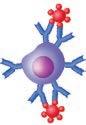
• Macrophage
Capillary
• Fibroblast
• Lymphocyte (a type of white blood cell)
• Fat cell
• Neutrophil (a type of white blood cell)
Fibers
Figure 4.9 Areolar connective tissue: A prototype (model) connective tissue. This tissue underlies epithelia and surrounds capillaries. (See Figure 4.11a for a micrograph.)
830 UNIT 4 Maintenance of the Body
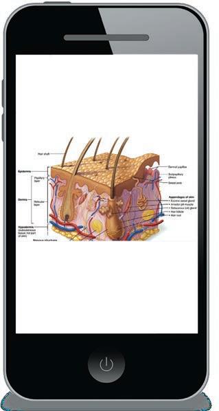
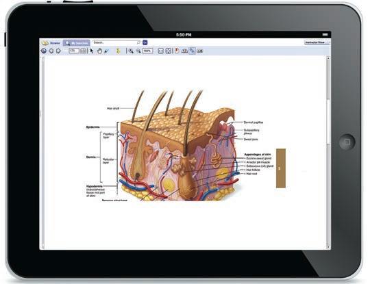
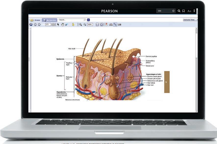
Access the Complete Textbook Using the Pearson eText
You can read your textbook without having to add weight to your bookbag! Videos and animations in the eText bring key concepts to life, helping you place what you are reading into context.
Powerful interactive and customization functions include instructor and student notetaking, highlighting, bookmarking, search, and links to glossary terms.
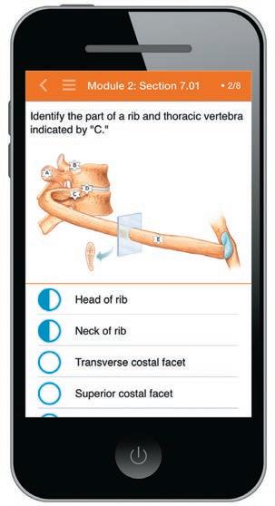
Get Online Practice and Coaching with Mastering A&P®
Mastering A&P® provides tutorials and review questions that you can access before, during, and after class.
EXPANDED! Interactive Physiology 2.0 Coaching Activities teach complex physiology processes using exceptionally clear animations, interactive tutorials, games, and quizzes. IP2 features new graphics, quicker navigation, and a mobile-friendly design. New topics include Pulmonary Ventilation, Tubular Reabsorption and Secretion, and Urine Concentration and Volume. IP2 and IP animations can be assigned from the Mastering A&P® item library or accessed through the Study Area.
NEW! 10 Histology Videos provide short, focused walkthroughs of some of the most commonly covered tissue types in A&P.
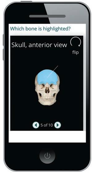
NEW and UPDATED! Bone and Organ Dissection Videos, with 23 UPDATED Bone Videos and 3 NEW Bone Videos for fetal skull, cervical vertebrae, and male and female pelves, cover major bone and organ dissections to help you prepare for lecture and lab.
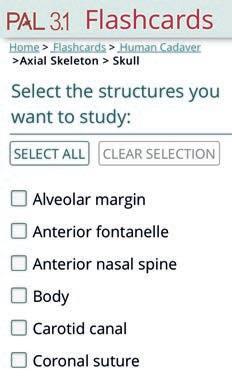
PAL 3.1 Customizable Flashcards allow you to create a personalized, mobile-friendly deck of flashcards and quizzes using images from Practice Anatomy Lab. Use the checklist to select only those structures covered in your course.
Dynamic Study Modules are manageable, mobilefriendly sets of questions with extensive feedback for you to test, learn, and retest yourself on basic concepts. Instructors can select or deselect specific questions for assignments from more than 3,000 questions, organized by chapter section.
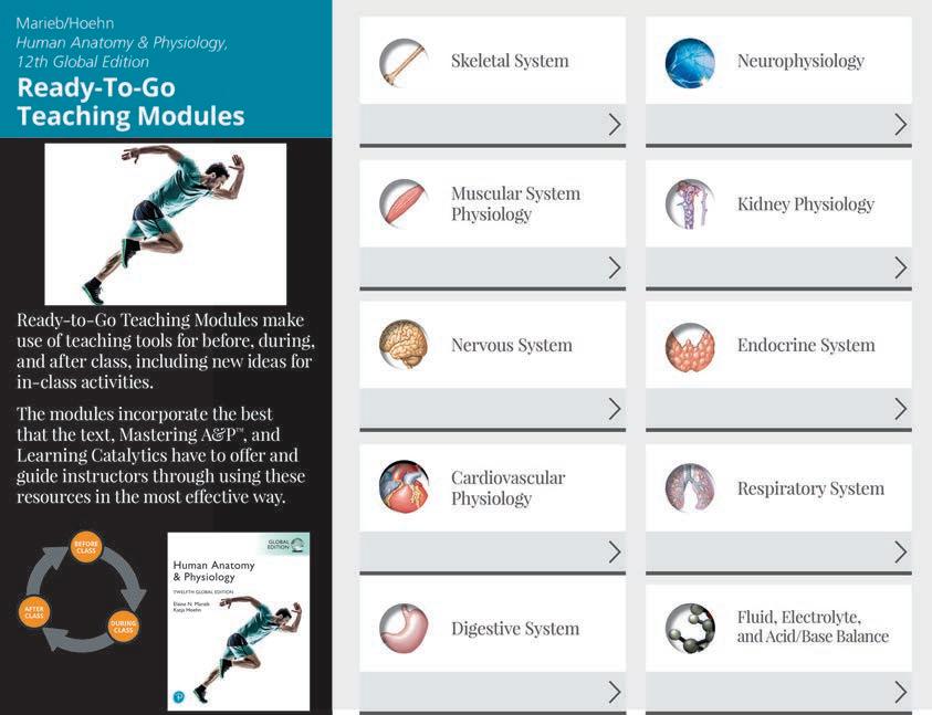
Resources for Instructors: Ready-to-Go Teaching Modules
Ready-to-Go Teaching Modules help instructors efficiently make use of the best teaching tools before, during, and after class. Accessed through the Instructor Resources area of Mastering A&P® and prepared by expert A&P instructors, each module includes a variety of teaching ideas and ready-to-use resources for teaching 10 challenging course topics.
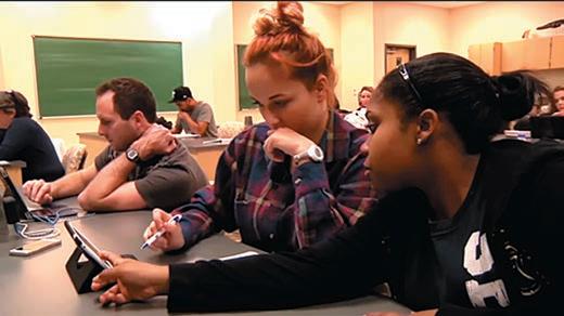
Learning Catalytics allows students to use their smartphone, tablet, or laptop to respond to questions in class. Visit learningcatalytics.com to learn more.
Additional Support for Students & Instructors
Mastering A&P® offers thousands of tutorials, activities, and questions that can be assigned for homework and practice. Highlights of the assignment options include:
• Building Vocabulary Coaching Activities give you practice learning and using word roots in context as you learn new A&P terms.
• Focus Figure “Mini-Animation” Coaching Activities bring some of the Focus Figures to life and include assessment questions.
• Concept Map Coaching Activities support the concept maps in the text without requiring students to submit their own concept map for grading.
• NCLEX-Style Questions give students practice with the kinds of questions that will eventually appear on a licensing exam.
The Mastering A&P® Instructor Resources Area includes the following downloadable tools for instructors who adopt the Twelfth Edition for their classes:
• Ready-to-Go Teaching Modules provide teaching tools for 10 challenging topics in A&P.
• Customizable PowerPoint® lecture outlines include customizable images and provide a springboard for lecture prep.
• The figures, photos, and tables from the text are available in JPEG and PowerPoint® formats, in labeled and unlabeled versions, and with customizable labels and leader lines.
• Test bank provides thousands of customizable questions across Bloom’s Taxonomy levels. Each question is tagged to chapter learning outcomes that can also be tracked within Mastering A&P® assessments. Available in Microsoft® Word and TestGen® formats.
• Animations and videos bring A&P concepts to life and include A&P Flix 3-D Animations.
Human Anatomy & Physiology Laboratory Manual
by Elaine Marieb & Lori Smith
Main 12/e ISBN 9781292442259
Cat 13/e ISBN 9781292442099
Fetal Pig 13/e ISBN 9781292441948
A Photographic Atlas for Anatomy & Physiology by Nora Hebert, Ruth E. Heisler, et al. ISBN 9780321869258
Human Anatomy & Physiology
TWELFTH GLOBAL EDITION
Elaine N. Marieb, R.N., Ph.D.
Holyoke Community College
Katja Hoehn, M.D., Ph.D.
Mount Royal University
Product Management: Gargi Banerjee
Content Strategy: Shabnam Dohutia and Amrita Naskar
Product Marketing: Wendy Gordon and Ashish Jain
Supplements: Bedasree Das
Digital Studio: Vikram Medepalli and Jayaprakash Kothandapani
Rights and Permissions: Anjali Singh
Cover Photo Credit: OSTILL is Franck Camhi/Shutterstock
Please contact https://support.pearson.com/getsupport/s/contactsupport with any queries on this content.
PearsonEducation Limited KAO Two KAO Park Harlow CM17 9SR United Kingdom
and Associated Companies throughout the world
Visit us on the World Wide Web at: www.pearsonglobaleditions.com
The rights of Elaine N. Marieb and Katja Hoehn to be identified as the authors of this work have been asserted by them in accordance with the Copyright, Designs and Patents Act 1988.
All rights reserved. No part of this publication may be reproduced, stored in a retrieval system, or transmitted in any form or by any means, electronic, mechanical, photocopying, recording or otherwise, without either the prior written permission of the publisher or a license permitting restricted copying in the United Kingdom issued by the Copyright Licensing Agency Ltd, Saffron House, 6–10 Kirby Street, London EC 1N 8TS.
All trademarks used herein are the property of their respective owners. The use of any trademark in this text does not vest in the author or publisher any trademark ownership rights in such trademarks, nor does the use of such trademarks imply any affiliation with or endorsement of this book by such owners.
Acknowledgments of third-party content appear on page 1225, which constitutes an extension of this copyright page.
PEARSON, ALWAYS LEARNING®, Mastering A&P®, Practice Anatomy Lab™, PAL™, Interactive Physiology®, and A&P Flix® are exclusive trademarks in the U.S. and/or other countries owned by Pearson Education, Inc., or its affiliates.
Unless otherwise indicated herein, any third-party trademarks that may appear in this work are the property of their respective owners and any references to third-party trademarks, logos or other trade dress are for demonstrative or descriptive purposes only. Such references are not intended to imply any sponsorship, endorsement, authorization, or promotion of Pearson’s products by the owners of such marks, or any relationship between the owner and Pearson Education, Inc. or its affiliates, authors, licensees or distributors.
Slinky® is a registered trademark of Poof-Slinky Inc.
Ziploc® is a registered trademark of S. C. Johnson & Son Inc.
Velcro® is a registered trademark of Velcro BVBA.
FedEx® is a registered trademark of FedEx Corporation.
Viagra® is a registered trademark of Pfizer Inc.
OxyContin® is a registered trademark of Purdue Pharma L.P.
Ventolin® is a registered trademark of Glaxo Group Limited.
Drāno® is a registered trademark of S. C. Johnson & Son Inc.
Lego® is a registered trademark of Lego Juris A/S.
South Beach Diet® is a registered trademark of SBD Enterprises LLC.
Atkins Diet™ is a trademark of Atkins Nutritionals Inc.
Weight Watchers® is a registered trademark of Weight Watchers International, Inc.
Etch a Sketch® is a registered trademark of Spin Master Ltd.
PowerPoint® is a registered trademark of Microsoft Corporation.
Microsoft® is a registered trademark of Microsoft Corporation.
TestGen® is a registered trademark of Tamarack Software Inc.
This eBook may be available as a standalone product or integrated with other Pearson digital products like MyLab and Mastering. This eBook may or may not include all assets that were part of the print version. The publisher reserves the right to remove any material in this eBook at any time.
Print ISBN 10: 1-292-42180-0; Print ISBN 13: 978-1-292-42180-3 (Paperback)
Print ISBN 10: 1-292-42181-9; Print ISBN 13: 978-1-292-42181-0 (Hardback)
eBook ISBN 13: 978-1-292-42178-0
British Library Cataloguing-in-Publication Data
A catalogue record for this book is available from the British Library
eBook formatted by B2R Technologies
About the Authors
We dedicate this work to our students both present and past, who always inspire us to “push the envelope.”
Elaine N. Marieb
After receiving her Ph.D. in zoology from the University of Massachusetts at Amherst, Elaine N. Marieb joined the faculty of the Biological Science Division of Holyoke Community College. While teaching at Holyoke Community College, where many of her students were pursuing nursing degrees, she developed a desire to better understand the relationship between the scientific study of the human body and the clinical aspects of the nursing practice. To that end, while continuing to teach full time, Dr. Marieb pursued her nursing education, which culminated in a Master of Science degree with a clinical specialization in gerontology from the University of Massachusetts. It is this experience that informed the development of the unique perspective and accessibility for which her publications are known.
Dr. Marieb gave generously to provide opportunities for students to further their education. She funded the E.N. Marieb
Katja Hoehn
Dr. Katja Hoehn is a professor in the Department of Biology at Mount Royal University in Calgary, Canada. Dr. Hoehn’s first love is teaching. Her teaching excellence has been recognized by several awards during her 24 years at Mount Royal University. These include a PanCanadian Educational Technology Faculty Award (1999), a Teaching Excellence Award from the Students’ Association of Mount Royal (2001), and the Mount Royal Distinguished Faculty Teaching Award (2004).
Dr. Hoehn received her M.D. (with Distinction) from the University of Saskatchewan, and her Ph.D. in Pharmacology from Dalhousie University. In 1991, the Dalhousie Medical Research Foundation presented her with the Max Forman (Jr.) Prize for excellence in medical research. During her Ph.D. and postdoctoral studies, she also pursued her passion for teaching by presenting guest lectures to first- and second-year medical students at Dalhousie University and at the University of Calgary.
Dr. Hoehn has been a contributor to several books, written numerous research papers in Neuroscience and Pharmacology,
Science Research Awards at Mount Holyoke College, which promotes research by undergraduate science majors, and underwrote renovation of the biology labs in Clapp Laboratory at that college. Dr. Marieb also contributed to the University of Massachusetts at Amherst, where she provided funding for reconstruction and instrumentation of a cutting-edge cytology research laboratory. Recognizing the severe national shortage of nursing faculty, she underwrote the Nursing Scholars of the Future Grant Program at the university.
In 2012 and 2017, Dr. Marieb gave generous philanthropic support to Florida Gulf Coast University as a long-term investment in education, research, and training for healthcare and human services professionals in the local community. In honor of her contributions, the university is now home to the Elaine Nicpon Marieb College of Health and Human Services.
and has co-authored some of the previous editions of this textbook. For many years, she has also reviewed and authored electronic media that accompanies Pearson anatomy and physiology books.
Following Dr. Marieb’s example, Dr. Hoehn provides financial support for students in the form of a scholarship that she established in 2006 for nursing students at Mount Royal University.
Dr. Hoehn is also actively involved in the Human Anatomy and Physiology Society (HAPS) and is a member of the American Association of Anatomists. When not teaching, she likes to spend time outdoors with her husband and two sons. She also enjoys competing in long-course triathlons, and playing Irish flute down at the local pub.
Preface
Today’s students have access to an enormous amount of information about anatomy and physiology. As educators, our biggest challenge is to help students focus on mastering the basic concepts of this field. Providing this firm foundation will help students to become lifelong learners who can critically evaluate new information, connect that information to the
Unifying Themes
Three unifying themes that have helped to organize and set the tone of this textbook continue to be valid and are retained in this edition. These themes are:
Interrelationships of body organ systems. This theme emphasizes the fact that nearly all regulatory mechanisms have interactions with several organ systems. The respiratory system, for example, cannot carry out its role of gas exchange in the body if there are problems with the cardiovascular system that prevent the normal delivery of blood throughout the body. The System Connections feature and Make Connections questions throughout the book help students connect new information to old information and think of the body as a community of dynamic parts instead of a number of independent units.
Homeostasis. Homeostasis is the normal and most desirable condition of the body. Its loss is always associated with past or present pathology. This theme is not included to emphasize pathological conditions, but rather to illustrate what happens in the body “when things go wrong” and homeostasis is lost. Whenever students see a red balance beam symbol accompanied by an associated clinical topic, their understanding of how the body works to stay in balance is reinforced.
Complementarity of structure and function. This theme encourages students to understand the structure of some body part (ranging from a molecule to an organ) in order to understand the function of that structure. For example, muscle cells can produce movement because they are contractile cells.
foundation they have already established, and apply it in a clinical setting. How can we help students build a strong foundation in anatomy and physiology? We believe that this new edition of our textbook will help learners by building on the strengths of previous editions while using new and innovative ways to help students visualize connections between various concepts.
Key Features of the Twelfth Edition
The following are the key features of the Twelfth Edition:
NEW! Boxes on scientists feature details about the lives and works of eminent scientists. These will show students the human side of science. Please refer to p. 16 for a list of these boxes.
NEW! Homeostatic Imbalance discussions have been added on Marfan syndrome, brittle bone disease, tetanus, and anxiety disorders.
NEW! We have added tables on the following topics:
• Functions of neurons and neuroglia
• Focal versus diffuse brain injuries
• Antihypertensive medications and renin-angiotensinaldosterone system
NEW! We have updated the content with information on recent developments on topics such as sperm centrioles, using fibroblasts as stem cells, 3D bioprinting used for skin grafts, how COVID-19 causes loss of smell, and juxtacrines.
NEW! We have added an A Closer Look feature on COVID-19.
To help students make connections between new and previously learned material. In order for students to master new concepts, they must link these new concepts with concepts they already understand. In this edition, we help them do this by adding:
• Text recall icons ( ). These icons direct the student back to the specific pages where a concept was first introduced.
• Make Connections questions. We’ve added more of this type of question to the Check Your Understanding review questions that follow each module within a chapter. To answer these questions, the student must employ concepts learned previously (most often in previous chapters).
• New kinds of higher-level questions. Each chapter has at least five higher-level questions that require students to think more deeply, pulling together strands from multiple concepts. These questions are clearly identified as APPLY , DRAW , PREDICT , MAKE CONNECTIONS , and WHAT IF? questions.
To enhance students’ visual literacy. Anatomy is and has always been taught principally through images. Increasingly, however, physiological data is also represented as images, whether it be molecular interactions or graphical descriptions of processes. Throughout their future health care careers, students will need to be able to understand and interpret information presented visually. In this edition, we help them do this through:
• Focus figures. Focus figures are illustrations that use a “big picture” layout and dramatic art to guide the student through difficult physiological processes in a step-by-step way. Our Focus figures have been a hit with both students and instructors.
• DRAW questions in each chapter. Students often think that they understand an illustration simply by looking at it, but to truly comprehend an illustration and cement its concepts requires a more active learning approach. For this reason we include at least one higher-level review question within each chapter that requires a student either to draw an illustration or to add to an existing diagram.
• Questions about illustrations. To help students practice their visual literacy skills, we have added Check Your Understanding questions that include an illustration as part of the question. Some of these are as simple as labeling exercises, but many require more advanced interpretation.
• Updated art. Today’s students are accustomed to seeing sophisticated photorealistically rendered images. However, many students are not adept at extracting, and thinking
critically about, the relevant information contained in such illustrations. With this in mind we continue to refine and update our illustrations as students’ needs change, improving their ability to teach important concepts.
• In-line figures. These are small (less than a half-column wide) illustrations or photos strategically located within the text that discuss the concept they illustrate. This edition has 31 such in-line figures.
To help students clinically apply what they have learned
• Updated Homeostatic Imbalance features. Many of the Homeostatic Imbalance features have been updated. All have been reviewed for accuracy and relevancy. In addition, the updated book design makes these features stand out more clearly.
• Clinical Case Studies in Chapters 5-29 with ✚ NCLEX-STYLE questions. The end-of-chapter review questions, which are now organized into three levels of difficulty based on Bloom’s Taxonomy categories, culminate in a clinical case study that allows students to apply some of the concepts they have learned to a clinical scenario. Each case study has two questions that are similar in style to those in the NCLEX exam.
• Clinically relevant photos. We have a number of photos that have clinical relevance (procedures, conditions, etc.) that will help students apply what they are reading to real-life situations and to their future careers.
As in the previous edition, we have taken painstaking care to ensure that almost all the text and the associated art are covered on the same two-page spread. Although this sounds like a simple goal, it actually takes a great deal of work and has not usually been achieved by other textbooks. We make this effort because it is invaluable to student learning to not have to flip pages back and forth between art and text. Finally, you will notice the appearance of icons referencing Mastering A&P® interspersed within the text. This guides students to go to the relevant on-line activities to supplement their learning.
Notable Scientists
CHAPTER 1 Marie Curie 47
CHAPTER 3 Reiji and Tsuneko Okazaki 127
CHAPTER 4 Ernst Ruska 147
CHAPTER 6 Andreas Vesalius 206
CHAPTER 7 Kristopher Kilian 277
CHAPTER 8 Hugh Herr 304
CHAPTER 9 Claude Bernard 320
CHAPTER 10 Archimedes 358
CHAPTER 11 Santiago Ramón y Cajal 426
CHAPTER 12 Roger Sperry 476
CHAPTER 13 Filippo Pacini 525
CHAPTER 14 Hermona Soreq 575
CHAPTER 15 Alcmaeon of Croton 586 Robert Bárány 622
CHAPTER 16 Dora Jacobsohn 642
CHAPTER 17 Karl Landsteiner 695
CHAPTER 18 W illem Einthoven 722
CHAPTER 19 Nikolai Korotkoff 749
Rhian Touyz 756
CHAPTER 21 Susumu Tonegawa 830
CHAPTER 22 John Scott Haldane 881
CHAPTER 23 John C. Brown 939
CHAPTER 24 Gerty Cori 978
CHAPTER 25 Eduardo Braun-Menéndez 1023
CHAPTER 26 Peter Agre 1052
CHAPTER 27 Neena B. Schwartz 1110
CHAPTER 28 Robert G. Edwards 1153
CHAPTER 29 Emanuelle Charpentier 1169
Acknowledgments
Producing a new edition of this book is an enormous undertaking. Let us take you through the steps and introduce you to the people behind the scenes that have helped make this book what it is. Every new edition begins with a revision plan. We’d like to thank all of the students and instructors who have provided the feedback (gathered by our editorial team) that forms the basis of this plan. Once this plan was in place, Barbara Price (our text Development Editor) scoured each chapter. This was Barbara’s first exposure to the book and her fresh eyes on the text found opportunities to further clarify the presentation. In addition, she noted places where additional chunking of the text (such as bulleted lists) would help the students. Her excellent work has made this text better. We incorporated her ideas, and reviewer feedback, together with our own updates and ideas for reorganization of the text and art. Thanks to Patricia Bowne for contributing to the Clinical Case Studies and Wendy Mercier for reviewing all of the Case Studies. We also very much appreciate the help of Karen Dougherty, who used her expertise as a physician and educator to review all of the Homeostatic Imbalance features and help us revise and update them.
We then laid out each chapter to maintain text-art correlation before passing the manuscript off to Michele Mangelli. Michele wore many different hats during this revision. She was both the Program Manager for the editorial side of things as well as the Goddess of Production. She reviewed the revised manuscript before she sent it to ace copyeditor Anita Hueftle. Anita saved us on many occasions from public embarrassment by finding our spelling and grammar errors, our logical lapses, and various other inconsistencies. We can’t thank Anita enough for her meticulous and outstanding work! (Any remaining errors are our fault.)
At the same time the text was in revision, the art program was going through a similar process. This book would not be what it is without the help of Laura Southworth, our superb Art Development Editor. Laura’s creativity, attention to detail, and her sense of what will teach well and what won’t have helped us immensely. She has worked tirelessly to make our Focus figures and other art even better. Finding good, usable photos is never easy, and we are grateful for the hard work of Kristin Piljay (Photo Researcher). It was also a pleasure to work with Jean Lake again, who expertly juggled the administrative aspects of the art program and kept us all on track. This team ensured that the artists at Imagineering had all the information they needed to produce beautiful final art products. As the manuscript made the transition from Editorial to Production, Michelle Mangelli (wearing a different hat—this
one as the Production and Design Manager) took over again. As head honcho and skilled handler of all aspects of production, everyone answered to her from this point on. Kudos to our excellent production coordinator, Karen Gulliver, who did much of the hands-on handling, routing, and scheduling of the manuscript. We’d also like to thank Martha Ghent (Proofreader), Betsy Dietrich (Art Proofreader), Sallie Steele (Indexer), Alicia Elliot (Project Manager at Imagineering), and Cenveo (Compositor). Izak Paul meticulously read every chapter for scientific accuracy, and we are very grateful for his careful work. Thanks also to Gary Hespenheide for his stunning design work on the cover, chapter opening pages, and the text.
It was a pleasure to work with Lauren Harp, our Acquisitions Editor. Her extensive knowledge of the needs of both faculty and students in anatomy and physiology has helped inform this revision. Her enthusiasm for this book is infectious, her choice for the cover is inspired, and we are delighted to have her on board! Before Lauren became part of the team, Serina Beauparlant, our Editor-in-Chief, stepped up to helm the planning phase of this revision. Fiercely dedicated to making this book and its associated media resources the best teaching tools that they can be, Serina has been invaluable in shaping this revision. We deeply appreciate all she has done for us and this book. Lauren and Serina were competently aided by Editorial Assistant Dapinder Dosanjh (and before her, Nicky Montalvo).
Other members of our team with whom we have less contact but who are nonetheless vital are: Barbara Yien, Director of Content Development, Stacey Weinberger (our Senior Manufacturing Buyer), and Derek Perrigo (our top-notch Marketing Manager). We appreciate the hard work of our media production team headed by Lauren Chen, Lauren Hill, Laura Tommasi, Sarah Young Dualan, and Cheryl Chi, and also wish to thank Eric Leaver for his astute observations on certain figures.
Kudos to our entire team. We feel we have once again prepared a superb textbook. We hope you agree.
Many people reviewed parts of this text—both professors and students, either individually or in focus groups—and we would like to thank them. Input from the following reviewers has contributed to the continued excellence and accuracy of this text and its accompanying Mastering A&P® assignment options, including Interactive Physiology 2.0:
Matthew Abbott, Des Moines Area Community College
Emily Allen, Rowan College at Gloucester County Lynne Anderson, Meridian Community College
Acknowledgments
David C. Ansardi, Calhoun Community College
Martin W. Asobayire, Essex Community College
David Babb, West Hills College Lemoore
Yvonne Baptiste-Szymanski, Niagara County Community College
Claudia Barreto, University of New Mexico–Valencia
Jerry Barton, Tarrant County College
Shawn Bearden, Idaho State University
Charles Benton, Madison Area Technical College
J. Gordon Betts, Tyler Junior College
Diana Bourke, Community College of Allegheny County
Sherry Bowen, Indian River State College
Michael Brady, Columbia Basin College
Betsy Brantley, Valencia College
Beth Braun, Truman College
Carol A. Britson, University of Mississippi
C. Steven Cahill, West Kentucky Community and Technical College
Christie Campbell, Ozarks Technical Community College
Maria C. Carles, Northern Essex Community College
Tamyra Carmona, Cosumnes River College
Marien Cendon, Miami Dade College
Brendon Chastain, West Kentucky Community Technical College
Sam Chen, Moraine Valley Community College
Alexander G. Cheroske, Mesa Community College–Red Mountain
Brandi Childress, Georgia Perimeter College
William M. Clark, Lone Star College–Kingwood
Joseph Comber, Villanova University
Teresa Cowan, Baker College–Auburn Hills
Donna Crapanzano, Stony Brook University
Maurice M. Culver, Florida State College at Jacksonville
Jason Dechant, University of Pittsburgh
Smruti A. Desai, Lone Star College–CyFair
Karen H. Dougherty, Hopkinsville Community College
Sondra Dubowsky, McLennan Community College
Karen Dunbar Kareiva, Ivy Tech Community College
Kathryn Durham, Lorain County Community College
Karen Eastman, Chattanooga State Community College
Sharon S. Ellerton, Queensborough Community College–CUNY
Paul Emerick, Monroe Community College
Elyce Ervin, University of Toledo
Martha Eshleman, Pulaski Technical College
Colin Everhart, St. Petersburg Community College
Brian D. Feige, Mott Community College
Michele Finn, Monroe Community College
John E. Fishback, Ozarks Technical Community College
Maria Florez, Lone Star College–CyFair
Reza Forough, Bellevue College
Juanita A. Forrester, Chattahoochee Technical College
Aaron Fried, Mohawk Valley Community College
Dean Furbish, Wake Technical Community College
Marie Gabbard, College of Western Idaho
Sophia Garcia, Tarrant County College
Jane E. Gavin, University of South Dakota
Peter Germroth, Hillsborough Community College
Emily K. Getty, Ivy Tech Community College
Amy Giesecke, Chattahoochee Technical College
Anna Gilletly, Central New Mexico Community College
Gary Glaser, Genesee Community College
Richard Gonzalez-Diaz, Seminole State College of Florida
Abigail Goosie, Walters State Community College
Pattie S. Green, Tacoma Community College
Edwin Griff, University of Cincinnati
George G. Hanak, Pasco-Hernando State College
Mary Beth Hanlin, Des Moines Area Community College–Boone
Heidi Hawkins, College of Southern Idaho
Martie Heath-Sinclair, Hawkeye Community College
Nora Hebert, Red Rocks Community College
Nadia Hedhli, Hudson County Community College
D.J. Hennager, Kirkwood Community College
Jennifer Hill, Montgomery College–Takoma Park-Silver Spring
Shannon K. Hill, Temple College
Mark Hollier, Georgia Perimeter College
H. Rodney Holmes, Waubonsee Community College
Mark J. Hubley, Prince George’s Community College
Carolyn Huffman, Wichita Area Technical College
Julie Huggins, Arkansas State University
Jason Hunt, Brigham Young University–Idaho
Alexander Ibe, Weatherford College
Alexander Imholtz, Prince George’s Community College
Virginia Irintcheva, Black Hawk College
Brian E. Jordan, C.S. Mott Community College
Thomas Jordan, Pima Community College
Christopher Jung, University of Alaska Anchorage
William M. Karkow, University of Dubuque
Suzanne Keller, Indian Hills Community College
Michael Kielb, Eastern Michigan University
Marta Klesath, North Carolina State University
Nelson H. Kraus, University of Indianapolis
Paul M. Lea IV, Northern Virginia Community College
Steven Lewis, Metropolitan Community College–Penn Valley
Juanita Limas, Kirkwood Community College
Jerri K. Lindsey, Tarrant County College–Northeast
Chelsea Loafman, Central Texas College
Paul Luyster, Tarrant County College
Ken Malachowsky, Florence-Darlington Technical College
Theresa Martin, College of San Mateo
Nicole Mashburn, Calhoun Community College
Abdallah M. Matari, Hudson County Community College
Bhavya Mathur, Chattahoochee Technical College
Tiffany Beth McFalls-Smith, Elizabethtown Community and Technical College
Jennifer Menon, Johnson County Community College
Jaime Mergliano, John Tyler Community College
Sharon Miles, Itawamba Community College
Todd Miller, Hunter College of CUNY
Louise Millis, North Hennepin Community College
Justin Moore, American River College
Christine Morin, Prince George’s Community College
Qian F. Moss, Des Moines Area Community College
Regina Munro, Chandler-Gilbert Community College
Necia Nicholas, Calhoun Community College
Maria Oehler, Florida State College–Jacksonville
Betsy Ott, Tyler Junior College
Ellen Ott-Reeves, Blinn College–Bryan
Stephen Page, Community College of Baltimore County & Townson University
Vikash Patel, Nevada State College
Dennis Pearson, Morton College
Diane Pelletier, Green River Community College
Jessica Petersen, Pensacola State College
Jason Pienaar, University of Alabama
Becky Pierce, Delta College
Gilbert Pitts, Austin Peay State University
Renee Prenitzer, Greenville Technical College
Fernando Prince, Laredo Community College
Sarah A. Pugh, Shelton State Community College
Suzanne Pundt, University of Texas at Tyler
Rolando J. Ramirez, The University of Akron
Wendy Rappazzo, Harford Community College
Terrence J. Ravine, University of South Alabama
Christine S. Rigsby, Middle Georgia State University
Laura H. Ritt, Burlington County College
Cynthia Robison, Wallace Community College
Susan Rohde, Triton College
Brian Sailer, Central New Mexico Community College
Sharon Schapel, Mott Community College
Mark Schmidt, Clark State Community College
Michael W. Sipala, Bristol Community College
Amy Skibiel, Auburn University
Lori Smith, American River College–Los Rios
Kerry Smith, Oakland Community College–Auburn Hills
Tom Sobat, Ivy Tech Community College
Kay Sourbeer, Tidewater Community College
Ashley Spring-Beerensson, Eastern Florida State College
Justin R. St. Juliana, Ivy Tech Community College
Cindy Stanfield, University of South Alabama
Laura Steele, Ivy Tech Community College–Northeast
George A. Steer, Jefferson College of Health Sciences
Michelle Stettner, Meridian Community College
Sherry Stewart, Navarro College
Susan E. Tappen, Central New Mexico Community College
Dean Thornton, South Georgia State College
Rita A. Thrasher, Pensacola State College
Brenda Tondi, George Mason University
Sheela Vemu, Waubonsee Community College
Khursheed Wankadiya, Central Piedmont Community College
Chad Wayne, University of Houston
Kira L. Wennstrom, Shoreline Community College
Shirley A. Whitescarver, Bluegrass Community and Technical College–KCTCS
John Whitlock, Hillsborough Community College
Patricia Wilhelm, Johnson and Wales University
Luann Wilkinson, Marion Technical College
Selwyn A. Williams, Miami Dade College
Darrellyn Williams, Pulaski Technical College
Peggie Williamson, Central Texas College
Heather Wilson-Ashworth, Utah Valley University
MaryJo A. Witz, Monroe Community College
Jackie Wright, South Plains College
James Robert Yount, Brevard Community College
We would like to acknowledge the following group who reviewed various iterations of the new Focus figures: Matthew Abbott, David Ansardi, Jake Dechant, Karen Dougherty, Peter Germroth, Gary Glaser, Suzanne Keller, Gilbert Pitts, Terry Ravine, Michelle Stettner, and Rita Thrasher.
We would also like to acknowledge the support of Katja’s colleagues at Mount Royal University (Trevor Day, Sarah Hewitt, Tracy O’Connor, Sarah Orton, Izak Paul, Lorraine Royal, Karen Sheedy, Kartika Tjandra, and Margot Williams); Department Chairs (Ruth Pickett-Seltner and Melanie Rathburn); and Deans (Jeffrey Goldberg and Jonathan Withey). Thanks also to Katja’s husband, Dr. Lawrence Haynes, a fellow physiologist who has worked together with Katja and has been involved in all aspects of this revision. We would like to thank Katja and Larry’s sons, Eric and Stefan Haynes, for putting up with their parents through many revisions of this book and for continuing to be an inspiration and a joy.
We really would appreciate hearing from you concerning your opinion—suggestions and constructive criticisms—of this text. It is this type of feedback that will help us in the next revision and underlies the continued improvement of this text.
Elaine Marieb Katja Hoehn
Global Edition Acknowledgments
Pearson would like to thank the following for contributing to the Global Edition:
Richard Brooksbank, University of the Witwatersrand
Snezana Kusljic, The University of Melbourne
Liana Maree, University of the Western Cape
Christiane Van den Branden, Vrije Universiteit Brussel
Pearson would also like to thank the following for reviewing the Global Edition:
Sarah Allsop, University of Bristol
Richard Brooksbank, University of the Witwatersrand
Snezana Kusljic, The University of Melbourne
Peace Mabeta, University of Pretoria
Hemant Mehta, Australian Catholic University
Shahed Nalla, University of Johannesburg
Puspha Sinnayah, Victoria University
Carine Smith, Stellenbosch University
Eva Strandell, Halmstad University
UNIT 1 Organization of the Body
1 The Human Body: An Orientation 31
1.1 Form (anatomy) determines function (physiology) 32
1.2 The body’s organization ranges from atoms to the entire organism 34
1.3 What are the requirements for life? 35
1.4 Homeostasis is maintained by negative feedback 39
1.5 Anatomical terms describe body directions, regions, and planes 42
A CLOSER LOOK Medical Imaging: Illuminating the Body 46
1.6 Many internal organs lie in membrane-lined body cavities 48
2 Chemistry Comes Alive 53
PART 1 B ASIC CHEMISTRY 54
2.1 Matter is the stuff of the universe and energy moves matter 54
2.2 The properties of an element depend on the structure of its atoms 55
2.3 Atoms bound together form molecules; different molecules can make mixtures 58
2.4 Three types of chemical bonds are ionic, covalent, and hydrogen 61
2.5 Chemical reactions occur when electrons are shared, gained, or lost 65
PART 2 BIOCHEMISTRY 68
2.6 Inorganic compounds include water, salts, and many acids and bases 68
2.7 Organic compounds are made by dehydration synthesis and broken down by hydrolysis 71
2.8 Carbohydrates provide an easily used energy source for the body 73
2.9 Lipids insulate body organs, build cell membranes, and provide stored energy 75
2.10 Proteins are the body’s basic structural material and have many vital functions 78
2.11 DNA and RNA store, transmit, and help express genetic information 83
2.12 ATP transfers energy to other compounds 85
3 Cells: The Living Units 90
3.1 Cells are the smallest unit of life 91
PART 1 PLASMA MEMBRANE 93
3.2 The plasma membrane is a double layer of phospholipids with embedded proteins 93
FOCUS FIGURE 3.1 The Plasma Membrane 94
3.3 Passive membrane transport is diffusion of molecules down their concentration gradient 98
3.4 Active membrane transport directly or indirectly uses ATP 103
FOCUS FIGURE 3.2 Primary Active Transport: The Na+-K+ Pump 104
3.5 Selective diffusion establishes the membrane potential 109
3.6 Cell adhesion molecules and membrane receptors allow the cell to interact with its environment 111
FOCUS FIGURE 3.3 G Proteins 112
PART 2 THE CYTOPLASM 113
3.7 Cytoplasmic organelles each perform a specialized task 113
3.8 Cilia and microvilli are two main types of cellular extensions 120
PART 3 NUCLEUS 121
3.9 The nucleus includes the nuclear envelope, the nucleolus, and chromatin 121
3.10 The cell cycle consists of interphase and a mitotic phase 126
3.11 Messenger RNA carries instructions from DNA for building proteins 128
FOCUS FIGURE 3.4 Mitosis 130
FOCUS FIGURE 3.5 Translation 136
3.12 Autophagy and proteasomes dispose of unneeded organelles and proteins; apoptosis disposes of unneeded cells 138
DEVELOPMENTAL ASPECTS of Cells 139
4 Tissue: The Living Fabric 145
4.1 Tissue samples are fixed, sliced, and stained for microscopy 147
4.2 Epithelial tissue covers body surfaces, lines cavities, and forms glands 147
4.3 Connective tissue is the most abundant and widely distributed tissue in the body 156
4.4 Muscle tissue is responsible for body movement 168
4.5 Nervous tissue is a specialized tissue of the nervous system 170
4.6 The cutaneous membrane is dry; mucous and serous membranes are wet 171
4.7 Tissue repair involves inflammation, organization, and regeneration 172
A CLOSER LOOK Cancer—The Intimate Enemy 174
DEVELOPMENTAL ASPECTS of Tissues 176
UNIT 2 Covering, Support, and Movement of the Body
5 The Integumentary System 180
5.1 The skin consists of two layers: the epidermis and dermis 180
5.2 The epidermis is a keratinized stratified squamous epithelium 182
5.3 The dermis consists of papillary dermis and reticular dermis 184
5.4 Melanin, carotene, and hemoglobin determine skin color 186
5.5 Hair consists of dead, keratinized cells 187
5.6 Nails are scale-like modifications of the epidermis 190
5.7 Sweat glands help control body temperature, and sebaceous glands secrete sebum 191
5.8 First and foremost, the skin is a barrier 193
5.9 Skin cancer and burns are major challenges to the body 195
DEVELOPMENTAL ASPECTS of the Integumentary System 197
SYSTEM CONNECTIONS 198
6 Bones and Skeletal Tissues 203
6.1 Hyaline, elastic, and fibrocartilage help form the skeleton 204
6.2 Bones perform several important functions 205
6.3 Bones are classified by their location and shape 206
6.4 The gross structure of all bones consists of compact bone sandwiching spongy bone 206
6.5 Bones develop either by intramembranous or endochondral ossification 214
6.6 Bone remodeling involves bone deposition and removal 218
6.7 Bone repair involves hematoma and callus formation, and remodeling 220
6.8 Bone disorders result from abnormal bone deposition and resorption 223
DEVELOPMENTAL ASPECTS of Bones 224
SYSTEM CONNECTIONS 226
7 The Skeleton 229
PART 1 THE AXIAL SKELETON 229
7.1 The skull consists of 8 cranial bones and 14 facial bones 231
7.2 The vertebral column is a flexible, curved support structure 248
7.3 The thoracic cage is the bony structure of the chest 254
PART 2 THE APPENDICULAR SKELETON 257
7.4 Each pectoral girdle consists of a clavicle and a scapula 257
7.5 The upper limb consists of the arm, forearm, and hand 260
7.6 The hip bones attach to the sacrum, forming the pelvic girdle 266
7.7 The lower limb consists of the thigh, leg, and foot 270
DEVELOPMENTAL ASPECTS of the Skeleton 276
8 Joints 281
8.1 Joints are classified into three structural and three functional categories 281
8.2 In fibrous joints, the bones are connected by fibrous tissue 282
8.3 In cartilaginous joints, the bones are connected by cartilage 283
8.4 Synovial joints have a fluid-filled joint cavity 284
FOCUS FIGURE 8.1 Synovial Joints 292
8.5 Five examples illustrate the diversity of synovial joints 294
8.6 Joints are easily damaged by injury, inflammation, and degeneration 302
A CLOSER LOOK Joints: From Medieval Armor to Bionic Humans 304
DEVELOPMENTAL ASPECTS of Joints 305
9 Muscles and Muscle Tissue 309
9.1 There are three types of muscle tissue 310
9.2 A skeletal muscle is made up of muscle fibers, nerves, blood vessels, and connective tissues 311