HistologyandCellBiology:AnIntroductionto Pathology5thEditionAbrahamLKierszenbaum

https://ebookmass.com/product/histology-and-cell-biology-anintroduction-to-pathology-5th-edition-abraham-lkierszenbaum/
Instant digital products (PDF, ePub, MOBI) ready for you
Download now and discover formats that fit your needs...
Histology and Cell Biology: An Introduction to Pathology 5th Edition Abraham L Kierszenbaum M.D. Ph.D.
https://ebookmass.com/product/histology-and-cell-biology-anintroduction-to-pathology-5th-edition-abraham-l-kierszenbaum-m-d-ph-d/
ebookmass.com
Histología y biología celular, ed. 4 (Spanish Edition)
Abraham L Kierszenbaum & Laura Tres [Kierszenbaum
https://ebookmass.com/product/histologia-y-biologia-celulared-4-spanish-edition-abraham-l-kierszenbaum-laura-tres-kierszenbaum/
ebookmass.com
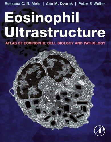
Eosinophil Ultrastructure: Atlas of Eosinophil Cell Biology and Pathology 1st Edition Rossana C.N. Melo
https://ebookmass.com/product/eosinophil-ultrastructure-atlas-ofeosinophil-cell-biology-and-pathology-1st-edition-rossana-c-n-melo/ ebookmass.com
Boss Witch Ann Aguirre
https://ebookmass.com/product/boss-witch-ann-aguirre-4/


ebookmass.com
The Rise of China and International Law: Taking Chinese Exceptionalism Seriously Congyan Cai
https://ebookmass.com/product/the-rise-of-china-and-international-lawtaking-chinese-exceptionalism-seriously-congyan-cai/
ebookmass.com
Management Accounting: Information for Creating and Managing Value 8th Edition Kim Langfield-Smith
https://ebookmass.com/product/management-accounting-information-forcreating-and-managing-value-8th-edition-kim-langfield-smith/
ebookmass.com
Youth Employment in Bangladesh: Creating Opportunities—Reaping Dividends 1st ed. 2020 Edition
Fahmida Khatun
https://ebookmass.com/product/youth-employment-in-bangladesh-creatingopportunities-reaping-dividends-1st-ed-2020-edition-fahmida-khatun/
ebookmass.com
roceeding of 5 th International Conference on Recent Trends in Engineering and Technology ICRTET’2016 Volume 1 Computer Engineering Information Technology Civil Engineering M. M. Rathore
https://ebookmass.com/product/roceeding-of-5-th-internationalconference-on-recent-trends-in-engineering-and-technologyicrtet2016-volume-1-computer-engineering-information-technology-civilengineering-m-m-rathore/ ebookmass.com
Anders gedacht: Text and Context in the German Speaking World (World Languages) 3rd Edition, (Ebook PDF)
https://ebookmass.com/product/anders-gedacht-text-and-context-in-thegerman-speaking-world-world-languages-3rd-edition-ebook-pdf/ ebookmass.com



13th Edition Roger Leroy Miller
https://ebookmass.com/product/business-law-today-comprehensivemindtap-course-list-13th-edition-roger-leroy-miller/
ebookmass.com



HISTOLOGY AND CELL BIOLOGY
An Introduction to Pathology
This page intentionally left blank
HISTOLOGY AND CELL BIOLOGY
An Introduction to Pathology
Fifth Edition
Abraham L. Kierszenbaum, M.D., Ph.D.
Medical (Clinical) Professor Emeritus
Former Chair of the Department of Cell Biology and Anatomy
The Sophie Davis School of Biomedical Education*
The City University of New York New York, New York USA
Laura L. Tres, M.D., Ph.D.
Medical (Clinical) Professor Emerita
The Sophie Davis School of Biomedical Education*
The City University of New York New York, New York USA
*Now CUNY School of Medicine/ Sophie Davis Biomedical Education Program
Elsevier 1600 John F. Kennedy Blvd. Ste 1600 Philadelphia, PA 19103-1899
HISTOLOGY AND CELL BIOLOGY: ISBN: 978-0-323-67321-1 AN INTRODUCTION TO PATHOLOGY, FIFTH EDITION
Copyright © 2020 by Elsevier, Inc. All rights reserved
No part of this publication may be reproduced or transmitted in any form or by any means, electronic or mechanical, including photocopying, recording, or any information storage and retrieval system, without permission in writing from the publisher. Details on how to seek permission, further information about the Publisher’s permissions policies and our arrangements with organizations such as the Copyright Clearance Center and the Copyright Licensing Agency, can be found at our website: www.elsevier.com/permissions.
This book and the individual contributions contained in it are protected under copyright by the Publisher (other than as may be noted herein).
Notice
Practitioners and researchers must always rely on their own experience and knowledge in evaluating and using any information, methods, compounds or experiments described herein. Because of rapid advances in the medical sciences, in particular, independent verification of diagnoses and drug dosages should be made. To the fullest extent of the law, no responsibility is assumed by Elsevier, authors, editors or contributors for any injury and/or damage to persons or property as a matter of products liability, negligence or otherwise, or from any use or operation of any methods, products, instructions, or ideas contained in the material herein.
Previous editions copyrighted 2016, 2012, 2007, and 2002.
Library of Congress Control Number: 2019940508
Content Strategist: Alexandra Mortimer
Senior Content Development Specialist: Ann Ruzycka Anderson
Publishing Services Manager: Catherine Jackson
Senior Project Manager: Daniel Fitzgerald
Printed in Canada.
Last digit is the print number: 9 8 7 6 5 4 3 2 1
To our daughters, Adriana and Silvia
To our grandchildren, Ryan, Trevor, Kyle and Marielle
To the beloved memory of our parents

This book is also dedicated to you, the teacher, who transmits enthusiastically the significance of knowledge in a way that goes beyond what is being taught; and to you, the student, who transforms the act of learning into the passion of learning.
This page intentionally left blank
New in the fifth edition are Primers Each Primer transmits information about a specific topic in a concise and visual format to transcend concepts and stimulate further exploration.
PREFACE
The fifth edition of Histology and Cell Biology: An Introduction to Pathology contains substantial revisions and additions. They strengthen the visual approach to learning histology within the context of cell biology and pathology introduced in the previous editions. New in the fifth edition are a number of Primers included in most chapters. Each Primer transmits information about a specific topic in a concise and visual format to transcend concepts and stimulate further exploration. The convergence of histology–cell biology–pathology intends to prepare medical students for the forthcoming learning of pathophysiology and clinical medicine. The practice of medicine changes relentlessly as new knowledge emerges. Future physicians can find in this book the basis for continuing education to better help their patients by constantly integrating basic and clinical sciences.
The visual approach presented in this book emerged from many years of practicing pathology and teaching cell biology, histology and pathology to medical students. The convergence of histology, cell biology and pathology promotes unity in diversity. Diversity leads to the transforming power of new knowledge. The cell biology and pathology components, although not complete, provide the necessary foundation for further learning and integration with medical sciences. Pathology students and residents may find this book useful for refreshing basic concepts of histology and cell biology. Histology and pathology are visually oriented sciences, and the visual cues included in this book can facilitate interpretation opportunities in clinical practice.
Similar to the previous editions, the fifth edition consists of six parts, bringing together histology, cell biology and general pathology within the context of the basic tissues and organ systems. Chapter 3, Cell Signaling | Cell Biology | Pathology, is an uncommon section in a histology book. It serves to unify the concept that the study of tissues and organs cannot be separated from the increasing impact of molecular biology in the practice of medicine.
New in the fifth edition is the use of a light green background to identify sections presenting essential concepts of histology and cell biology, a starting point for further learning.
Each Concept Mapping provides a basic framework of interconnected concepts arranged in a hierarchical form leading to integration and critical thinking.
Each Concept Mapping provides a basic framework of interconnected concepts arranged in a hierarchical form, leading to integration and critical thinking.
Students may find the online animation version of Concept Mappings convenient for group interaction, transforming the passivity of learning into a dynamic and collective activity.
New in this edition is the use of a light green background to identify in each chapter sections presenting essential concepts of histology. A number of teachers may find this offering useful as a starting point for further learning. The expectation is that the additional material could stir curiosity to unfold the indispensable complement of fragmented knowledge. All the information is presented in a clear, concise and student-friendly manner using color graphics and photographs that are meant to be studied. In some cases the graphics reiterate the succinct text; in others they add new information complementing or extending the text. Several boxes dispersed in all chapters introduce students to clinical and pathological conditions and to recent and evolving molecular and biochemical knowledge.
Most chapters include one or more Concept Mappings. Each Concept Mapping provides a basic framework of interconnected concepts arranged in a hierarchical form, leading to integration and critical thinking. Concept Mapping and Essential Concepts highlight key issues to review and integrate when the time of in-course and board examinations arrives. Students may find the online animation version of Concept Mappings convenient for group interaction, transforming the passivity of learning into a dynamic and collective activity. Activity inspired by a new mode of communication, depending not only on content but also the integrative vision and shared values of the information.
There are many people to be acknowledged and thanked. We are grateful for the numerous suggestions, comments and encouragements from faculty and students. We thank publishers who made available to faculty and students the Chinese, French, Greek, Japanese, Portuguese, Spanish and Turkish editions. We thank the British Medical Association for awarding the First Prize in Basic and Medical Sciences to the second edition. Our special appreciation goes to Alexandra Mortimer, Ann Ruzycka Anderson and Daniel Fitzgerald in London, New York and St. Louis offices for their magnificent effort in making sure that the fifth edition met high publishing standards.
Abraham
L. Kierszenbaum | Laura L. Tres
This page intentionally left blank
BASIC TISSUES | CELL BIOLOGY
Chapter 1 | EPITHELIUM | CELL BIOLOGY
Classification of epithelia, 2
Epithelial cell polarity, 2
Box 1-A | General characteristics of epithelia, 2
Concept Mapping | Epithelium, 3
Apical domain, 3
Cilia, 3
Multiple motile cilia, 3
Single or primary non-motile cilium, 3
Microvilli, 7
Stereocilia (stereovilli), 7
Cell adhesion molecules and cell junctions, 7
Ca2+-dependent molecules, 7
Cadherins, 7
Selectins (C-type lectins), 8
Primer 1-A | Ciliogenesis. Primary cilium and Hedgehog signaling, 9
Ca2+-independent molecules, 9
Superfamily of immunoglobulin-like cell adhesion molecules (Ig-CAMs), 9
Integrins,11
Primer 1-B | Transmigration of leukocytes through the endothelial barrier, 12-13
Cell junctions, 12
Tight junctions, 13
Adhering junctions, 14
Primer 1-C | ADAM, a member of the family of sheddase proteins, 15
Hemidesmosomes, 17
Box 1-B | Tight junctions and disease, 18
Gap junctions or communicating junctions, 19
Connexin mutations, 20
Basement membrane, 21
Box 1-C | Periodic acid–Schiff (PAS) reaction, 21
Primer 1-D | Cell adhesion molecules, cell junction and basement membrane, 22
Cytoskeleton, 23
Microfilaments, 23
Primer 1-E | Actin microfilaments: Assembly and disassembly, 25
Microtubules, 26
Box 1-D | Wiskott-Aldrich syndrome, 26
Centrosomes, 27
Axoneme, 29
Microtubule-targeting agents, 29
Box 1-E | Centrosomes, centromeres and kinetochores, 29
Ciliopathies, 30
Box 1-F | Bardet-Biedl syndrome, 30
Cargo transport and motor proteins, 30
Primer 1-F | Intraciliary and axonal (neuronal) transport, 31
Intraciliary transport, 31
Axonal (neuronal) transport, 32
Myosin motor proteins, 32
Primer 1-G | Myosin motor proteins, 33
Smooth muscle and myosin light-chain kinase, 34
Intermediate filaments, 35
Box 1-G | Summary: Intermediate filament proteins, 36
Hemidesmosomes and skin blistering diseases, 37
Cell nucleus, 38
Nuclear lamina, 39
Laminopathies, 40
Box 1-H | Clinical aspects of several laminopathies, 40
Chromatin, 40
Chromatin condensation and transcription, 42
Nucleolus, 43
Protein nuclear import and export, 44
X chromosome inactivation, 44
Cellular localization of nucleic acids, 44
Primer 1-H | Ran GTPase directs nucleocytoplasmic bidirectional transport, 45
Cell cycle, 46
Autoradiography and FACS, 47
Box 1-I | PAS and Feulgen reactions, 47
Box 1-J | Basophilia and acidophilia, 47
Disassembly of the nuclear envelope, 47
The mitotic cycle, 47
Karyotyping (chromosome analysis), 47
Box 1-K | Cytochemistry and histochemistry methods used in Histology and Pathology, 49
Retinoblastoma (Rb) protein, 50
Retinoblastoma tumors, 51
Primer 1-I | Disassembly and reassembly of the nuclear envelope, 52
Box 1-L | Summary of cell division, 54
p53 protein, a transcription regulator, 55
Box 1-M | Li-Fraumeni syndrome, 56
Telomerase: Aging, senescence and cancer, 56
Basic concepts of Medical Genetics, 58
Chromosomal disorders, 58
Concept Mapping | A glossary of Human Genetics, 59
Mendelian inheritance, 60
Box 1-N | Pedigree analysis, 60
Non-mendelian inheritance, 61
Concept Mapping | Epithelial Differentiations, 62 Essential Concepts | Epithelium, 62
Chapter 2 | EPITHELIAL GLANDS | CELL BIOLOGY
Epithelial glands, 68
Types of epithelial glands, 68
Components of exocrine glands, 68
Composition of secretion of an exocrine gland, 69
Components of a branched (compound) gland, 72
Mechanisms of secretion of an exocrine gland, 72
Plasma membrane and cytomembranes, 73
Plasma membrane, 73
Types of lipids and lipid domains, 73
Box 2-A | Lipid rafts, 73
Plasma membrane proteins, 74
Transporter and channel proteins, 75
Box 2-B | Glycocalyx, 75
Endoplasmic reticulum, 75
Primer 2-A | Freeze-fracture, 76
Freeze-fracture technique, 77
Protein synthesis and sorting, 77
Golgi apparatus, 79
Functions of the Golgi apparatus, 79
Primer 2-B | Protein synthesis, 80
Vesicle transport, 81
Lysosomal sorting pathway, 82
Cholesterol uptake by receptor-mediated endocytosis, 82
Sorting of clathrin- and COP-coated vesicles, 83
Box 2-C | Familial hypercholesterolemia, 83
Primer 2-C | Clathrin- and COP-mediated vesicle transport and targeting of transporting vesicles, 84-85
Lysosomes, 84
Box 2-D | Macroautophagy and autophagy, 85
Primer 2-D | Lysosomes, 86
Phagocytosis, endocytosis and macroautophagy, 87
Lysosome storage disorders, 87
Box 2-E | Lysosome hydrolytic enzymes can be secreted, 87
Primer 2-E | Lysosome storage disorders: Tay-Sachs disease and Gaucher’s disease, 88
Mitochondria, 89
Mitochondria participate in apoptosis, steroidogenesis and thermogenesis, 91
Mitochondria maternal inheritance, 92
Mitochondria replacement therapies, 93
Peroxisomes, 93
Peroxisome biogenesis, 94
Peroxisome biogenesis disorders (PBDs), 95
Concept Mapping | Epithelial Glands, 96
Essential Concepts | Epithelial Glands, 96
Chapter 3 | CELL SIGNALING | CELL BIOLOGY | PATHOLOGY
Cell signaling mechanisms, 100
Cell signaling and feedback action, 100
Types of signaling molecules and their ligands, 100
Steroid hormones, 100
Box 3-A | Steroid hormones, 101
Peptide hormones and growth factors, 102
Nitric oxide, 102
Box 3-B | Peptide hormones, 102
Receptor and non-receptor tyrosine kinases, 103
Neurotransmitters, 104
Eicosanoids, 104
Box 3-C | Eicosanoids, 104
Cell surface receptors, 104
G protein–coupled receptors, 104
Cytokine receptors, 105
Receptors with tyrosine phosphatases activity, 106
Major signal transduction pathways, 106
The cAMP pathway, 106
The cGMP pathway, 107
The phospholipid–calcium pathway, 107
The calcium–calmodulin pathway, 108
The Ras–Raf / MAP kinase (MEK–ERK) pathway, 108
The JAK-STAT pathway, 109
NF- B transcription factor pathway, 109
The integrin-actin pathway, 109
Specific signaling pathways, 110
Stem cell niches and stemness, 110
Primer 3-A | Specific cell signaling pathways, 111
Primer 3-B | Specific cell signaling pathways, 112
Regenerative medicine and cell plasticity, 112
Box 3-D | Epithelial-mesenchymal transition (EMT), 113
Cell culture, 115
Cell and tissue injury, 115
Concept Mapping | Cell death, necrosis and apoptosis, 116
Necrosis, 116
Apoptosis, 117
What a nematode worm told us about apoptosis, 117
Extrinsic and intrinsic signaling of apoptosis, 118
Caspases: Initiators and executioners of cell death, 119
Intrinsic pathway: Mitochondria cytochrome c, 120
Apoptosis and the immune system, 120
Apoptosis and neurodegenerative diseases, 120
Necroptosis, 121
Primer 3-C | Necroptosis, 122
Mitochondria permeability transition, 123
Intracellular degradation, 123
Autophagy pathway, 123
Ubiquitin–proteasome pathway, 124
Mitophagy signaling pathway, 125
Neoplasia, 125
Proto-oncogenes, oncogenes and tumor suppressor genes, 126
Concept Mapping | Neoplasia, 127
Box 3-E | Proto-oncogenes and tumor suppressor proteins, 128
Concept Mapping | Oncogenes and tumor suppressor genes, 129
Identification of oncogenes in retroviruses, 130
Box 3-F | Proto-oncogenes and oncogenes, 130
Concept Mapping | Cell Signaling, 131
Essential Concepts | Cell Signaling, 131
Chapter 4 | CONNECTIVE TISSUE
Classification, 136
Box 4-A | Types of collagens, 136
Components of connective tissue, 137
Fibroblast, 137
Box 4-B | Diverse cell sources of collagen, 138
Synthesis, secretion and assembly of collagen, 139
Ehlers-Danlos syndrome, 140
Elastic fibers, 140
Box 4-C | Characteristics of collagens, 142
Marfan’s syndrome, 143
Macrophages, 145
Box 4-D | Metachromasia, 145
Mast cells, 146
Plasma cells, 146
Box 4-E | Allergic hypersensitivity reactions, 146
Extracellular matrix (ECM), 147
Degradation of the ECM, 148
Molecular biology of tumor invasion, 149
Primer 4-A | Tumor invasion and metastasis, 150-151
Adipose tissue or fat, 151
Adipogenesis, 152
Lipid storage and breakdown (lipolysis), 154
Box 4-F | Visualization of fat in histology sections, 154
Leptin and obesity, 154
Cartilage, 155
Box 4-G | Survival of chondrocytes, 155
Box 4-H | Cartilage repair after injury, 155
Chondrogenesis, 155
Types of cartilage, 156
Box 4-I | Cartilage of the joints, 160
Box 4-J | Sox9 transcription factor, 160
Bone, 160
Macroscopic structure of mature bone, 160
Microscopic structure of mature bone, 161
Periosteum and endosteum, 161
Bone matrix, 162
Cellular components of bone, 163
The osteoblast, 163
Primer 4-B | Genes involved in osteoblast differentiation, 166
Differentiation of preosteoblasts to osteoblasts to osteocytes, 166
The osteoclast, 167
Osteoclastogenesis, 169
Osteoporosis, 170
Osteopetrosis and osteomalacia, 171
Concept Mapping | Connective Tissue, 172
Essential Concepts | Connective Tissue, 172
Chapter 5 | OSTEOGENESIS
Osteogenesis (Bone development or ossification), 178
Intramembranous ossification, 178
Endochondral ossification, 178
Secondary centers of ossification, 180
Zones of endochondral ossification, 181
Growth in length of the diaphysis, 181
Hedgehog signaling: The epiphyseal growth plate and dwarfism, 182
Conversion of trabecular bone into osteons, 184
Box 5-A | Metaphyseal chondrodysplasia, 184
Bone remodeling, 188
Bone fracture and healing, 189
Primer 5-A | Bone fracture and healing, 190
Bone disorders, 191
Concept Mapping | Metabolic and hereditary bone disorders, 192
Joints, 192
Rheumatoid arthritis, 192
Concept Mapping | Osteogenesis, 195
Essential Concepts | Osteogenesis, 195
Chapter 6 | BLOOD AND HEMATOPOIESIS
Blood, 200
Plasma, 200
Red blood cells (RBC; erythrocytes), 200
Cytoskeletal and hemoglobin abnormalities of red blood cells, 200
Hemoglobin A1c (glycated hemoglobin) and diabetes mellitus, 201
Erythroblastosis fetalis, 201
Box 6-A | Hemolysis in erythroblastosis fetalis, 202
Leukocytes, 202
Granulocytes, 202
Neutrophils, 202
Eosinophils, 203
Box 6-B | Blood cells / L or mm3, 203
Box 6-C | Primary and specific granules, 204
Basophils, 204
Box 6-D | Eosinophilic esophagitis, 205
Agranulocytes, 205
Lymphocytes, 205
Monocytes, 206
Leukocyte recruitment and inflammation, 207
Box 6-E | Leukocyte adhesion deficiency (LAD), 208
Mast cell, eosinophil and asthma, 208
Platelets, 208
Coagulation disorders, 211
Hemostasis and blood clotting, 211
Box 6-F | Hemophilia, 211
Primer 6-A | Blood clotting pathways, 212
Hematopoiesis, 213
The vascular niche, 213
The endosteal niche, 213
Hematopoietic cell populations, 216
Hematopoietic growth factors, 216
Erythroid lineage, 216
Leukopoiesis, 218
Granulocytes, 219
Box 6-G | Anemia, 219
Agranulocytes: Lymphocytes, 221
Monocytes, 224
Colony-stimulating factors and interleukins, 225
c-kit receptor and its ligand stem cell factor, 225
Leukemias, 227
Megakaryocytes and platelets, 229
Iron-overload disorders, 229
Primer 6-B | Uptake of iron by internalization of transferrin and iron-linked disorders, 230
Megaloblastic anemia, 232
Concept Mapping | Blood and Hematopoiesis, 233
Essential Concepts | Blood and Hematopoiesis, 233
Chapter 7 | MUSCLE TISSUE
Skeletal muscle, 238
Skeletal muscle cell or fiber, 238
The sarcomere, 240
Components of the sarcomere, 241
Sarcomere changes during muscle contraction, 243
Creatine phosphate, 244
Neuromuscular junction: Motor end plate, 244
Disorders of neuromuscular synaptic transmission, 244
Box 7-A | Myasthenia gravis, 244
T tubules, calcium ions and muscle contraction, 245
Box 7-B | Functional types of muscle fibers, 245
Muscular dystrophies, 246
Satellite cells and muscle repair, 249
Neuromuscular spindle and Golgi tendon organ, 249
Primer 7-A | Satellite cells and muscle repair, 250
Cardiac muscle, 253
Transport proteins and the sarcolemma, 253
Myocardial infarction, 253
Smooth muscle, 255
Mechanism of smooth muscle contraction, 256
Concept Mapping | Muscle Tissue, 258
Essential Concepts | Muscle Tissue, 258
Chapter 8 | NERVOUS TISSUE
Development of the nervous system, 262
Box 8-A | Ectoderm germ cell layer, 263
Cell types: Neurons, 263
Box 8-B | Brain development, 264
Types of neurons, 264
Designation of neurons and axons, 264
Synaptic terminals and synapses, 265
Axonal transport, 265
Box 8-C | Neural tube defects, 265
Box 8-D | Neuronal migration, 265
Box 8-E | Cerebral cortex, 267
Glial cells, 270
Astrocytes, 271
Box 8-F | Neurotransmitters: Mechanisms of action, 271
Oligodendrocytes, 272
Myelinization, 272
Myelin, 274
Box 8-G | Charcot-Marie-Tooth disease, 275
Unmyelinated nerve fibers, 275
Demyelinating diseases, 277
Neurodegenerative diseases, 254
Box 8-H | Amyloid deposits, 281
Microglia, 281
Function of microglia, 281
Primer 8-A | Microglia, 282
Ependyma, 283
Choroid plexus, 283
Cerebrospinal fluid, 283
Brain permeability barriers, 285
Peripheral nervous system, 287
Structure of a peripheral nerve, 287
Segmental demyelination and axonal degeneration, 287
Autonomic nervous system, 291
Enteric nervous system, 291
Sympathetic and parasympathetic nervous divisions, 291
Box 8-I | Neurotrophins, 291
Box 8-J | Schwannomas, 291
Autonomic (sympathetic) ganglia, 293
Sensory (spinal) ganglia, 293
Neurohistochemistry, 293
Box 8-K | Neurotransmitters: Classification, 295
Concept Mapping | Nervous Tissue, 295
Essential Concepts | Nervous Tissue, 296
Chapter 9 | SENSORY ORGANS: VISION AND HEARING
Eye, 300
Development of the eye, 300
Outer tunic: Sclera and cornea, 301
Cornea, 301
Box 9-A | Development of the cornea, 301
Middle tunic: Uvea, 302
Box 9-B | Cornea transplantation, 302
Box 9-C | Uvea, 303
Three chambers of the eye, 305
Lens, 309
Box 9-D | Cataracts, 312
Accommodation, 312
Inner layer: Retina, 312
Cell layers of the retina, 313
Box 9-E | Detachment of the retina, 313
Photoreceptor neurons: Rods and cones, 313
Box 9-F | The retina, 314
Conducting neurons: Bipolar and ganglion cells, 314
Association neurons: Horizontal and amacrine cells, 317
Supporting glial cells: Müller cells, 317
Fovea centralis and optic disk, 317
Box 9-G | The synaptic ribbon, 321
The eyelids, conjunctiva and the lacrimal gland, 321
Box 9-H | Retinitis pigmentosa, 322
Box 9-I | Red eye and conjunctivitis, 323
Ear, 325
External ear, 326
Middle ear, 326
Inner ear: Development of the inner ear, 327
General structure of the inner ear, 328
Vestibular system, 328
Semicircular canals, 328
Otolithic organs: Utricle and saccule, 331
Box 9-J | Ménière’s disease, 332
Cochlea, 332
Organ of Corti, 334
Molecular and mechanical aspects of the hearing process, 334
Deafness and balance, 338
Concept Mapping | Sensory Organs: Eye, 338
Essential Concepts | Sensory Organs: Vision and Hearing, 339
Concept Mapping | Sensory Organs: Ear, 341
Chapter 10 | IMMUNE-LYMPHATIC SYSTEM
Components of the immune–lymphatic system, 344
Types of immunity, 345
Box 10-A | Toll-like receptors, 345
Properties of adaptive or acquired immunity, 346
Development and maturation of B cells in bone marrow, 346
Major histocompatibility complex (MHC) and the human–equivalent leukocyte antigens (HLA), 347
Box 10-B | CD antigens, 347
T-cell receptor, 348
LCK and CD4 and CD8 coreceptors, 348
Box 10-C | The immune synapse, 348
Thymocyte maturation in the thymus: Positive and negative selection, 348
Primer 10-A | Structure of the T cell receptor and class I and II major histocompatibility complex (MHC), 349
Box 10-D | Immunoglobulins, 351
CD4+ T-cell subsets: TH1, TH2, TH17 and TFH cells, 351
How do CD4+ helper T cells help?, 352
Box 10-E | Multiple myeloma, 352
How do CD8+ cytolytic T cells kill?, 353
Natural killer cells, 353
Acquired immunodeficiency syndrome (AIDS), 354
Box 10-F | HIV reproductive cycle, 355
Hypersensitivity reactions, 355
Primer 10-B | Immune system and HIV infection, 356
Complement system, 358
Primer 10-C | Complement system, 359
Inflammation, 360
Acute inflammation, 360
Concept Mapping | Acute inflammation, 360
Concept Mapping | Acute and chronic inflammation compared, 362
Resolution of acute inflammation, 363
Types of acute inflammation, 363
Chronic inflammation, 363
Lymphoid organs, 365
Lymph nodes, 365
Lymphadenitis and lymphomas, 368
Box 10-G | Lymph flow and dendritic cell migration, 368
Thymus, 368
Development of the thymus, 368
Development of thymic epithelial cells, 369
Box 10-H | Aire gene and autoimmunity, 369
Structure of the thymus, 370
Box 10-I | DiGeorge syndrome, 370
Spleen, 375
Vascularization of the spleen, 377
White pulp, 379
Red pulp, 379
Sickle cell anemia, 379
Asplenia, 380
Cancer immunotherapy, 380
Tumor cells secrete exosomes carrying PDL1, 382
Concept Mapping | Immune-Lymphatic System, 383
Essential Concepts | Immune-Lymphatic System, 383
Chapter 11 | INTEGUMENTARY SYSTEM
Organization and types of skin, 390
Epidermis, 390
Differentiation of keratinocytes, 391
Melanocytes, 392
Melanin production by melanocytes, 394
Box 11-A | Cornified cell envelope disorders, 394
Box 11-B | Disorders of keratinization, 397
Box 11-C | Differentiation of melanocytes, 398
Langerhans cells (dendritic cells), 399
Merkel cell, 400
Dermis, 400
Box 11-D | Leprosy, 400
Wound healing, 401
Concept Mapping | Wound healing, 401
Psoriasis, 402
Tumors of the epidermis, 404
Box 11-E | Tumors of the epidermis, 404
Epithelial antimicrobial proteins, 405
Skin: Blood and lymphatic supply, 405
Sensory receptors of the skin, 406
Box 11-F | Vascular disorders of the skin, 407
Hypodermis (superficial fascia), 409
Development of the hair follicle, 409
Structure of the hair follicle, 409
Lgr5+ stem cell pathways, 410
Glands of the skin. Sebaceous glands, 412
Sweat glands, 414
Cystic fibrosis, 415
Fingernails, 415
Concept Mapping | Integumentary System, 417
Essential Concepts | Integumentary System, 417
ORGAN SYSTEMS | BLOOD CIRCULATORY SYSTEMS
Chapter 12 | CARDIOVASCULAR SYSTEM
The cardiovascular system, 422
Heart, 422
Conductive system of the heart, 422
Purkinje fibers, 422
Blood vessels, 423
Arteries, 423
Large elastic arteries (conducting vessels), 424
Aortic aneurysms, 424
Muscular arteries (distributing vessels), 424
Arterioles (resistance vessels), 425
Capillaries (exchange vessels), 425
Types of capillaries, 426
Arterial and venous portal systems, 427
Veins (capacitance, or reservoir, vessels), 428
Vasculitis, 430
Box 12-A | Capillary endothelial barriers, 430
Lymphatic vessels, 432
Primer 12-A | Vasculitis, 433
Box 12-B | Lymphatic vascular disorders, 433
Edema, 434
Hemorrhage, 435
Atherosclerosis, 435
Functions of the endothelium, 435
Primer 12-B | Atherosclerosis, 436
Primer 12-C | Vasculogenesis and angiogenesis, 438-439
Vasculogenesis and angiogenesis, 439
Angiogenesis and tumor progression, 440
Box 12-C | Kaposi’s sarcoma, 440
Concept Mapping | Cardiovascular pathogenesis, 441
Thrombosis, embolism and infarction, 441
Hypertension, 442
Concept Mapping | Hypertension, 442
Concept Mapping | Cardiovascular System, 443
Essential Concepts | Cardiovascular System, 443
Chapter 13 | RESPIRATORY SYSTEM
The respiratory system, 448
Nasal cavities and paranasal sinuses, 448
Nasopharynx, 448
Olfactory epithelium, 449
Box 13-A | Olfactory epithelium, 449
Larynx, 451
Box 13-B | True vocal cords or folds, 451
Trachea and primary bronchi, 453
Box 13-C | Airway mucus, 453
Cystic fibrosis, 453
Box 13-D | Cystic fibrosis gene, 455
Segmentation of the bronchial tree, 455
Pulmonary lobule and pulmonary acinus, 458
Club cells (formerly Clara cells), 460
Respiratory portion of the lung, 460
The alveolus, 460
Alveolar type 2 (AT2) cells, 464
Bronchopulmonary diseases, 467
Asthma, 467
Chronic obstructive pulmonary disease, 467
Acute respiratory distress syndrome, 468
Primer 13-A | Asthma, 468-469
Lung cancer, 472
Box 13-E | Lung cancer immunotherapy, 473
Pleura, 473
Disorders of the pleura, 473
Essential Concepts | Respiratory System, 475
Concept Mapping | Respiratory System, 476
Chapter 14 | URINARY SYSTEM
The kidneys, 480
The renal vascular system, 480
Vasa recta, 480
Renal medullary pyramid, renal lobe and renal lobule, 482
The uriniferous tubule, 482
The renal corpuscle, 483
Glomerular filtration barrier, 484
Pathology of the GBM, 485
Box 14-A | Acute kidney injury, 485
Mesangium, 489
Podocyte injury, 490
Primer 14-A | Pathology of the renal corpuscle:
Glomerulonephritis, 492
Juxtaglomerular apparatus, 493
Proximal convoluted tubule, 493
Loop of Henle, 495
Box 14-B | Fibroblast growth factor (FGF) 23, the kidneys and phosphate metabolism, 495
Distal convoluted tubule, 495
Collecting tubule/duct, 497
Renal interstitium, 497
Excretory passages of urine, 501
Box 14-C | Osmoregulation, 501
Regulation of water and NaCl absorption, 501
Primer 14-B | Renin-angiotensin system (RAS), 502
Renin-angiotensin system (RAS), 503
Countercurrent multiplier and exchanger, 504
Mechanism of action of diuretics, 506
Essential Concepts | Urinary System, 507
Concept Mapping | Urinary System, 507
ORGAN SYSTEMS | THE ALIMENTARY SYSTEM
Chapter 15 | UPPER DIGESTIVE SEGMENT
Mouth (oral cavity), 512
Lips, 512
Gingiva, hard and soft palate, 513
Tongue, 513
Types and function of taste receptor cells, 513
Tooth, 516
Tooth development, 517
Dental pulp, 517
Periodontium, 517
Odontoblasts, 519
Ameloblasts, 519
Non-neoplastic and neoplastic lesions of the oral mucosa, 520
General organization of the digestive tube, 521
Box 15-A | Peptic ulcer disease (PUD), 522
Microvasculature of the stomach, 522
Enteric nervous system (ENS), 523
Esophagus, 524
Box 15-B | Gastroesophageal reflux disease (GERD), 526
Stomach, 526
Cardia region of the stomach, 528
Fundus-body region of the stomach, 529
Chief cells and parietal cells, 531
Secretion of hydrochloric acid, 531
Box 15-C | Ménétrier’s disease, 531
Box 15-D | Autoimmune gastritis, 531
Secretion of hydrochloric acid, 531
Infection with Helicobacter pylori, 533
Primer 15-A | Helicobacter pylori, chronic gastric inflammation and ulcers, 534
Gastroenteroendocrine cells, 535
Box 15-E | Zollinger-Ellison syndrome, 537
Pyloric region of the stomach, 537
Concept Mapping | Upper Digestive Segment, 538
Essential Concepts | Upper Digestive Segment, 538
Chapter 16 | LOWER DIGESTIVE SEGMENT
Small intestine, 544
The peritoneum, 544
Intestinal wall, 544
Microcirculation of the small intestine, 545
Innervation and motility of the small intestine, 546
Histologic differences between the duodenum, jejunum and ileum, 547
Villi and crypts of Lieberkühn, 547
Enterocytes: Absorptive cells, 547
Trafficking of peptides and sugars, 549
Trafficking of lipids and cholesterol, 551
Goblet cells, 553
Enteroendocrine cells, 553
Tuft cells, 553
Intestinal stem cells (ISCs), 553
Protection of the small intestine, 554
Intestinal tight junction barrier, 554
Peyer’s patches, 555
Follicle-associated epithelium (FAE), 556
Box 16-A | Development of Peyer’s patches, 558
Polymeric IgA, 559
Paneth cells, 560
Intestinal antimicrobial proteins (AMPs), 561
Box 16-B | Lgr5+-intestinal stem cells are regulated by FoxL1+-telocytes located in the lamina propria, 561
Inflammatory bowel diseases, 562
Malabsorption syndromes, 563
Large intestine, 563
The appendix, 566
The rectum, 566
Hirschsprung’s disease, 568
Colorectal tumorigenesis, 568
Primer 16-A | APC (adenomatous polyposis coli) and cancer of the colon, 569
Box 16-C | Lynch syndrome, 570
Concept Mapping | Lower Digestive Segment, 571
Essential Concepts | Lower Digestive Segment, 571
Chapter 17 | DIGESTIVE GLANDS
Structure of a salivary gland, 576
Box 17-A | Classification of exocrine glands, 576
Saliva, 576
Parotid gland, 576
Submandibular (submaxillary) gland, 577
Sublingual gland, 577
Box 17-B | Parotid gland: Mumps, rabies, autoimmunity and tumors, 579
Exocrine pancreas, 579
Pancreatic tumors, 582
Functions of the pancreatic acinus, 585
Pancreatitis and cystic fibrosis, 586
Liver, 587
Organization of the hepatic lobule, 587
Concepts of the hepatic lobule, 589
Hepatocyte, 590
Peroxisomes, 594
Perisinusoidal cells, 594
Perisinusoidal cells and chronic liver disease, 594
Box 17-C | Liver iron-overload disorders, 594
Alcoholism and fatty liver (alcoholic steatohepatitis), 596
Chronic hepatitis and cirrhosis, 596
Box 17-D | Liver regeneration, 596
Primer 17-A | Metabolism of bilirubin, 598
Metabolism of bilirubin, 598
Gallbladder, 599
Hyperbilirubinemia, 599
Mechanism of bile secretion, 599
Composition of the bile, 601
Conditions affecting bile secretion, 602
Concept Mapping | Digestive Glands, 602
Essential Concepts | Digestive Glands, 602
ORGAN SYSTEMS | THE ENDOCRINE SYSTEM
Chapter 18 | NEUROENDOCRINE SYSTEM
Hypophysis, 608
Embryologic origin of the hypophysis, 608
Hypothalamohypophyseal portal circulation, 608
Histology of the pars distalis (anterior lobe), 611
Hormones secreted by acidophils: Growth hormone and prolactin, 612
Growth hormone, 612
Gigantism (in children) and acromegaly (in adults), 613
Prolactin, 613
Hyperprolactinemia, 615
Hormones secreted by basophils: Gonadotropins, TSH and ACTH, 615
Gonadotropins: Follicle-stimulating hormone and luteinizing hormone, 615
Infertility, 616
Thyroid-stimulating hormone (thyrotropin), 616
Hypothyroidism, 617
Adrenocorticotropic hormone, 617
Cushing’s disease, 618
Neurohypophysis, 618
Histology of the neurohypohysis, 618
Function of VP/ADH and oxytocin, 619
Hypothalamic diabetes insipidus, 621
Pineal gland, 622
Development of the pineal gland, 622
Histology of the pineal gland, 625
Pinealocytes secrete melatonin, 625
Light is a regulator of circadian rhythms, 625
Pinealomas, 627
Concept Mapping | Neuroendocrine System, 628
Essential Concepts | Neuroendocrine System, 628
Chapter 19 | ENDOCRINE SYSTEM
Thyroid gland, 632
Development of the thyroid gland, 632
Histologic organization of the thyroid gland, 632
Function of the thyroid gland, 632
Graves’ disease and hypothyroidism, 636
Box 19-A | Pathology of the thyroid gland, 636
Calcium regulation, 638
Parathyroid glands, 639
Development of the parathyroid glands, 639
Histology of the parathyroid glands, 639
Signal transduction mediated by CaSR, 639
Functions of the parathyroid hormone, 639
Dysfunction of the parathyroid glands, 641
CaSR and Gq/11 mutations, 641
Box 19-B | Rickets and osteomalacia, 641
C cells (thyroid follicle), 641
Vitamin D (calcitriol), 643
Adrenal (suprarenal) glands, 644
Development of the adrenal gland, 644
Functions of the fetal adrenal cortex, 644
Histology of the adrenal cortex, 644
Zona glomerulosa, 648
Zona fasciculata, 648
Zona reticularis, 650
Dysfunction of the adrenal cortex, 650
Box 19-C | Congenital adrenal hyperplasia, 650
Adrenal medulla, 650
Blood supply to the adrenal gland, 653
Box 19-D | Pheochromocytoma, 653
Endocrine pancreas, 653
Development of the pancreas, 653
Islets of Langerhans, 653
Peptides produced by cells of the islets of Langerhans, 656
Cell entry and fate of insulin, 656
Cell entry and fate of glucose, 656
Diabetes mellitus, 658
Box 19-E | The Nrf2–Keap1 pathway and diabetes, 659
Concept Mapping | Endocrine System, 660
Essential Concepts | Endocrine System, 660
ORGAN SYSTEMS | THE REPRODUCTIVE SYSTEM
Chapter 20 | SPERMATOGENESIS
The testes, 664
Seminiferous epithelium, 664
Basal and adluminal compartments, 666
The spermatogenic cell progeny, 666
Primer 20-A | The spermatogenic cell progeny, 667
Sertoli cells, 670
Box 20-A | Androgens and spermatogenesis, 671
Spermatogonia, 671
Box 20-B | Sertoli cell–only syndrome (SCOS), 672
Regulation of spermatogonia cell function, 672
Spermatocytes, 673
Meiosis, 673
Primer 20-B | Meiosis I: Prophase I (from leptotene to pachytene), 675
Primer 20-C | Meiosis I: Prophase I (from diplotene to diakinesis), 676
Primer 20-D | Molecular structure of the synaptonemal complex, 677
Spermatids, 678
Box 20-C | Acroplaxome, 678
Primer 20-E | Manchette and acroplaxome, 682
Shaping spermatids into fertilizing sperm, 683
Box 20-D | Intramanchette transport (IMT), 683
Completion of spermiogenesis, 683
Structure of the sperm, 683
Conditions affecting male fertility, 685
Box 20-E | Semen analysis, 685
Temperature, 685
Cryptorchidism, 685
Inguinal hernia, cysts and hydrocele, 685
Cancer chemotherapy, 685
Viral orchitis, 686
Spermatic cord torsion, 687
Varicocele, 687
Leydig cells, 687
Steroidogenic acute regulatory protein (StAR), 687
Bioregulation of spermatogenesis, 688
Box 20-F | Actions of testosterone in the male reproductive system, 688
The spermatogenic cycle, 689
Epigenetics reprogramming, 692
Testicular tumors, 695
Essential Concepts | Spermatogenesis, 696
Concept Mapping | Spermatogenesis, 697
Chapter 21 | SPERM TRANSPORT AND MATURATION
Development of the gonads, 702
Development of the testes, 702
Primer 21-A | Migration of primordial germinal cells from the yolk sac to the gonadal ridges, 703
Development of internal genitalia, 704
Box 21-A | Development of internal genitalia, 704
Testicular descent, 704
Androgen insensitivity syndrome (AIS), 704
Steroid 5 -reductase 2 deficiency, 705
Sperm maturation pathway, 705
The epididymal ducts, 707
Box 21-B | Epididymal duct, 709
Vas deferens, spermatic cord and ejaculatory duct, 709
The azoospermia factor (AZF), 710
Box 21-C | Klinefelter’s syndrome, 710
Accessory genital glands, 710
Seminal vesicles, 711
Box 21-D | Seminal fluid (semen), 711
Prostate gland, 711
Benign prostatic hyperplasia, 714
The androgen receptor, 715
Prostate cancer and tumor suppressor genes, 716
Male and female urethra, 716
Penis, 717
Box 21-E | Erectile dysfunction, 718
Bulbourethral glands, 719
Concept Mapping | Sperm Transport and Maturation, 719
Essential Concepts | Sperm Transport and Maturation, 719
Chapter 22 | FOLLICULOGENESIS AND MENSTRUAL CYCLE
Development of the female reproductive tract, 724
Development of the ovary, 724
Development of the female genital ducts, 724
Box 22-A | Turner’s syndrome, 724
Box 22-B | Müllerian duct development. The role of antimüllerian hormone in folliculogenesis, 724
Primer 22-A | From the indifferent gonad to the ovary and testis, 725
Development of the external genitalia, 726
The ovaries, 726
Box 22-C | Lgr5+ stem cells in the ovarian surface epithelium, 726
The ovarian cycle, 726
Granulosa cell–primary oocyte interaction, 729
Primer 22-B | Granulosa cell–primary oocyte interaction, 730
Box 22-D | Polycystic ovary syndrome, 731
Box 22-E | Ovarian hormones, 731
Theca interna–granulosa cell interaction, 731
Follicular atresia or degeneration, 732
Ovulatory phase, 732
Box 22-F | Follicular atresia, 733
Luteal phase: Luteinization and luteolysis, 733
Hormonal regulation of the menstrual cycle, 736
Oviduct, fallopian or uterine tube, 737
The uterus, 737
Box 22-G | Decidualization, 743
Hypogonadotropic hypogonadism and GnRH, 743
Endometriosis, 744
Cervix and vagina, 744
Cervical cancer and high-risk human papillomavirus infection, 746
Diagnostic cytopathology, 748
Mons pubis, labia majora and labia minora, 748
Female urethra and glands (paraurethral glands and Bartholin’s glands), 748
Concept Mapping | Folliculogenesis and Menstrual Cycle, 749
Essential concepts | Folliculogenesis and Menstrual Cycle, 749
Chapter 23 | FERTILIZATION, PLACENTATION AND LACTATION
Fertilization, 756
Sperm capacitation, 756
Acrosome reaction and sperm-egg fusion, 756
Box 23-A | Tetraspanins, 757
Box 23-B | Oocyte activation, 757
Conditions leading to fertilization, 759
Box 23-C | Fertilization in vitro, 759
Implantation of the blastocyst, 760
Box 23-D | Timetable of implantation, 760
Differentiation of the trophoblast, 760
Immunoprotective decidua during implantation, 761
Primary, secondary and tertiary villi, 762
Box 23-E | Trophoblast cells, 763
Structure of the placenta, 763
Decidua basalis and chorion, 764
Placental blood circulation, 764
Structure of the chorionic villus, 767
Functions of the placenta, 769
Exchange of gases, 769
Transfer of maternal immunoglobulins, 769
Rh (D antigen) isoimmunization, 769
The fetoplacental unit, 769
The luteal-placental shift, 769
Active transport of ions and glucose, 770
Fetal alcohol syndrome, 770
Infectious agents, 770
Placenta and fetal tissues and the maternal immune system, 770
Abnormal placentation, 771
Box 23-F | Ectopic pregnancy, 771
Box 23-G | Hydramnios, 771
Gestational trophoblastic diseases, 772
Box 23-H | Placenta previa, 772
Lactation, 773
The mammary glands, 773
Morphogenesis of the mammary glands, 774
Mammary gland development, 774
Mammary glands during puberty and pregnancy, 776
Histology of the mammary glands, 776
Suckling during lactation, 776
Mammary cell lineages and the branched epithelial ductal tree, 778
Primer 23-A | Distinct cell lineages form the branched epithelial ductal tree of the mammary glands, 779
Box 23-I | Lactation, 780
Benign breast diseases and breast cancer, 781
Concept Mapping | Fertilization, Placentation and Lactation, 783
Essential Concepts | Fertilization, Placentation and Lactation, 783
INDEX, 787
HISTOLOGY AND CELL BIOLOGY
An Introduction to Pathology
This page intentionally left blank

