
https://ebookmass.com/product/head-and-neck-pathology-a-
Essentials of Nursing Leadership & Management 7th Edition, (Ebook PDF)
https://ebookmass.com/product/essentials-of-nursing-leadershipmanagement-7th-edition-ebook-pdf/
ebookmass.com


https://ebookmass.com/product/head-and-neck-pathology-a-
Essentials of Nursing Leadership & Management 7th Edition, (Ebook PDF)
https://ebookmass.com/product/essentials-of-nursing-leadershipmanagement-7th-edition-ebook-pdf/
ebookmass.com


Busam: Dermatopathology, 2e 9780323261913
Folpe and Inwards: Bone and Soft Tissue Pathology 9780443066887
Hsi: Hematopathology, 3e 9780323479134
Iacobuzio-Donahue and Montgomery: Gastrointestinal and Liver Pathology, 2e 9781437709254
Marchevsky, Abdul-Karim, and Balzer: Intraoperative Consultation 9781455748235
Nucci and Oliva: Gynecologic Pathology 9780443069208
O’Malley, Pinder, and Mulligan: Breast Pathology, 2e 9781437717570
Prayson: Neuropathology, 2e 9781437709490
Procop and Pritt: Pathology of Infectious Diseases 9781437707625
Zhou and Magi-Galluzzi: Genitourinary Pathology, 2e 9780323188272
The study and practice of anatomic pathology are both exciting and somewhat overwhelming, as surgical pathology (and cytopathology) have become increasingly complex and sophisticated. It is simply not possible for any individual to master all of the skills and knowledge required to perform the daily tasks at the highest level. Simply being able to make a correct diagnosis is challenging enough, but the standard of care has far surpassed merely providing an accurate diagnosis. Pathologists are now asked to provide huge amounts of ancillary information, both diagnostic and prognostic, often on small amounts of tissue, a task that can be daunting even to the most experienced surgical pathologists.
Although large general surgical pathology textbooks remain useful resources, by necessity they cannot possibly cover many of the aspects that diagnostic pathologists need to know and include in their daily surgical pathology reports. As such, the concept behind Foundations in Diagnostic Pathology was born. This series is designated to cover the major areas of surgical pathology, and each volume is focused on one major topic. The goal of every book in this series is to provide the essential information that any pathologist, whether general or subspecialized, in training or in practice, would find useful in the evaluation of virtually any type of specimen encountered.
Dr. Lester Thompson and Dr. Justin Bishop, both renowned and highly prolific head and neck pathologists, have edited an outstanding state-of-the-art book on the essentials of head and neck pathology. In fact, this area is one of the most common topics encountered by any surgical pathologist, but very few pathologists actually
have formal training in this area. As such, a comprehensive reference such as this has great practical value in the day-to-day practice of any surgical pathologist. The list of contributors, as usual, includes some of the most renowned pathologists in this area, all of whom have significant expertise as practicing pathologists, researchers, and renowned educators on this topic. Each chapter is organized in an easy-to-follow manner, the writing is concise, tables are practical, and the photomicrographs are of high quality. There are thorough discussions pertaining to the handling of biopsy and resection specimens as well as frozen sections, which can be notoriously challenging in this field.
The book is organized into 29 chapters, including separate chapters that provide thorough overviews of non-neoplastic, benign, and malignant neoplasms of the larynx, hypopharynx, trachea, nasal cavity, nasopharynx, paranasal sinuses, oral cavity, oropharynx, salivary glands, ear and temporal bone, gnathic bones, and neck. Similarly, chapters describing the non-neoplastic, benign, and malignant neoplasms of the thyroid gland, parathyroid gland, and paraganglia system are included.
I am truly grateful to Dr. Thompson and Dr. Bishop as well as to all of the contributors who put forth tremendous effort to allow this book to come to fruition. It is yet another outstanding edition in the Foundations in Diagnostic Pathology series, and I sincerely hope you enjoy this comprehensive textbook and find it useful in your everyday practice of head and neck pathology.
John R. Goldblum, MD
There is an axiom in computing called Moore’s law that states the computing speed of processors doubles every 2 years while the cost halves. However, if you actually read the fine print, it is the number of transistors in an average computer that would double every 2 years—a corollary if you will. Thus, the average CPU in a computer now has 904 million transistors, which clearly contributes to the overall speed, even though perhaps the “law” has slowed down.
How does this apply to pathology and medicine? Well, it seems that there is a tremendous increase in the number of discoveries, new entities being carved out of old ones, new diagnostic tools to achieve even greater precision in diagnostic terms and clinical prognostication. Even with this staggering volume of data, it must always be harnessed by a mind willing to synthesize all of the data points into a meaningful and actionable diagnosis that a clinician and patient alike can use to treat the disease and achieve the best outcome for the patient.
It is the aim of this edition to highlight several of the new diagnostic entities within the anatomic confines of the larynx, sinonasal tract, ear and temporal bone, salivary gland, oral, oropharynx, nasopharynx, gnathic, and neck regions. Clearly, the unlimited nature of the internet with countless webpages of information cannot be contained within a single book without requiring a forklift to move it around. Thus, the reader is encouraged to use this book as a starting point to make a meaningful diagnosis of the most common and frequent diagnoses that may beset a busy surgical pathologist in daily practice, while using the references and other materials to lead to greater understanding. Use the pertinent clinical, imaging, laboratory, macroscopic, microscopic, histochemical, immunohistochemical, ultrastructural, and molecular results presented herein to reach a meaningful, useful, and actionable diagnosis.
Lester D.R. Thompson, MD, and Justin A. Bishop, MD
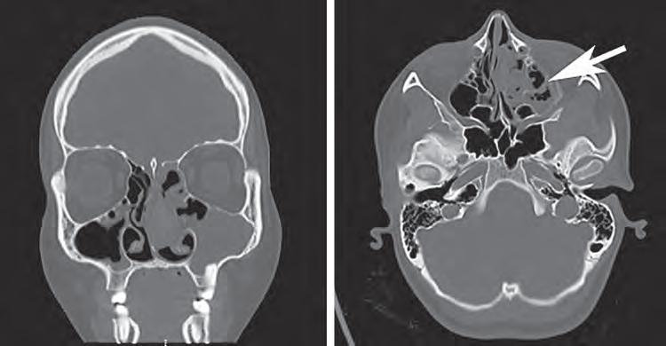
FIGURE 1.1
This computed tomography scan demonstrates radiographic features of both acute and chronic sinusitis. The left maxillary sinus demonstrates near complete opacification (A), and air-fluid levels are noted (arrow) in the left ethmoid sinus (B).
MICROSCOPIC FINDINGS
Rhinosinusitis exhibits sinonasal mucosa with a submucosal inflammatory infiltrate. The inflammatory cells are generally composed of lymphocytes, plasma cells, macrophages, and eosinophils, which predominate in allergic disease (Fig. 1.2). Acute rhinosinusitis is characterized by increased neutrophils, especially when associated with a bacterial etiology. There is often a component of stromal edema, which leads to the development of inflammatory polyps (described in detail in the next topic). The surface epithelium may also demonstrate changes, including inflammation, squamous metaplasia (Fig. 1.3A), or reactive papillary hyperplasia (so-called papillary sinusitis) (see Fig. 1.3B).
The diagnosis of rhinosinusitis is usually not difficult. Many of the changes overlap with sinonasal inflammatory polyps, and the distinction between the two entities is not important. In cases with squamous metaplasia and/ or reactive papillary hyperplasia of the surface epithelium, sinonasal papilloma can enter the differential diagnosis. Sinonasal papillomas have squamous or squamoid epithelium that is also thickened, proliferative with endophytic and/or exophytic growth, and infiltrated by neutrophils with microabscesses. Rarely, adenocarcinoma may enter the differential diagnosis when there is a reactive proliferation of seromucinous glands.
PROGNOSIS AND THERAPY
Acute viral rhinosinusitis is treated symptomatically, whereas bacterial disease requires antimicrobials. Chronic
Gross Findings
■ Nonspecific
Microscopic Findings
■ Submucosal infiltrate of lymphocytes, plasma cells, neutrophils, eosinophils, often with edema
■ Surface epithelium may demonstrate squamous metaplasia, inflammation, or reactive papillary hyperplasia
Pathologic Differential Diagnosis
■ Inflammatory polyps, sinonasal papilloma, adenocarcinoma
allergic sinusitis is treated with antihistamines, intranasal corticosteroids, and/or allergic desensitization. Patients with chronic rhinosinusitis refractory to medical therapy may require endoscopic surgery. Rhinosinusitis is generally not life-threatening, with the rare exception of untreated bacterial infection that can lead to infection of the orbit or meninges.
Sinonasal inflammatory polyps are common non-neoplastic masses of sinonasal tissue that essentially result from edema within the submucosa.
Inflammatory polyps are associated with many conditions. They are most often seen in the setting of allergic rhinosinusitis but may also be seen in the setting of infections,
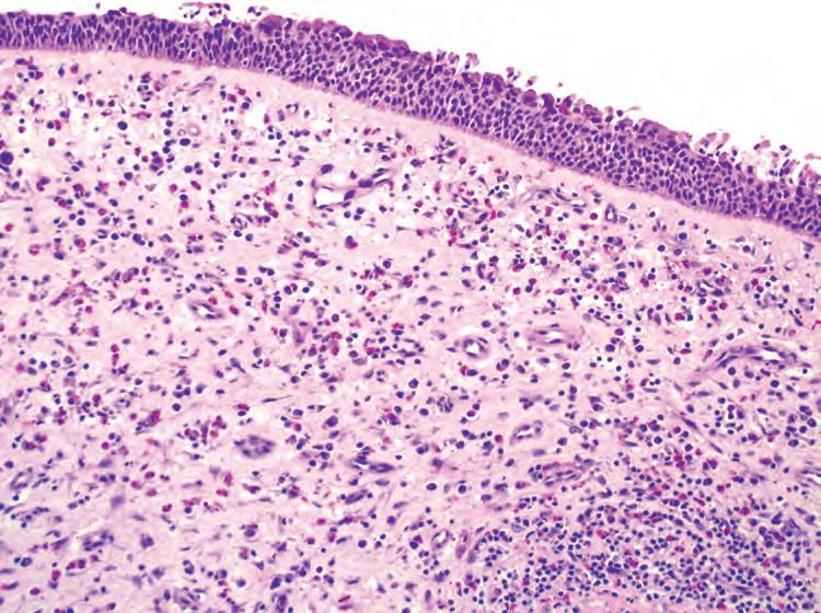
1.2
Chronic sinusitis is histologically characterized by a submucosal infiltrate of chronic inflammatory cells including lymphocytes, plasma cells, and eosinophils, which tend to predominate in allergic sinusitis.
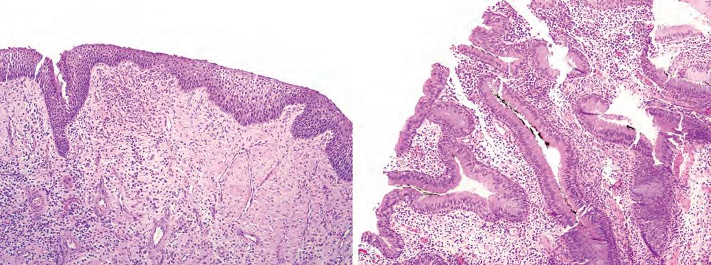
1.3
Some cases of chronic sinusitis can demonstrate foci of surface epithelial squamous metaplasia (A). In addition, chronic sinusitis occasionally exhibits papillary surface epithelial hyperplasia as a reactive change. When prominent, this finding can be confused with other lesions such as respiratory epithelial adenomatoid hyperplasia or sinonasal papilloma (B).
asthma, aspirin intolerance, cystic fibrosis, diabetes mellitus, and other conditions. Inflammatory polyps are typically seen in adults (except for cystic fibrosis-associated polyps), with no sex predilection. They involve the nasal cavity (especially the lateral wall) and maxillary and ethmoid sinuses and are usually bilateral (Fig. 1.4A). In addition to the symptoms of the underlying condition (e.g., allergies), sinonasal inflammatory polyps may cause nasal obstruction and pain. A subtype of inflammatory polyp known as antrochoanal polyp arises from the maxillary antrum and extends through the sinus ostia into the nasal cavity, nasopharynx, or oral cavity (see Fig. 1.4B). Antrochoanal
polyps are usually seen in younger patients (teenagers and young adults), usually males, and are typically unilateral.
Inflammatory polyps are typically translucent and mucoid in appearance (see Fig. 1.4A). Antrochoanal polyps tend to be elongated with a stalk and fibrotic.
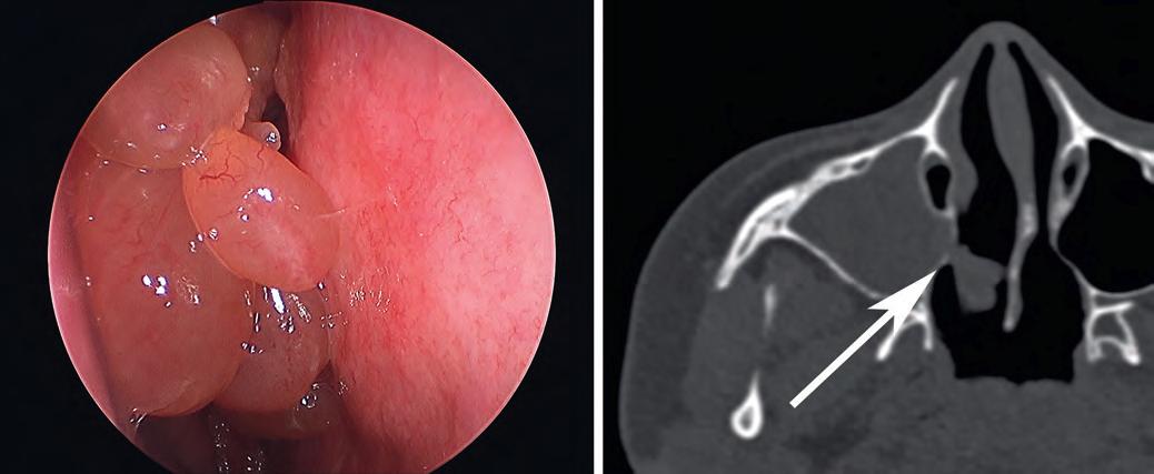
The typical clinical appearance of inflammatory polyps is that of bilateral, multiple mucoid polypoid masses with a translucent appearance involving the nasal cavity (A). The antrochoanal polyp is a subtype of inflammatory polyp arising from the maxillary antrum and protruding into the nasal cavity via a stalk (arrow) through the nasal choana (B). (A, Courtesy of Dr. Douglas Reh.)
SINONASAL INFLAMMATORY POLYPS—DISEASE FACT SHEET
Definition
■ Polypoid growths of sinonasal mucosa that result primarily from submucosal edema
■ An allergic etiology is most common
Incidence
■ Common
■ Nasal cavity and paranasal sinuses, often bilateral
■ Antrochoanal polyp is a subtype that arises from the maxillary antrum and protrudes through the sinus ostium, usually unilateral
Morbidity and Mortality
■ Usually minimal, although rarely may lead to bone erosion or remodeling
Sex and Age Distribution
■ Typically adults (except antrochoanal polyps in teenagers/young adults and cystic fibrosis polyps in children)
Clinical Features
■ Symptoms of underlying disease (e.g., rhinorrhea, nasal stuffiness, headaches in allergic polyps)
■ Nasal obstruction and epistaxis
Treatment and Prognosis
■ Endoscopic removal
■ Treatment of underlying disease (e.g., nasal steroids for allergic polyps)
SINONASAL
Gross Findings
■ Translucent, glistening, and mucoid
■ Antrochoanal polyps have a long stalk and are fibrotic
Microscopic Findings
■ Polypoid fragments of sinonasal mucosa with abundant stromal edema
■ Chronic inflammatory cell infiltrate with numerous eosinophils
■ Epithelial basement membrane is usually hyalinized
■ Secondary changes including infarction, hemorrhage, and fibrin deposition can be seen.
■ Antrochoanal polyps are less edematous, more fibrotic, fewer eosinophils, and minimal basement membrane hyalinization.
Pathologic Differential Diagnosis
■ Amyloidosis, hemangioma, lymphangioma, infections, respiratory epithelial adenomatoid hamartoma, nasopharyngeal angiofibroma, sinonasal papilloma, embryonal rhabdomyosarcoma
MICROSCOPIC FINDINGS
The most prominent feature of a sinonasal inflammatory polyp is submucosal edema beneath an intact respiratory epithelium (Fig. 1.5A). The subepithelial basement membrane is typically hyalinized (see Fig. 1.5B). There is usually a mild to moderate infiltrate of chronic inflammatory cells with a predominance of eosinophils (see Fig. 1.5B). Scattered stellate or spindled fibroblasts are seen, some of which may exhibit enlarged, hyperchromatic nuclei (see Fig. 1.5C). Larger inflammatory polyps may demonstrate prominent submucosal hemorrhage with fibrin
1.7
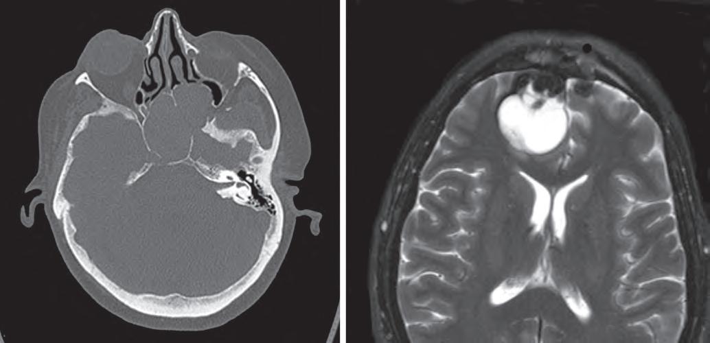
This computed tomography scan demonstrates a sphenoid sinus mucocele, with expansion of the sinus with secretions and thinning and remodeling of the surrounding bones (A). This T2-weighted magnetic resonance imaging scan shows a fluid-filled mucocele involving the brain (B).
PARANASAL SINUS MUCOCELE—PATHOLOGIC FEATURES
Gross Findings
■ Abundant mucin, otherwise nonspecific
Microscopic Findings
■ Very nonspecific
■ Sinonasal mucosa with inflammation, sometimes attenuation, squamous metaplasia, scarring, reactive bone, or cholesterol granulomas
Pathologic Differential Diagnosis
■ Normal sinonasal mucosa, inflammatory polyps, unsampled neoplasm leading to obstruction
of the orbit or cranial cavity (see Fig. 1.7B). Given these dramatic symptoms and radiographic features, a neoplastic process is often suspected clinically.
PATHOLOGIC FEATURES
GROSS FINDINGS
Abundant mucin is generally apparent grossly or reported intraoperatively (if suction has removed all of the contents).
The microscopic features of mucoceles are typically underwhelming (particularly in the setting that is suspicious for malignancy) and closely mimic normal sinonasal tissue. The sinonasal tissue sometimes has an attenuated appearance resembling a cyst lining (Fig. 1.8A and B). Epithelial squamous metaplasia, fibrosis, a rim of reactive bone, or cholesterol granuloma formation can also be seen. Because of their nonspecific nature, a definitive diagnosis cannot be made on histologic grounds without clinical or radiographic input.
The main diagnostic consideration is normal sinonasal tissue. Clinical and radiographic correlation is needed to make the distinction. Sinonasal polyps or a salivary gland mucocele may also be in the differential. An unsampled neoplasm may be the cause of the obstruction leading to a mucocele.
Sinus mucoceles are treated by surgical excision. The underlying cause of the obstruction (e.g., chronic sinusitis) should also be addressed. The prognosis is excellent.
(Fig. 1.11A). Finally, AFS must be distinguished from acute or chronic forms of invasive fungal sinusitis, in which fungal elements invade stroma with frequent involvement of vessels (see Fig. 1.11B).
PROGNOSIS AND THERAPY
Treatment includes removal of the mucus as a means to restore mucociliary function. Intranasal steroids are frequently used. Fungal desensitization may also be used as a treatment option. There does not appear to be a role for antifungal agents. Prognosis is good, although longterm therapy may be needed to control relapses in some patients.
Nasal glial heterotopia is a benign condition resulting from the failure of the developing frontal lobe to completely retract into the cranial cavity during fetal development. Because it is not a neoplasm, the historical term “nasal glioma” should not be used.
ALLERGIC FUNGAL SINUSITIS—PATHOLOGIC FEATURES
Gross Findings
■ Thick, viscous mucin that may resemble putty or peanut butter
Microscopic Findings
■ Allergic mucin: a striated mixture of mucin, inflammatory cells, Charcot-Leyden crystals, and other debris
■ Fungal hyphae seen in approximately half of cases with special stains
■ Fungi often scarce and have a degenerated appearance
Pathologic Differential Diagnosis
■ Nonspecific rhinosinusitis, sinonasal polyp, mycetoma (fungus ball), invasive fungal sinusitis (acute or chronic)
Nasal glial heterotopia usually affects infants, although it can occasionally be encountered in older patients. There is no predilection for either sex. Glial heterotopia presents as a firm nodule that can be extranasal (60%) on the bridge or side of the nose, intranasal within the nasal cavity (30%), or both intranasal and extranasal (10%). Patients often have nasal obstruction and infants may show difficulty feeding as a result of the mass. By radiology, there is no connection to the intracranial cavity, a crucial feature that distinguishes glial heterotopia from an encephalocele (Fig. 1.12A and B).
PATHOLOGIC FEATURES
GROSS FINDINGS
Well-circumscribed nodule of firm soft tissue, 1 to 3 cm in size, with a glistening cut surface.
Mycetoma (fungus ball) is a form of noninvasive fungal sinusitis consisting of a matted collection of degenerating fungal hyphae growing within the sinus, with no tissue invasion (A). In contrast, fulminant invasive fungal sinusitis is characterized by invasion of tissues with necrosis and a limited inflammatory reaction (B).
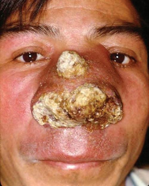
FIGURE 1.14
A clinical photograph of a 42-year-old man with rhinoscleroma presenting as several years of progressive nasal ulceration, along with palatal perforation, yielding marked nasal and mid-facial distortion. (Courtesy of Dr. R. Carlos.)
MICROSCOPIC FINDINGS
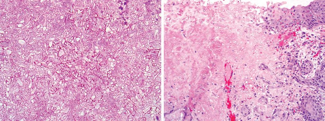
Rhinoscleroma is most often biopsied in the florid stage, where the submucosa is expanded by an inflammatory infiltrate including lymphocytes, plasma cells, neutrophils, and histiocytes. As in any disease with abundant plasma cells, Russell bodies—large cytoplasmic inclusions composed of immunoglobulin—are frequent. The diagnostic microscopic finding is the presence of “Mikulicz cells”—large histiocytes with abundant, clear, vacuolated cytoplasm (Fig. 1.15). As rhinoscleroma progresses, lesions become increasingly fibrotic and less inflammatory.
A Warthin-Starry stain highlights rod-shaped Klebsiella organisms within the Mikulicz cells (see Fig. 1.15).
Rhinoscleroma can mimic Rosai-Dorfman disease; however, in rhinoscleroma emperipolesis is not observed
Definition
■ Infectious disease caused by Klebsiella rhinoscleromatis, a gram-negative coccobacillus bacterium
Incidence and Location
■ Rare
■ Endemic in parts of South America, Central America, Africa, India, and Indonesia
Morbidity and Mortality
■ Can cause marked facial deformity and nasal stenosis
Sex and Age Distribution
■ Second and third decades
■ Slight female predilection
Clinical Features
■ Three clinical stages: rhinitic (exudative) with abundant foul-smelling mucopurulent secretions; florid (proliferative) with numerous small friable nodules causing obstruction and deformity; and fibrotic (cicatrical) with marked scarring and stenosis
Treatment and Prognosis
■ Long-term antibiotics and possibly surgical débridement
■ High relapse rates necessitate long-term follow-up
RHINOSCLEROMA—PATHOLOGIC FEATURES
Gross Findings
■ Nonspecific, or friable polyps, or dense sclerosis
Microscopic Findings
■ Marked chronic inflammation with lymphocytes, plasma cells, neutrophils, and histiocytes in the sinonasal submucosa
■ The diagnostic finding is the “Mikulicz cell”—large histiocytes with clear, vacuolated cytoplasm
Ancillary Studies
■ Warthin-Starry stain highlights the rod-shaped organisms within the Mikulicz cells
Pathologic Differential Diagnosis
■ Rosai-Dorfman disease, infections (atypical mycobacteria, leprosy, syphilis), granulomatosis with polyangiitis, clear cell epithelial neoplasms
(Fig. 1.16). Moreover, although Mikulicz cells are positive for CD68, they are negative for S100 protein. In some cases of rhinoscleroma, the Mikulicz cells can be so prominent that the lesion may be mistaken as a clear cell epithelial neoplasm such as mucoepidermoid
RHINOSPORIDIOSIS—PATHOLOGIC FEATURES
Gross Findings
■ Friable polyps or masses
Microscopic Findings
■ Variably sized cysts up to 300 µm, predominantly subepithelial
■ The largest cysts contain small endospores
■ Background nonspecific chronic inflammation and edema, acute inflammation if cysts rupture
Ancillary Studies
■ GMS and PAS highlight organisms, though usually not needed for diagnosis
Pathologic Differential Diagnosis
■ Oncocytic sinonasal papilloma, coccidiomycosis
GMS, Gomori methenamine silver; PAS, periodic acid–Schiff.
Special studies are not generally needed as the cysts are typically numerous and visible on routine stains, but microorganisms can be highlighted with PAS and GMS stains.
The oncocytic type of sinonasal papilloma exhibits numerous intraepithelial microcysts that can be confused with the cysts of rhinosporidiosis. However, in oncocytic sinonasal papilloma the microcysts are confined to the epithelium. The cysts of rhinosporidiosis can be confused with the spherules of Coccidioides immitis, but these spherules are much smaller (up to 60 µm) and accompanied by a granulomatous inflammatory infiltrate.
PROGNOSIS AND THERAPY
Rhinosporidiosis is treated by complete surgical excision. Antibiotics are not effective. The prognosis is excellent, with only occasional recurrences. The disease is not infectious to other individuals.
term Wegener granulomatosis is still widely used, many organizations (e.g., American College of Rheumatology, the European League against Rheumatism, and the American Society of Nephrology) recommend avoiding it due to a trend against eponyms.
GPA tends to affect middle-aged adults, with a slight male predominance. GPA classically affects the head and neck (especially sinonasal tract), lung, and kidney, but it can be localized to only one or two of these areas. Affected patients complain of nasal discharge, nasal obstruction, nosebleeds, and pain. On clinical examination, patients have a nasal septum ulcer with crusting, which can sometimes progress to perforation and collapse of the nasal cartilages (Fig. 1.18A). Respiratory disease manifests as hemoptysis, lung infiltrates, or cavitary masses, whereas renal disease results in glomerulonephritis.
GROSS FINDINGS
The gross appearance is often a nonspecific appearing ulcer.
MICROSCOPIC FEATURES
The histologic triad of GPA is biocollagenolytic (necrobiotic) necrosis, granulomatous inflammation, and vasculitis. “Biocollagenolytic” or “necrobiotic” necrosis refers to zones of geographic basophilic necrosis with granular, cellular debris (see Fig. 1.18B). The granulomatous inflammation of GPA is typically poorly formed, sometimes simply consisting of scattered giant cells (see Fig. 1.18C). Vasculitis of small to medium-sized vessels is the most specific finding but is often focal or absent. Unfortunately, most patients with GPA have biopsies that show nonspecific acute and chronic inflammation with eosinophils and sometimes neutrophilic microabscesses, and multiple biopsies may be required to establish a pathologic diagnosis.
■ GRANULOMATOSIS WITH POLYANGIITIS
Granulomatosis with polyangiitis (GPA) is a systemic immune complex vasculitis of unknown etiology that often affects the sinonasal tract. Although the synonymous
Elastic stains may be helpful by highlighting vessels that are involved by vasculitis (see Fig. 1.18D). Special stains for microorganisms are negative. Patients with GPA have positive serum cytoplasmic antineurtrophil cytoplasmic antibodies (c-ANCA) and proteinase 3 (PR3) antibodies in approximately 80% of cases (Fig. 1.19).
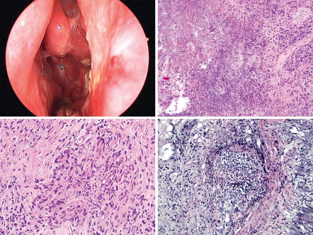
FIGURE 1.18
Granulomatosis with polyangiitis clinically presents as nasal erythema, crusting, ulcer, and perforation (A). Histologically a classic feature of granulomatosis with polyangiitis is “biocollagenolytic necrosis” (or “necrobiosis”), which is basophilic necrosis with nuclear debris (B). The granulomas of granulomatosis with polyangiitis are typically not well formed and may consist simply of giant cells (C). An elastic stain can highlight foci of vasculitis (D). (A, Courtesy of Dr. Douglas Reh.)
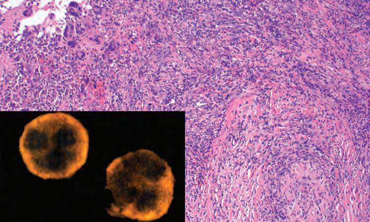
FIGURE 1.19
In granulomatosis with polyangiitis, giant cells may be present (upper left), but well-formed granulomas are absent. Note the vessel wall in the lower right, with destruction by the inflammatory process in an example of vasculitis. Inset: A c-ANCA shows a granular cytoplasmic pattern in a case of granulomatosis with polyangiitis.