Contributors
Alexandre Arkader, MD
Associate Professor of Orthopaedic Surgery
Perelman School of Medicine at University of Pediatrics
Pediatric Orthopedic and Orthopedic Oncology Surgeon
Children’s Hospital of Philadelphia (CHOP) Philadelphia, Pennsylvania
Richard E. Bowen, MD
Clinical Professor
Orthopaedic Surgery
David Geffen School of Medicine at UCLA
Los Angeles, California
Orthopaedic Institute for Children Los Angeles, California
Andrea C. Bracikowski, MD
Associate Professor
Orthopedics and Rehabilitation
Vanderbilt University School of Medicine
Nashville, Tennessee
Associate Professor
Pediatrics
Vanderbilt Children’s Hospital Nashville, Tennessee
Associate Professor
Emergency Medicine
Vanderbilt University School of Medicine Nashville, Tennessee
Kevin M. Dale, MD
Assistant Professor of Orthopaedics and Rehabilitation
Department of Orthopaedic Surgery
Vanderbilt University School of Medicine Nashville, Tennessee
Jaime R. Denning, MD, MS
Assistant Professor
Orthopaedic Surgery
Cincinnati Children’s Hospital Medical Center
University of Cincinnati School of Medicine Cincinnati, Ohio
Aleksei B. Dingel, BS
Research Coordinator
Department of Orthopaedic Surgery
Stanford University School of Medicine
Stanford, California
Eric W. Edmonds, MD, FAOA
Professor of Orthopedic Surgery
University of California San Diego
Director of Orthopedic Research and Sports Medicine
Division of Orthopedic Surgery
Rady Children’s Hospital San Diego San Diego, California
John B. Erickson, DO
Assistant Professor
Pediatric Orthopedic Surgery
Medical College of Wisconsin Milwaukee, Wisconsin
Steven L. Frick, MD
Professor and Vice Chairman
Orthopaedic Surgery
Stanford University School of Medicine
Stanford, California
Chief, Pediatric Orthopaedics
Orthopaedic Surgery
Lucile Packard Children’s Hospital Stanford Stanford, California
Christopher Greeley, MD, MS
Professor Pediatrics
Baylor College of Medicine Houston, Texas
Chief
Section of Public Health Pediatrics
Texas Children’s Hospital Houston, Texas
Nathan L. Grimm, MD
Orthopaedic Surgery Pediatric & Adult Sports Medicine Fellow
UConn Health Center
Department of Orthopaedic Surgery
Farmington, Connecticut
Christina K. Hardesty, MD
Assistant Professor of Orthopaedic Surgery
Pediatric Orthopaedic Surgery
Rainbow Babies and Children’s Hospitals at Case Western Reserve University Cleveland, Ohio
Ginger E. Holt, MD
Professor and Vice Chair, Orthopaedic Surgery
Vanderbilt University Medical Center Nashville, Tennessee
Elizabeth W. Hubbard, MD
Assistant Professor of Orthopaedic Surgery
Duke University School of Medicine Durham, North Carolina
Megan E. Johnson, MD
Assistant Professor Orthopaedics
Vanderbilt University Medical Center Nashville, Tennessee
Sheila M. Jones, MD
Medical Director, Pediatric Emergency Department
Centennial Women’s & Children’s Hospital Nashville, Tennessee
Adjunct Assistant Professor of Pediatrics
Vanderbilt Children’s Hospital Nashville, Tennessee
Kevin E. Klingele, MD
Chief
Orthopedic Surgery
Nationwide Children’s Hospital Columbus, Ohio
Scott H. Kozin, MD
Clinical Professor Orthopedics Surgery
Lewis Katz School of Medicine
Temple University
Philadelphia, Pennsylvania
Clinical Professor
Orthopaedic Surgery
Sidney Kimmel Medical College
Thomas Jefferson University
Philadelphia, Pennsylvania
Chief of Staff
Shriners Hospital for Children Philadelphia, Pennsylvania
Ying Li, MD
Associate Professor
Department of Orthopaedic Surgery
C.S. Mott Children’s Hospital Ann Arbor, Michigan
Kristin Livingston, MD
Assistant Professor of Orthopaedic Surgery
University of California, San Francisco San Francisco, California
Raymond W. Liu, MD
Associate Professor and Victor M. Goldberg Endowed Chair in Orthopaedics
Pediatric Orthopaedic Surgery
Rainbow Babies and Children’s Hospitals at Case Western Reserve University
Cleveland, Ohio
Steven Lovejoy, BS, MD
Assistant Professor
Pediatric Orthopedic Surgery
Vanderbilt Children’s Hospital Nashville, Tennessee
Jeffrey E. Martus, MD, MS
Associate Professor
Pediatric Orthopaedic Surgery
Vanderbilt University Medical Center
Nashville, Tennessee
Gregory A. Mencio, MD
Neil E. Green Professor and Vice Chairman
Department of Orthopaedics
Vanderbilt University Medical Center
Chief, Pediatric Orthopaedics
Monroe Carell, Jr. Children’s Hospital at Vanderbilt Nashville, Tennessee
Amirhossein Misaghi, MD
Orthopedic Surgeon
CHOC (Children’s Hospital of Orange County) Orange, California
James F. Mooney III, MD
Chief of Staff
Medical Affairs
Shriners Hospital for Children-Springfield Springfield, Massachusetts
Robert F. Murphy, MD
Assistant Professor
Department of Orthopaedics
Medical University of South Carolina Charleston, South Carolina
Unni G. Narayanan, MBBS, MSc, FRCS(C) Professor
Department of Surgery & Rehabilitation Sciences
Institute
University of Toronto
Divisions of Orthopaedic Surgery and Child Health Evaluative Sciences
The Hospital for Sick Children
Toronto, Ontario
Canada
James P. Norris IV, MD
Clinical Instructor
Orthopaedic Surgery
Vanderbilt University Medical Center Nashville, Tennessee
Shital N. Parikh, MD
Professor
Orthopaedic Surgery
Cincinnati Children’s Hospital Medical Center
University of Cincinnati School of Medicine
Cincinnati, Ohio
Anthony I. Riccio, MD
Associate Professor of Orthopaedic Surgery
University of Texas Southwestern Medical Center
Dallas, Texas
Sanjeev Sabharwal, MD, MPH Professor Orthopedics
University of California San Francisco Oakland, California
Walter Samora, MD
Assistant Professor
Orthopedic Surgery
Nationwide Children’s Hospital Columbus, Ohio
Brian Scannell, MD
Associate Professor Department of Orthopaedic Surgery
Atrium Health
Charlotte, North Carolina
Physician
OrthoCarolina
Charlotte, North Carolina
Jonathan G. Schoenecker, MD, PhD
Associate Professor and Mast Chair of Pediatric Trauma and Hip Surgery
Vanderbilt University Medical Center
Monroe Carell, Jr. Children’s Hospital at Vanderbilt Nashville, Tennessee
Herbert S. Schwartz, MD
Professor and Chair Emeritus
Orthopaedic Surgery
Vanderbilt University Medical Center Nashville, Tennessee
Kevin G. Shea, MD
Orthopedic Surgeon
Sports Medicine
Stanford University
Stanford, California
Professor
Orthopaedics
Stanford University
Stanford, California
Jeffrey Shilt, MD
Chief of Community Surgery
The Woodlands
Texas Children’s Hospital
The Woodlands, Texas
Associate Professor
Department of Orthopaedics & Scoliosis Surgery
Baylor School of Medicine
Houston, Texas
Medical Director of Motion Analysis & Human Performance
Texas Children’s Hospital Houston, Texas
Eric D. Shirley, MD
Pediatric Orthopaedic Surgeon
Pediatric Orthopaedic Associates
Woodstock, Georgia
Mauricio Silva, MD
Medical Director
Orthopaedic Institute for Children
Los Angeles, California
Clinical Professor
UCLA-Orthopaedic Hospital Department of Orthopaedic Surgery
David Geffen School of Medicine
University of California Los Angeles
Los Angeles, California
Louise Z. Spierre, MD
Director Pediatric Rehabilitation
Wolfson Children’s Hospital
Assistant Professor
Department of Pediatrics
University of Florida Health Science Center Jacksonville, Florida
Chris Stutz, MD
Assistant Professor
Orthopedic, Hand, and Microvascular Surgery
Texas Scottish Rite Hospital for Children Dallas, Texas
George H. Thompson, MD Director
Division of Pediatric Orthopaedic Surgery
Rainbow Babies and Children’s Hospital
Cleveland, Ohio
Professor, Orthopaedic Surgery and Pediatrics
Case Western Reserve University Cleveland, Ohio
Rachel M. Thompson, MD
Assistant Professor-in-Residence
Orthopaedic Surgery
UCLA/OIC
Los Angeles, California
Dan A. Zlotolow, MD
Adjunct Clinical Associate Professor of Orthopaedics
The Hospital for Special Surgery
New York, New York
Attending Physician
Shriners Hospital for Children
Philadelphia, Pennsylvania
1
INTRODUCTION
Consideration of growth potential is the major difference in treating injuries in children as compared with adults. Pediatric skeletal trauma can result in enhanced or diminished growth. Future growth is usually helpful because some angular and length deformities can correct themselves as the child grows. Loss of growth potential can be one of the more difficult problems to treat. Adult bone is dynamic; it is constantly involved in bone turnover and remodeling in response to aging and changes in stress on the skeleton. The pediatric skeleton not only remodels in response to alterations in stress but also grows in length and width and changes shape, alignment, and rotation as it matures. Understanding growth potential and the changing forces after skeletal trauma in children are important in determining the appropriate treatment for injured bones.
The following are the most common clinical questions in caring for children with fractures: (1) is the physis injured with an accompanying risk of growth disturbance, and (2) is the length and alignment of the fracture acceptable or unacceptable (i.e., will it improve with growth enough that function and cosmesis will not be adversely affected)? If the answer is no, a reduction is indicated. The response to these two questions requires knowledge of normal growth mechanisms and studies of fractures in children (the science), whereas applying this knowledge to an individual patient and making decisions about how to care for the fracture require an assessment of multiple factors related to the child and the fracture (the art).
Principles of fracture treatment are the same for all ages—the goal is to achieve restoration of normal length, alignment, rotation, and the anatomic reduction of articular surfaces. In children, attempting to preserve normal growth potential is also critical; thus, assessment of the integrity and alignment of the physis is important. Although some angulation is acceptable when treating fractures in children, it is best to keep the amount of angulation as small as possible by closed fracture treatment methods, regardless of the patient’s age. On the other hand, multiple attempts at anatomic reduction in a child, particularly in fractures involving the physis, may cause harm and should be avoided. The small amount of angulation associated with torus or so-called buckle fractures in children is almost always acceptable. Marked bowing that causes clinical deformity,
which can be seen in plastic deformation and “greenstick” fractures in the forearm, should usually be corrected.1,2
Bone healing in children is generally rapid, primarily because of the thickened, extremely osteogenic periosteum. The age of the patient directly affects the rate of healing of any fracture: the younger the child, the more rapidly the fracture heals. The periosteum thins as the child grows older and has less osteogenic capability. Injuries to the growth plate heal more rapidly than shaft fractures do. Physeal injuries, in almost all parts of the body, heal in approximately 3 weeks.3
Treatment of trauma to the pediatric skeleton is generally straightforward. Dislocations and ligamentous injuries are uncommon in children in comparison with adults because the physis and bones in children are usually weaker mechanical links in the system and thus more susceptible to injury. Ligamentous injuries may occur, especially in older children, as physiologic physeodeses begin to occur, resulting in more secure attachments of the epiphyseal and metaphyseal regions.4,5 Most injuries, though, are simple fracture patterns caused by low-velocity trauma such as falls. In most cases, closed reduction followed by a short period of immobilization restores normal function to a pediatric extremity. However, a number of pitfalls can make treatment of pediatric fractures, particularly fractures of the growth plate, difficult and demanding.
HISTORY, DIAGNOSIS, AND INJURY MECHANISMS
In infants, skeletal trauma may be related to the birthing process or may be the only sign of child abuse because young children are at higher risk for abuse.6 The presenting sign may be deformity, swelling, or lack of movement in an extremity. Caregivers should be questioned about the circumstances of the injury, and a lack of a plausible mechanism of injury should prompt an evaluation for nonaccidental trauma. Radiographs of an infant can be difficult to obtain and interpret, especially those of bones in the elbow and hip region, which may require comparison views. Anteroposterior and lateral views, including the joints above and below the injured area, constitute a minimal radiographic evaluation. Usually, routine radiographs coupled with a good physical examination can establish the diagnosis. Arthrograms, ultrasonography, or magnetic resonance imaging (MRI) can be
useful as a diagnostic aid when radiographs are confusing.3,7 Additionally, a skeletal survey can be used in the young patient because unsuspected fractures may be present up to 20% of the time.8
Children with multiple trauma or head injuries or both can have occult axial fractures and physeal injuries that may not be suspected or may be difficult to diagnose, even with a good physical examination. This is more commonly seen in patients with a lower Glasgow Coma Scale and higher Injury Severity Score.9 In these children, historically, a bone scan assisted in diagnosing fractures unidentified by routine screening radiographs10; however, they can be difficult to obtain in multiply injured children. More recently, radiographic skeletal surveys and multiplanar imaging with computed tomography or MRI are favored for identifying occult injuries.11
Fractures through the growth plate in children can be difficult to interpret if the fracture is not displaced. A thorough physical examination can usually identify this type of injury; the sign is swelling and maximal tenderness occurring over the injured physis, which occurs most commonly at the distal end of the radius or fibula. Palpation at or distal to the tip of the lateral malleolus usually identifies a ligamentous injury; swelling and tenderness at the growth plate may suggest a fracture undetected by radiographs. However, studies evaluating children with lateral ankle pain after injury but normal radiographs were not found to frequently have a physeal fracture by ultrasound5 or MRI.12 Another recent MRI study challenges the perception that distal fibular physeal injuries are common after twisting ankle injuries in skeletally immature patients.4 Often, a small metaphyseal fragment on the radiograph suggests physeal injury. Repeated radiographs in 1 to 2 weeks can confirm the physeal injury because the healing physis appears wider, and periosteal reaction may be seen.
Each age group has typical injury mechanisms and common fractures. Most infants and newborns (≤12 months of age) sustain fractures by having someone else injure them. When children are older, walking and running, accidental injuries are more common. Children most commonly fracture the forearm, usually the distal end of the radius.13–15 Clavicle fractures are common in infancy and in the preschool age group, but their incidence decreases with increasing age. Elbow hyperextension in early and midchildhood predisposes children in these age groups to supracondylar humerus fractures. Forearm fractures, although common in young children, show a progressive increase into the teenage years.
Most injuries occur when the child falls. Severe, high-energy injuries are less common in children and are frequently caused by automobiles, lawn mowers, or motorcycles/all-terrain vehicles. As a child approaches the midteens, injuries are much like those of an adult. The age at which the growth plates close varies greatly and depends on hereditary factors and hormonal variation. Skeletal age is an important factor in the consideration of injuries in children, in that the closer the child is to the end of growth, the less prominent the role of the growth plate in treatment of the injury and the less the remodeling potential. Healing capacity is inversely related to age.
Finally, child abuse must be considered in all children’s injuries, and, as noted earlier, it should especially be considered
when treating very young children with fractures.6,16 Care must be taken to ensure that the child is checked for signs of abuse on the initial assessment and for possible subsequent injuries during follow-up. Repeating a skeletal survey at 2 to 3 weeks is strongly recommended in young patients to increase diagnostic yield in patients with suspected abuse injuries.17 Parents or guardians of children who are not brought back for follow-up appointments for fractures should be contacted and asked to schedule a return visit.
FORMATION OF BONE
Embryonic bone forms through either membranous or endochondral ossification. In the former, mesenchymal cells proliferate to form membranes primarily in the region in which flat bones are fabricated.18,19 Endochondral ossification is bony replacement of a cartilage model and is the mode of formation of long bones.
MEMBRANOUS BONE FORMATION
Membranous bone formation increases the diameter of long bones and is responsible for the creation of flat bones such as the scapula, skull, and, in part, the clavicle and pelvis. Flat bones are formed as mesenchymal cells condense into sheets that eventually differentiate into osteoblasts. Surface cells become the periosteum. Primary bone is remodeled and transformed into cancellous bone, to which the periosteum adds a compact cortical bone cover. This type of growth is independent of a cartilage model.
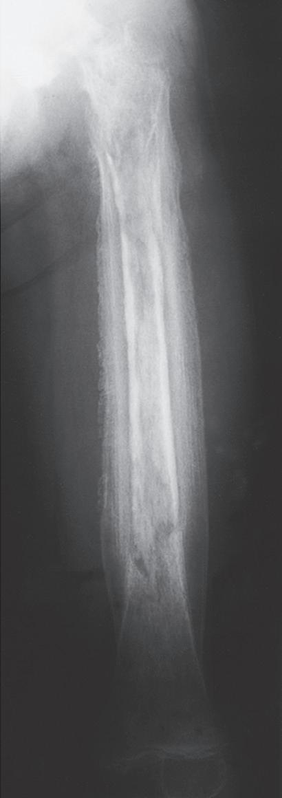
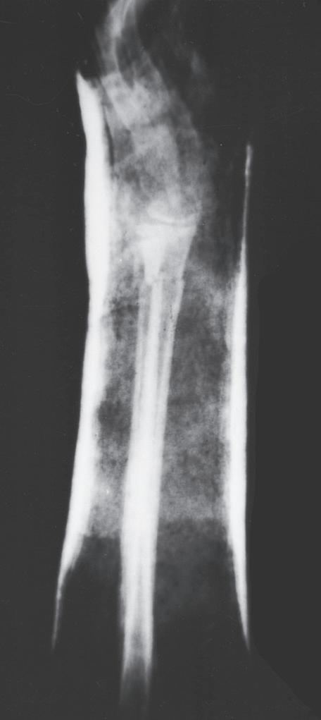
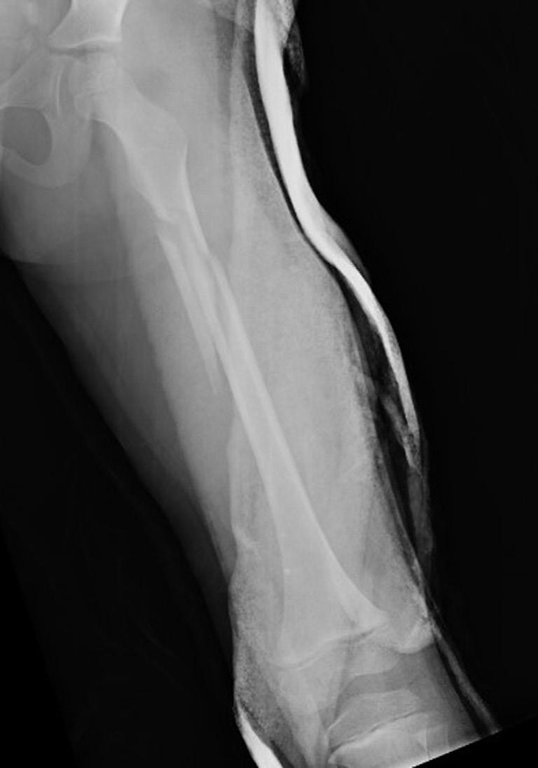
As endochondral ossification lengthens bones, proliferation of bone occurs beneath the periosteum through membranous bone formation, thus enlarging the diameter of the diaphysis in long bones. This type of bone formation is also apparent in subperiosteal infection and after bone injury when periosteal bone forms around a fracture hematoma (Fig. 1.1). The osteogenic periosteum of children contributes to rapid healing because the callus and periosteal new bone increase the diameter of the bone and provide early biomechanical strength.
ENDOCHONDRAL OSSIFICATION
Endochondral ossification requires the presence of a cartilage anlage. Early in gestation, mesenchymal cells aggregate to form models of the future long bones. A cartilage model develops, and the peripheral cells organize into a perichondrium.18,19 Cartilage cells enlarge and degenerate, and the matrix surrounding them calcifies. This calcification begins in the center of the diaphysis and becomes the primary ossification center. Vascular buds enter the ossification center and transport new mesenchymal cells capable of differentiating into osteoblasts, chondroclasts, and osteoclasts. These cells align themselves on the calcified cartilage and deposit bone. Primary cancellous bone is thus formed, and ossification expands toward the metaphyseal regions.
Long bone growth continues as the terminal ends of the cartilage model keep growing in length by cartilage cell proliferation, hypertrophy, and production of extracellular matrix. This growth continues in this manner until after birth, when secondary ossification centers (epiphyses) develop.
A B
Fig. 1.1 (A) Radiograph of a healing supracondylar fracture illustrating the periosteum stripped (arrow) to nearly the midshaft of the humerus. The bridging periosteal bone stabilizes this fracture in about 3 weeks. (B) The large periosteal involucrum surrounds the former bone (sequestrum) in a femur with osteomyelitis. The periosteum can be stimulated to remanufacture an entire cortex around this area, such that when the sequestered bone is removed, the periosteal bone will form a new (larger-diameter) femoral diaphysis.
The mass of cartilage found between the epiphyseal and diaphyseal bones in later postnatal development thins to become the epiphyseal plate, which continues as the principal contributor to the growth (in length) of long bones until maturation is reached. The girth of the long bone is provided by the cambium layer of the periosteum.13,18 Successive surfaces of compact bone are added to the exterior while remodeling by resorption of the interior (endosteal) surface takes place.
Once the physis is established between the epiphysis and metaphysis, the periosteal ring becomes relatively firmly attached at the level of the zone of hypertrophied cells. This periphyseal periosteal collar is referred to as the fibrous ring of LaCroix.3,13,18 The zone of Ranvier, the cellular segment responsible for growth in diameter of the physis,13 is located in the same area. The periosteum is firmly attached at this level. Even when the periosteum is torn over the metaphysis or diaphysis, it usually remains attached at the physis.
REGULATION OF GROWTH AND DEVELOPMENT
Factors affecting skeletal growth vary and are incompletely understood. Although commonly used growth curve charts suggest that growth is smoothly continuous throughout childhood, a saltation and stasis model of human growth is now recognized,20,21 with bursts of growth in length
(growth spurts) occurring after prolonged periods of stasis. Growth in length occurs only at the physis and can occur through three mechanisms: an increase in the number of cells, an increase in the size of cells, or an increase in the amount of extracellular matrix. The physis responds to various growth-regulating hormones (e.g., growth hormone, thyroxine, estrogen, and testosterone), parathyroid hormone, and corticosteroids, as well as the peptide-signaling proteins—transforming growth factor β (TGF-β), platelet-derived growth factor (PDGF), and bone morphogenetic proteins (BMPs)—and immunoregulatory cytokines (interleukin-1 [IL-1] and IL-6).22–26 Further research is still needed to better understand these growth pathways. However, recently it was suggested that differential expression of gene pathways specifically for BMP-2 and BMP-6 may contribute to further physeal growth.27
Local paracrine regulators have recently been identified as critical in controlling bone development and remodeling. An important feedback loop controlling chondrocyte development involves Indian hedgehog protein (Ihh) and parathyroid hormone-related peptide. These paracrine factors control the decision for chondrocytes to leave the proliferative pool and undergo hypertrophic differentiation.28,29 Fibroblast growth factor (FGF) signaling also appears crucial in regulating chondrocyte proliferation and differentiation in the physis and appears to have an opposite effect from the BMPs, decreasing chondrocyte proliferation, increasing production of Ihh, and accelerating differentiation of hypertrophic chondrocytes.29 An example of abnormal growth related to FGF signaling is in achondroplasia. A gene mutation affecting FGF receptor 3 suppresses proliferation and maturation of growth plate chondrocytes, causing decreased growth plate size and decreased bone elongation.30 Thyroxine is also involved in the cellular and molecular events of terminal chondrocyte differentiation and morphogenesis of columnar cartilage.31
Diurnal variation in the growth of bone has been shown to reflect the levels of the different hormones, and animal studies suggest mechanical factors may also be critical because 90% of growth occurred during periods of recumbency in a study of growth in sheep.21 Physeal growth is slowed by excessive compression and accelerated by distraction, recognized in the American literature as the HueterVolkmann law,32 but noted earlier by Delpech in 1829.33 This theory affects growth but also fracture healing, which is one of many differences between pediatric and adult fracture healing.
Growth in length ceases at skeletal maturity with fusion of the physes and occurs at different times in individual bones; it also varies based on gender, hereditary factors, and hormone levels. Physiologic physiodesis is the normal, gradual replacement of the growth plate by bone during adolescence, and physeal closure is induced at skeletal maturity by estrogen levels in both males and females.28
BIOLOGY OF FRACTURE HEALING
Fracture healing is usually divided into three stages: (1) inflammatory, (2) reparative, and (3) remodeling. Fracture healing involves both membranous and endochondral ossification. Injuries to the pediatric skeleton always involve
a variable amount of surrounding soft tissue injury. Unlike the soft tissues, which heal by replacement of the injured tissue with collagen scar tissue, bone heals by replacing the area that is injured with normal bony tissue.
The blood supply to the bone is an important part of fracture healing, and significant soft tissue injury delays healing because the blood supply to bone enters at sites of soft tissue attachment. The normal process of fracture healing in any part of the bone follows a set chronologic order. Any of these phases may be disrupted or delayed by excessive adjacent soft tissue injury.
INFLAMMATORY PHASE
The inflammatory phase of fracture healing “sets the stage” for cartilage and bone formation by supplying the building blocks necessary for repair and remodeling. When bone is injured, the bone, periosteum, and soft tissue (mostly muscle) around the fracture begin to bleed. Hematomas form at the fracture site, both inside and outside the bone. The hematoma may dissect along the periosteum, which is easily elevated or was elevated at the time that the fracture was maximally displaced. The more severe the soft tissue injury, the more displaced the fracture; in addition, the more the periosteum is torn, the larger the area that fills with the hematoma. The role of a hematoma is to serve as a source of signaling agents capable of initiating cellular events critical to fracture healing. This also explains why some minimally displaced or greenstick fractures (minimal to no hematoma) may be slow to heal.
Research has focused on factors controlling fracture healing in two groups: peptide-signaling proteins (TGF-β, FGF, PDGF, and BMPs) and immunoregulatory cytokines (IL-1 and IL-6).25 The peptide-signaling proteins are derived from platelets and extracellular bone matrix and are critical for regulation of cell proliferation and mesenchymal stem cell differentiation. TGF-β is a multifunctional growth factor that controls tissue differentiation in fracture repair. FGFs increase the proliferation of osteoblasts and chondrocytes and may stimulate the formation of new blood vessels. PDGF acts on mesenchymal cell precursors to stimulate osteoblast differentiation. BMPs are a class of proteins produced in the early stages of fracture repair and strongly stimulate endochondral ossification. The sole criterion for BMP classification is the induction of bone formation in a standard in vivo rodent assay, and at least 14 BMPs, grouped in the TGF superfamily of growth and differentiation factors, have been identified. BMPs are present in bone matrix in a form that allows for presentation to marrow stromal cells to induce differentiation into osteoblasts. Furthermore, osteoblasts have been shown to synthesize and secrete BMPs. Cells that synthesize new bone during fractures also have been shown to be targets of BMPs and to possess BMP receptors. BMPs (BMP 2, 3, 4, 5, and 8) and BMP receptors are upregulated in the periosteum as early as 3 days after fracture.34
Studies utilizing microarray analysis of the genetic response to a fracture demonstrate that the genomic response to a fracture is complex and involves thousands of genes, including the BMPs and other growth factors noted earlier, as well as immunoregulatory cytokines.10,34–36 The immunoregulatory cytokines are released from inflammatory cells present in the hematoma and serve to regulate the early events in fracture healing.
Pediatric bone is more vascular than that of an adult and is able to generate a greater hyperemic and inflammatory response. The more mature (less porous) the cortex, the slower the vascular response to injury. Vasodilatation and the cellular inflammatory response begin shortly after a fracture, and the injured area is filled with inflammatory cells such as polymorphonuclear leukocytes and macrophages. The hematoma and inflammatory response also incite the release of molecules such as growth factors and cytokines from the platelets.37 In the initial phase of fracture healing, after the hematoma has formed, a scaffolding of fibrovascular tissue replaces the clot with collagen fibers. These fibers eventually become the collagen of the woven bone of the primary callus that forms around the fracture.
The primary callus is later ossified as the microvascular supply returns to the area. However, the bone, for at least a millimeter or two directly adjacent to the fracture site, loses its blood supply early in the inflammatory stage. After initial reabsorption of the dead bone along the fracture line, the fracture line in children usually becomes more visible radiographically 2 or 3 weeks after injury. The dead bone at the fracture surface is revascularized in a process that occurs faster in more vascular areas such as the metaphysis (as compared with the diaphysis).
The vascular response aids in initiating the cellular response to the fracture. A number of TGF-β subtypes help mediate cellular and tissue responses to inflammation and tissue repair.37 During the inflammatory phase of fracture healing, TGF-β from the extracellular matrix of bone and also from platelets controls the mesenchymal precursor cells that may form osteoblasts and osteoclasts. The maximal cellular response is ongoing within 24 hours of injury and occurs first in the subperiosteal region of the fracture.38,39
Osteogenic induction is stimulation by growth factors to convert the multipotential cells into osteoprogenitor cells. The osteoprogenitor cells on the undersurface of the periosteum help form periosteal bone. The osteogenic cells that originate from the periosteum help manufacture the external callus. Endochondral bone formation from the endosteal areas combines with subperiosteal bone formation to bridge the fracture.
The subperiosteal callus in children initially stabilizes the area so that the external callus may clinically heal the fracture by the end of the reparative phase. During remodeling, this callus decreases and is replaced with the endochondral ossified bone that has formed at the fracture surface.
REPARATIVE PHASE
The reparative phase of fracture healing is highlighted by the development of new blood vessels and the onset of cartilage formation. The surrounding soft tissue provides vascular ingrowth initially to the periosteal area and subsequently to the endosteal area. Before the fracture, the cortical blood supply was primarily from endosteal bone and branched out radially from inside the medullary canal. During the reparative phase, most of the blood supply to the cortex arises from outside the bone rather than inside.
Rat models of fracture healing reveal that intramembranous and endochondral bone formation is initiated during the first 10 days. Inflammatory mediators in the fracture hematoma recruit chondrocytes capable of producing fracture
callus. The hematoma is eventually replaced by the ingrowth of fibrovascular tissue. This developing construct provides structural support to stabilize the bone ends. This primitive tissue is eventually replaced through endochondral and intramembranous bone formation.
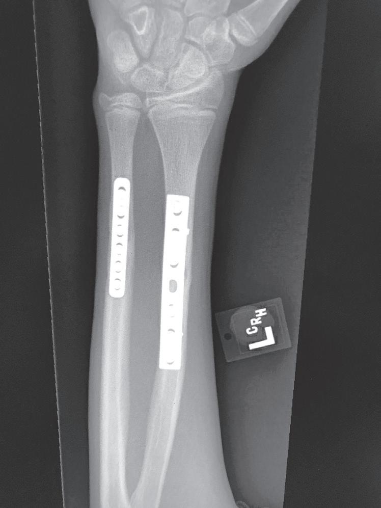
Tissue differentiation during the reparative phase is strongly influenced by local mechanical factors. Fracture stability has a critical effect on bone healing. Fracture healing is classically divided into primary and secondary healing. Primary healing results from rigid stabilization (i.e., plate immobilization) and involves a direct attempt by the cortex to bridge the fracture gap. Bridging occurs through direct haversian remodeling by intramembranous bone formation (Fig. 1.2A, B).
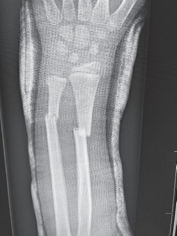
Secondary healing results from treatment of fractures with less rigid methods (i.e., fracture bracing, casts). In secondary healing, more motion at the fracture site leads to lower
Fig. 1.2 Anteroposterior (A) and lateral (B) radiographs of a forearm in a 15-year-old male who underwent open reduction and internal fixation with plates/screws. The fracture healing demonstrates primary bone healing with rigid fixation. An anteroposterior radiograph of a forearm in a 5-year-old at the time of original cast immobilization (C) and at 10 weeks (D) with complete fracture union demonstrates secondary bone healing. The motion that occurs with secondary bone healing results in abundant callus formation.
oxygen tension, and more cartilage is formed. Motion at the fracture site, the presence of a fracture gap, and an intact soft tissue envelope all encourage the formation of abundant callus (Fig. 1.2C, D). The increased diameter of the callus enhances biomechanical stability because the rigidity of the bone is proportional to its radius. The callus formed subsequently undergoes endochondral ossification. Ideal fracture treatment involves enough rigidity to ensure adequate vessel ingrowth, followed by progressive loading and motion to stimulate ample callus formation.36,40
As the periosteum produces bone beneath it, the periosteum is pushed away from the bone and makes a collar of bone around the area of injury. Initially, this tissue is more cartilaginous and fibrous and is not very well ossified. It may not show up well on a radiograph until the blood supply is adequate enough to allow mineralization and conversion to bone.
An important process that occurs between the reparative and remodeling phases is clinical union of the fracture, which takes place when the bony callus surrounds the fracture fragments and joins the callus coming from the other side. At this point, the bone may be stable clinically, and although some plastic deformation is still possible with force, the bone is usually strong enough that the patient can begin to use the extremity in a more normal way.
Although there are many ways suggested in the literature to determine union, clinical examination with radiographic evidence of healing is the most important in assessing union.41 Clinical union has occurred when the fracture site is no longer tender and does not move during examination and when physiologic loading does not cause pain. Radiographic union occurs later when radiographs demonstrate bone bridging across the fracture. This point demarcates the end of the reparative phase and the beginning of the remodeling phase.
REMODELING PHASE
Remodeling is the final phase of bone healing. It may last for a short time in a young child, or continue throughout growth or even beyond the end of growth in an older child. Once the bone is clinically stabilized, the ongoing stresses and strains on the bone that normally cause modeling are responsible for remodeling this early soft woven bone. After fractures in children, the bone usually returns to normal radiographically and clinically.
One complete skeletal turnover occurs during a child’s first year of life. This turnover declines to about 10% per year in late childhood and continues at about this rate or a little slower for life.24 Remodeling does not result from the activity of a single type of cell, such as osteoclasts or osteoblasts, but rather results from coordinated absorption and formation of bone over large regions around the fracture. The control mechanisms for the remodeling phase of bone are believed to be the bioelectric behavior that is responsible for modeling bone, according to Wolff’s law. As bone is subjected to the stresses of use during normal activities, the bone remodels appropriately for those stresses. Because a child’s bone is normally modeling anyway (actively changing in response to growth and stress), a child’s bone remodels significantly faster than an adult’s. This remodeling typically involves addition of bone to the concavity of angular deformities (compression side) and subtraction of bone from the convexity (tension side), resulting in a “rounding off” of the angle.
Systemic factors can affect the rate of bone healing. In addition to the age of the patient, hormonal factors that may help promote bone healing are growth hormone, thyroid hormone, calcitonin, insulin, anabolic steroids, and vitamins A and B.19
Factors that have been shown to discourage bone healing are diabetes, corticosteroids, exposure to cigarette smoke, and certain endocrinopathies. Denervation, irradiation, and high doses of hyperbaric oxygen may also slow the healing of fractures.
Historically, nonsteroidal antiinflammatory medications in children were avoided because of concern for fracture healing. However, more recent basic science and clinical studies suggest that it is unlikely to affect healing of fractures in children.42–44
PHYSEAL FRACTURE HEALING
Cartilage does not heal in the same phases as bone. When the physis is injured, it does not heal by the formation of callus within the physis. Inflammatory and reparative phases occur in cartilage healing, but cartilage healing has no remodeling phase.19,23 In 1958, Dale and Harris used a rhesus monkey model of physeal fractures and described the process of physeal fracture healing: initially the gap in the physis is filled with fibrin, and new bone formation ceases. The calcified cartilage cells on the metaphyseal side of the fracture line persist unaltered, while the cells on the epiphyseal side of the fracture continue to grow. These two processes lead to a temporary but pronounced increase in the thickness of the physeal plate in healing physeal fractures, which creates widening at the physis radiographically. Finally, callus grows from the metaphysis and periosteum of the shaft across the physeal fracture gap and reunites the epiphysis to the metaphysis and shaft. Once this occurs, the vascular supply is restored and normal endochondral ossification resumes; the physeal thickness rapidly returns to normal as the dead and dying chondrocytes on the metaphyseal side of the physis are calcified, and the calcified cartilage is then replaced with bone.45
Most physeal fractures heal uneventfully, and normal growth resumes. Occasionally, however, physeal bars form after fractures through the physis, and shortening or angular deformity develops. There are a few theories for physeal bar etiology: (1) axial compression causes injury to germinal chondrocytes;33,46 (2) anastomoses between epiphyseal and metaphyseal blood supplies lead to bone formation between the two;16 and (3) fractures extending to the physealepiphyseal border may disrupt the vascular supply to the physis.47 The axial compression theory seems less likely because chondrocytes are better able to withstand compressive loads than immature bone, and metaphyseal bone would likely fail first. Occasionally, fractures occur that result in metaphyseal bone contacting epiphyseal bone; in addition, some authors have suggested that repeated attempts at closed reduction may result in “grinding away” the physis14 or predisposing to growth arrest.48 The vascular theories differentiate physeal fracture prognosis based on the plane of the fracture within the physis. Previously, it was believed that physeal fractures almost always occurred within the zone of hypertrophy of the physis,46 but now the variability of the fracture plane has been noted.19,47
Basic science studies of physeal bar formation demonstrate that bars form by primary ossification47,49 along vertical septa created when fractures extend to or through the physeal-epiphyseal border.47 Some clinical research suggests that periosteum interposed in physeal fractures may contribute to bar development,50 although basic science work shows only minor shortening without an increase in bar formation.51 Physeal arrest appears to be less likely to occur when it involves only the hypertrophic zone but more likely to occur when involving the basement plate of the physis.52 Anatomic reduction of displaced physeal fractures seems to decrease the rate of premature physeal closure, especially for fractures that involve the epiphyseal-articular surface (Salter-Harris types III and IV),16,53 and perhaps for some physeal-metaphyseal fractures (Salter-Harris type II).50 This
is controversial because surgical reduction of distal tibial fractures was not shown to reduce the incidence of premature physeal closure, which still remained high at 43%.54 Physeal fracture healing in clinical and basic science studies is rapid; almost all fractures heal within 3 weeks.
DIFFERENCES BETWEEN PEDIATRIC AND ADULT FRACTURE HEALING
One of the primary differences between pediatric and adult bone is that the periosteum in children is very thick. The periosteum around the fracture site walls off the hematoma and is stripped from the bone as bleeding occurs—a primary factor in the amount of new bone formed around a fracture. The area of bone necrosis on either side of the fracture surface must be replaced by viable bone through the process of bone resorption and deposition. This process leads to an initial radiographic appearance of sclerosis at the fracture site because new bone is being formed on the existing necrotic bone. The area around the necrotic bone elicits an inflammatory response. Because pediatric bone is more vascular than adult bone, the inflammatory (hyperemic) response is more rapid and significant. Temperatures as high as 40°C may be noted after major long bone fractures. This hyperemic inflammatory reaction may also be responsible for growth stimulation, which may result in overgrowth of the bone. The early stage of fracture healing is shorter in a child than in an adult.14,19
The major reason for the increased speed of healing of children’s fractures is the periosteum, which contributes the largest part of new bone formation around a fracture. Children have significantly greater osteoblastic activity in this area because bone is already being formed beneath the periosteum as part of normal growth. This already active process is readily accelerated after a fracture. Periosteal callus bridges fractures in children long before the underlying hematoma forms a cartilage anlage that goes on to ossify. Once cellular organization from the hematoma has passed through the inflammatory process, repair of the bone begins in the area of the fracture. In most children, by 10 days to 2 weeks after the fracture, a rubberlike bone forms around the fracture and makes it difficult to manipulate. The fracture site is still tender, however, and not yet ready for mobilization of the adjacent joints.
As part of the reparative phase, cartilage formed as the hematoma organizes is eventually replaced by bone through the process of endochondral bone formation. Fracture healing is a recapitulation of bone development that, as noted previously, involves a complex interaction of multiple cell types and cellular processes.55
The remodeling phase of fracture healing may continue for some time, particularly in more displaced fractures. The motion of the adjacent joints and the use of the extremity accelerate remodeling. The stresses and strains of regular use of the bone directly promote remodeling of the fractured bone into a bone that closely resembles the original structure.
Children also vary from adults as bone overgrowth has been seen in pediatric fracture healing. Fractures distant from the physis can also result in changes in growth patterns. This is particularly evident in overgrowth after femoral shaft
fractures; some investigators have hypothesized that disruption of the periosteal sleeve or increasing vascularity of the bone after a fracture increases longitudinal growth.19,23 This phenomenon most frequently compensates for fractures that heal with shortening but occasionally results in the injured limb being longer. The same cells and processes that govern normal growth are involved in fracture healing.55 Studying growth mechanisms with microarray technology demonstrates the complexity of the genetic response to a fracture. In a study of femoral overgrowth mechanisms in a rat model, more than 5000 genes in the proximal femoral physis were noted to respond significantly to fractures. Genes related to vascular development and growth were downregulated, which casts doubt on the widely held assertion that femoral overgrowth is a consequence of increased vascularity in the limb after a fracture.35 It has been postulated that mechanical factors, such as tension within the surrounding periosteum, may have some control over the growth rate.18,19 This is clinically best exemplified by posttraumatic tibia valgus after proximal tibial metaphyseal fractures in children (the Cozen phenomenon).56–58 Recent basic science data by Halanski et al59 confirmed that disruption of the periosteum would accelerate growth. This pattern of overgrowth is not observed in adult fracture healing, and thus, fracture shortening at union will be permanent.
GROWTH ARREST LINES OR GROWTH SLOWDOWN LINES
In radiographs of bones that were fractured several weeks to months previously, transverse lines may be seen in the metaphyseal region. These lines are usually referred to as Harris growth arrest lines, or the transverse lines of Park,60 and are unique to children’s bones after a fracture or injury. These transversely oriented trabeculae occur in bones that are normally growing rapidly (e.g., femur or tibia) and in those in which the trabeculae are predominantly longitudinally oriented (Fig. 1.3). When growth deceleration occurs, as happens immediately after a fracture of an extremity, the bone is, in effect, standing still and making transversely oriented trabeculae. The calcified cartilage and bone formed has increased density and is evident radiographically after further growth. Arrest lines should parallel the physeal contour if the physis is growing normally. After a fracture, these lines are typically visible 6 to 12 weeks after injury and can provide the orthopedist with the ability to assess and predict abnormal growth.60 A specific effort should be made to look for these arrest lines during radiographic evaluation of childhood fractures, especially those involving the physes, because lines that do not parallel the physis indicate an area of physeal damage or an osseous bridge.60,61 Arrest lines that do not parallel the physis point to an area of abnormal physeal growth.
The physes that grow more rapidly (e.g., the distal end of the femur or the proximal end of the tibia) have arrest lines farthest from the physis. In the metaphyseal areas of bones, where the slowest growth occurs, transverse trabeculae may be difficult to see radiographically or may not form at all.
Transversely oriented Harris lines may also result from any type of stress on the bone that causes a temporary slowdown in the formation of longitudinally oriented bone. Such stresses include systemic illness, fever, and starvation,
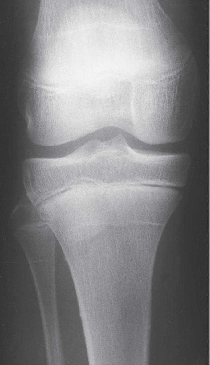
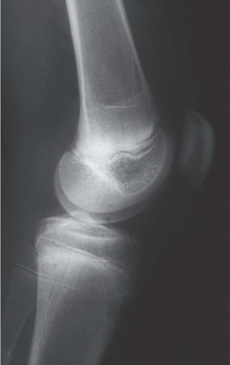
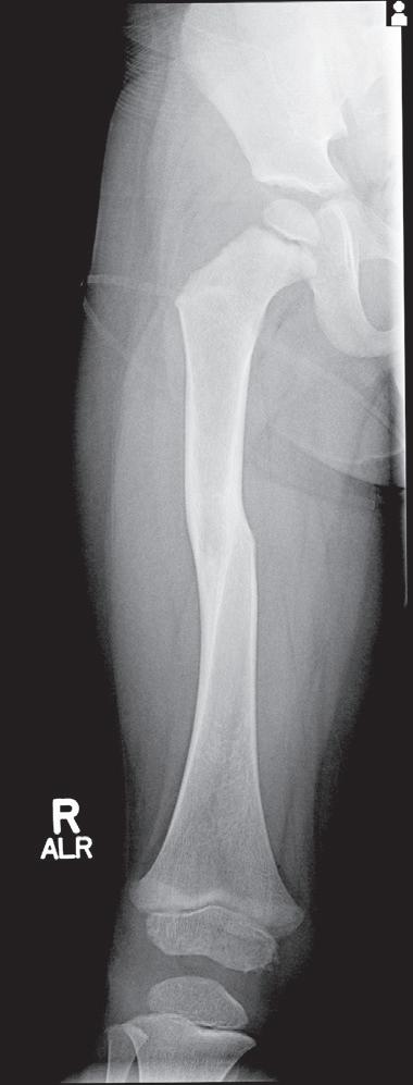
Fig. 1.3 Anteroposterior (A) and lateral (B) radiographs of the knee of a 12-year-old child 6 months after a femoral shaft fracture. Arrest lines parallel to the physis can be seen in the metaphyseal area of the distal end of the femur and proximal tibia (arrows). The temporary depression of growth at the time of injury results in more horizontal trabeculae being laid down, thereby increasing the density of bone at that level.
as well as skeletal trauma.62 Similar lines can be seen in skeletally immature patients being treated with bisphosphonates, and are called zebra lines in osteogenesis imperfecta patients.63,64
REMODELING AFTER A FRACTURE IN CHILDHOOD
The remodeling ability of bone in children may make reduction accuracy less important than it is in adults. Remodeling is a commonly used term in pediatric fracture care, implying that the child has the ability to straighten and correct residual deformity with growth. This capability depends not only on the mechanisms of bone remodeling described earlier during the remodeling phase of fracture healing (Wolff’s law) but also on reorientation of the physis by asymmetric growth after a fracture (Hueter-Volkmann law or Delpech’s law). Younger children have greater remodeling potential. The amount depends on the age of the child, location of the injury in the bone (proximity to the physis), degree of deformity, and whether the deformity is in the plane of motion of the adjacent joint.3,65,66 Clinical judgment and experience are required to guide decision-making regarding defining “acceptable” reductions, but obtaining the best reduction possible during initial treatment is advisable because it will lessen reliance on remodeling. Remodeling does not occur in displaced intraarticular fractures; thus reduction, usually by open methods, is needed. In children, remodeling is often relied on for the treatment of proximal humeral and distal radial injuries because these physes contribute greatly to the length of the respective segment, and the joints have wide ranges of motion. Remarkable remodeling has been documented in cases of these fractures (Fig. 1.4).
The effect of growth on fracture healing usually aids in fracture treatment because some angulation and deformity remodels with growth. Remodeling of angular deformities of
immature long bones occurs at the growth plate and along the shaft.67 Accelerated growth of the injured bone (as well as surrounding bones) can occur, leading to limb length discrepancy (usually the femur or humerus).68 Growth, however, can produce deformity if the growth plate is injured or if trauma has altered muscle forces on an extremity, as may occur after a spinal cord injury or traumatic brain injury.
Remodeling may occur readily in the plane of a joint (Fig. 1.5), but it occurs far less readily, if at all, in children with rotational deformity or angular deformity not in the plane of the joint.14,65,66,69 Abraham67 studied the remodeling potential of immature monkeys and found that remodeling occurred at the growth plate and along the concavity of the shaft deformity, with minimal resorption on the convexity of the shaft. Diaphyseal remodeling and physeal reorientation with growth contributed similar amounts to the degree of remodeling. In femoral shaft fractures in children, 75% of the remodeling of angular deformities takes place in the physis, and 25% comes from appositional remodeling of the diaphysis.70 The physis adjacent to a fracture realigns itself with asymmetric growth to become perpendicular to the forces acting through the bone, and most authors believe this is the primary mechanism for remodeling.66
Significant angulation in the midportion of long bones is not usually acceptable and does not remodel very well, depending on the age of the child. In children younger than 8 years, residual angulation is more acceptable. If the angulation is less than 30 degrees and is within the plane of the joint, remodeling toward normal alignment can be expected.2,19 The potential for remodeling to an acceptable functional and cosmetic outcome depends on many factors, including which bone is fractured, how close the fracture is to a joint, the orientation to the joint axis, and the amount of growth remaining for the child.66 Side-to-side (bayonet or
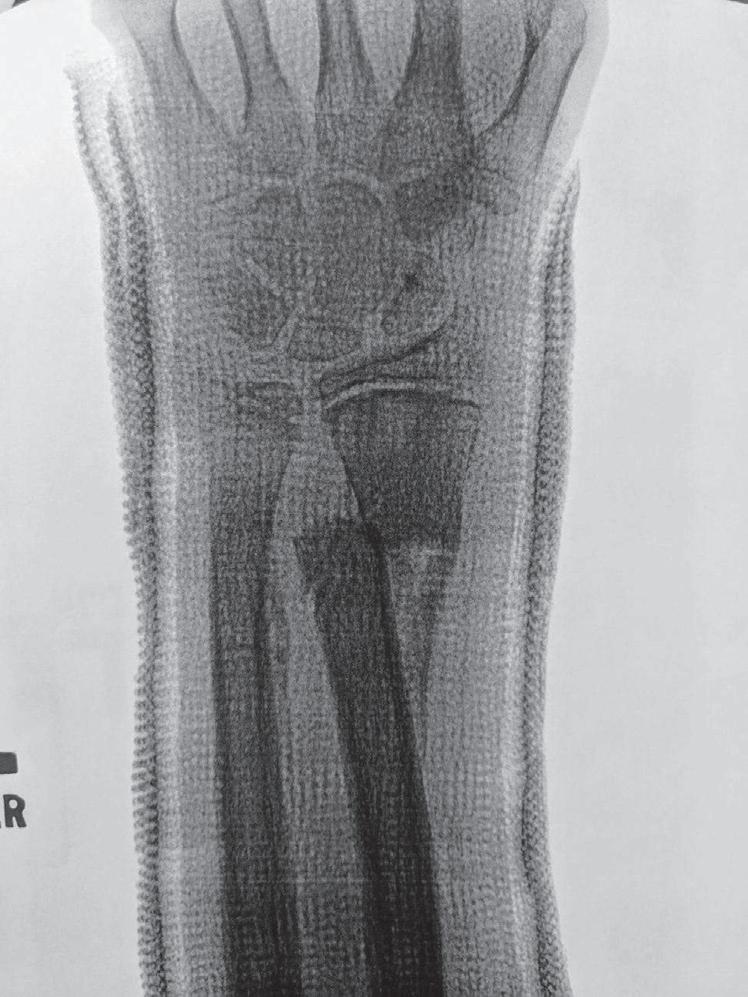
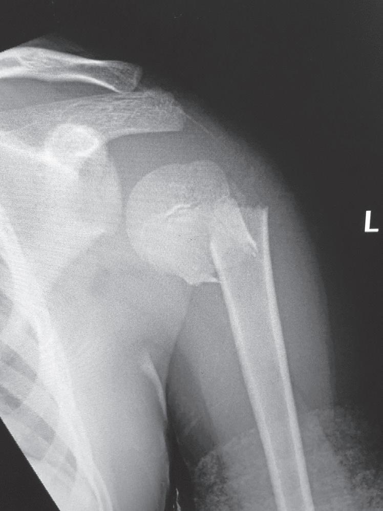
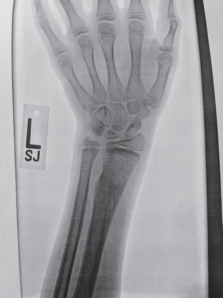
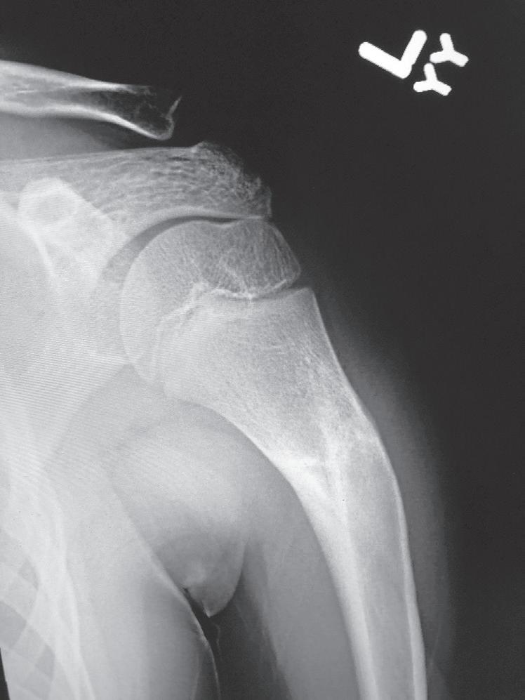
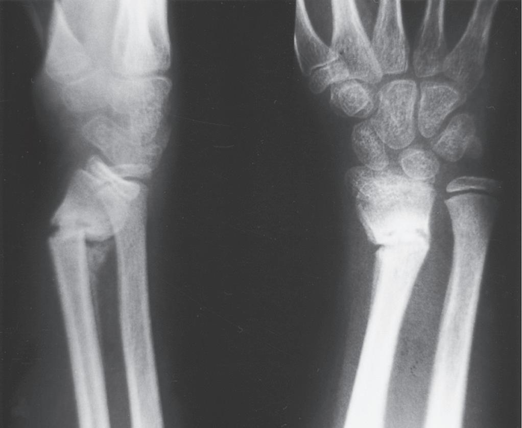
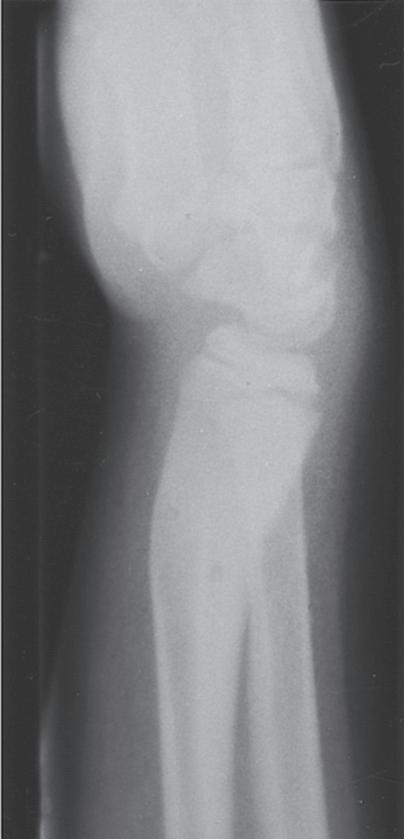
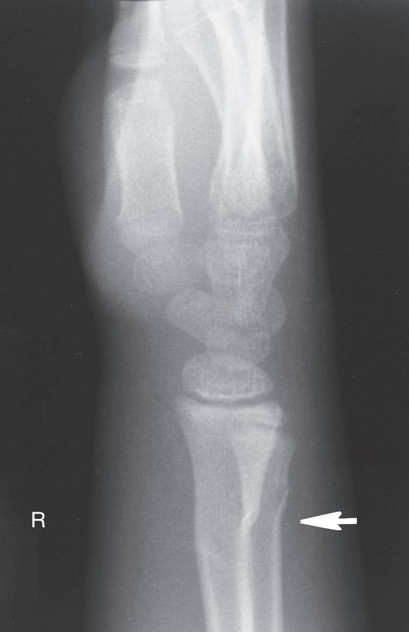
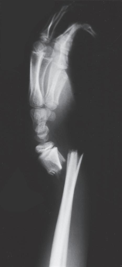
Fig. 1.4 Anteroposterior of a 9-yearold with a distal radius fracture that was pinned in poor position (A), now 4 weeks out from pinning (x-ray after pin removal). Anteroposterior of the same distal radius fracture (B) at 12 months from the injury with complete remodeling. Anteroposterior of a proximal humerus fracture in an 11-year-old at the time of injury (C) and at 6 months (D). Similar to the distal radius, these anatomic locations have tremendous remodeling potential secondary to the large contribution of growth from adjacent physes.
Fig. 1.5 (A) Radiograph of the distal portion of the radius in an 11-year-old girl at the time of cast removal 6 weeks after injury. (B) A lateral radiograph taken 3 months later shows considerable remodeling of the fracture in the plane of the joint.
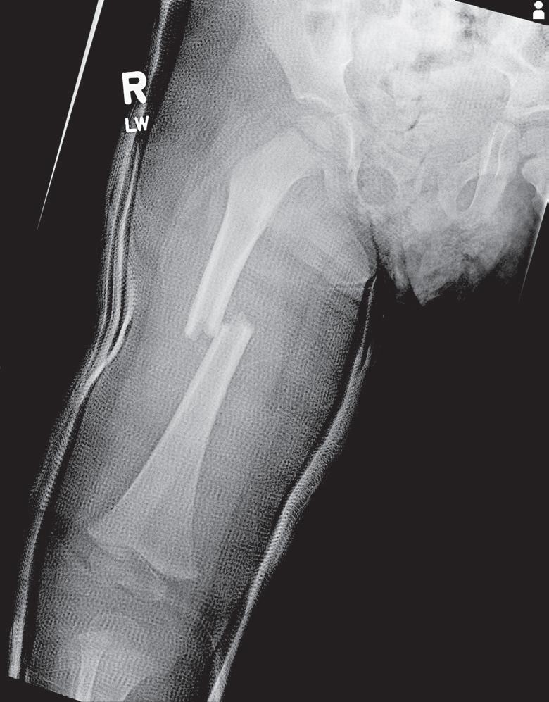
Fig. 1.6 Radiograph of a femoral shaft fracture in a 2-yearold boy in a spica cast at the time of injury (A) and at 12 months postinjury (B). The fracture demonstrates bayonet apposition with otherwise good alignment. The bayonet apposition remodeled within 12 months in this young child.
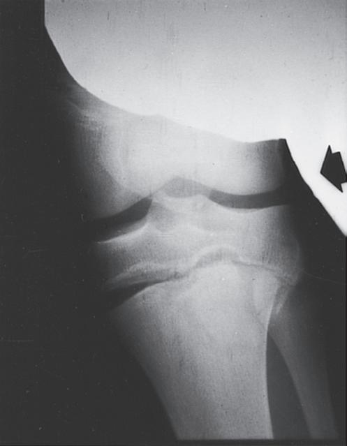
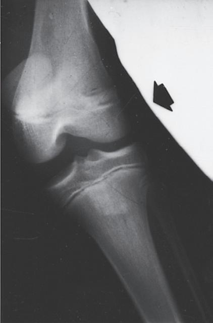
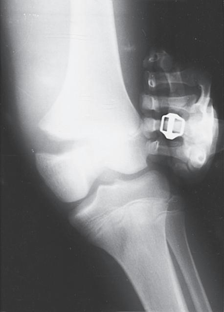
Fig. 1.7 Stress films illustrating injuries to the proximal tibial physis (A), the medial collateral ligament (B), and the distal femoral physis (C) in skeletally immature children. Stress films are no longer recommended. The diagnosis of a nondisplaced Salter I physeal fracture is made based on local tenderness and swelling over the physis.
overriding) apposition of bone is acceptable as long as alignment is accurate (Fig. 1.6). This position leads to prompt, strong union with solid periosteal bone bridging.57
COMPLICATIONS OF FRACTURES IN CHILDREN OTHER THAN PHYSEAL ARREST
Delayed union and nonunion rarely occur in healthy children. In a series of more than 2000 fractures in children, not a single case of nonunion was seen.71 Lateral condyle fractures of the distal humerus are one of the few childhood injuries with a predilection for nonunion, but displaced fractures treated with accurate reduction and fixation rarely fail to heal.72 Exceptions to uneventful fracture healing occur in older children with open injuries that have severe soft tissue injury or that become infected. Refracture is uncommon, although in malaligned forearm fractures, refracture may occur after mobilization.73 Myositis ossificans and stiffness in joints secondary to fractures are rare. Physical therapy to regain motion is seldom necessary in children because return of motion and function is typical as the child resumes normal activities and play.
ANATOMIC DIFFERENCES OF PEDIATRIC BONES
As the skeleton of a child grows, it develops from a relatively elastic and rubbery type of biomechanical material to the more rigid structure of an adult skeleton. Because of the amount of radiolucent cartilaginous material in pediatric bone, comparison films are sometimes necessary to determine whether a radiograph is abnormal, and this lack of clarity in the radiograph can make diagnosis of fractures difficult. The types of injuries may also be different in children; for example, ligamentous injuries and dislocations are rare. Injuries around the knee frequently lead to ligamentous and meniscal injuries in adults. In children, the distal femoral or proximal tibial physis is more likely to be injured because it is the weak link (Fig. 1.7). Previously, stress radiographs were recommended, but these are usually unnecessary because the diagnosis can be made by a complete history and physical examination and confirmed at follow-up when radiographs demonstrate a widened physis consistent with a healing growth plate injury. Ligamentous injuries in skeletally immature children are uncommon, but they do occur and become more frequent in adolescence as
the transition to skeletal maturity occurs.5 As noted earlier, traditional teaching that twisting ankle injuries cause distal fibular physeal injuries more commonly than ligamentous injuries in children has been challenged by studies using advanced imaging techniques.4,5
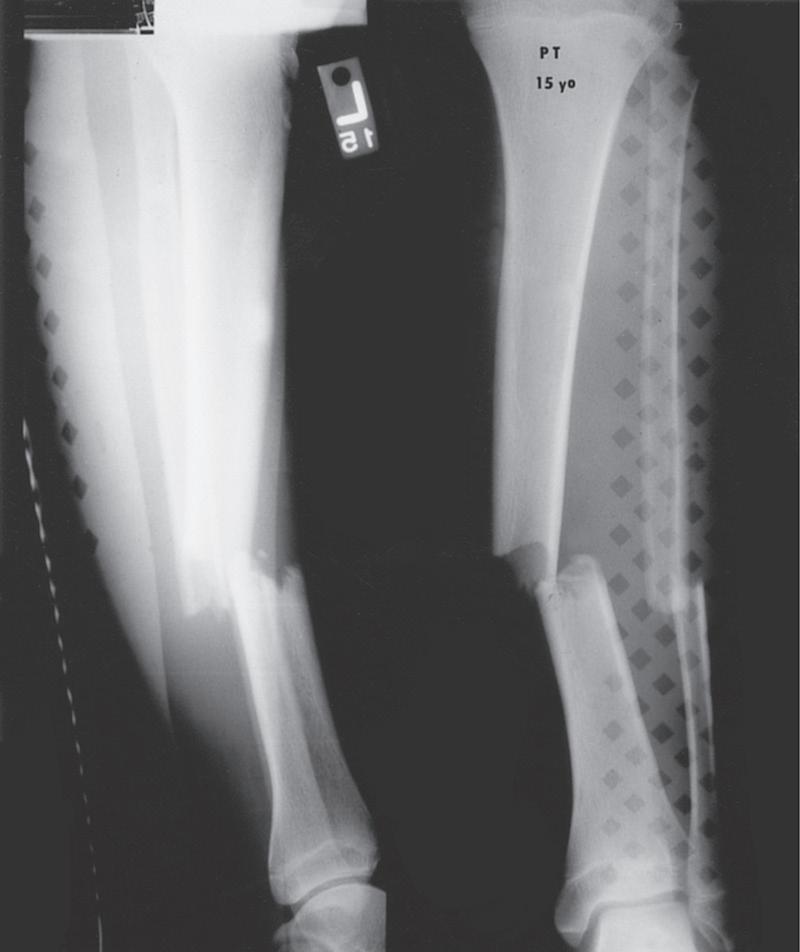
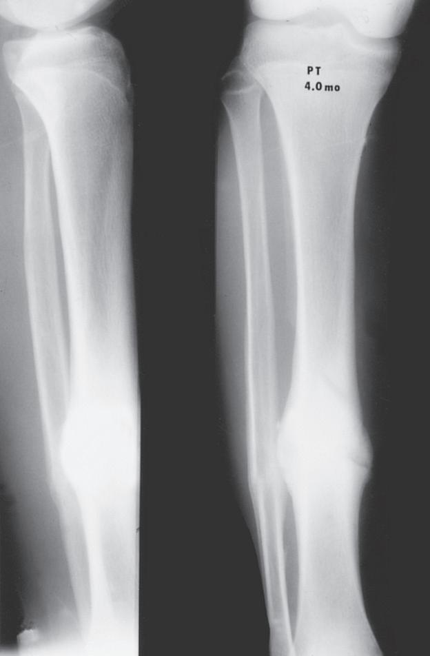
Pediatric bony injuries are more often treated by closed reduction than by open reduction because of the short time to union and the ease of obtaining and maintaining near-anatomic reductions, as well as the potential for remodeling (Fig. 1.8). The quality of anesthesia/analgesia provided to the child is strongly correlated with the quality of the reduction.22
Fig. 1.8 (A) Anteroposterior and lateral radiographs of a 15-year-old boy who sustained a displaced transverse fracture of the diaphysis of his tibia. (B) Follow-up at 4 months shows abundant periosteal healing, although a portion of the fracture line is still evident. It is characteristic for pediatric long bone fractures to heal early with periosteal callus; secondarily, the diaphyseal cortex heals and remodels.
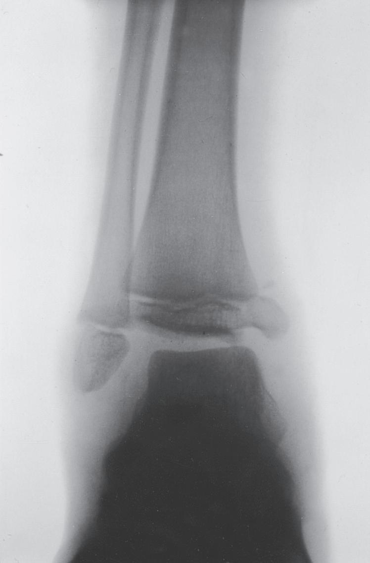
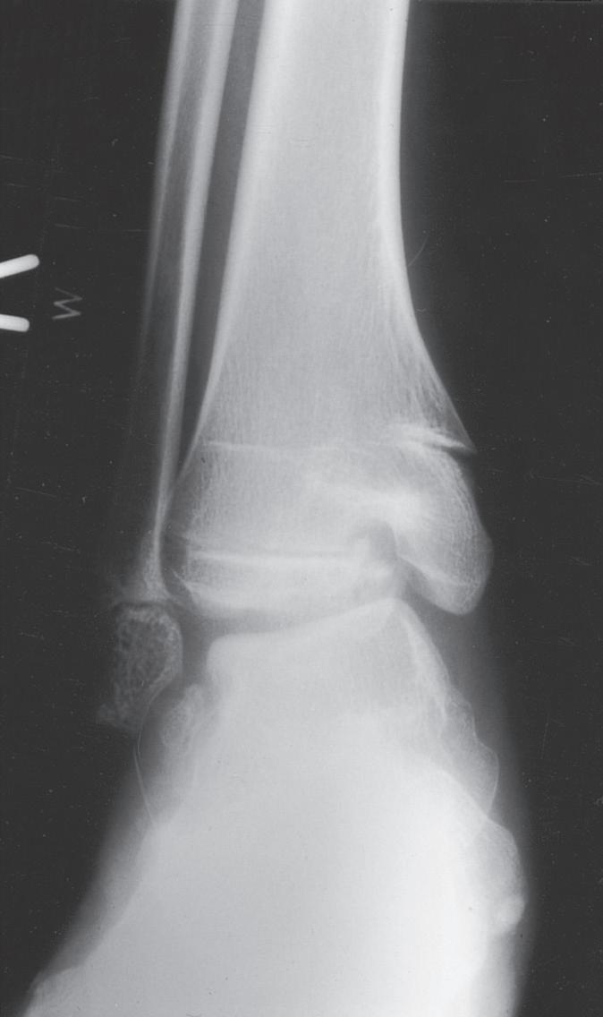
Fig. 1.9 (A) Radiograph of a 7-year-old boy who sustained a fracture of the medial malleolus, as shown in this mortise view. The fracture was treated with closed reduction and application of a long leg cast. (B) Mortise view of the ankle of the same patient 4 years after his injury. The medial malleolus portion of the epiphysis has healed to the metaphyseal area of this Salter-Harris type IV injury. He has not only an incongruous growth plate but also an incongruous ankle joint (arrows). Intraarticular fractures such as this one should be treated with open reduction and internal fixation to anatomically restore both the joint surface and the growth plate.
The most obvious anatomic differences in the pediatric skeleton are the presence of growth plates and the thick periosteum. Growth plate injuries and epiphyseal injuries can lead to growth disturbance that may be significant (Fig. 1.9). Treatment of injury to the growth plate and epiphysis parallels adult intraarticular injuries in that pediatric articular injuries require anatomic reduction to preserve joint function and growth potential. As noted earlier, the periosteum in children is much thicker, more active, less readily torn, and more easily stripped from the bone than in adults. The periosteum helps both in reduction (where intact periosteum on the concavity of the deformity serves as a hinge) and
in maintenance of reduction and contributes immensely to rapid fracture healing. The intact periosteum helps reduce the amount of displacement and is the primary reason for more stable fractures in children.
EPIPHYSIS
At birth, most epiphyses are completely cartilaginous structures. The length of time for formation of the secondary ossification center within the epiphysis varies, with the distal portion of the femur being formed first.65 A global type of growth plate is present in the epiphysis, as evidenced by columns of chondrocytes and growth potential at the physis occurring between the epiphysis and metaphysis and also just beneath the articular surface. When the epiphysis is entirely cartilaginous, it is almost completely protected from injury; traumatic forces tend to fracture the diaphysis or metaphysis, or infrequently, they may disrupt the physis, as is seen in distal humeral physeal separations in infants. Once bone has formed within the epiphysis, it is more likely to be broken, but epiphyseal fractures are much less common than fractures of the diaphysis and metaphysis. When the epiphysis is nearly all bone, it is subject to injury, much like the remainder of the bones.
PHYSIS
The growth plate remains cartilaginous throughout development. As the child grows older, the physis becomes thinner, and it is easier to disrupt the growth plate by injury. The most common location of injury in Salter-Harris type I injuries is classically described as through the lower hypertrophic zone of the physis, but variation in the plane of physeal fractures has been noted.47 Physeal anatomy changes markedly with growth. Infants and newborns have fewer mammillary processes that stabilize the epiphysis on the metaphysis. However, with further growth, particularly in the distal femoral region, prominent mammillary processes help the physis secure the epiphysis to the metaphysis, which is likely a response to physiologic demand and the need to resist torsional forces. The proximal femoral physis changes considerably with growth and eventually forms two separate physeal areas: the capital femoral epiphysis and, below it, the trochanteric physis.
METAPHYSIS
The metaphysis is the trumpet-shaped end of long bones. It has a thinner cortical area and increased trabecular bone and is wider than the corresponding diaphyseal part of the bone. Porosity in the metaphyseal area is greater than in the diaphyseal area, and the periosteum is more firmly attached in the metaphyseal area as it gets closer to the physis.
Much bone remodeling occurs in the metaphyseal region of a bone after a fracture. Periosteal bone forms in the area joining the diaphysis to the epiphysis. This area progressively transforms back into a trumpet-shaped metaphyseal cortex with longitudinal growth.
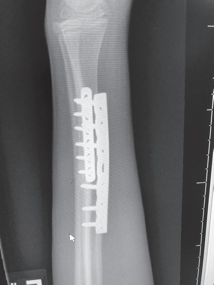
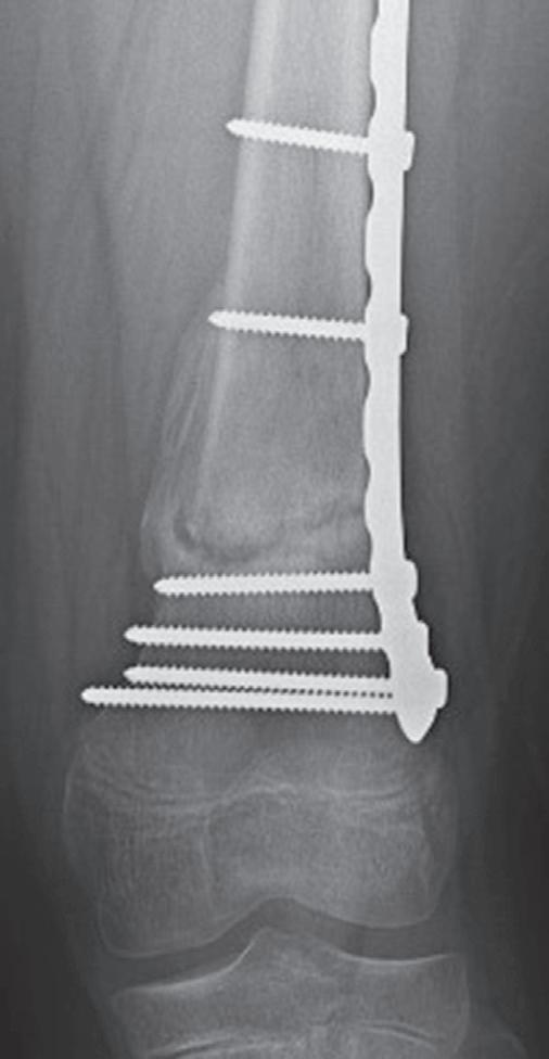
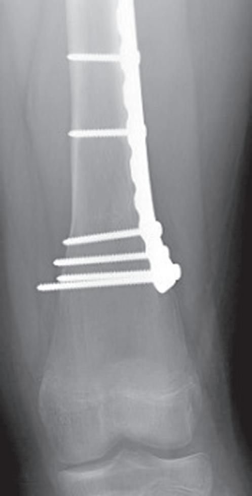
The remodeling of the metaphyseal region to create the trumpet shape can be an issue after plates and screws are placed in the metaphysis for fractures. Physeal growth results in the physis “growing away” from the plate, and the “cut-back” remodeling can result in long screws that are prominent medially (Fig. 1.10).74 In addition to this problem, lateral plates placed on the distal femur adjacent to the
AB
Fig. 1.10 Anteroposterior radiographs of 12-year old male who underwent open reduction and internal fixation of a supracondylar femur fracture at 1 month (A) and 18 months (B) postoperative. The significant growth of the distal femur resulted in physis “growing away” from the screws and the screws becoming prominent on the medial side.
physis that are left in growing patients can result in substantial valgus deformity.75,76
DIAPHYSIS
The diaphysis is the principal portion of the long bone and is extremely vascular in the newborn. With further growth it becomes less vascular, and the cortical bone thickens. The diaphysis grows in diameter by periosteum-mediated membranous bone formation.
BIOMECHANICAL DIFFERENCES AND CHANGES WITH GROWTH
Pediatric bone is less dense, more porous, and is penetrated by more vascular channels than adult bone.62 It has a comparatively lower modulus of elasticity, lower bending strength, and lower mineral content.77 Immature bone has greater porosity on cross section, and immature cortical bone has a greater number of osteon systems traversing the cortex than mature bone. The increased porosity of pediatric bone helps prevent propagation of fracture lines, explaining the infrequency of comminuted fractures in children. A comparison of load deformation curves of fractures in pediatric and adult bone shows a long plastic phase in children.77 The porosity and rough mechanical fracture surface prolong the time and energy absorption before bone is broken. Adult bone almost always fails in tension, whereas bone in children can fail either in tension or in compression (buckle or torus fractures).62 When bones are bent, stress on the tension side is about the same as on the compression side. Because bone has a lower yield stress in tension than in compression, bone yields first on the tension side. As the bending continues, a crack travels across the bone from the tension side toward the
compression side. Depending on the amount of energy to be absorbed, the large pores in growing bone may stop propagation of the fracture line, which may leave a portion of the cortex intact on the compression side and result in a greenstick fracture.1
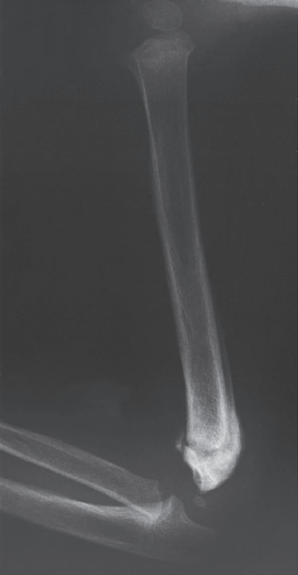
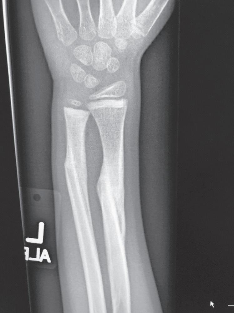
Bone is said to be elastic if it returns to its original shape after the load is removed. If bone does not return to its original shape and residual deformity remains after the load is released, bone has undergone plastic deformation. Incomplete failure in tension in which the fracture line does not propagate through bone results in plastic deformity of bone (Fig. 1.11). If enough plastic deformity is present in the remaining cortex, it may be necessary to complete the fracture as part of treatment. Completing the fracture is usually done by reversing the deformity so that the remaining cortex is placed under tension until it fails.1
CLASSIFICATION OF CHILDREN’S FRACTURES
The anatomic and biomechanical differences in the pediatric skeleton necessitate different classification systems to describe children’s fractures. Pediatric fractures can be classified into five types: (1) plastic deformation, (2) buckle fractures (near the metaphysis), (3) greenstick fractures, (4) complete fractures, and (5) physeal fractures.
PLASTIC DEFORMATION
Plastic deformation of bone is essentially unique to children. It is most commonly seen in the ulna and, occasionally, the fibula. If no hematoma is formed, periosteal elevation and significant callus formation may not occur to promote remodeling, and the bone may be permanently deformed. If the deformity occurs in a child younger than 4 years or if the deformation is less than 20 degrees, the angulation usually corrects with growth.1 Plastic deformation that produces clinically evident deformity should usually be reduced because remodeling can be unreliable.
BUCKLE FRACTURES
A buckle fracture, also an injury primarily of childhood, is a compression failure of bone that usually occurs at the junction of the metaphysis and diaphysis. In the metaphysis, where porosity is greatest, bone in compression may be buckled by the denser bone of the diaphysis (Fig. 1.12). The more cortical diaphyseal bone may be pushed into the more porous metaphyseal bone and may create a torus fracture, so named because of its similarity to the raised band around the base of a classical Greek column.62
GREENSTICK FRACTURES
Greenstick fractures occur when a bone is bent, and the tension side of the bone fails. The bone begins to fracture, but the fracture line does not propagate entirely through the bone. Incomplete failure on the compression side of
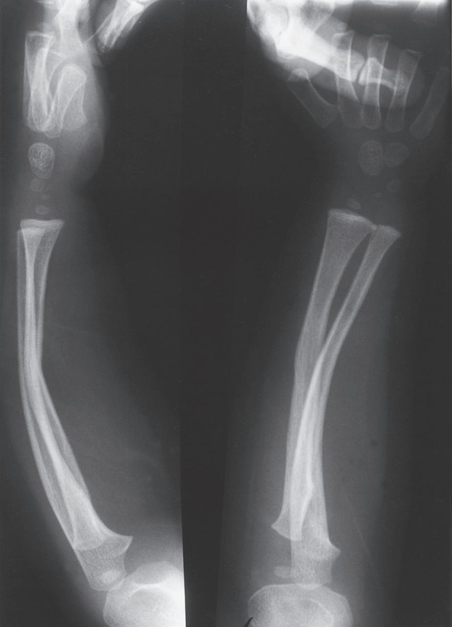
Fig. 1.11 Plastic deformation in the radius and ulna of a 2-year-old patient after a fall. The bones are plastically deformed at the midshaft, with volar compression and dorsal tension failure, but without fracture propagation.
Fig. 1.12 Torus fractures usually occur at the junction (arrow) of metaphyseal and diaphyseal bone. The more porous metaphyseal bone fails in compression.
the bone allows plastic deformity to occur. In children, if bone undergoes plastic deformation, it does not recoil to an anatomic position and usually must be completely broken to restore normal alignment. Bone is viscoelastic, meaning that its response to loading depends on the rate at which the load is applied. Remembering this when correcting plastic deformation and greenstick fractures can be helpful because slow application of steadily increasing amounts of force over a fulcrum can result in gradual return of more normal alignment.
COMPLETE FRACTURES
Fractures that propagate completely through a bone may be described in several ways, based on the pattern of the fracture.
SPIRAL FRACTURES
Spiral fractures are usually created by a rotational force on the bone. They are typically low-velocity injuries. An intact periosteal hinge enables reduction of the fracture by reversing the rotational injury.
OBLIQUE FRACTURES
Oblique fractures occur diagonally across diaphyseal bone, usually at about 30 degrees to the axis of the bone. Analogous to complete fractures in an adult, these injuries usually cause more significant disruption of the soft tissues, including the periosteum. Because these fractures are unstable and may be difficult to hold in anatomic reduction, alignment is important. Fracture reduction is attempted by immobilizing the extremity while applying traction.
TRANSVERSE FRACTURES
Transverse fractures through pediatric bone usually occur from three-point bending and are readily reduced by use of the periosteum on the concave side of the fracture force. The periosteum on the side opposite the force is typically torn. The three-point mold type of immobilization usually maintains this diaphyseal fracture in a reduced position (Fig. 1.13).
Butterfly fragments are not common in pediatric injuries but result from a mechanism similar to that causing a transverse fracture; the butterfly fragment remains on the side of the apical force of the three-point bend. This injury occurs in the highly cortical area of the diaphysis—usually in the midshaft of the femur, tibia, or ulna (Fig. 1.14).
PHYSEAL FRACTURES
Injuries to the epiphysis of a bone almost always involve the growth plate, but most physeal fractures do not involve the epiphysis (or therefore the articular surface). Problems after injury to the growth plate are not common, but any time the physis is injured, the potential for growth disturbance exists. The distal radial physis is often cited as the most frequently injured physis.15 Usually, the growth plate repairs well and rapidly, and most physeal injuries heal within 3 weeks. The rapid healing provides a limited window for reduction of deformity because late reduction (later than 1 week) after early physeal healing has occurred may cause physeal damage.48,78 Damage to the plate can occur by crushing, vascular
compromise of the physis, or bone growth bridging from the metaphysis to the bony portion of the epiphysis. The damage can result in progressive angular deformity, limb length discrepancy, or joint incongruity.
Injury to the physis has been studied extensively.3,14,45,78 These studies show an age-dependent change in the stability
Fig. 1.13 (A and B) Lateral radiographs of a dorsally displaced transverse distal radial and ulnar fracture, which is easily reduced by use of the intact dorsal periosteum to aid in locking the distal fragments in place.
Fig. 1.14 Radiograph of the femur in a 7-year-old patient involved in a motor vehicle collision. The butterfly fragment typically lies on the side of the apical force of the three-point bend.
of the epiphysis on the metaphysis. The physis and epiphyses are firmly connected externally by periosteum and internally by the mammillary processes. The physis is a hard, rubbery material that is more susceptible to injury by rotation than by angulation or traction.
Injuries involving the growth plate usually occur at the junction of calcifying cartilage cells or those that are uncalcified, although recent studies demonstrate the variability of the fracture plane within the physis.46,47 With epiphyseal injury, the growth plate is generally attached to the epiphyseal side of the fracture, and anatomic reduction of the joint surface usually results in anatomic reduction of the growth plate. The fracture plane does not always propagate directly through the hypertrophic zone but may at some places undulate into the germinal zone of the physis or into the epiphysis or metaphysis. Changes in physeal contour are caused by the mammillary processes extending into the metaphysis and play a role in determining the fracture plane. The distal femoral growth plate is shaped such that fragments of the metaphysis are often broken off when the growth plate is injured, and its propensity for physeal bar formation may be related to its large number of mammillary processes.
Physeal injuries are usually classified by the Salter-Harris classification system,46 which is based on the radiographic appearance of the fracture (Fig. 1.15). Injury may occur to the epiphysis, growth plate, metaphysis, or perichondrial ring.
In a type I fracture, the epiphysis separates completely from the metaphysis without any radiographically evident fracture through ossified bone. The periosteum usually remains attached to the growth plate, thereby preventing significant displacement of the epiphysis from the metaphysis. In patients with very little periosteal disruption, a slight widening of the physis may be the only radiographic sign of an injury through the physis. Although type I injuries are not usually associated with vascular change, complete separation of the capital femoral epiphysis can result in avascular necrosis and growth arrest of the proximal end of the femur. The larger the ossification center, the greater the tendency of the injury to produce a metaphyseal fragment on the compression side of the injury.
In a type II fracture, the most common Salter-Harris fracture pattern,79 the injury passes through the growth plate and out through a portion of the metaphysis. The periosteum is usually damaged on the tension side, but the fracture leaves the periosteum intact in the region of the metaphyseal fragment. As in a type I injury, the line of fracture separation usually occurs along the hypertrophic and calcified zones of the physis. However, propagation along this junction is
Fig. 1.15 Illustration of the Salter-Harris classification of epiphyseal injuries (see text). (From Salter RB, Harris WR. Injuries involving the epiphyseal plate. J Bone Joint Surg Am. 1963;45:587.)
more variable in a type II injury. As the fracture line courses toward the compression side of the injury, it propagates through the metaphyseal area. The periosteal attachment along the metaphyseal fragment can be used to aid in reduction of the injury.
Growth disruption secondary to type I and type II injuries is infrequent, although it can occur, particularly if the circulation to the epiphysis is disrupted. Anatomic reduction is not generally required with type I and type II injuries, although one study in distal tibia fractures describes persistent gaps (>3 mm) after reduction as correlated with an increased incidence of premature physeal closure,50 but a later study by the same group did not confirm this finding.54 These injuries are adjacent to the joint, and the entire epiphysis is intact.
A type III fracture is intraarticular and passes through the epiphysis until it reaches the growth plate. The fracture line then courses through the growth plate to the periosteal surface. This type of fracture usually occurs when the growth plate is beginning to undergo closure. As such, problems pertaining to growth arrest may not be major. With anatomic reduction of the articular surface, the physis is usually anatomically reduced as well, and growth arrest after anatomic reduction usually does not occur.
A type IV injury is also intra-articular and involves the epiphysis as well as the metaphysis. The fracture line crosses through the growth plate. The injury is similar to a type III fracture in that the articular surface must be anatomically reduced. A more vertical split of all zones of the physis occurs, and the physis must be anatomically reduced to restore the architecture of the growth plate and minimize the risk of osseous bridge formation.
Considerable debate exists concerning type V injuries. The original type V injury as described by Salter was a crush injury to the growth plate.14 A type V fracture cannot be recognized on initial radiographs because it appears to be a type I injury. These injuries are very uncommon, but any injury accompanied by clinical swelling and tenderness around the growth plate and associated with considerable axial load could possibly become a type V fracture, as evidenced by early closure of the physis after injury.
The Salter-Harris classification is useful as a rapid means of describing a physeal injury based on radiographic interpretation. A more complex and inclusive classification scheme was proposed by Ogden.19,80 It includes nine types of fractures that are further divided into subtypes A through D. Peterson also developed a physeal injury classification.19,79–82 The Ogden and Peterson classifications have not been used as often.