Contact your Elsevier Sales Representative for teaching resources for this title—including image collections—or request these supporting materials at http://evolve.elsevier.com.
Richard L. Drake, PhD, FAAA
Director of Anatomy
Professor of Surgery
Cleveland Clinic Lerner College of Medicine
Case Western Reserve University Cleveland, Ohio
A. Wayne Vogl, PhD, FAAA
Professor of Anatomy and Cell Biology
Department of Cellular and Physiological Sciences
Faculty of Medicine
University of British Columbia Vancouver, British Columbia
Adam W.M. Mitchell, MBBS, FRCS, FRCR
Consultant Radiologist
Chelsea and Westminster Hospital
Honorary Senior Lecturer Imperial College London, UK
Illustrations by Richard Tibbitts and Paul Richardson
Photographs by Ansell Horn
SECOND EDITION
GRAY’S BASIC ANATOMY
1600 John F. Kennedy Blvd. Ste. 1800 Philadelphia, PA 19103-2899
Previous edition copyrighted 2012 by Churchill Livingstone, an imprint of Elsevier, Inc.
All rights reserved. No part of this publication may be reproduced or transmitted in any form or by any means, electronic or mechanical, including photocopying, recording, or any information storage and retrieval system, without permission in writing from the publisher. Permissions may be sought directly from Elsevier’s Rights Department: phone: (+1) 215 239 3804 (US) or (+44) 1865 843830 (UK); fax: (+44) 1865 853333; e-mail: healthpermissions@elsevier.com. You may also complete your request on-line via the Elsevier website at http://www.elsevier.com/permissions
Notice
Knowledge and best practice in this field are constantly changing. As new research and experience broaden our knowledge, changes in practice, treatment and drug therapy may become necessary or appropriate. Readers are advised to check the most current information provided (i) on procedures featured or (ii) by the manufacturer of each product to be administered, to verify the recommended dose or formula, the method and duration of administration, and contraindications. It is the responsibility of the practitioner, relying on their own experience and knowledge of the patient, to make diagnoses, to determine dosages and the best treatment for each individual patient, and to take all appropriate safety precautions. To the fullest extent of the law, neither the Publisher nor the Authors assume any liability for any injury and/or damage to persons or property arising out of or related to any use of the material contained in this book.
To my parents who guide me, To my wife who supports me, To my students who challenge me
RLD
To my family, my colleagues, my mentors, and my students
AWV
To all my family, Cathy, Max and Elsa, and my colleagues
AWMM
Acknowledgments
Any book, no matter the size, is a major undertaking, and we want to thank all of the individuals who have helped move this project to completion. These include William Schmitt and Rebecca Gruliow, who both helped in initially evaluating the need for this type of concise textbook and in moving the first edition forward to completion. Madelene Hyde and Rebecca Gruliow guided this second edition through to completion. We also appreciate the contributions of our illustrators, Richard Tibbitts and Paul Richardson, who did all of the artwork.
We’d also like to thank Professor Richard A. Buckingham of the Abraham Lincoln School of Medicine, University of
Illinois, for Fig. 8.97B; Dr. Murray Morrison, Dr. Joanne Matsubara, Dr. Brian Westerberg, Laura Hall, and Jing Cui for contributing images for the head and neck chapter; and Dr. Bruce Crawford and Logan Lee for help with images for the surface anatomy of the upper limb.
Finally, we are very appreciative of the numerous individuals, anatomists, and educators who provided feedback on the first edition and whose suggestions were included in this second edition.
Richard L. Drake A. Wayne Vogl
Adam W.M. Mitchell
Preface
Gray’s Basic Anatomy was developed in response to students and colleagues from around the world who requested a more concise description of anatomy than that presented in Gray’s Anatomy for Students. To accomplish this goal, we reworked the material to focus mainly on regional anatomy and integrated the clinical material, imaging, and surface anatomy information directly into the text as:
• Clinical apps, which give students context for why a strong anatomical background helps facilitate the solving of clinical problems;
• Imaging apps, which offer students a great introduction to the different techniques and modalities available for imaging relevant anatomy; and
• Surface anatomy boxes, which help students visualize the relationship between anatomical structures and surface landmarks necessary for any kind of patient examination.
In addition, at the beginning of each chapter students are directed to additional learning resources available on Student Consult (Elsevier’s online educational website).
Summarizing, Gray’s Basic Anatomy uses a regional approach, similar to Gray’s Anatomy for Students, with eight
chapters: The Body, Back, Thorax, Abdomen, Pelvis and Perineum, Lower Limb, Upper Limb, and Head and Neck. The artwork presents the same familiar illustrations from Gray’s Anatomy for Students, but they have been resized to fit within a smaller format while retaining a close physical location to the text with which each figure is associated. Finally, while some verbiage has been sacrificed in keeping with the goal of presenting a concise textbook of anatomy (e.g., muscle descriptions have for the most part been incorporated into tables with no loss of content), additional clinical and imaging material has been added to enhance learning in context.
This second edition includes numerous edits resulting from reader feedback, some new and revised figures, and revisions based on current research in the field of the anatomical sciences.
We hope you will continue to find this new edition a useful and valuable resource whether you are an educator or a student.
Richard L. Drake A. Wayne Vogl Adam W.M. Mitchell
Index of Clinical Apps, Imaging Apps, and Surface Anatomy Boxes
1 The Body
CLINICAL APPS
Bone marrow transplants 9
Bone fractures 10
Avascular necrosis 10
Osteoporosis 10
Epiphyseal fractures 10
Joint replacement 13
Degenerative joint disease 13
Arthroscopy 13
The importance of fascias 14
Muscle paralysis 14
Muscle atrophy 14
Muscle injuries and strains 15
Atherosclerosis 15
Varicose veins 15
Anastomoses and collateral circulation 16
Lymph nodes 17
Dermatomes and myotomes 21
Referred pain 29
IMAGING APPS
Determination of skeletal age 9
2 Back
CLINICAL APPS
Spina bifida 38
Vertebroplasty 39
Scoliosis 39
Kyphosis 39
Lordosis 39
Variation in vertebral numbers 39
The vertebrae and cancer 39
Osteoporosis 39
Back pain 41
Herniation of intervertebral discs 41
Joint diseases 41
Ligamenta flava 42
Vertebral fractures 43
Pars interarticularis fractures 43
Surgical procedures on the back 43
Nerve injuries affecting superficial back muscles 45
Lumbar cerebrospinal fluid tap 54
Anesthesia within the vertebral canal 54
Herpes zoster 55
IMAGING APPS
Typical cervical vertebrae 32
Typical thoracic vertebrae 33
Typical lumbar vertebrae 33
Articulation between atlas and axis 35
SURFACE ANATOMY
How to identify specific vertebral spinous processes 36
Primary and secondary curvatures in the sagittal plane 38
Visualizing the inferior ends of the spinal cord and subarachnoid space 52
3 Thorax
CLINICAL APPS
Axillary process of breast 59
Breast cancer 60
Thoracic outlet syndrome 62
Cervical ribs 65
Rib fractures 65
Collection of sternal bone marrow 67
The manubriosternal joint as reference 68
Surgical access to the chest 74
Index of Clinical Apps, Imaging Apps, and Surface Anatomy Boxes
Thoracostomy (chest) tube insertion 75
Intercostal nerve block 75
The arrangement of pleural cavities is clinically significant 78
Innervation of parietal and visceral pleura 79
Pleural recesses 80
Pleural effusion 81
Pneumothorax 81
Inhaled objects 86
Lung cancer 93
Pneumonia 93
Pericardial innervation 96
Pericarditis 96
Pericardial effusion 96
Constrictive pericarditis 96
Valve disease 106
Common congenital heart defects 107
Cardiac auscultation 107
Clinical terminology for coronary arteries 110
Cardiac conduction system 110
Heart attack 114
Classic symptoms of heart attack 114
Referred pain 114
Are heart attack symptoms the same in men and women? 115
Ectopic parathyroid glands in the thymus 116
Left brachiocephalic vein 118
Venous access for central and dialysis lines 118
Using the superior vena cava to access the inferior vena cava 119
Coarctation of the aorta 120
Traumatic injury to the aorta 120
Aortic dissection 121
Aortic arch and its anomalies 121
Abnormal origin of great vessels 121
The vagus nerves, recurrent laryngeal nerves, and hoarseness 123
Esophagus constrictions 124
Esophageal cancer 125
Esophageal rupture 125
IMAGING APPS
Visualizing the pulmonary trunk by computed tomography 88
Visualizing the lungs 90
Plain chest radiograph 92
Visualizing the heart 99
Visualizing the chambers of the heart 100
Visualizing the right atrium and pulmonary veins 103
Visualizing structures in the superior mediastinum 117
Visualizing structures at the TIV vertebral level 120
Visualizing the mediastinum in the axial plane 130
SURFACE ANATOMY
The breast in women 60
How to count ribs 69
Visualizing structures at the TIV/V vertebral level 69
Visualizing the pleural cavities and lungs, pleural recesses, and lung lobes and fissures 84
Where to listen for lung sounds 85
Visualizing the margins of the heart 100
Where to listen for heart sounds 106
Visualizing structures in the superior mediastinum 123
4 Abdomen
CLINICAL APPS
Preperitoneal vs. retroperitoneal 140
Surgical incisions 147
Cremasteric reflex 148
Masses around the groin 148
Inguinal hernias 149
Indirect inguinal hernias 149
Direct inguinal hernias 149
Femoral hernias 149
Umbilical hernias 150
Incisional hernias 150
Sportsmen’s groin/sportsmen’s hernia 150
Other hernias 150
Potential problem of hernias 150
The peritoneum 151
Innervation of peritoneum 151
Ventriculoperitoneal shunts 151
Hemodialysis and peritoneal dialysis 152
Peritoneal spread of disease 152
Perforated bowel 152
The greater omentum 153
Epithelial transition between the abdominal esophagus and stomach 158
Surgery for obesity 158
Duodenal ulceration 158
Examination of the bowel lumen 159
Meckel’s diverticulum 159
Carcinoma of the stomach 159
Appendicitis 162
Congenital disorders of the gastrointestinal tract 165
Bowel obstruction 165
Diverticular disease 165
Ostomies 168
Index of Clinical Apps, Imaging Apps, and Surface Anatomy Boxes
Segmental anatomy of the liver 168
Annular pancreas 169
Pancreatic cancer 171
Gallstones 171
Jaundice 172
Spleen disorders 173
Vascular supply to the gastrointestinal system 178
Hepatic cirrhosis 180
Portosystemic anastomosis 180
Psoas muscle abscess 190
Diaphragmatic hernias 190
Hiatal hernia 190
Urinary tract stones 194
Urinary tract cancer 194
Kidney transplant 194
Abdominal aortic stent graft 197
Inferior vena cava filter 199
Retroperitoneal lymph node surgery 201
IMAGING APPS
Visualizing the stomach 155
Visualizing the jejunum and ileum 157
Endoscopic examination of the abdominal gastrointestinal tract 159
Visualizing the liver 167
Visualizing the pancreas 170
Visualizing the diaphragm 189
Investigation of the urinary tract 195
SURFACE ANATOMY
Using abdominal quadrants to locate major viscera 135
Defining surface regions to which pain from the gut is referred 135
How to find the superficial inguinal ring 148
Visualizing the position of major blood vessels 199
5 Pelvis and Perineum
CLINICAL APPS
Bone marrow biopsy 210
Common problems with the sacro-iliac joints 212
Pelvic fracture 213
Pelvic measurements in obstetrics 216
Defecation 217
Episiotomy 219
Digital rectal examination 221
Carcinoma of the colon and rectum 221
Bladder cancer 222
Bladder stones 223
Suprapubic catheterization 223
Bladder infection 224
Urethral catheterization 225
Undescended testes 225
Hydrocele of the testis 225
Testicular tumors 225
Vasectomy 226
Prostate problems 226
Ovarian cancer 228
Hysterectomy 228
Tubal ligation 229
Carcinoma of the cervix and uterus 229
The recto-uterine pouch 232
Pudendal block 236
Prostatectomy and impotence 239
Hemorrhoids 244
Abscesses in the ischio-anal fossae 246
Urethral rupture 252
SURFACE ANATOMY
Defining the margins of the perineum 245
Superficial features of the external genitalia in women 248
Superficial features of the external genitalia in men 250
6 Lower Limb
CLINICAL APPS
Pelvic fractures 269
Femoral neck fractures 271
Intertrochanteric fractures 272
Femoral shaft fractures 272
Varicose veins 277
Deep vein thrombosis 277
Harvesting veins for grafts 277
Vascular access in the lower limb 281
Trendelenburg’s sign 282
Intramuscular injection in the gluteal region: avoiding the sciatic nerve 285
Shin splints 291
Quadriceps injury 293
Hamstring injuries 294
Compartment syndrome 294
Index of Clinical Apps, Imaging Apps, and Surface Anatomy Boxes
Peripheral vascular disease 298
Soft tissue injuries to the knee 305
Clinical tests for tears in the cruciate ligaments: anterior drawer test, posterior drawer test 305
Arthroscopy 305
Foot drop 315
Fracture of the talus 317
Achilles tendon rupture 318
Ankle injuries 323
Bunions 325
Morton’s neuroma 335
Dermatomes and myotomes in the lower limb 336
Testing sensory innervation carried by major peripheral nerves in the lower limb 337
Tendon taps in the lower limb 338
Gait and gait defects 338
IMAGING APPS
Visualizing the hip joint 275
Visualizing the knee joint 304
Visualizing the bones of the foot 319
Visualizing the ankle joint 322
SURFACE ANATOMY
Finding the femoral artery in the femoral triangle 281
Visualizing the contents of the popliteal fossa 307
Finding the tarsal tunnel—the gateway to the foot 326
Finding the dorsalis pedis artery 333
Pulse points 339
7 Upper Limb
CLINICAL APPS
Fracture of the proximal humerus 346
Fractures of the clavicle and dislocations of the acromioclavicular and sternoclavicular joints 350
Dislocations of the glenohumeral joint 350
Rotator cuff disorders 351
Quadrangular space syndrome 354
“Winging” of the scapula 358
Trauma to the arteries in and around the axilla 362
Central venous access via the subclavian/axillary vein 362
Damage to long thoracic nerve 366
Injuries to the brachial plexus 370
Lymphatic drainage and breast cancer 370
Rupture of biceps tendon 373
Median nerve injury in the arm 375
Radial nerve injury in the arm 375
Supracondylar fracture of the humerus 379
Pulled elbow 379
Fracture of the head of radius 379
“Tennis” and “golfer’s” elbow (epicondylitis) 379
Ulnar nerve injury at the elbow 380
Construction of a dialysis fistula 381
Blood pressure measurement 381
Fractures of the radius and ulna 384
Fracture of the scaphoid and avascular necrosis of the proximal scaphoid 397
‘Double-jointed’ 398
‘Knuckle cracking’ 398
De Quervain syndrome 399
Carpal tunnel syndrome 399
Tenosynovitis 401
Trigger finger 401
Allen’s test 406
Ulnar nerve injury 407
Radial nerve injury 410
Dermatomes and myotomes in the upper limb 410
Tendon taps in the upper limb 410
Testing sensory innervation carried by major peripheral nerves in the upper limb 411
IMAGING APPS
Visualizing the sternoclavicular joint 347
Visualizing the acromioclavicular joint 347
Visualizing the glenohumeral joint 348
Visualizing the rotator cuff muscles 349
Developmental changes in the elbow joint 378
Visualizing the elbow joint 379
Visualizing the forearm 382
Visualizing the hand and wrist joint 396
SURFACE ANATOMY
Locating the brachial artery in the arm 376
Identifying tendons and locating major vessels and nerves in the distal forearm 392
Position of the flexor retinaculum and the recurrent branch of the median nerve 399
Motor function of the median and ulnar nerves in the hand 409
Pulse points 412
Index of Clinical Apps, Imaging Apps, and Surface Anatomy Boxes
8
Head and Neck
CLINICAL APPS
Medical imaging of the head 428
Fractures of the skull vault 430
Cerebrospinal fluid leak 434
Hydrocephalus 434
Meningitis 434
Endarterectomy 436
Stroke 437
Intracerebral aneurysms 437
Emissary veins 439
Head injury 441
Concussion 442
Cranial nerve lesions 447
Overview of cranial nerves 447
Parotid gland—tumors and stones 456
Trigeminal neuralgia 458
Facial nerve [VII] palsy (Bell’s palsy) 462
Scalp laceration 466
Orbital fracture 468
Full and partial ptosis 470
Horner’s syndrome 470
Ophthalmoscopy 484
Glaucoma 485
Cataracts 485
Swimmer’s ear 489
Surfer’s ear 489
Examination of the ear 490
Mastoiditis 492
Temporomandibular joint disorders 503
Lingual nerve injury 508
Anesthesia of the inferior alveolar nerve 508
Middle meningeal artery and extradural hematoma 513
Spread of infection from the pterygoid plexus into the cranial cavity 513
Dry eye 518
Spread of neck infections 522
Central venous access 523
Tracheobronchial injury 531
Thyroidectomy 533
Goiter 533
Hyperparathyroidism 534
Ectopic parathyroid glands 534
Recurrent laryngeal nerve palsy 541
Clinical lymphatic drainage of the head and neck 544
Laryngoscopy 552
Cricothyrotomy 556
Tracheostomy 562
Deviated nasal septum 565
Surgical approach to the pituitary gland 568
Epistaxis 572
Oral cancer 575
Test for cranial nerve XII 582
Test for cranial nerve X 588
IMAGING APPS
Visualizing the skull—anterior view 417
Visualizing the skull—lateral view 419
Visualizing the internal carotid and vertebral arteries 435
Visualizing the muscles of the eyeball 477
Visualizing the nasal cavities and paranasal sinuses 567
SURFACE ANATOMY
Anatomical position of the head and major landmarks 420
Estimating the position of the middle meningeal artery 430
Major features of the face 463
The eye and lacrimal apparatus 472
How to outline the anterior and posterior triangles of the neck 520
How to find the thyroid gland 532
How to locate the cricothyroid ligament 556
Pulse points 596
1 The Body
What is anatomy? 2
How can gross anatomy be studied? 2
Important anatomical terms 2
Imaging 3
Diagnostic imaging techniques 3
Image interpretation 6
Plain radiography 6
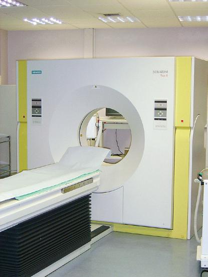
Computed tomography 7
Magnetic resonance imaging 7
Nuclear medicine imaging 7
Safety in imaging 8
Body systems 8
Skeletal system 8
Cartilage 8
Bone 8
Joints 10
Skin and fascias 13
Skin 13
Fascia 14
Muscular system 14
Cardiovascular system 15
Lymphatic system 16
Lymphatic vessels 16
Lymph nodes 16
Lymphatic trunks and ducts 17
Nervous system 18
Central nervous system 18
Functional subdivisions of the CNS 19
Other systems 30
2 Back
Regional anatomy 32
Skeletal framework 32
Vertebrae 32
Intervertebral foramina 37
Posterior spaces between vertebral arches 38
Curvatures of vertebral column 38
Joints 40
Joints between vertebrae in the back 40
Ligaments 41
Anterior and posterior longitudinal ligaments 41
Ligamenta flava 42
Supraspinous ligament and ligamentum nuchae 42
Interspinous ligaments 42
Back musculature 43
Superficial group of back muscles 43
Intermediate group of back muscles 45
Deep group of back muscles 46
Thoracolumbar fascia 48
Spinal cord 49
Vasculature 50
Meninges 52
Arrangement of structures in the vertebral canal 53
Spinal nerves 53
3 Thorax
Regional anatomy 58
Pectoral region 58
Breast 58
Muscles of the pectoral region 60
Thoracic wall 60
Superior thoracic aperture 61
Inferior thoracic aperture 62
Skeletal framework 62
Intercostal spaces 71
Diaphragm 75
Venous drainage 77
Innervation 77
Movements of the thoracic wall and diaphragm during breathing 77
Pleural cavities 78
Pleura 78
Lungs 81
Mediastinum 93
Middle mediastinum 94
Superior mediastinum 116
Posterior mediastinum 123
Anterior mediastinum 129
4 Abdomen
Regional anatomy 134
Surface topography 134
Four-quadrant pattern 134
Nine-region pattern 134
Abdominal wall 135
Superficial fascia 136
Anterolateral muscles 136
Extraperitoneal fascia 140
Peritoneum 141
Innervation 141
Arterial supply and venous drainage 142
Lymphatic drainage 143
Groin 143
Inguinal canal 144
Abdominal viscera 150
Peritoneum 150
Peritoneal cavity 151
Organs 154
Arterial supply to the gastrointestinal tract 173
Venous drainage 178
Lymphatics 181
Innervation 181
Posterior abdominal region 186
Posterior abdominal wall 186
Viscera 190
Vasculature 196
Lymphatic system 200
Nervous system in the posterior abdominal region 201
Sympathetic trunks and splanchnic nerves 201
5 Pelvis and Perineum
Regional anatomy 208
Pelvis 208
Bones 208
Joints 211
Orientation 212
Gender differences 212
True pelvis 212
Viscera 220
Fascia 230
Peritoneum 230
Nerves 232
Blood vessels 239
Lymphatics 243
Perineum 244
Borders and ceiling 244
Ischio-anal fossae and their anterior recesses 245
Anal triangle 246
Urogenital triangle 246
Somatic nerves 252
Visceral nerves 253
Blood vessels 253
Veins 255
Lymphatics 255
6
Lower Limb
Regional anatomy 266
The Hip 267
Bony pelvis 267
Proximal femur 270
Hip joint 272
Gateways to the lower limb 274
Nerves 276
Arteries 276
Veins 276
Lymphatics 278
Deep fascia and the saphenous opening 279
Femoral triangle 280
Gluteal region 281
Muscles 282
Nerves 283
Arteries 286
Veins 287
Lymphatics 287
Thigh 287
Bones 288
Muscles 291
Arteries 294
Veins 298
Nerves 298
Knee joint 300
Tibiofibular joint 305
Popliteal fossa 306
Leg 307
Bones 308
Joints 309
Posterior compartment of leg 309
Lateral compartment of leg 313
Anterior compartment of leg 314
Foot 315
Bones 316
Joints 319
Tarsal tunnel, retinacula, and arrangement of major structures at the ankle 326
Arches of the foot 327
Plantar aponeurosis 328
Fibrous sheaths of toes 328
Extensor hoods 328
Intrinsic muscles 329
Arteries 332
Veins 334
Nerves 334
7 Upper Limb
Regional anatomy 342
Shoulder 343
Bones 344
Joints 346
Muscles 351
Posterior scapular region 351
Gateways to the posterior scapular region 351
Nerves 354
Arteries and veins 354
Axilla 355
Axillary inlet 355
Anterior wall 356
Medial wall 357
Lateral wall 358
Posterior wall 358
Gateways in the posterior wall 359
Floor 359
Contents of the axilla 360
Arm 370
Bones 370
Muscles 373
Nerves 374
Arteries and veins 375
Elbow joint 377
Cubital fossa 380
Forearm 382
Bones 383
Joints 384
Anterior compartment of the forearm 385
Muscles 385
Arteries and veins 387
Nerves 388
Posterior compartment of forearm 390
Muscles 390
Arteries and veins 392
Nerves 392
Hand 394
Bones 394
Joints 397
Carpal tunnel and structures at the wrist 398
Palmar aponeurosis 399
Anatomical snuffbox 399
Fibrous digital sheaths 400
Extensor hoods 401
Muscles 402
Arteries and veins 403
Nerves 407
8
Head and Neck
Regional anatomy 415
Head 416
Neck 416
Skull 416
Anterior view 416
Lateral view 418
Posterior view 420
Superior view 421
Inferior view 421
Cranial cavity 424
Roof 424
Floor 425
Meninges 430
Cranial dura mater 430
Arachnoid mater 433
Pia mater 433
Meningeal spaces 433
Brain and its blood supply 434
Brain 434
Blood supply 435
Venous drainage 437
Cranial nerves 442
Olfactory nerve [I] 442
Optic nerve [II] 443
Oculomotor nerve [III] 443
Trochlear nerve [IV] 444
Trigeminal nerve [V] 444
Ophthalmic nerve [V1] 445
Maxillary nerve [V2] 445
Mandibular nerve [V3] 446
Abducent nerve [VI] 446
Facial nerve [VII] 446
Vestibulocochlear nerve [VIII] 446
Glossopharyngeal nerve [IX] 447
Vagus nerve [X] 450
Contents
Accessory nerve [XI] 450
Hypoglossal nerve [XII] 450
Face 450
Muscles 450
Parotid gland 455
Innervation 457
Vessels 460
Scalp 463
Layers 463
Innervation 464
Vessels 465
Lymphatic drainage 466
Orbit 467
Bony orbit 467
Eyelids 468
Lacrimal apparatus 471
Sensory innervation 473
Fissures and foramina 473
Fascial specializations 475
Muscles 476
Vessels 479
Innervation 480
Eyeball 483
Ear 486
External ear 487
Middle ear 491
Internal ear 495
Temporal and infratemporal fossae 499
Bony framework 499
Temporomandibular joints 501
Masseter muscle 503
Temporal fossa 504
Infratemporal fossa 505
Pterygopalatine fossa 513
Skeletal framework 514
Gateways 515
Contents 515
Neck 519
Fascia 520
Superficial venous drainage 523
Anterior triangle of the neck 523
Posterior triangle of the neck 534
Root of the neck 538
Pharynx 544
Skeletal framework 545
Pharyngeal wall 546
Fascia 547
Gaps in the pharyngeal wall and structures passing through them 547
Nasopharynx 548
Oropharynx 549
Laryngopharynx 549
Tonsils 549
Vessels 549
Nerves 551
Larynx 552
Laryngeal cartilages 553
Extrinsic ligaments 555
Intrinsic ligaments 556
Laryngeal joints 557
Cavity of the larynx 558
Intrinsic muscles 558
Function of the larynx 560
Vessels 562
Nerves 563
Nasal cavities 563
Lateral wall 564
Regions 564
Skeletal framework 565
External nose 566
Paranasal sinuses 566
Walls, floor, and roof 568
Choanae 570
Gateways 570
Vessels 571
Innervation 572
Oral cavity 575
Skeletal framework 575
Walls: the cheeks 578
Floor 578
Salivary glands 584
Roof—palate 587
Oral fissure and lips 590
Oropharyngeal isthmus 590
Teeth and gingivae 591
ADDITIONAL LEARNING RESOURCES FOR CHAPTER 1, THE BODY, ON STUDENT CONSULT (www.studentconsult.com):
n Short Questions—These are questions requiring short responses, Chapter 1
n Clinical Case Appendicitis
The Body
What is anatomy? 2
How can gross anatomy be studied? 2 Important anatomical terms 2 Imaging 3
Diagnostic imaging techniques 3
Image interpretation 6
Plain radiography 6
Computed tomography 7
Magnetic resonance imaging 7
Nuclear medicine imaging 7
Safety in imaging 8
Body systems 8
Skeletal system 8
Cartilage 8
Bone 8
Joints 10
Skin and fascias 13
Skin 13
Fascia 14
Muscular system 14
Cardiovascular system 15
Lymphatic system 16
Lymphatic vessels 16
Lymph nodes 16
Lymphatic trunks and ducts 17
Nervous system 18
Central nervous system 18
Functional subdivisions of the CNS 19
Other systems 30
The Body
What is anatomy?
Anatomy includes those structures that can be seen grossly (without the aid of magnification) and microscopically (with the aid of magnification). Typically, when used by itself, the term anatomy tends to mean gross or macroscopic anatomy—that is, the study of structures that can be seen without using a microscopic. Microscopic anatomy, also called histology, is the study of cells and tissues using a microscope.
Observation and visualization are the primary techniques a student should use to learn anatomy. Anatomy is much more than just memorization of lists of names. Although the language of anatomy is important, the network of information needed to visualize the position of physical structures in a patient goes far beyond simple memorization. Knowing the names of the various branches of the external carotid artery is not the same as being able to visualize the course of the lingual artery from its origin in the neck to its termination in the tongue. An understanding of anatomy requires an understanding of the context in which the terminology can be remembered.
HOW CAN GROSS ANATOMY BE STUDIED?
The term anatomy is derived from the Greek word temnein, meaning “to cut.” Clearly, at its root, the study of anatomy is linked to dissection. Dissection of cadavers by students is now augmented, or even in some cases replaced, by viewing prosected (previously dissected) material and plastic models, or using computer teaching modules and other learning aids.
Anatomy can be studied following either a regional or a systemic approach.
n With a regional approach, each region of the body is studied separately and all aspects of that region are studied at the same time. For example, if the thorax is to be studied, all of its structures are examined. This includes the vasculature, nerves, bones, muscles, and all other structures and organs located in the region of the body defined as the thorax. After studying this region, the other regions of the body (i.e., the abdomen, pelvis, lower limb, upper limb, back, head, and neck) are studied in a similar fashion.
n In contrast, in a systemic approach, each system of the body is studied and followed throughout the entire body. For example, a study of the cardiovascular system looks at the heart and all of the blood vessels in the body. This approach continues for the whole body until every system, including the nervous, skeletal, muscular, gastrointestinal, respiratory, lymphatic, and reproductive systems, has been studied.
IMPORTANT ANATOMICAL TERMS
The anatomical position
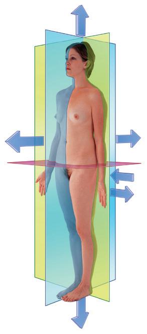
The anatomical position is the standard reference position of the body used to describe the location of structures (Fig. 1.1). The body is in the anatomical position when standing upright with feet together, hands by the side, and
face looking forward. The mouth is closed and the facial expression is neutral. The rim of bone under the eyes is in the same horizontal plane as the top of the opening to the ear, and the eyes are open and focused on something in the distance. The palms of the hands face forward with the fingers straight and together and with the pad of the thumb turned 90° to the pads of the fingers. The toes point forward.
Anatomical planes
Three major groups of planes pass through the body in the anatomical position (Fig. 1.1).
n Coronal planes are oriented vertically and divide the body into anterior and posterior parts.
n Sagittal planes also are oriented vertically, but are at right angles to the coronal planes and divide the body into right and left parts. The plane that passes through the center of the body dividing it into equal right and left halves is termed the median sagittal plane
n Transverse, horizontal, or axial planes divide the body into superior and inferior parts.
Terms to describe location
Anterior (ventral) and posterior (dorsal), medial and lateral, superior and inferior
Three major pairs of terms are used to describe the location of structures relative to the body as a whole or to other structures (Fig. 1.1).
Inferior margin of orbit level with top of external auditory meatus
Face looking forward
Feet together, toes forward Hands by sides, palms forward
Transverse, horizontal, or axial plane
1.1 The anatomical position, planes, and terms of location and orientation.
Fig.
n Anterior (or ventral) and posterior (or dorsal) describe the position of structures relative to the “front” and “back” of the body. For example, the nose is an anterior (ventral) structure, whereas the vertebral column is a posterior (dorsal) structure.
n Medial and lateral describe the position of structures relative to the median sagittal plane and the sides of the body. For example, the thumb is lateral to the little finger.
n Superior and inferior describe structures in reference to the vertical axis of the body. For example, the head is superior to the shoulders.
Proximal and distal, cranial and caudal, and rostral
Other terms used to describe positions include proximal and distal, cranial and caudal, and rostral.
n Proximal and distal are used with reference to being closer to or farther from a structure’s origin, particularly in the limbs. For example, the hand is distal to the elbow joint. These terms are also used to describe the relative positions of branches along the course of linear structures, such as airways, vessels, and nerves. For example, distal branches occur farther away toward the ends, whereas proximal branches occur closer to and toward the origin.
n Cranial (toward the head) and caudal (toward the tail) are sometimes used instead of superior and inferior, respectively.
n Rostral is used, particularly in the head, to describe the position of a structure with reference to the nose. For example, the forebrain is rostral to the hindbrain.
Superficial and deep
Two other terms used to describe the position of structures in the body are superficial and deep. These terms are used to describe the relative positions of two structures with respect to the surface of the body. For example, the sternum is superficial to the heart.
DIAGNOSTIC IMAGING TECHNIQUES
In 1895 Wilhelm Röntgen used the X-rays from a cathode ray tube to expose a photographic plate and produce the first radiographic exposure of his wife’s hand. Over the past 35 years there has been a revolution in medical imaging, which has been paralleled by developments in computer technology.
Plain radiography
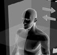
The basic physics of X-ray generation has not changed. X-rays are photons (a type of electromagnetic radiation) and are generated from a complex X-ray tube, which is a type of cathode ray tube (Fig. 1.2). The X-rays are then collimated (i.e., directed through lead-lined shutters to stop them from fanning out) to the appropriate area, as determined by the radiographic technician. As the X-rays pass through the body they are attenuated (reduced in energy)
by the tissues. Those X-rays that pass through the tissues interact with the photographic film.
In the body:
n Air attenuates X-rays a little.
n Fat attenuates X-rays more than air but less than water.
n Bone attenuates X-rays the most.
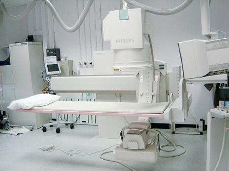
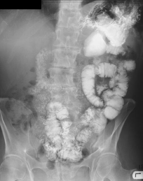
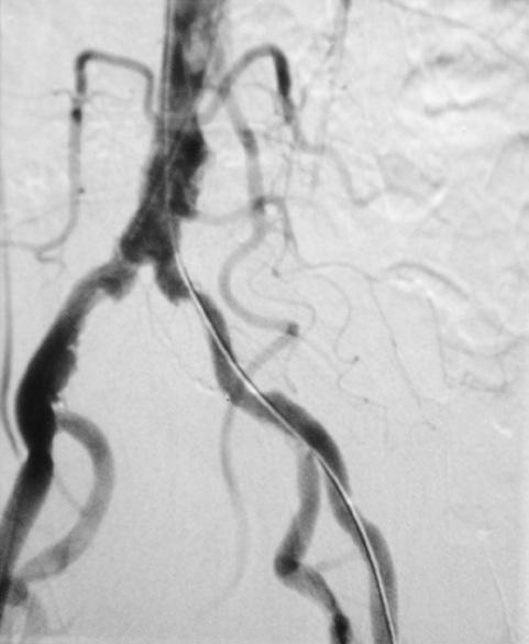
These differences in attenuation result in differences in the level of exposure of the film. When the photographic film is developed, bone appears white on the film because this region of the film has been exposed to the least amount of X-rays. Air appears dark on the film because these regions were exposed to the greatest number of X-rays. Modifications to this X-ray technique allow a continuous stream of X-rays to be produced from the X-ray tube and collected on an input screen to allow real-time visualization of moving anatomical structures, barium studies, angiography, and fluoroscopy (Fig. 1.3).
Contrast
agents
To demonstrate specific structures, such as bowel loops or arteries, it may be necessary to fill these structures with a substance that attenuates X-rays more than bowel loops or
Fig. 1.2 Cathode ray tube for the production of X-rays.