SECOND SOUTH ASIA EDITION (VOLUME-ONE & VOLUME-TWO)
Richard L. Drake, PhD, FAAA
Director of Anatomy, Professor of Surgery, Cleveland Clinic Lerner College of Medicine, Case Western Reserve University, Cleveland, Ohio
A. Wayne Vogl, PhD, FAAA
Professor of Anatomy and Cell Biology, Department of Cellular and Physiological Sciences, Faculty of Medicine, University of British Columbia, Vancouver, British Columbia, Canada
Adam W.M. Mitchell, MB BS, FRCS, FRCR
Consultant Radiologist, Director of Radiology, Fortius Clinic, London, United Kingdom
Illustrations by
Richard Tibbi�s and Paul Richardson
Photographs by Ansell Horn
Adaptation Editors
Raveendranath Veeramani, MBBS, MD
Associate Professor of Anatomy, Jawaharlal Institute of Postgraduate Medical Education and Research (JIPMER), Puducherry, India
Sunil Jonathan Holla, MBBS, MS
Professor of Anatomy, Christian Medical College, Vellore, India
Table of Contents
Cover image
Title page
Copyright
Dedication
Acknowledgments to the Second South Asia Edition
Acknowledgments to the Fourth International Edition
Previous editions copyrighted 2014, 2010, 2005 by Churchill Livingstone, an imprint of Elsevier Inc.
All rights reserved.
ISBN: 978-0-323-39304-1
This adaptation of chapters 1-9 from GRAY’S ANATOMY FOR STUDENTS, 4e by Richard L. Drake, A. Wayne Vogl, and Adam W. M. Mitchell was undertaken by RELX India Private Limited and is published by arrangement with Elsevier Inc.
GRAY’S ANATOMY FOR STUDENTS, Second South Asia Edition (Volume-01 & Volume-02)
Adaptation Editors: Raveendranath Veeramani and Sunil Jonathan Holla
All rights reserved. No part of this publication may be reproduced or transmi�ed in any form or by any means, electronic or mechanical, including photocopying, recording, or any information storage and retrieval system, without permission in writing from the publisher. Details on how to seek permission, further information about the Publisher’s permissions policies and our arrangements with organizations such as the Copyright Clearance Center and the Copyright Licensing Agency, can be found at our website: www.elsevier.com/permissions.
This book and the individual contributions contained in it are protected under copyright by the Publisher.
Notice
The adaptation has been undertaken by RELX India Private Limited at its sole responsibility. Practitioners and researchers must always rely on their own experience and knowledge in evaluating and using any information, methods, compounds or experiments described herein. Because of rapid advances in the medical sciences, in particular, independent verification of diagnoses and drug dosages should be made. To the fullest extent of the law, no responsibility is assumed by Elsevier, authors, editors or contributors in relation to the adaptation or for any injury and/or damage to persons or property as a ma�er of products liability, negligence or otherwise, or from any use or operation of any methods, products, instructions, or ideas contained in the material herein.
This publication is licensed for sale in India, Bangladesh, Bhutan, Maldives, Nepal, Pakistan and Sri Lanka only. Circulation of this version outside these territories is unauthorized and illegal.
Content Strategist: Arvind Koul
Content Project Manager: Ayan Dhar
Cover Designer: Raman Kumar
Printed in India by.........
Dedication
To my wife, Cheryl, who has supported me; and my parents, who have guided me.
RLD
To my family, to my professional colleagues and role models, and to my students this book is for you.
AWV
To Max, Elsa, and Cathy. And the man that made us all, Timothy Ianthor Mitchell!
AWMM
The almighty for the opportunity given and the capability for the completion of the work.
I would like to thank my wife, Dr Umamageswari and my son Charvesh who encouraged and
supported me in my work.
Raveendranath Veeramani
Jesus Christ, my Lord, who enabled me.
Muriel, my wife, for her inspiration.
Jonathan and David my sons, for their understanding.
Sunil Jonathan Holla
Acknowledgments to the Second South Asia Edition
We acknowledge Dr Nachiket Shankar, St John’s Medical College, for his valuable inputs in planning the format of the outlines in the book.
Many thanks to Dr Umamageswari, radiologist at Sri Manakula Vinayagar Medical College, Puducherry, for her opinion and constant support on the radiology content of the book.
We are indebted to Dr Aby S. Charles and Mr Rajkumar of Christian Medical College, Vellore, for their contribution in making the line diagrams (LDs) which will help students understand anatomical concepts and be able to illustrate them. Many thanks to Dr Suganthy Rabi, the head of the Department of Anatomy at CMC Vellore, for giving permission to do this project.
We are thankful to Mr Arvind Kaul, Ms Shabina Nasim, Ms Shivani Pal, and Mr Ayan Dhar, the Elsevier team members, for their assistance and guidance throughout the project.
Raveendranath Veeramani
Sunil Jonathan Holla
Acknowledgments to the Fourth International Edition
First, we collectively thank those who have reviewed drafts of various editions of this book anatomists, educators, and student members of the editorial review board from around the world. Your input was invaluable.
We also thank Richard Tibbi�s and Paul Richardson for their skills in turning our visual ideas into a reality that is not only a foundation for the acquisition of anatomical knowledge but also beautiful. Thanks must also go to Madelene Hyde, Jeremy Bowes, Bill Schmi�, Rebecca Gruliow, John Casey, and all the team at Elsevier for guiding us through the preparation of this book.
We also thank Professor Richard A. Buckingham of the Abraham Lincoln School of Medicine, University of Illinois, for the provision of Fig. 8.112. Finally, because we worked separately, distanced by, in some cases, thousands of miles, there are various people who gave local support, whom we would like to make mention of individually. We have gratefully listed them here:
Dr Leonard Epp, Dr Carl Morgan, Dr Robert Shellhamer, and Dr Robert Cardell, who profoundly influenced my career as a scientist and an educator.
Richard L. Drake
Dr Sydney Friedman, Dr Elio Raviola, and Dr Charles Slonecker, for their inspiration and support and for instilling in me a passion for the discipline of anatomy.
Dr Murray Morrison, Dr Joanne Matsubara, Dr Brian Westerberg, Laura Hall, and Jing Cui, for contributing images for the chapter on
the head and neck.
Dr Bruce Crawford and Logan Lee, for help with images for the surface anatomy of the upper limb.
Professor Elizabeth Akesson and Dr Donna Ford, for their enthusiastic support and valuable critiques.
Dr Sam Wiseman, for contributing surgical and other images in the chapters on abdomen and head and neck.
Dr Rosemary Basson, for writing the “Erectile Dysfunction In the Clinic” in the chapter on pelvis and perineum.
A. Wayne Vogl
Anatomy changes! We see it through new “glasses” X-rays, CT, MRI, and now AI... what’s next (I do not know, but it’s exciting). We need new eyes and new blood, and I am delighted to have been helped by two inspirational colleagues, Dr Monika Rowe and Dr Rajat Choudhury with the clinical. They are stars of the future.
Dr Justin Lee and Dr Gajan Rajeswaran are the greatest colleagues who challenge me daily with MSK anatomy.
Many thanks for constant support from Mr Andrew Williams, Prof James Calder, and Lucy Ball. They are all awesome!
Adam W.M. Mitchell
Preface to the Second South Asia Edition
The Second South Asia Edition builds on the past and looks to the future. It maintains the goals and objectives of First South Asia Edition. In this new edition, the content has been revised considering the feedback received. New content has been added on the basis of updates in the Fourth International Edition including the addition of a new chapter on neuroanatomy.
The language has been simplified. The use of outlines, as in First South Asia Edition, has been continued. The innovative features of the First South Asia Edition such as Set Inductions, Outlines, and Flowcharts have been improved. Students are encouraged to use online resources available on MedEnact.
The Second South Asia Edition contains a new chapter on neuroanatomy. The addition of this chapter is based on the eFiles of the Fourth International Edition. This chapter is organized in keeping with the pa�ern of the existing chapters in the First South Asia Edition, with set inductions, specific learning objectives, multimodal approach covering recall, demonstrate structures, analyze concepts, apply and solve problems. Neuroanatomical connections are represented in the form of flowcharts and maps.
A unique feature of this edition is that each chapter contains “line diagrams,” abbreviated as LDs. These line diagrams are sketches which are easy to draw during an examination and can help students to acquire anatomical concepts and do well in assessment.
Competencies have been added in all the chapters since the curriculum is becoming competency based.
Based on feedback from students, this book has been split as follows: Volume One The Body, Upper Limb, Lower Limb, Abdomen, Pelvis and Perineum; and Volume Two Thorax, Back, Head and Neck, and Neuroanatomy. This division is based on the examination pa�ern followed in several medical colleges and will enable easy handling and use of the book.
In the outlines in each chapter, the structures to be demonstrated in a prosected specimen are given as Level I, II, and III which represent the Must Know, Desirable to Know, and Nice to Know content, respectively. This helps to emphasize the core content.
The editors are open to and welcome feedback that will help improve this book.
Raveendranath Veeramani
Sunil Jonathan Holla
Preface to the Fourth International Edition
The 4th edition of Gray’s Anatomy for Students maintains the goals and objectives of the 1st, 2nd, and 3rd editions while at the same time continuing to incorporate input from our readers and adjust the content to align with the evolving educational environment.
One of the major focuses of our a�ention as we prepared the 4th edition was adjusting print content to accommodate the evolving capacity to move material onto e-learning platforms. We have expanded the number of “In the Clinic” boxes in the printed book but have moved some of the “Clinical Cases” in earlier editions of the book from the print version to the online platform. Moving some of the clinical cases to the e-learning platform has allowed us to add new material to the print version without expanding the size of the book or compromising the basic goals and objectives of the work. New material added has included new imaging to reflect recent advances in the field of radiology and, in response to reader feedback, we have added simple summary line diagrams (“Quick draw sketches”) to some of the key figures that can be easily replicated by students.
A new feature in the 4th edition is an accompanying online e-book organized systemically and referred to as Gray’s Systemic Anatomy. This e-book has chapters on the cardiovascular, respiratory, gastrointestinal, nervous, urogenital, and lymphatic systems. We feel students may find this material useful as many medical school curriculums now use a systems-based, integrated approach.
A second new feature to the 4th edition is the inclusion of a neuroanatomy chapter. We hope that this eChapter will assist
students as they advance their knowledge of neuroanatomy.
As was the case in previous editions, review materials/study aids are available on Student Consult as an online resource with the appropriate review materials for each chapter listed at the beginning of that chapter. This information includes an online anatomy and embryology self-study course, medical clinical cases, physical therapy clinical cases, self-assessment questions, an interactive surface anatomy module, and short answer questions. We believe that with these changes the 4th edition of Gray’s Anatomy for Students is an improved version of the 3rd edition and hope that the book will continue to be a valuable learning resource for students.
October 2018
Richard L. Drake
A. Wayne Vogl
Adam W.M. Mitchell
About the book
The idea
In the past 20 years or so, there have been many changes that have shaped how students learn human anatomy in medical and dental schools and in allied health programs, with curricula becoming either more integrated or more system based. In addition, instructional methods focus on the use of small group activities with the goals of increasing the amount of self-directed learning and acquiring the skills for the life-long acquisition of knowledge. An explosion of information in every discipline has also been a force in driving curricular change as it increases the amount to be learned without necessarily increasing the time available. With these changes, we felt it was time for a new book to be wri�en that would allow students to learn anatomy within the context of many different curricular designs and within ever-increasing time constraints. We began in the fall of 2001 by considering the various approaches and formats that we might adopt, eventually deciding upon a regional approach to anatomy with each chapter having four sections. From the beginning, we wanted the book to be designed with multiple entry points, to be targeted at introductory level students in a broad spectrum of fields, and to be a student-oriented companion text for Gray’s Anatomy, which is aimed at a more professional audience. We wrote the text first and subsequently constructed all the artwork and illustrations to complement and augment the words. Preliminary drafts of chapters, when complete, were distributed to an international editorial board of anatomists, educators, and anatomy students for review. Their comments were then considered carefully in the preparation of the final book.
The text is not meant to be exhaustive in coverage, but to present enough anatomy to provide students with a structural and functional context in which to add further detail as they progress through their careers. Gray’s Anatomy was used as the major reference, both for the text and for the illustrations during the
preparation of this book, and it is the recommended source for acquiring additional detail.
The book
Gray’s Anatomy for Students is a clinically oriented, student-friendly textbook of human anatomy. It has been prepared primarily for students in a variety of professional programs (e.g., medical, dental, chiropractic, and physical therapy programs). It can be used by students in traditional, systemic, combined traditional/systemic, and problem-based curricula, and will be particularly useful to students when lectures and laboratories in gross anatomy are minimal.
Organization
Using a regional approach, Gray’s Anatomy for Students progresses through the body in a logical fashion, building on the body’s complexities as the reader becomes more comfortable with the subject ma�er. Each chapter can be used as an independent learning module, and varying the sequence will not affect the quality of the educational experience.
The Second South Asia Edition of this book has been split into two volumes: Volume One: The Body, Upper Limb, Lower Limb, Abdomen, Pelvis and Perineum; and Volume Two: Thorax, Back, Head and Neck, and Neuroanatomy. The just-mentioned division is based on the examination pa�ern followed in several medical colleges and will enable easy handling and best use of the book. We begin with “The Body,” which contains an overview of the discipline of gross anatomy and an introduction to imaging modalities and general body systems. We follow this with the “Upper Limb” followed by the “Lower Limb,” as they are easy to understand at the beginning of the course. Next begins a progression through the cavities of the body. “Abdomen,” “Pelvis and Perineum” followed logically to the “Thorax” and “Back.” The last regions discussed are the “Head and Neck” and “Neuroanatomy.” These regions contain some of the most difficult anatomy in the body. Covering all the
other regions first gives the student the opportunity to build a strong foundation on which to understand these complex regions.
Content
Each regional anatomy chapter consists of four consecutive sections: conceptual overview, regional anatomy, surface anatomy, and clinical cases.
The conceptual overview provides the basis on which information in the later sections is built. This section can be read independently of the rest of the text by students who require only a basic level of understanding and can also be read as a summary of important concepts after the regional anatomy has been mastered.
The regional anatomy section provides more detailed anatomy along with a substantial amount of relevant clinical correlations. It is not an exhaustive discussion but instead provides information to a level that we feel is necessary for understanding the organization of the region. Throughout this section, two levels of clinical material are provided. Clinical hooks are fully integrated with the main anatomical text and function to relate (“hook”) the anatomy discussed directly to a clinical application without taking students out of their train of thought and without disrupting the flow of the text. Although fully integrated with the anatomical text, these passages are differentiated from it by the use of green highlighting. Early clinical exposure sections are concise descriptions that provide students with useful and relevant clinical information demonstrating how the application of anatomical knowledge facilitates solving of clinical problems. These are spread throughout the text close to the most relevant anatomical discussion.
Surface anatomy assists students in visualizing the relationship between anatomical structures and surface landmarks. This section also provides students with practical applications of the anatomical information, combining visual inspection with functional assessment, as occurs during any type of patient examination.
The final section of each chapter consists of clinical cases. These cases represent the third level of clinical material in the book. In these cases, a clinical problem is described, and a step-by-step process of questions and answers leads the reader to the resolution of the case. The inclusion of these cases in each chapter provides students with the opportunity to apply an understanding of anatomy to the resolution of a clinical problem.
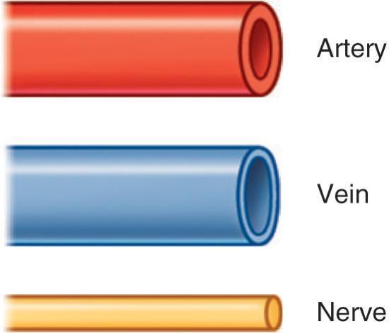
Illustrations are an integral part of any anatomy text. They must present the reader with a visual image that brings the text to life and presents views that will assist in the understanding and comprehension of the anatomy. The artwork in this textbook accomplishes all of these goals. The illustrations are original and vibrant, and many views are unique. They have been designed to integrate with the text, to present the anatomy in new ways, to deal with the issues that students find particularly difficult, and to provide a conceptual framework for building further understanding. To ensure that the illustrations of the book work together and to enable students to cross-refer from one illustration to another, we have used standard colors throughout the book, except where indicated otherwise. This edition has an additional feature of line diagrams (LDs) along with questions and answers. These will help in acquiring important anatomical concepts and will be useful for students in assessment for learning and assessment of learning.