No part of this publication may be reproduced or transmitted in any form or by any means, electronic or mechanical, including photocopying, recording, or any information storage and retrieval system, without permission in writing from the publisher. Details on how to seek permission, further information about the Publisher’s permissions policies and our arrangements with organizations such as the Copyright Clearance Center and the Copyright Licensing Agency, can be found at our website: www.elsevier.com/permissions
This book and the individual contributions contained in it are protected under copyright by the Publisher (other than as may be noted herein).
Notices
Knowledge and best practice in this field are constantly changing. As new research and experience broaden our understanding, changes in research methods, professional practices, or medical treatment may become necessary.
Practitioners and researchers must always rely on their own experience and knowledge in evaluating and using any information, methods, compounds, or experiments described herein. In using such information or methods they should be mindful of their own safety and the safety of others, including parties for whom they have a professional responsibility.
With respect to any drug or pharmaceutical products identified, readers are advised to check the most current information provided (i) on procedures featured or (ii) by the manufacturer of each product to be administered, to verify the recommended dose or formula, the method and duration of administration, and contraindications. It is the responsibility of practitioners, relying on their own experience and knowledge of their patients, to make diagnoses, to determine dosages and the best treatment for each individual patient, and to take all appropriate safety precautions.
To the fullest extent of the law, neither the Publisher nor the authors, contributors, or editors, assume any liability for any injury and/or damage to persons or property as a matter of products liability, negligence or otherwise, or from any use or operation of any methods, products, instructions, or ideas contained in the material herein.
Library of Congress Cataloging-in-Publication Data
Names: Kloss, Brian T., author, editor. | Bruce, Travis, illustrator.
Title: Graphic guide to infectious disease / Brian Kloss, Travis Bruce.
Description: Philadelphia, PA : Elsevier, [2019] | Includes bibliographical references and index.
Identifiers: LCCN 2018006370 | ISBN 9780323442145 (hardcover : alk. paper)
Subjects: | MESH: Communicable Diseases | Infection | Pictorial Works
Classification: LCC RC111 | NLM WC 17 | DDC 616.9--dc23 LC record available at https://lccn.loc.gov/2018006370
Executive Content Strategist: James Merritt
Senior Content Development Manager: Kathryn DeFrancesco
Content Development Specialist: Angie Breckon
Book Production Manager: Jeff Patterson
Project Manager: Lisa A. P. Bushey
Book Designer: Patrick Ferguson
LIST OF REVIEWERS/ CONTRIBUTOR
CONTRIBUTING AUTHOR:
Hernan Rincon-Choles, MD, MSCI
Assistant Professor
Department of Nephrology and Hypertension
Cleveland Clinic Lerner College of Medicine of the Case Western Reserve University
Medical Director, Ohio Renal Care Group Huron Dialysis Center
Attending Physician, Glickman Urological and Kidney Institute
Topics written by Hernan Rincon:
Echinococcosis
Melioidosis
Granuloma Inguinale
Tularemia
Plague
Legionellosis
Scabies
Rabies
Ebola
Rift Valley Fever
Hanta Virus Pulmonary Syndrome
Hemorrhagic Fever with Renal Syndrome
Crimea Congo Hemorrhagic Fever
Colorado Tick Fever
Rocky Mountain Spotted Fever
Lyme Disease
Babesiosis
Ehrlichiosis
Anaplasmosis
GUEST REVIEWERS FOR DIAGNOSTIC TESTING:
Kanish Mirchia, MD Department of Pathology SUNY Upstate Medical University
Rochelle Nagales-Nagamos, MD, MBA Department of Pathology SUNY Upstate Medical University
Alexandria Smith-Hannah, MD, MS, MPH Department of Pathology SUNY Upstate Medical University
ACKNOWLEDGEMENTS
The authors would like to thank our friends, family, and all the staff at Elsevier for their support.
Thanks to Alex Seldes and Sabre Mrkva for being great friends and permitting us to draw Cleo with all those pediatric illnesses. Thanks to Karen Cyndari, MD/PhD candidate, for website development and support. Thanks to Kara Welch and David Rothman for assistance with the references and suggested reading section.
Very special thanks to Zubin Damania, MD, a.k.a. ZDoggMD, for making a cameo in our Zika Fever illustration; Mike Cadogan, MD, from the Life in the Fastlane Emergency Medicine website and blog for making a cameo in our Melioidosis illustration; and Jawad Kassem, MD, for making a cameo in our Middle Eastern Respiratory Syndrome illustration. Lastly, many thanks to Rob Guillory, Eisner Award-winning comic book artist for Chew, for his support and serving as a guest illustrator for our Avian Flu illustration. The guest appearances, celebrity cameos, and pop culture references contained in this textbook are intended to be works of satire and parody.
INTRODUCTION
Kloss and Bruce combine real medical education with comic book–style illustrations to create beautiful artistic images that enhance learning. Realizing that many medical professionals are visual learners, Kloss and Bruce enhance learning by breaking down complex medical conditions and diseases into illustrated scenes. As children of the eighties and with a passion for comic books, pop culture, nostalgia, and humor, their illustrations exceed boundaries set by other medical illustrations. Irreverent, provocative, and unconventional are terms that have been used to describe their campy, tongue-in-cheek approach to medical education. Their visual aids are colorful, comical, and boundary pushing, all of which make learning more fun and memorable. Dr. Brian Kloss, a professor and emergency medicine physician, and Travis Bruce, a talented illustrator and designer, aim to educate physicians, physician assistants, nurses, medical students, and other healthcare providers using humor and comic book–style illustrations.
Their process is simple. First, Dr. Kloss pencils a rough draft of a medical syndrome, disease, or illness and hands it over to Travis. Travis then draws out the illustration and adds clarity and color. The end result is a helpful educational tool that is both comical and informative.
The dynamic duo has been collaborating since connecting at a house party in Brooklyn during the turn of the century. Their first educational product, Toxicology in a Box, was published by McGraw-Hill in 2013 and is available in Kindle version on Amazon.com. Toxicology in a Box is a set of 150 full-color flashcards geared toward teaching medical providers how to recognize and treat various toxic exposures ranging from the bizarre to the mundane.
Kloss and Bruce, in collaboration with Elsevier, are pleased to present their latest work: Graphic Guide to Infectious Disease. This body of text and illustrations represents 4 years of late nights, highly caffeinated beverages, deadline extensions, revisions, and more deadline extensions. While they’re admittedly no Rick and Morty, they needed a few deadline extensions.
Their only request is that you loosen your collar, sit back, relax, and enjoy learning high-yield medicine via a truly unique medium. Welcome to Kloss and Bruce: Medical Education Through Comic Illustration.
Brian Kloss, DO, JD, PA-C Associate Professor
Department of Emergency Medicine SUNY Upstate Medical University
Travis Bruce, BFA
To learn more about us, please visit www.KlossandBruce.com
ABOUT THE AUTHORS
Brian Kloss, DO, JD, PA-C, is an Emergency Medicine Physician and Associate Professor at the SUNY Upstate Medical University and VA Medical Center in Syracuse, New York. He holds a Certificate in Radiologic Technology from Morristown Memorial Hospital in New Jersey, an Associate of Science in Chemistry from the County College of Morris, a Bachelor of Science in Physician Assistant Studies from Gannon University, a Juris Doctor for the University at Buffalo School of Law, and a Doctor of Osteopathic Medicine from UMDNJ-SOM (Rowan). He completed a postgraduate Physician Assistant Fellowship in Gastroenterology, is Board Certified in Emergency Medicine, and completed a Wilderness Medicine Fellowship at SUNY Upstate. Brian likes vintage video games, comic books, action figures, and old-school hip hop.
Travis Bruce is an artist, illustrator, and designer living in Queens, New York. He graduated with a BFA in illustration from the School of Visual Arts, with a focus on graphic narratives and children’s books. Along with illustration, he has designed tabletop products and giftware for the past 15 years.
PART 10 | RAT-, FLEA-, LOUSE-, AND CHIGGER-BORNE ILLNESSES
Chapter 10.1 Hemorrhagic Fever with Renal Syndrome 236
Chapter 10.2 Hantavirus Pulmonary Syndrome 238
Chapter 10.3 Plague 240
Chapter 10.4 Leptospirosis 242
Chapter 10.5 Rat Bite Fever 244
Chapter 10.6 Trench Fever 246
Chapter 10.7 Scrub Typhus 248
Chapter 10.8 Epidemic Typhus
250
Chapter 10.9 Endemic Typhus 252
Chapter 10.10 Arenaviridae 254
PART 11 | OROPHARYNGEAL INFECTIONS
Chapter
Chapter
PART 12 | VIRAL
Chapter
Chapter
Chapter
Chapter
Chapter
PART 13 | PARASITES AND PRIONS
PART 14 | BACTERIAL
BIBLIOGRAPHY AND SUGGESTED READING
PART 1 VIRAL HEPATITIS
Disease Name:
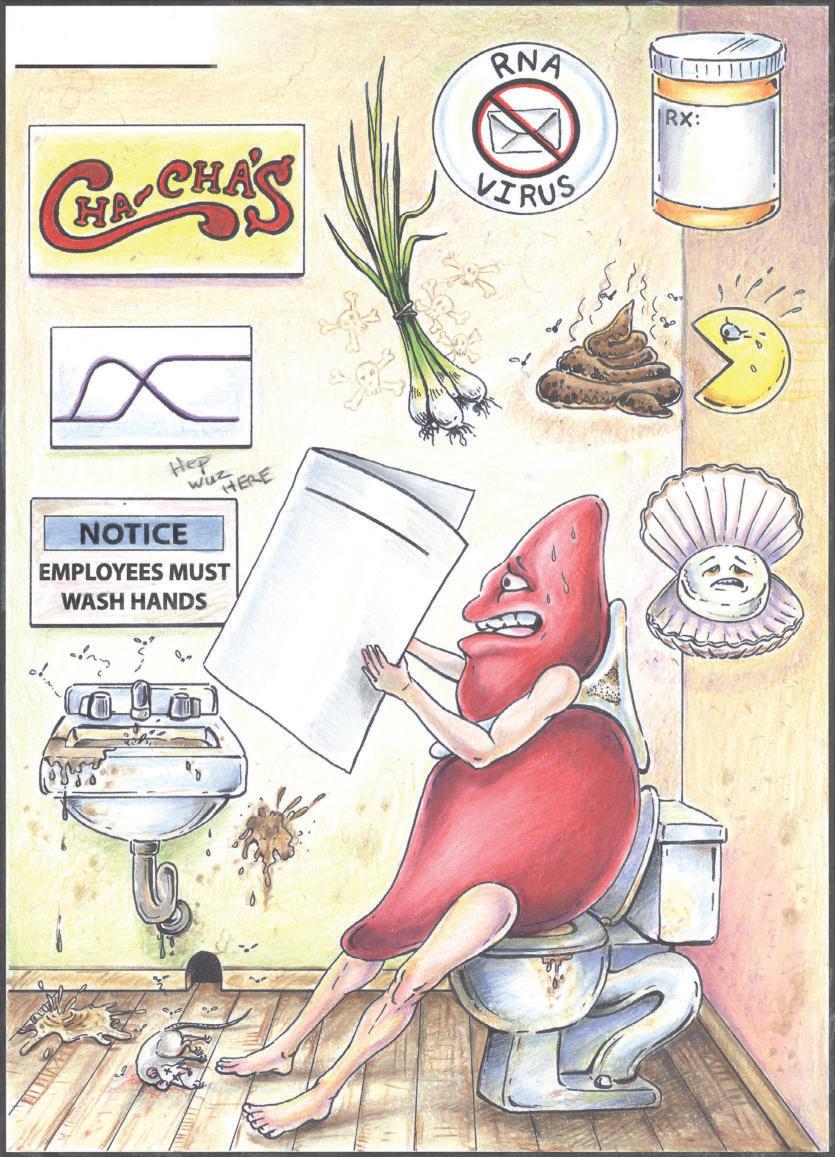
Hepatitis A
Synonyms: Infectious Hepatitis
Causative Agent: Hepatitis A Virus (HAV)
Reservoir: Humans
Incubation Period: 15-50 days; Average: 28 days
Geographic Regions Affected: Worldwide
Description: HAV is a vaccine-preventable fecal-orally transmitted RNA virus that causes acute hepatitis. Hepatitis A is never chronic, is often asymptomatic in younger patients, and causes fulminant hepatic failure in ,1% of cases. Risk factors include travel to endemic regions, ingestion of contaminated food or water (raw shellfish), work in day care centers (exposure to feces/diaper changing), close contact with infected patients, and men who have sex with men.
Signs and Symptoms: Typically, the younger the patient, the fewer symptoms he or she exhibits. Most infants will show little to no signs of infection, whereas most adults become symptomatic. Symptomatic patients experience nausea, vomiting, malaise, abdominal pain, and fever followed by scleral icterus and jaundice several days later. Symptoms often last for less than 2 months; however, the disease may be prolonged or can relapse over a 6-month period. Infection confers lifelong immunity.
Diagnostic Testing: Labs will reveal a hepatocellular pattern with ALT/AST elevations ,1000, rising before an increase in bilirubin and alkaline phosphatase is seen. ALT, being more specific to the liver, is often higher than AST. IgM rises in acute infection, and IgG begins to rise in convalescence. Fulminant hepatic failure, a serious consequence of infection characterized by altered mental status (hepatic encephalopathy) and elevations of PT/INR, is more common in older patients and those with preexisting liver disease (chronic hepatitis B and/or C).
Treatments: Supportive. The disease is preventable by two doses of a vaccine given at least 6 months apart. Depending on the manufacturer, the second dose of the vaccine can be given up to 12 or 18 months after the first dose. Postexposure vaccine can be given in healthy persons aged 1–40 years within 14 days to prevent infection. Postexposure immune globulin is recommended within 14 days for unvaccinated patients with immunodeficiency, chronic liver disease, adults .41 years, and children ,12 months old.
Pearls: There have been several food-related outbreaks of hepatitis A in the United States. Five hundred people became ill in 2003 after consuming salsa made with green onions at a now-bankrupt Mexican food chain restaurant.
Hepatitis A IgMIgG
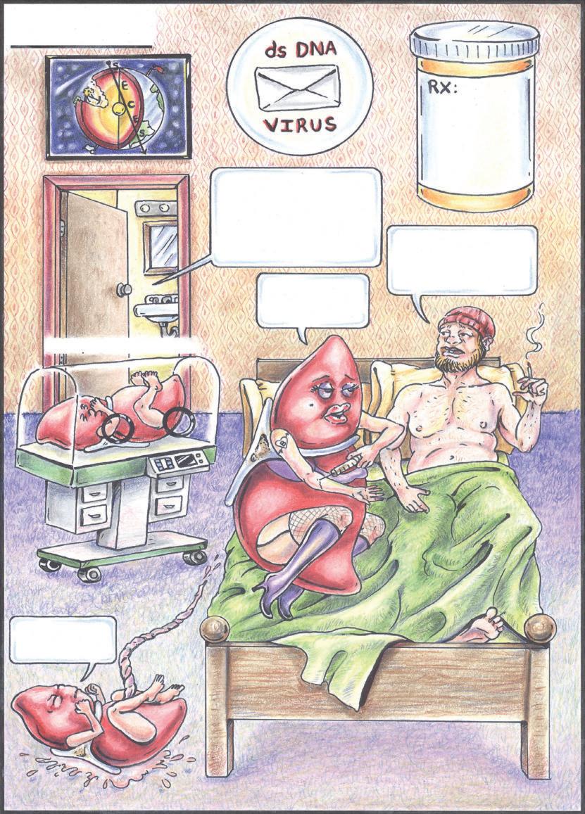
Disease Name: Hepatitis B
Synonyms: Serum Hepatitis
Causative Agent: Hepatitis B Virus (HBV)
Incubation: 60–150 days; Average: 90 days
Geographic Regions Affected: Worldwide, Higher Incidence in Asia
Reservoir: Humans
Description: HBV is a double-stranded DNA virus that causes acute and chronic hepatitis. The disease can be transmitted vertically at birth or through contact with infected bodily fluids, such as blood, semen, and vaginal secretions. Hepatitis B can be transmitted sexually and through IV drug use. Healthcare workers are at risk of infection from needlestick injuries.
Signs and Symptoms: Acute: Most infants and young children with acute infections are asymptomatic. Older patients are more likely to show symptoms, which include fever, malaise, anorexia, nausea, vomiting, abdominal pain, and jaundice. Approximately 70% of acutely infected adults will have symptoms. Symptoms can last for several weeks. The incidence of fulminant hepatic failure is ,1%. Chronic: Chronic hepatitis B is often asymptomatic but patients can have symptomatic flairs. If untreated, the disease can be spread to others, cause cirrhosis, and predispose patients to hepatocellular carcinoma. The likelihood of developing chronic hepatitis B is inversely proportional to age at time of infection. The risk of vertical transmission is very high and is dependent on the mother’s HBeAg/HBeAb status. Those women who are HBeAg1 and HBeAb are more likely to pass on the infection. The Centers for Disease Control and Prevention report that infected newborns develop chronic hepatitis B approximately 90% of the time, whereas children infected between the ages of 1 and 5 years have a 25%–50% chance of developing chronic disease. Older children and adults are more likely to clear the disease and have a 5%–10% chance of chronicity.
Diagnostic Testing: Acute and chronic hepatitis B are diagnosed by specific serum markers and HBV viral load testing using PCR. Chronic hepatitis B is defined as the presence of HBsAg detectable in serum for 6 months or longer after symptom onset. Please see our summary of hepatitis B markers for more information.
Treatments: Treatment of acute disease is supportive, except in cases of fulminant hepatic failure, wherein nucleoside/nucleotide analog medications are indicated. In chronic hepatitis B infections, treatment options and the decision to treat are highly dependent on individual patient factors, such as viral load, presence/absence of cirrhosis or hepatocellular cancer, HBeAg/HBeAb status, pregnancy status, patient age, and biochemical markers. Treatment options for chronic hepatitis B include PEGylated interferon or nucleoside/nucleotide analogs. Nucleoside/nucleotide analogs include lamivudine, adefovir, entecavir, telbivudine, and tenofovir. The authors recommend referencing current American Association for the Study of Liver Diseases (AASLD) and/or European Association for the Study of the Liver (EASL) guidelines prior to initiating treatment. Hepatitis B is vaccine preventable, and infection after acute exposure can be prevented with vaccination and immune globulin.
Hepatitis B
High Rate of Vertical Transmission
I'm going to get the postexposure immune globulin and start the vaccine series <12 hours after needle stick or <2 weeks after sexual exposure.
By the way, I have Hepatitis B!
I was vaccinated Day 0, 1 M & 6 M and I am HBsAb+
Give me vaccine & immune globulin ≤12 hours.
Disease Name: Hepatitis B Serum Markers
Hepatitis B Surface Antigen (HBsAg):
Hepatitis B e Antigen (HBeAg):
HBsAg is the first serum marker to appear and is detectable prior to the onset of clinical symptoms. It peaks approximately 12 weeks after exposure and should become undetectable within 24 weeks (6 months). The elimination of HBsAg and seroconversion to hepatitis B surface antibody (HBsAb) indicates that the infection has resolved. If HBsAg persists in the serum for .6 months, the patient has a chronic infection.
HBeAg is associated with viral replication and is detectable within 6–14 weeks after exposure. In chronic hepatitis B, HBeAg serves as a marker of both disease activity and infectivity. Patients who are HBeAg+ (HBeAb ) have higher viral loads and greater disease activity and are more infectious. In pregnancy, HBsAg+ women are more likely to pass the infection on to their infants. Any infant born to a mother with chronic HBV should be vaccinated and given immune globulin, ideally within 12 hours.
Hepatitis B Virus Core Antibody (HBcAb):
Hepatitis B e Antibody (HBeAb):
HBcAb is the first antibody to be detected via serum. Although there is a core antigen, it is intracellular and cannot be detected in serum. HBcAb exists as both IgM (signifying acute infection or significant reactivation of chronic disease) and IgG (indicating a past exposure to the hepatitis B disease). Because HBcAb can only exist in those exposed to the disease, it is used when screening donated blood and/or distinguishing whether a person is immune from disease exposure or vaccination. Because the HBV vaccine consists only of HBsAg, those patients who have been successfully vaccinated will test HBsAb1 and HBcAb . This indicates that they have antibodies to the hepatitis B surface antigen but have never been exposed to the full virus. Patients who are HBsAb1 and HBcAb1 have been exposed to the full hepatitis B virus, had the disease to some extent, and are now immune.
Antibodies to the e antigen are associated with decreased viral loads and herald convalescence. Seroconversion to HBeAb occurs early in acute disease but can be delayed for years in chronic infection. Those patients with chronic hepatitis B that are HBeAb seroconvert to HBeAb1 at a rate of approximately 0.5% per year.
HBsAb: HBsAb confirms immunity, either via vaccination or resolved infection. HBsAb begins to elevate after HBsAg levels taper off. Again, presence of HBsAg and absence of HBsAb at 6 months indicates chronic infection. In some cases, there is a window when neither HBsAg nor HBsAb can be detected. In this case an HBcAb IgM level can be obtained to evaluate for acute infection.
Hepatitis B Serum DNA: PCR can be used in either a qualitative (yes or no) or quantitative (How much?) fashion when determining the presence or absence of HBV DNA in serum samples. In patients who contract HBV infection and seroconvert to an HBsAb1 state, HBV DNA should be 100% cleared from their serum. Those patients with chronic hepatitis B will have detectable HBV DNA in their serum, the amount highly dependent on their HBeAg/HBeAb status. HBV DNA quantitative studies, HBeAg/HBeAb status, and liver biopsy results (to grade liver damage) are important in determining treatment eligibility and options.
Hepatitis B Markers
Chronology of Serum Markers
S
HepBsAg
E (Surface) (Surface) (Earth) (Core) (Earth)
HepBeAg
C E
HepBcAb
HepBeAb
HepBsAb
S
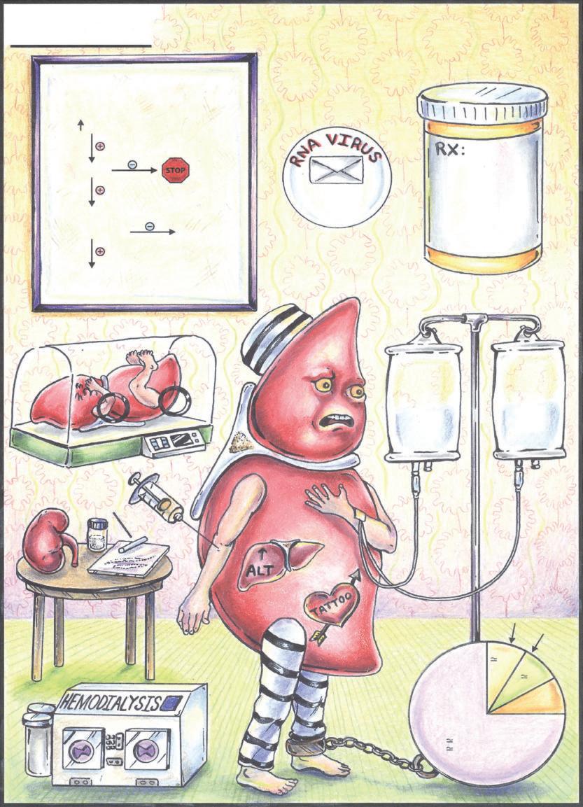
Disease Name: Hepatitis C
Causative Agent: Hepatitis C Virus, HCV
Incubation: 14–180 days; Average: 45 days
Geographic Regions Affected: Worldwide
Reservoir: Humans
Description: HCV is a single-stranded RNA virus responsible for both acute and chronic viral hepatitis, with up to 85% of infections becoming chronic. Risk factors for HCV transmission include IV drug use, blood transfusions and organ transplantation before July 2012, receiving clotting factors before 1987, intranasal drug use (cocaine), long-term hemodialysis, unsterile tattoos (e.g., prison), and vertical transmission. Sexual and household transmission can occur but is low.
Signs and Symptoms: Acute: Approximately 85% of acute infections are asymptomatic, and about 85% of hepatitis C infections become chronic. Acute symptoms can include malaise, fatigue, nausea, vomiting, abdominal pain, and jaundice. Chronic: Often asymptomatic but can cause malaise and fatigue. People with chronic HCV infections can develop cirrhosis and are predisposed to developing primary hepatocellular carcinoma. Extrahepatic manifestations of chronic HCV can include diabetes, cryoglobulinemia, and glomuleronephritis.
Diagnostic Testing: People with chronic hepatitis C may have normal liver enzymes. Hepatitis C antibody testing screens for the disease, and positive results should be followed by PCR testing for viral load and genotype. Positive HCV antibody tests with negative viral load may indicate a false-positive antibody test or identify a patient who previously had hepatitis C and cleared the virus either spontaneously or via treatment.
Treatments: Since the treatment for hepatitis C is continuously evolving, the authors recommend consulting the American Association for the Study of Liver Disease (AASLD) and/or the Infectious Disease Society of America (IDSA) for current guidelines. Initially, PEGylated interferon combined with oral ribavirin provided about 50% sustained virologic response (SVR) or “cure” rate. Direct-acting antivirals (DAAs) are now the preferred treatment and include oral agents: elbasvir/grazoprevir, ledipasvir/sofosbuvir, simeprevir/sofosbuvir, and sofosbuvir/velpatasvir.
Treatment is often based on genotype, viral load, presence/absence of cirrhosis, previous treatment failures, and insurance formularies. New drugs are constantly entering the development pipeline, and many patients may be offered enrollment in clinical trials. All patients with chronic hepatitis C should be vaccinated against hepatitis A virus and hepatitis B virus to prevent additional liver disease from these infections.
Hepatitis C
Simplified Testing Algorithm
HCV Risk Factors ALT
HCV Ab
HCV Viral Load Quant HCV Genotype
Consider Liver Biopsy Referral to ID/GI
= 2% Vertical Transmission
Consider Repeating in 3-6 Months
6 Main Genotypes 1–6
Blood Transfusion < July 1992 Clotting Factors < 1987
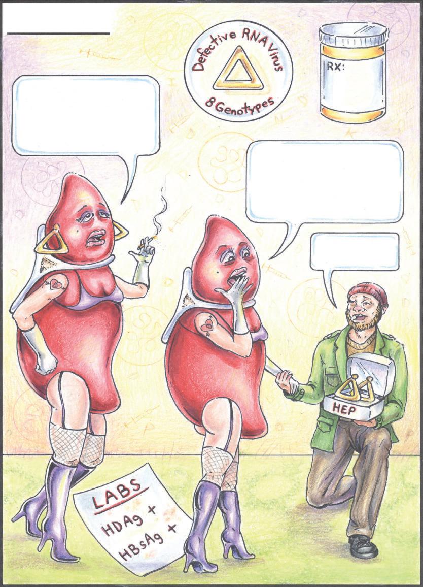
Disease Name:
Hepatitis D
Causative Agent: Hepatitis D Virus (HDV), Delta Virus
Geographic Regions Affected: Worldwide; Higher incidences where hepatitis B is endemic. Hepatitis D is rare in the United States.
Reservoir: Humans with hepatitis B virus (HBV)/HDV coinfections.
Description: Hepatitis D is a “defective” single-stranded RNA virus that requires the machinery and assistance of hepatitis B to replicate. HDV is acquired as either a coinfection (simultaneously acquired with hepatitis B) or as a superinfection (acquired by a person who already has chronic hepatitis B).
Signs and Symptoms: When acquired as a coinfection, HDV can increase the likelihood of severe illness and fulminant hepatic failure. Coinfections are less likely to result in chronic hepatitis D. Superinfections are more likely to cause chronic hepatitis D and can worsen preexisting liver disease.
Diagnostic Testing: Since patients cannot have hepatitis D without hepatitis B, patients will test positive for hepatitis B serum markers. IgM and IgG anti-HDV antibody testing is available in the United States, and an elevated anti-HDV IgG titer can be seen in chronic hepatitis D infections. Patients testing positive for HDV antibodies can be followed up with HDV viral levels via PCR technology.
Treatments: Vaccination against hepatitis B is also preventative against hepatitis D. PEGylated interferon is the only treatment that is effective for chronic hepatitis D, but successful clearance of the virus with treatment is low (approximately 20%–25%).
Pearls: HDV infections cannot exist without hepatitis B. The incubation period for hepatitis D coinfection is identical to that of acute hepatitis B (the HDV infection occurs concurrently with HBV). The presence of HBsAg and IgM anti-HBc is essential for the diagnosis of HDV infections.
Hepatitis D
Co-infection occurs when Hep B + D arrive simultaneously. It’s indistinguishable from acute Hep B.
Super-infection occurs when D is introduced into a chronic HBsAg carrier and presents as severe acute hepatitis or exacerbation of a chronic hepatitis.
Miss B, may I super-infect you with D?
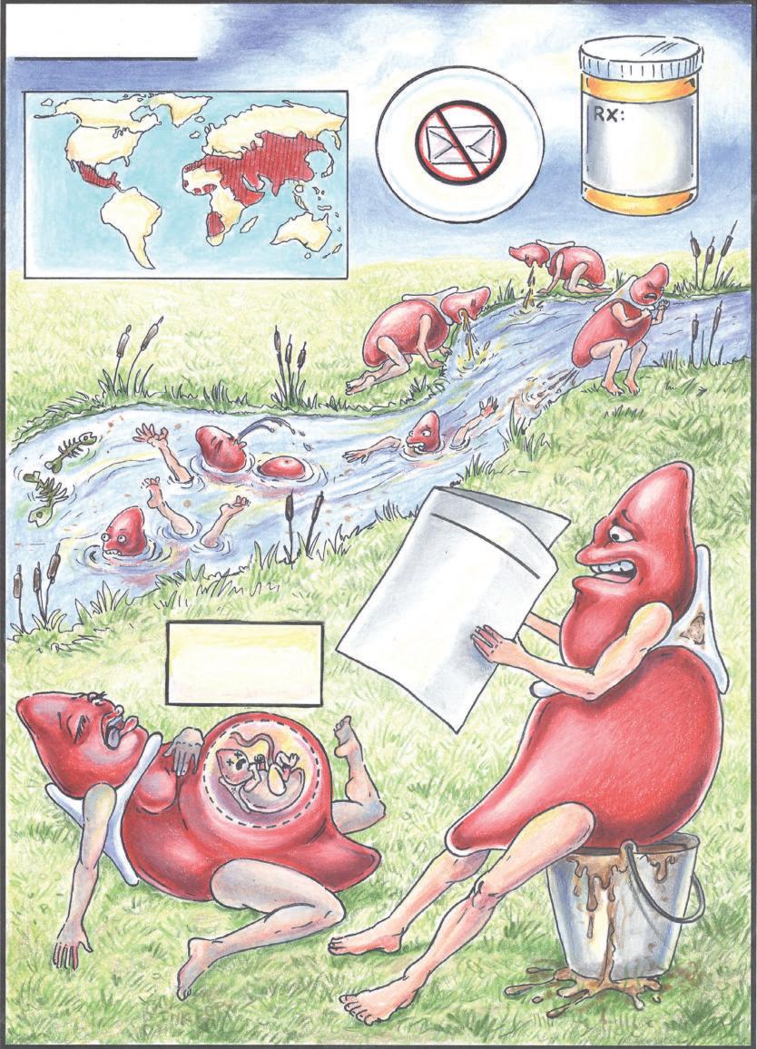
Disease Name: Hepatitis E
Causative Agent: Hepatitis E Virus, HEV
Incubation: 15–60 days; Average: 40 days
Geographic Regions Affected: Central America, Africa, Middle East, India, and Asia
Reservoir: Swine and small rodents may serve as an animal reservoir.
Description: HEV is a fecal-oral (genotype 1 and 2) and foodborne (genotype 3 and 4) single-stranded RNA virus responsible for acute viral hepatitis. There are four major viral genotypes, each associated with specific regions and unique clinical presentations. Genotype 3 is found in developed countries, tends to affect those patients .40 years old or immunocompromised, and can cause chronic infections. Genotypes 1, 2, and 4 more commonly affect younger adults. HEV infections can occur as sporadic outbreaks affecting many people or as isolated cases affecting isolated individuals.
Signs and Symptoms: Most cases are asymptomatic, with fewer than 5% of patients showing signs of acute infection. When symptomatic, patients may exhibit malaise, fever, nausea, vomiting, abdominal pain, arthralgia, and jaundice. Fulminant hepatic failure can occur in up to 3% of infections and is more common in pregnant females and in those with preexisting liver disease.
Diagnostic Testing: There are no commercially approved tests for HEV in the United States. Some countries have access to IgM, IgG, and HEV PCR testing capabilities. Acute infection would be determined by a positive IgM and HEV PCR viral load. IgM is elevated in the acute setting and indicates recent exposure to the HEV virus, whereas IgG increases during convalescence and confirms past exposure. In chronic HEV infections, HEV RNA will be detectable via PCR in serum or stool more than 6 months after initial infection.
Treatments: Supportive. Ribavirin may be beneficial in the treatment of chronic HEV infection. There is an HEV vaccine that is licensed in China.
Pearls: Acute hepatitis E infection during pregnancy has a high mortality rate (up to 25%).