GARDNER AND SUTHERLAND’S CHROMOSOME ABNORMALITIES AND GENETIC COUNSELING
OXFORD MONOGRAPHS ON MEDICAL GENETICS
General Editors:
JUDITH G HALL PETER S HARPER LOUANNE HUDGKINS EVAN EICHLER CHARLES J EPSTEIN (DECEASED 2011) ARNO G. MOTULSKY (RESIGNED 2011)
1 R B McConnell: The genetics of gastrointestinal disorders
2. A. C. Kopéc: The distribution of the blood groups in the United Kingdom
3. E. Slater and V. A. Cowie: The genetics of mental disorders
4. C. O. Carter and T. J. Fairbank: The genetics of locomotor disorders
5. A. E. Mourant, A. C. Kopéc, and K. Domaniewska-Sobezak: The distribution of the human blood groups and other polymorphisms
6. A. E. Mourant, A. C. Kopéc, and K. Domaniewska-Sobezak: Blood groups and diseases
7. A. G. Steinbert and C. E. Cook: The distribution of the human immunoglobulin allotypes
8 D Tills, A C Kopéc, and R E Tills: The distribution of the human blood groups and other polymorphisms: Supplement I
10. D. Z. Loesch: Quantitative dermatoglyphics: Classification, genetics, and pathology
11. D. J. Bond and A. C. Chandley: Aneuploidy
12. P. F. Benson and A. H. Fensom: Genetic biochemical disorders
13. G. R. Sutherland and F. Hecht: Fragile sites on human chromosomes
14. M. d’A Crawfurd: The genetics of renal tract disorders
16. C. R. Scriver and B. Child: Garrod’s inborn factors in disease
18. M. Baraitser: The genetics of neurological disorders
19 R J Gorlin, M M Cohen, Jr , and L S Levin: Syndromes of the head and neck, third edition
21. D. Warburton, J. Byrne, and N. Canki: Chromosome anomalies and prenatal development: An atlas
22. J. J. Nora, K. Berg, and A. H. Nora: Cardiovascular disease: Genetics, epidemiology, and prevention
24. A. E. H. Emery: Duchenne muscular dystrophy, second edition
25. E. G. D. Tuddenham and D. N. Cooper: The molecular genetics of haemostasis and its inherited disorders
26. A. Boué: Foetal medicine
27 R E Stevenson, J G Hall, and R M Goodman: Human malformations
28. R. J. Gorlin, H. V. Toriello, and M. M. Cohen, Jr.: Hereditary hearing loss and its syndromes
29. R. J. M. Gardner and G. R. Sutherland: Chromosomes abnormalities and genetic counseling, second edition
30. A. S. Teebi and T. I. Farag: Genetic disorders among Arab populations
31. M. M. Cohen, Jr.: The child with multiple birth defects
32. W. W. Weber: Pharmacogenetics
33. V. P. Sybert: Genetic skin disorders
34. M. Baraitser: Genetics of neurological disorders, third edition
35. H. Ostrer: Non- Mendelian genetics in humans
36 E Traboulsi: Genetic factors in human disease
37. G. L. Semenza: Transcription factors and human disease
38. L. Pinsky, R. P. Erickson, and R. N. Schimke: Genetic disorders of human sexual development
39. R. E. Stevenson, C. E. Schwartz, and R. J. Schroer: X- linked mental retardation
40. M. J. Khoury, W. Burke, and E. Thomson: Genetics and public health in the 21st century
41. J. Weil: Psychosocial genetic counseling
42. R. J. Gorlin, M. M. Cohen, Jr., and R. C. M. Hennekam: Syndromes of the head and neck, fourth edition
43. M. M. Cohen, Jr., G. Neri, and R. Weksberg: Overgrowth syndromes
44. R. A. King, J. I. Rotter, and A. G. Motulsky: Genetic basis of common diseases, second edition
45. G. P. Bates, P. S. Harper, and L. Jones: Huntington’s disease, third edition
46. R. J. M. Gardner and G. R. Sutherland: Chromosome abnormalities and genetic counseling, third edition
47. I. J. Holt: Genetics of mitochondrial disease
48. F. Flinter, E. Maher, and A. Saggar- Malik: Genetics of renal disease
49. C. J. Epstein, R. P. Erickson, and A. Wynshaw-Boris: Inborn errors of development: The molecular basis of clinical disorders of morphogenesis
50. H. V. Toriello, W. Reardon, and R. J. Gorlin: Hereditary hearing loss and its syndromes, second edition
51. P. S. Harper: Landmarks in medical genetics
52. R. E. Stevenson and J. G. Hall: Human malformations and related anomalies, second edition
53. D. Kumar and S. D. Weatherall: Genomics and clinical medicine
54. C. J. Epstein, R. P. Erickson, and A. Wynshaw-Boris: Inborn errors of development: The molecular basis of clinical disorders of morphogenesis, second edition
55 W Weber: Pharmacogenetics, second edition
56. P. L. Beales, I. S. Farooqi, and S. O’Rahilly: The genetics of obesity syndromes
57. P. S. Harper: A short history of medical genetics
58. R. C. M. Hennekam, I. D. Krantz, and J. E. Allanson: Gorlin’s syndromes of the head and neck, fifth edition
59. D. Kumar and P. Elliot: Principles and practices of cardiovascular genetics
60. V. P. Sybert: Genetic skin disorders, second edition
61. R. J. M. Gardner, G. R. Sutherland, and L. C. Shaffer: Chromosome abnormalities and genetic counseling, fourth edition
62. D. Kumar: Genomics and health in the developing world
63 G Bates, S Tabrizi, and L Jones: Huntington’s disease, fourth edition
64. B. Lee and F. Scaglia: Inborn errors of metabolism: From neonatal screening to metabolic pathways
65. D. Kumar and C. Eng: Genomic medicine, second edition
66 R Stevenson, J Hall, D Everman, and B Solomon: Human malformations and related anomalies, third edition
67. R. Erickson and A. Wynshaw-Boris: Epstein’s inborn errors of development: The molecular basis of clinical disorders of morphogenesis, third edition
68. C. Hollak and R. Lachmann: Inherited metabolic disease in adults: A clinical guide
69. V. P. Sybert: Genetic skin disorders, third edition
70. R. J. M. Gardner and D. J. Amor: Gardner and Sutherland’s chromosome abnormalities and genetic counseling, fifth edition
GARDNER AND SUTHERLAND’S
Chromosome Abnormalities and Genetic Counseling
FIFTH EDITION
R. J. McKinlay GARDNER
ADJUNCT PROFESSOR
CLINICAL GENETICS GROUP
UNIVERSITY OF OTAGO, DUNEDIN, NEW ZEALAND
David J. AMOR
LORENZO AND PAMELA GALLI CHAIR
UNIVERSITY OF MELBOURNE
VICTORIAN CLINICAL GENETICS SERVICES
MURDOCH CHILDREN’S RESEARCH INSTITUTE
ROYAL CHILDREN’S HOSPITAL, MELBOURNE, AUSTRALIA
Oxford University Press is a department of the University of Oxford. It furthers the University’s objective of excellence in research, scholarship, and education by publishing worldwide. Oxford is a registered trade mark of Oxford University Press in the UK and certain other countries.
Published in the United States of America by Oxford University Press 198 Madison Avenue, New York, NY 10016, United States of America.
All rights reserved. No part of this publication may be reproduced, stored in a retrieval system, or transmitted, in any form or by any means, without the prior permission in writing of Oxford University Press, or as expressly permitted by law, by license, or under terms agreed with the appropriate reproduction rights organization. Inquiries concerning reproduction outside the scope of the above should be sent to the Rights Department, Oxford University Press, at the address above.
You must not circulate this work in any other form and you must impose this same condition on any acquirer.
Library of Congress Cataloging-in-Publication Data
Names: Gardner, R J M , author | Amor, David J , author
Title: Gardner and Sutherland’s chromosome abnormalities and genetic counseling / R J McKinlay Gardner, David J. Amor.
Other titles: Chromosome abnormalities and genetic counseling | Oxford monographs on medical genetics ; no. 70.
Description: Fifth edition. | Oxford ; New York : Oxford University Press, [2018] | Series: Oxford monographs on medical genetics ; no 70 | Preceded by Chromosome abnormalities and genetic counseling / R J McKinlay Gardner, Grant R Sutherland, Lisa G Shaffer c2012 | Includes bibliographical references and index
Identifiers: LCCN 2017034126 | ISBN 9780199329007 (hardcover : alk. paper) | ISBN 9780199329021 (epub)
Classification: LCC RB155.7 | NLM QS 677 | DDC 616/.042 dc23 LC record available at https://lccn loc gov/2017034126
This material is not intended to be, and should not be considered, a substitute for medical or other professional advice. Treatment for the conditions described in this material is highly dependent on the individual circumstances. And, while this material is designed to offer accurate information with respect to the subject matter covered and to be current as of the time it was written, research and knowledge about medical and health issues is constantly evolving and dose schedules for medications are being revised continually, with new side effects recognized and accounted for regularly Readers must therefore always check the product information and clinical procedures with the most up-to-date published product information and data sheets provided by the manufacturers and the most recent codes of conduct and safety regulation. The publisher and the authors make no representations or warranties to readers, express or implied, as to the accuracy or completeness of this material. Without limiting the foregoing, the publisher and the authors make no representations or warranties as to the accuracy or efficacy of the drug dosages mentioned in the material The authors and the publisher do not accept, and expressly disclaim, any responsibility for any liability, loss or risk that may be claimed or incurred as a consequence of the use and/or application of any of the contents of this material.
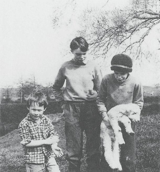
This book is dedicated to Jocelyn, Geoffrey, and Craig, their parents, and all other families who seek our “chromosomal advice.”
Jocelyn and Geoffrey (with lamb) have a partial trisomy for chromosome 4 long arm, and Craig, the youngest, had a 46,XY result on amniocentesis. Their father is a translocation carrier (see Fig. 5–1, lower). Craig, since married, came to the genetic clinic
for confirmatory advice about his low genetic risk.
Heredity
Inescapably, this is me the diagnosis is cause for anger at those who brightly say we choose our destinies. There is no store of courage, wit or will can save me from myself and I must face my children, feeling like that wicked fairy, uninvited at the christening, bestowing on my own, amidst murmurs of apprehension, a most unwanted gift that of a blighted mind. No one could tell me of this curse when I was young and dreamt of children and the graces they would bear. Later, it seemed that a chill morning revealed deeper layers of truth. For my romancing there is a price to pay perhaps my children’s children will pass this tollgate after me. My grandmothers gaze down from their frames on my wall, sadly wondering.
Dear DNA
In real life you’re just a tangle of white filaments captured in a test-tube, and your first photo is not flattering: grey smudges like tractor tracks, or a rusty screw. Yet many say you are beautiful. Online for a night with a hundred fantastic portraits
Meg Campbell
and I’m head over heels In love with you, DNA, bewitched by your billions coiled in my cells, transcribing, replicating, mutating. I see your never-ending dance. A length of twisted ladder briefly unwinds, both strands duplicate, each copy drifts away on its secret mission to make a thought, feel sunshine, or digest this morning’s porridge. Two winding parallel threads, a tiny tangle of gossamer designing my life. DNA, you are astonishing and I am yours truly.
Genes pass on our kind But our selves are transmitted In words left behind.
Winifred Kavalieris
J. Patrick Gookin
Curiosity is a virtue, perhaps an unsung and undervalued virtue, which should be the energizing fuel to the thinking geneticist.
Willie Reardon
Where is the wisdom we have lost in knowledge? Where is the knowledge we have lost in information?
T. S. Eliot
PREFACE TO THE FIFTH EDITION
Chromosomal disorders have been, and will always be, with us; that is a given. What is changing is our ability to recognize and detect them: detection both in terms of the subtlety of abnormalities and of the means we can use to find them. Classical cytogenetics has now well and truly given way to “molecular karyotyping,” and this has been the extraordinary development of the early twenty-first century. Readers will now be as accustomed to molecular nomenclature in defining a segment, such as chr5:1-18,500,000, as they had been to the classical description, 5p14.1→pter.
The very small deletions and duplications which molecular karyotyping can now reveal have become familiar to the clinicians and counselors who see patients and families in the clinic. A large number of these are now on record, many attracting the nomenclature “copy number variant”: Some are very well understood, others becoming so, and yet quite a few variants of uncertain significance, the acronym “VOUS” in daily parlance whose roles in human pathology are imperfectly appreciated. Many are not in the same mold as the deletions and duplications of classical cytogenetics, in which the single defect sufficed to cause a particular phenotype, and always did so: We now need to take account of the concept of incomplete penetrance, with some microdeletions or duplications not, of themselves, always leading to an abnormal phenotype. Apparently clinically normal parents may carry the same alteration as their child with an abnormal phenotype. Digenic, or “two-hit,” mechanisms may now require consideration. These were not formerly notions much entering into the assessment of chromosomal disorders; discussion apropos in the clinic presents a new challenge.
The number of “new” del/dup syndromes increases almost with each issue of the clinical genetic journals. We include a mention of a considerable number of these here (Chapter 14), not intending to create an encyclopedic resource per se but believing that such a record may provide a useful first point of contact when these cases are encountered in the clinic. Copy number variants of uncertain significance, on the other hand, we mostly take only a broad rather than a detailed view (Chapter 17); the
reader will need to consult other repositories for fuller information, as their interpretations evolve.
The new (or now, established) laboratory methodologies blur the boundaries between what might have been regarded as the classic chromosomal abnormalities and Mendelian conditions. Some disorders recorded as being due not only to segmental deletion/duplication affecting a single locus but also to point mutation at that locus we continue to treat as “chromosomal”; and for most, their place in this book is secure. But one major category, the fragile X syndromes, are now seen as essentially Mendelian disorders, their historic cytogenetic-based nomenclature notwithstanding, and they no longer claim their chapter.
Peripheral blood and skin have been the tissues in common usage for chromosome analysis, with an increasing role for cells got from the convenient and painless “spit sample.” Prenatal diagnosis has been based on amniocentesis and chorionic villus sampling, but latterly blastomeres from early embryos, and fetal DNA in the maternal circulation, have become targets for testing. Now we can anticipate the potential for whole genome analysis to be applied to the prenatal diagnosis of the classic aneuploidies, from a simple maternal blood sample, and this would widen such testing very considerably. Questions such as these raise ethical issues, and a literature on “chromosomal ethics” is accumulating.
As we have previously written, however marvelous may be these new ways to test for chromosomes, the concerns of families remain essentially the same. We may reproduce here the final paragraph of the Preface of the first edition of this book, from 1989, as valid now as then:
Families pursue genetic counseling in an effort to demystify the mysterious If they did not want to “hear it all,” they would not bother with genetic counseling. Families want an honest evaluation of what is known and what is unknown, a clear explanation of all possibilities, both good and bad, and a sensitive exploration of all available information with which they can make knowledgeable decisions about future family planning. Thus, Bloch et al. (1979) succinctly convey the essence of why people go to the genetic counselor. We hope this book will assist counselors in their task.
Dunedin Melbourne February 2018
ACKNOWLEDGMENTS
We thank John Barber, Rachel Beddow, Amber Boys, Cyril Chapman, Jane Halliday, Jan Hodgson, Caroline Lintott, Nicole Martin, Belinda McLaren, Fiona Norris, Mamoru Ozaki, Mark Pertile, Jenny Rhodes, Sharyn Stock-Myer, and Jane Watt for their critical advice. We acknowledge Lisa Shaffer, who was a co-author of the previous edition, and much of whose work has flowed over into this edition. We have made much use of the ideograms created by Nicole Chia. The length of the Reference list, and the frequency with which we acknowledge, in legends to figures, the courtesy of colleagues whose work we use, speaks for the debt we owe to our colleagues in clinical cytogenetics worldwide. Belatedly, R.J.M.G. thanks Ngaire Adams and Dianne Grimaldi, whose need for chromosomal teaching at Dunedin Hospital in the 1980s provided the germination for writing this book. We have appreciated the wise guidance, and the patience and forbearance of Oxford University Press, from Jeff House when this book made its first appearance, through to Chad Zimmerman and Chloe Layman in this fifth edition. R.J.M.G. thanks his wife Kelley for her patient help, once again, in document management; and the front cover art, and most of the new illustrations in this edition, have been drawn, or redrawn, by her.
CONTENTS
PART ONE: BASIC CONCEPTS
1. Elements of Medical Cytogenetics
2. Chromosome Analysis
3. The Origins and Consequences of Chromosome Pathology
4. Deriving and Using a Risk Figure
PART TWO: PARENT OR CHILD WITH A CHROMOSOMAL ABNORMALITY
13. Down Syndrome, Other Full Aneuploidies, Polyploidy, and the Influence of Parental Age
14. Autosomal Structural Rearrangements: Deletions and Duplications
15. Sex Chromosome Aneuploidy and Structural Rearrangement
16. Chromosome Instability Syndromes
PART THREE: CHROMOSOME VARIANTS
17. Normal Chromosomal Variation
PART FOUR: DISORDERS ASSOCIATED WITH ABERRANT GENOMIC IMPRINTING
18. Uniparental Disomy and Disorders of Imprinting
PART FIVE: REPRODUCTIVE CYTOGENETICS
19. Reproductive Failure
20. Prenatal Testing Procedures
21. Chromosome Abnormalities Detected at Prenatal Diagnosis
22. Preimplantation Genetic Diagnosis
PART SIX: DISORDERS OF SEX DEVELOPMENT
23. Chromosomal Disorders of Sex Development
PART SEVEN: NOXIOUS AGENTS
24. Gonadal Cytogenetic Damage from Exposure to Extrinsic Agents
APPENDICES
A. Ideograms of Human Chromosomes, and Haploid Autosomal Lengths
B. Cytogenetic Abbreviations and Nomenclature
C. Determining 95 Percent Confidence Limits, and the Standard Error
References Index
PART ONE BASIC CONCEPTS
1
ELEMENTS OF MEDICAL CYTOGENETICS
CHROMOSOMES WERE first seen and named in the late nineteenth century. Chromosome is a combination of Greek words meaning colored (chrom) body (soma); the word was coined by the illustrious German anatomist Heinrich Wilhelm Gottfried von Waldeyer-Hartz. It was early appreciated that these brightly staining objects appearing in the cell nucleus must be the “stuff of heredity,” the very vessels of our genetic inheritance. Most observers had concluded, in the earlier part of the twentieth century, that the human chromosome count was 48. It was not until the 1950s, due to technical advances, and in particular the use of a hypotonic solution to swell the cells, giving an uncluttered view of the chromosomes, that Joe Hin Tjio and Albert Levan could recognize that 46 was the correct number. This discovery spurred research into conditions in which a chromosomal cause had hitherto been suspected, and in 1959 (“the wonderful year of human cytogenetics”) came the first demonstrations of a medical application of the new knowledge, with practically simultaneous discoveries of the chromosomal basis of Down syndrome, Klinefelter syndrome, and Turner syndrome (Lejeune et al.1 1959; Jacobs and Strong 1959; Ford et al. 1959); these were followed soon thereafter by the recognition of the other major aneuploidy syndromes. Harper (2006) records the history, and the personalities behind the history, in his book First Years of Human Chromosomes a book that should be read by every student of medical cytogenetics with an interest in how their discipline came to be. Harper points out that the practice of genetic counseling came into its own essentially upon the basis of these chromosomal discoveries: So to speak, geneticists now had “their organ.”
“Colored bodies” became an especially apt derivation with the development of various different staining techniques in the 1980s and 1990s, showing different parts of chromosomes in many different colors,
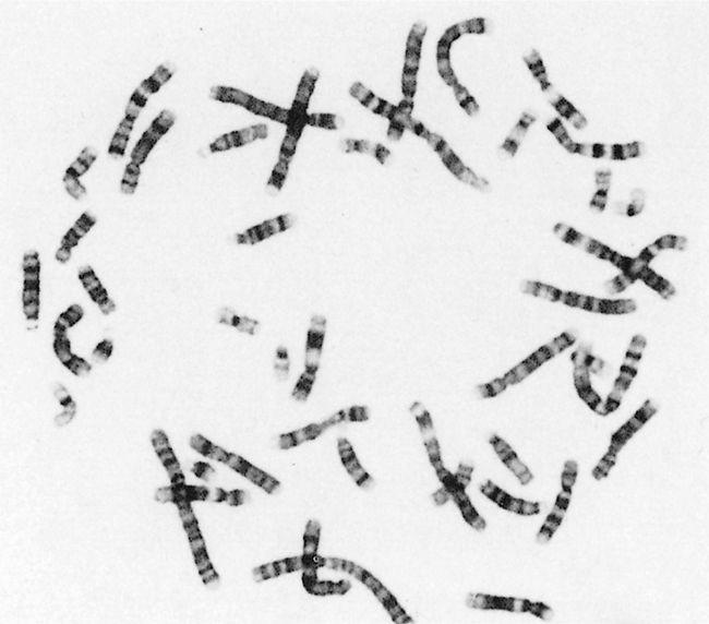
whether true or computer-generated false colors. The images produced by this kaleidoscopic karyotyping could be rather beautiful. Black-and-white photographs were less splendid but often sufficed (Figure 1–1). Albeit that molecular methodologies have substantially taken over from classical cytogenetics, and providing a different view of the genetic material, the word chromosome will surely last forever.
FIGURE 1–1 The appearance of banded chromosomes, from a classical cytogenetic study
Chromosomal Morphology
Chromosomes have a linear appearance: two arms that are continuous at the centromere. Reflecting the French influence in the establishment of the cytogenetic nomenclature, the shorter arm is designated p (for petit), and the longer is q (variously explained as being the next letter in the alphabet, a mistyping of g (for grand), for queue, or as the other letter in the formula
p + q = 1). In the early part of the cell cycle, each chromosome is present as a single structure, a chromatid, a single DNA molecule. During the cell cycle, the chromosomes replicate, and two sister chromatids form. Now the chromosome exists as a double-chromatid entity. Each chromatid contains exactly the same genetic material. This replication is in preparation for cell division so that, after the chromosome has separated into its two component chromatids, each daughter cell receives the full amount of genetic material. It is during mitosis that the chromosomes contract and become readily distinguishable on light microscopy.
Blood and buccal mucosal cells are the tissues from which DNA is extracted in routine chromosome analysis. From blood, the nucleated white cell is the tested component for microarray analysis, and in classical cytogenetic analysis, it is the lymphocyte. Buccal mucosal cells and white blood cells2 are obtained from a saliva sample. The chromosomal status of each small sample is taken as representative of the constitution of (essentially) every other cell of the body. In the case of invasive prenatal diagnosis, the cells from amniotic fluid or chorionic villi are the source material; these tissues are assumed (with certain caveats) to represent the fetal chromosomal constitution. Noninvasive prenatal diagnosis exploits the presence of fetal blood cells and DNA in the maternal circulation.
The 46 chromosomes come in 23 matching pairs and constitute the genome. One of each pair came from the mother, and one from the father. For 22 of the chromosome pairs, each member (each homolog) has the same morphology in each sex: These are the autosomes. The sex chromosome (or gonosome) constitution differs: The female has a pair of X chromosomes, and the male has an X and a Y chromosome. The single set of homologs one of each autosome plus one sex chromosome is the haploid set. The haploid number (n) is 23. The haploid complement exists, as such, only in the gametocytes (ovum and sperm). All other cells in the body the soma have a double set: the diploid complement (2n) of 46. If there is a difference between a pair of homologs, in the sense of one being structurally rearranged, the person is described as a heterozygote.
The chromosomes are classically distinguishable on the basis of their size, centromere position, and banding pattern. The centromere may be in the middle, off-center, or close to one end metacentric, submetacentric, and acrocentric, respectively. The chromosomes are numbered 1 through 22, and X and Y, and are also assigned to groups A through G, according to their general size and the position of the centromere. The diagrammatic representation of the banding pattern is the ideogram (Appendix A). The numbering is based on size, largest to smallest (to split hairs, this order is
not exact; for example, chromosomes 10 and 11 are shorter than chromosome 12, and chromosome 21 is smaller than 22).
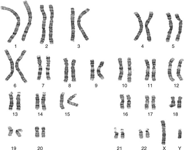
The classical format of a chromosome display, the “karyotype, ” has the chromosomes lined up with p arms upward, in their matching pairs (Figure 1–2). Those coming from a DNA-based view may see the chromosome lying on its side, and microarray reports usually show a horizontal depiction of the chromosome arms, with the graph indicating duplications and deletions by a rise or a fall compared to baseline, respectively (although no one is proposing that short and long arms be renamed as left and right!). Karyotypes are described according to a shorthand notation, the International System of Human Cytogenetic Nomenclature (ISCN 2016); an outline is given in Appendix B.
FIGURE 1–2 Chromosomes arranged as a formal karyotype, from a classical cytogenetic study.
Chromosomal Structure
Chromatin exists in differently condensed forms: the less condensed
euchromatin and the more condensed heterochromatin. Euchromatin contains the coding DNA the genes while heterochromatin comprises noncoding DNA. Chromosomes are capped at the terminal extremities of their long and short arms by telomeres, specialized DNA sequences comprising many repeats of the sequence TTAGGG, that can be thought of as sealing the chromatin and preventing its fusion with the chromatin of other chromosomes. The centromere 3 is a specialized region of DNA that, at mitosis, provides the site at which the spindle apparatus can be anchored and draw each separated chromatid to opposite poles of the dividing cell. Centromeric heterochromatin contains “satellite DNA,” so-called because these DNA species have different buoyant densities and produce distinct humps on a density gradient distribution. (These are not to be confused with the satellites on acrocentric chromosomes.) A separate issue, of considerable academic interest (but which we shall take no further here), is the “packaging question”: how the centimeters of DNA are compacted into micron-length chromosomes, and which parts of the nucleus each chromosome occupies (Annunziato 2008; Lieberman-Aiden et al. 2009).
CHROMOSOME ABNORMALITY
Chromosomes are distributed to each daughter cell during cell division in a very precise process precise, but prone to error. From our perspective, the two cell divisions of meiosis, during which the gametes are formed, are of central importance. Most of the discipline of medical cytogenetics focuses on the consequences of disordered meiosis having produced a chromosomally abnormal gamete, causing a chromosomal abnormality in the conceptus. A chromosome abnormality that is present from conception and involves the entire body is a constitutional abnormality. If an additional cell line with a different chromosomal complement arises before the basis of the body structure is formed (that is, in embryonic or preembryonic life) and becomes an integral part of the organism, constitutional mosaicism results. In this book, we concern ourselves practically solely with constitutional abnormalities. Acquired chromosomal abnormality of course exists, and indeed it is a major initiating and sustaining cause in most cancers, a fact first proposed by Boveri in 1914 and voluminously attested in the work of Mitelman et al. (2016); but this is more the field of study of the molecular pathologist than the genetic counselor.
An incorrect amount of genetic material carried by the conceptus disturbs and distorts its normal growth pattern (from zygote → blastocyst
→ embryo → fetus). In trisomy, there is three of a particular chromosome, instead of the normal two. In monosomy, only one member of the pair is present. Two of each is the only combination that works properly! It is scarcely surprising that a process as exquisitely complex as the development of the human form should be vulnerable to a confused outflow of genetic instruction from a nucleus with a redundant or incomplete database.
Trisomy and monosomy for a whole chromosome were the first cytogenetic mechanisms leading to an abnormal phenotype to be identified. More fully, we can list the following pathogenetic mechanisms that arise from chromosomal abnormalities:
1. A dosage effect, with a lack (deletion) or excess (duplication) of chromosomal material, whether for a whole chromosome or a part of a chromosome. This is by far the predominant category.
2. A direct damaging effect, with disruption of a gene at the breakpoint of a rearrangement
3. An effect due to the incongruent parental origin of a chromosome or chromosomal segment (genomic imprinting)
4. A position effect, whereby a gene in a new chromosomal environment functions inappropriately
5. Combinations of the above
We discuss these mechanisms in more detail in following chapters.
Autosomal Imbalance
STRUCTURAL IMBALANCE
As noted earlier, imbalance may involve the gain or loss of a whole chromosome full aneuploidy or of part of a chromosome partial aneuploidy. The abnormality may occur in the nonmosaic or mosaic state. Loss (that is, monosomy) of chromosomal material generally has a more devastating effect on growth of the conceptus than does an excess of material (that is, trisomy). Certain imbalances lead to certain abnormal phenotypes. The spectrum is listed in outline in Box 1–1. Most full autosomal trisomies and virtually all full autosomal monosomies set development of the conceptus so awry that, sooner or later, abortion occurs the embryo “self-destructs” and is expelled from the uterus. This issue is further explored in Chapter 19. A few full trisomies are not necessarily
lethal in utero, and many partial chromosomal aneuploidies are associated with survival through to the birth of an infant.
Box 1–1 The Spectrum of Effects, in Broad Outline, Resulting from Constitutional Chromosomal Abnormality
1. Devastation of blastogenesis, with transient implantation or nonimplantation of the conceptus
2. Devastation of embryogenesis, with spontaneous abortion, usually in the first trimester
3. Major disruption of normal intrauterine morphogenesis, with stillbirth or early neonatal death
4. Major disruption of normal intrauterine morphogenesis, but with some extrauterine survival
5. Moderate distortion of normal intrauterine development, with substantial extrauterine survival and severe mental retardation
6. Mild distortion of normal intrauterine development, with substantial extrauterine survival, and considerable intellectual compromise
7. Minimal physical phenotypic effect, varying degrees of intellectual compromise; possible compromise of fertility
8. No discernible physical phenotypic effect; cognitive function within the normal range, but less than expected from the family background
Characteristically, “survivable imbalances” produce a phenotype of widespread dysmorphogenesis, and there may be malformation of internal organs and limbs. It is often in the facial appearance (facies) that the most recognizable physical abnormality is seen, with Down syndrome the classic example, although the physical phenotype in some cases of subtler deletion or duplication may be rather “bland.” The most complex organ of all, the brain, is the most vulnerable to a less than optimal genetic constitution, and some compromise of mental and intellectual functioning, usually to the extent of an obvious deficit, is nearly invariable, at least in imbalances of classical size. With several of the (much smaller) imbalances due to copy number variants, developmental delay or mental retardation4 with an outwardly normal physical phenotype is well recognized as a chromosomal presentation. Thus, the central concern of most people seeking genetic counseling for a chromosomal condition is that of having a child who might have a physical, intellectual, or severe
social handicap.
Historically, the chromosomal basis of many syndromes was identified following analysis of groups of patients with similar phenotypes. This “phenotype-first” approach led to the identification of many of the wellknown microdeletion syndromes (and of course such classic conditions as Down syndrome). With the advent of microarray analysis, new syndromes came to be identified based on their DNA aberration, a “genotype-first” approach. Representative examples of these newer syndromes are reviewed in Chapter 14.
SEX CHROMOSOMAL ABNORMALITY
Sex chromosome (gonosome) imbalance has a much less deleterious effect on the phenotype than does autosomal aneuploidy. The X chromosome is one of the larger and is gene-dense; the Y is small, comprising mostly heterochromatin, and carries very few genes. In both male and female, one, and only one, completely functioning X chromosome is needed. X chromosomes in excess of one are inactivated, as the normal 46,XX female exemplifies; her second X does, however, maintain some segments genetically active. With X chromosome excess or deficiency, a partially successful buffering mechanism exists whereby the imbalance is counteracted, in an attempt to achieve the same effect as having a single active X. In such states as, for example, XXX, XXY, XXXX, XXYY, and XXXXX, excess X chromosomes are inactivated. In the 45,X state, the single X remaining is not subject to inactivation. If an abnormal X chromosome (e.g., an isochromosome, or a deleted X) is present, then, as a rule, cells containing this abnormal chromosome as the active X are selected against, perhaps due to preferential growth of those cells in which it is the normal X that is the active one. In X imbalance, the reproductive tract and brain are the organs predominantly affected. The effect may be minimal. As for Y chromosome excess, such as XYY, there is a rather limited phenotypic consequence, but again the brain may be a vulnerable organ.
FUNCTIONAL IMBALANCE
A correct amount of chromatin does not necessarily mean the phenotype will be normal. Inappropriate inactivation, or activation, of a segment of the genome can compromise the genetic message. Some segments of the genome require only monosomic expression, and the homologous segment
on the other chromosome is inactivated. If this control fails, both segments can become activated, or both inactivated, and the over- or underexpression of the contained loci can cause phenotypic abnormality. The classic example of this is genomic imprinting according to parent of origin, and we discuss this concept in Chapter 18. A rather specialized example arises with the X-autosome translocation. A segment of X chromosome can fail to be inactivated; or conversely, X-inactivation can spread into an autosomal segment (Chapter 6).
The Frequency and Impact of Cytogenetic Pathology
According to the window of observation, chromosomal disorders make a greater or lesser contribution to human mortality and morbidity. Looking at prenatal existence, the earliest window has been provided by the in vitro fertilization (IVF) clinic, from the procedure of preimplantation genetic diagnosis (Chapter 22), at which cells taken from 3- to 5-day-old embryos are subjected to genetic analysis; and an extraordinary fraction are chromosomally abnormal. After implantation (about day 6), and through the first trimester of pregnancy (to week 13), chromosomal mortality is very high, and aneuploidy is the major single cause of spontaneous abortion (Chapter 19). Perinatal and early infant death has a significant chromosomal component, of which trisomies 18 and 21 (although the latter less so in more recent times) are major elements.
As for morbidity, the brain, as mentioned above, is the most vulnerable organ, and chromosomal defects are the basis of a substantial fraction of all intellectual deficit, and many of these retarded individuals will also have structural malformations that cause functional physical disability. Among a mentally retarded population, Down syndrome is the predominant contributor in the fraction who have a classic chromosome abnormality (Phelan et al. 1996). Development of the heart is particularly susceptible to chromosomal imbalance, and in a population study from the US National Center on Birth Defects, 1 in 8 infants with a congenital heart defect had a chromosomal abnormality, with again trisomy 21 the most common of these (53%), followed by trisomy 18 (13%), 22q11.2 deletion (12%), and trisomy 13 (6%) (Hartman et al. 2011).
Adolescence is a period during which many sex chromosome defects come to light, when pubertal change fails to occur; and in young adulthood, chromosomal causes of infertility are recognized. Few new classic cytogenetic defects come to attention later in adult life, but many