Chief Quality Officer, Hospital-Based Services, Associate Radiologist-in-Chief, Department of Radiology, Texas Children’s Hospital, Professor of Radiology, Baylor University College of Medicine, Houston, Texas
Table of Contents
Cover image
Title page
Copyright
Contributors
Preface
Chapter 1 Special Considerations in Pediatric Imaging
▪Pediatric Radiology As a Potential Career
▪Introduction: Special Considerations in Pediatric Imaging
Chapter 2 Airway
▪Acute Upper Airway Obstruction
▪Lower Airway Obstruction
▪Congenital Airway Obstruction
Chapter 3. Chest
▪Neonatal Chest
▪Roles of Imaging in Pediatric Pneumonia
▪High-Resolution Computed Tomography in Children
Chapter 4 Cardiac
▪Imaging Modalities in Congenital Heart Disease
▪Categorization of Congenital Heart Disease
▪Abnormalities of Conotruncal Rotation
▪Surgeries for Congenital Heart Disease
Chapter 5. Gastrointestinal
▪Neonatal
▪Intestinal Obstruction in Children
▪Swallowed Foreign Bodies
▪Abnormalities of the Pediatric Mesentery
▪Neonatal Jaundice
▪Liver Masses
▪Blunt Abdominal Trauma
▪The Immunocompromised Child
▪Complications Related to Cystic Fibrosis
▪Inflammatory Bowel Disease in Children
▪Pediatric Obesity
Chapter 6 Genitourinary
▪Urinary Tract Infections
▪Evaluation of Prenatally Diagnosed Hydronephrosis
▪Renal Tumors
▪Pelvic Rhabdomyosarcoma
▪Sacrococcygeal Teratoma
▪Scrotum
▪Acute Pelvic Pain in Older Girls and Adolescents
Chapter 7. Musculoskeletal
▪Normal Variants and Common Benign Entities
▪Trauma
▪Periosteal Reaction in the Newborn
▪Lucent Permeative Lesions in Children
▪Focal Sclerotic Lesions in Children
▪Multifocal Bone Lesions in Children
▪Constitutional Disorders of Bone
▪Hip Disorders
▪Metabolic Disorders
▪Miscellaneous Disorders
▪Disorders Affecting Primarily Soft Tissues
Chapter 8 Neuro
▪Pediatric Neuroimaging Modalities: Magnetic Resonance, Computed Tomography, and Ultrasound
▪Basic Review of Advanced Magnetic Resonance Imaging Techniques in Pediatric Neuroimaging
▪Neonatal Head Ultrasound
▪Normal Myelination
▪Developmental Abnormalities
▪Sequelae of in Utero Insults
▪Neurocutaneous Syndromes
▪Metabolic and Degenerative Disorders
▪Infection
▪Tumors
▪Trauma
▪Hydrocephalus and Ventriculoperitoneal Shunts
▪Craniosynostosis
▪Lacunar Skull
▪Head and Neck Inflammatory and Infectious Processes
▪Retinoblastoma
▪Neck Masses
▪Congenital Vertebral Anomalies
▪Spinal Dysraphism
▪Spinal Trauma
▪Normal Variants and Congenital Anomalies of the Cervical Spine
▪Atlantoaxial Instability
▪Spondylolysis and Spondylolisthesis
Index
Copyright
1600 John F Kennedy Blvd
Ste 1800
Philadelphia, PA19103-2899
FUNDAMENTALS OF PEDIATRIC IMAGING, SECOND EDITION ISBN: 978-0-323-41619-1
No part of this publication may be reproduced or transmitted in any form or by any means, electronic or mechanical, including photocopying, recording, or any information storage and retrieval system, without permission in writing from the publisher. Details on howto seek permission, further information about the Publisher’s permissions policies, and our arrangements with organizations such as the Copyright Clearance Center and the Copyright Licensing Agency can be found at our website: wwwelseviercom/permissions
This book and the individual contributions contained in it are protected under copyright by the Publisher (other than as may be noted herein)
Notices
Knowledge and best practice in this field are constantly changing As newresearch and experience broaden our understanding, changes in research methods, professional practices, or medical treatment may become necessary.
Practitioners and researchers must always rely on their own experience and knowledge in evaluating and using any information, methods, compounds, or experiments described herein In using such information or methods, they should be mindful of their own safety and the safety of others, including parties for whom they have a professional responsibility.
With respect to any drug or pharmaceutical products identified, readers are advised to check the most current information provided (i) on procedures featured or (ii) by the manufacturer of each product to be administered, to verify the recommended dose or formula, the method and duration of administration, and contraindications. It is the responsibility of practitioners, relying on their own experience and knowledge of their patients, to make diagnoses, to determine dosages and the best treatment for each individual patient, and to take all appropriate safety precautions
To the fullest extent of the law, neither the Publisher nor the authors, contributors, or editors assume any liability for any injury and/or damage to persons or property as a matter of products liability, negligence, or otherwise or from any use or operation of any methods, products, instructions, or ideas contained in the material herein
Previous edition copyrighted 2009
Library of Congress Cataloging-in-Publication Data
Names: Donnelly, Lane F., editor. | Preceded by (work): Donnelly, Lane F. Pediatric imaging.
Title: Fundamentals of pediatric imaging / [edited by] Lane F Donnelly
Description: Edition 2 | Philadelphia, PA: Elsevier, Inc , [2017] | Preceded by Pediatric imaging : the fundamentals / Lane F Donnelly c2009. | Includes bibliographical references and index.
Identifiers: LCCN 2016029806 | ISBN 9780323416191 (pbk.)
LC record available at https://lccn loc gov/2016029806
Executive Content Strategist: Robin Carter
Content Development Specialist: Stacy Eastman
Publishing Services Manager: Catherine Jackson
Senior Project Manager: Doug Turner
Designer: Brian Salisbury
Printed in China
Last digit is the print number: 9 8 7 6 5 4 3 2 1
Contributors
Lane F. Donnelly, MD, Chief Quality Officer, Hospital-Based Services, Associate Radiologist-in-Chief, Department of Radiology, Texas Children’s Hospital, Professor of Radiology, Baylor University College of Medicine, Houston, Texas
Monica Epelman, MD, Vice-Chair, Department of Medical Imaging/Radiology, Nemours Children’s Health System/Nemours Children’s Hospital, Associate Professor, University of Central Florida College of Medicine, Associate Professor, Florida State University College of Medicine, Orlando, Florida
Carolina V. Guimaraes, MD, Assistant Professor, Department of Radiology, Texas Children’s Hospital, Houston, Texas
Daniel J. Podberesky, MD, Radiologist-in-Chief, Nemours Children’s Health System, Chair, Department of Radiology, Nemours Children’s Hospital, Associate Professor, Department of Radiology, University of Central Florida College of Medicine, Associate Professor, Department of Radiology, Florida State University College of Medicine, Orlando, Florida
Alexander J. Towbin, MD, Associate Radiologist-in-Chief, Clinical Operations and Radiology, Informatics, Neil D. Johnson Chair of Radiology Informatics, Department of Radiology, Cincinnati Children’s Hospital, Associate Professor, Cincinnati, Ohio
Preface
When I was a radiology resident at the University of Cincinnati, one of the senior musculoskeletal imaging faculty members was Dr. Aaron Weinstein. He had originally gone into surgery, but when he was the chief surgical resident at the University of Cincinnati, he developed rheumatoid arthritis and decided to switch to radiology As a senior radiology faculty person, Dr Weinstein ran Bone Conference every Thursday morning at 7 a m For Bone Conference, residents brought cases, often from the teaching files, and presented them as unknowns that other residents had to take Dr Weinstein commented on and critiqued the job done by the resident taking the unknown case (and often the resident who picked the case) and then offered his opinion of the case. Not only was Dr. Weinstein an expert in musculoskeletal imaging, but he also had been at the University of Cincinnati for so long that he had already seen every interesting musculoskeletal case there, usually multiple times; so he was impossible to stump. He was also a cantankerous old man (though much of it was a show), and he smoked constantly, even during Bone Conference, which gave the whole thing an added cinematic flare We usually just referred to him as “the old man ” The entire process was terrifying to me as a young resident I was always concerned about being humiliated in front of my peers and superiors So every Wednesday evening of my residency I read the little Fundamentals of Skeletal Radiology textbook by Clyde Helms cover to cover. I figured that having very good grasp of the basics of musculoskeletal imaging would minimize my chance of looking like an idiot. It worked pretty well. Certainly, I learned bone radiology. I loved that book, and I often wondered why really good, short practical books about the other radiology subspecialties did not exist. I knowthat I retained more useful information when I read short and basic books over and over than when I read longer, more detailed texts once
In the late 1990s, I was on the faculty in the Department of Radiology at Duke University and had the opportunity to work with and learn from Clyde Helms. I shared with him my love for his book, the story of howI read this book every Wednesday evening when I was in residency, and my disappointment that there were not other high quality fundamental books in the other subspecialties in our field. He encouraged me to write such a book on pediatric radiology and put me in contact with the folks at what was then WB Sanders (nowpart of Elsevier) I was in my early thirties and just a couple of years out of training at the time and probably had no business writing a textbook about anything However, I proceeded and that led to Fundamentals of Pediatric Radiology and subsequently to the first edition of Pediatric Imaging: The Fundamentals By medical textbook standards, the books have been very successful more than 20,000 copies have been sold. They have been particularly popular among radiology residents and fellows. It’s funny, but despite other accomplishments on which I have worked hard, my name is predominantly associated with these books.
The intention of the book you are nowreading is to serve as a basic introductory text on pediatric imaging It is written in prose, rather than as an outline, and is intended to be readable The emphasis is on commonly encountered imaging scenarios and pediatric diseases The topics included reflect questions commonly asked by residents on the pediatric radiology service, important issues that rotating residents often seem not to know, and commonly made mistakes. The book is intended to serve as an introduction or reviewfor a resident or medical student who is about to begin a rotation in pediatric radiology, as a resource to a general or pediatric radiologist who wishes to brush up on pediatric radiology, or as a guide for a pediatric resident or pediatrician who wants to learn more about pediatric radiology
Given the growing scope and complexity of pediatric imaging, for the first time I have brought in additional pediatric radiologists to contribute chapters in their areas of expertise. They include Drs. Monica Epelman (“Chest” and “Genitourinary”), Carolin V. Guimaraes (“Neuro”), Daniel J. Podberesky (“Cardiac”), and AlexJ Towbin (“Musculoskeletal”) I am deeply indebted to these authors for their
expertise, contributions, and help Dated portions of the text have been updated, older images replaced with more modern imaging, and suggested readings updated Thus I am pleased to present the second edition of Fundamentals of Pediatric Imaging I think you will find the same practicality and easy-to-read prose as in the previous addition.
Much of what appears in this book is the summation of what numerous radiologists have taught us, and I would like to thank them for their time and efforts I have had the honor and privilege to work at great organizations and for great leaders and mentors The case material in this book is the result of the hard work of the faculty, technologists, and trainees in those departments and the referring physicians who care for their patients. I would like to acknowledge their efforts, without which this book would not be possible. Finally, and most importantly, I would like to thank my wife, Carolina, and children, Piper, Griffin, and Enzo, for all of their love and support.
Best of luck with pediatric imaging
Lane
F. Donnelly, MD
1
Special Considerations in Pediatric Imaging
Lane F Donnelly
▪ PediatricRadiology AsaPotential Career
Most pediatric radiologists are very happy with both their jobs and career choice. There are a number of attractive aspects about pediatric radiology First, one of the most important elements of job satisfaction is the quality of the interactions one has with the people with whom one works In general the physicians who choose to go into pediatric subspecialties, as well as other health care workers who choose to work at pediatric institutions, tend to be nice people Aggressive, power-hungry people tend not to want to work with children This makes a huge difference in the quality of daily life In addition, pediatric subspecialists seem to rely on the opinions of pediatric radiologists more than many of their adult subspecialist counterparts. Similarly, pediatric radiology does not seem to have the same number of turf battles that many adult-oriented departments have.
Another unique feature of pediatric radiology is that one gets to be a “general specialist.” Pediatric radiology is a small part of medical imaging overall, and in this sense the pediatric radiologist is very much a subspecialist Compared with general radiologists who must have a working knowledge of a daunting amount of information, most pediatric radiologists feel comfortable that they have an adequate command of the knowledge they need to provide outstanding care At the same time, pediatric radiologists are generalists in the sense that many pediatric radiologists deal with all modalities and organ systems. They get the best of both worlds. It is also possible in pediatric radiology to become a sub-subspecialist, such as a pediatric neuroradiologist, pediatric interventional radiologist, pediatric cardiac imager, or pediatric fetal imager.
The most powerful and fulfilling aspect of becoming a pediatric health care provider is probably the satisfaction that comes from working with and for children Fewactivities are more rewarding than helping children and their families There are many other attractive aspects of pediatric care First, most kids recover from their illnesses, as compared with elderly adults Most pediatric illnesses are not self-induced. Pediatric diseases are highly varied and interesting. In addition, pediatric conditions are being increasingly recognized as important precursors to adult illnesses that cause significant morbidity and mortality obesity, osteoporosis, and glucose intolerance. Finally, children and their families are highly appreciative of pediatricians’help.
▪ Introduction: Special Considerationsin PediatricImaging
Many issues are unique to the imaging of children as compared with that of adults. Imaging examinations that are easily carried out in adults require special adjustments to be successfully achieved in children The rotating resident on a pediatric imaging rotation and the general radiologist who occasionally images children must be prepared to deal with these issues and to adjust imaging techniques to safely and successfully obtain imaging examinations In this introductory chapter, several of the general issues that can arise when imaging children are addressed briefly
Relationship Between Imager and Parents
In both pediatric and adult patient care situations, there are family members with whom the imager
must interact However, in the pediatric setting there are several unique features in the relationship among imager, patient, and family. When caring for children, communication more often takes place between the radiologist and parent than between the radiologist and patient. Obviously, communication directly with the child is also paramount to success. In addition, the degree of interaction between the imager and the child-parent unit may be greater in the pediatric setting than in the adult setting because of associated issues, such as the potential need for sedation, the need for consent from the parent rather than the child (if the child is a minor), and the need for intense explanation of the procedure on the levels of both the child and the parent Most people are also much more inquisitive and protective when their children are involved Because of these reasons, descriptions of what to expect during the visit to the imaging area may have to be more detailed when dealing with pediatric patients and their parents
The stress level of parents when their child is or may be ill is immense, and such stress often brings out both the best and worst in people. Because of the intense bonds between most parents and their children, the relationship between imager and parents is most successful when the radiologist exercises marked empathy, patience, professionalism, and effective communication.
Professionalism and Effective Communication
It is interesting to note that in pediatric health care most of the complaints by parents and families are not related to technical errors; they are more commonly related to issues of professionalism and communication. Of reported parent complaints 30% are related to poor communication and unprofessional behavior. In addition, practicing effective communication has been shown to have multiple positive outcomes, including better patient outcome, decreased cost, increased patient and family satisfaction, and decreased chance of litigation in the presence of adverse events.
Although physicians are referred to as health care professionals, historically they have not received formal training in professionalism and communication, have had poor role models, and have been seen as individual practitioners rather than as members of health care teams Radiology departments and individual radiologists must be proactive in making improvements in this area Having a program to improve and standardize interactions with families can be helpful. Scripting expected interactions can help improve patient and family interactions, such as defining howphysicians introduce themselves to patients and families (including stating positions and roles in the upcoming procedure), as well as behaviors to avoid (such as stating that the patient’s ordering physician does not knowhowto order or that one does not have time to talk to a referring physician because one is too busy) Scripting both the type of conversation and process in general is also very helpful for the delivery of difficult news, such as defining the process for communicating with the family when a child is diagnosed with a newtumor
Inability to Cooperate
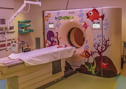
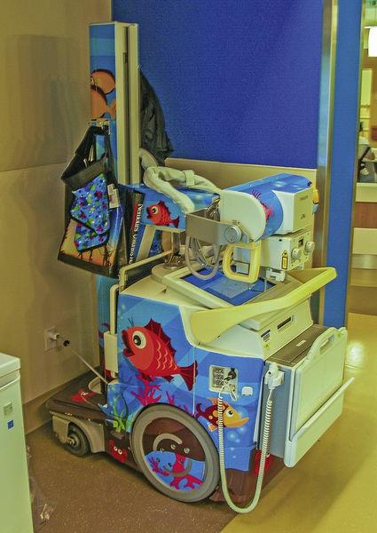
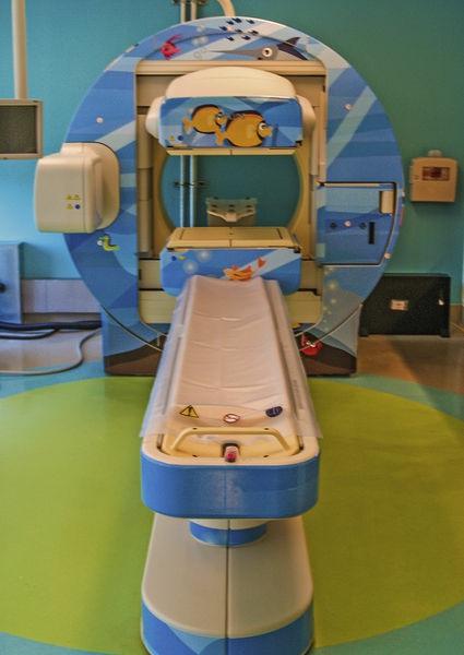
Infants and young children are commonly unable to cooperate with requirements that typically are easily met by adults For example, they may be unable to keep still, remain in a certain position, concentrate for more than a brief moment, or breath-hold Children of various ages have unique limitations. Infants and toddlers are unable to stay still, whereas 3-year-olds are more apt to refuse to cooperate. These limitations affect almost all pediatric imaging examinations: radiography, fluoroscopy, ultrasound, computed tomography (CT), magnetic resonance imaging (MRI), nuclear imaging, and interventional radiology. There are a number of potential solutions that can be helpful in these situations Commonly employed techniques include distracting the child, providing child-friendly surroundings (Figs 1-1 through 1-7), immobilization, and sedation
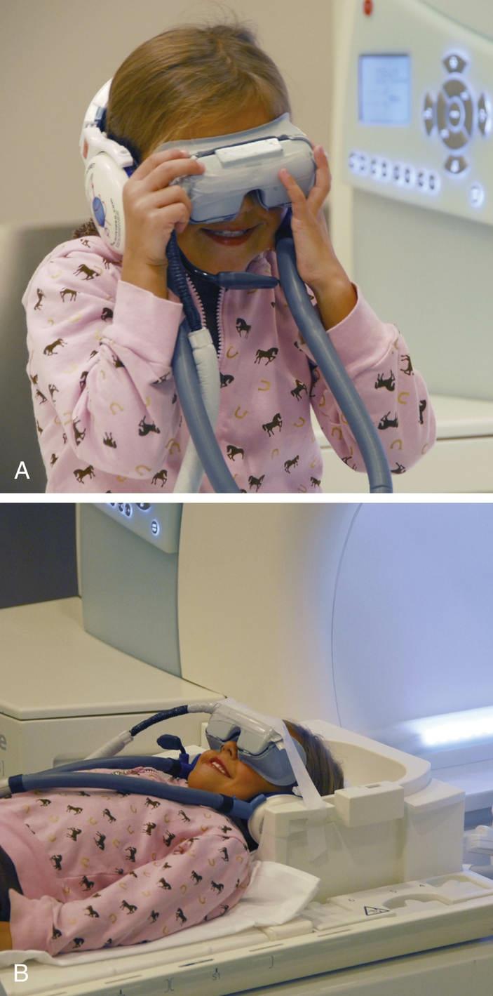
Distracting the child with something other than the procedure is often a simple and easy tactic to use Talking to older children about school and other activities can be helpful Certified child-life specialists are very successful in helping to coach and distract children so that they can complete imaging exams without sedation. They often use rattles and noise-making toys with very young children. Video players are a very useful distraction technique for ultrasound, fluoroscopy, and CT, and video goggles (see Fig. 1-7) have been very successful in decreasing sedation for MRI. Children can be encouraged to bring their own movies or choose from the department’s stock It is amazing how cooperative many children will be when they are able to watch television Using a combined program that includes the introduction of a child-life specialist, a combination of the tactics discussed earlier to calm infants and young children, and the promotion of a culture that avoids sedation whenever possible was shown to reduce the frequency of need for sedation in children less than 7 years of age by
▪ FIGURE 1-1 Colorful, child-friendly décor in pediatric waiting area Many children’s hospitals are now being decorated with modern, brightly colored, open areas without “cartoonish” themes.
▪ FIGURE 1-2 Child-friendly waiting area with dancing cows
▪ FIGURE 1-3 CT scanner decorated with child-friendly decals
▪ FIGURE 1-4 Portable radiograph unit decorated with child-friendly decals
▪ FIGURE 1-5 SPECT/CT unit decorated with child-friendly decals
▪ FIGURE 1-6 Imaging roomoutfitted with customlighting control, at this time turned to pink at request of child
Providing child-friendly surroundings may help to ease a young child’s anxiety and cause him or her to be more cooperative Paintings on the walls and equipment and cartoonish figures in the examination rooms can be helpful Eliminating or minimizing painful portions of the examination can also be very helpful in keeping a young child cooperative. The placement of an intravenous line often causes a great deal of patient anxiety and renders the child uncooperative for a subsequent imaging study, such as a CT scan. Using topical analgesia to decrease the pain of the intravenous line placement commonly makes this portion of the examination less traumatic. In addition, it is helpful to schedule appropriate sequencing of imaging examinations so that the most difficult exam is performed last For example, it can be much easier to perform a renal ultrasound before rather than after the child has experienced a voiding cystourethrogram
Immobilization is also a helpful technique Infants who are bundled or “papoosed” in a blanket are more apt to stay still than infants who are not. This may make the difference between needing or not needing sedation to obtain an examination. There are also a number of commercially available
immobilization devices that are helpful when performing certain examinations, such as the use of an octagon board when obtaining fluoroscopic studies of young children There are other devices that enhance proper positioning for specific modalities, such as chest radiographs Imaging departments that image children should consider making such equipment available.
In certain situations, distraction and immobilization may not be successful, and sedation or general anesthesia may be necessary to obtain imaging studies. Many children younger than 6 years of age require sedation for MRI studies because of the prolonged nature of the examination and the need for the patient to be completely still Sedation is needed much less often nowthan in the past for children undergoing CT examination because of the increased speed of acquisition by the newer CT scanners and the previously mentioned sedation reduction program
Other procedures that might require sedation include some nuclear medicine studies and most interventional procedures
▪ FIGURE 1-7 Video goggles can help young children to cooperate for MR examination, thus avoiding sedation. A, Video goggles on a child preparing for an MR examination. B, Video goggles with audio headphones in place as child is slid into scanner Note happy demeanor
Standards of care for conscious sedation are required by The Joint Commission and are based on standards published by several organizations, including the Committee on Drugs and the American Academy of Pediatrics Any imaging department planning to sedate children must have a defined sedation program that is in concordance with these guidelines The sedation program must have protocols for presedation preparation, sedative agents used, monitoring during sedation and during postsedation recovery, and discharge criteria. There has been a national shift concerning who provides sedation for pediatric imaging studies in pediatric radiology departments. In the 1990s’most pediatric radiology sedation programs were run by radiologists. Currently, multiple factors have led to such programs being run by anesthesiologists, emergency physicians, or intensive care physicians. At many institutions, such physicians have access to sedatives that are better for imaging sedation, such as propofol or dexmedetomidine
Variable Size and Physiology
Because of the size variability from infant to adult-sized children, many adaptations must be considered for pediatric imaging studies in relation to size. The doses of contrast and drugs used in imaging examinations need to be adjusted according to a child’s size, often on a per-weight (mg/kg) basis. Oral contrast dosing is also based on patient weight or age. Using CT as an illustrative example, other variables may also be affected by patient size In small children the largest possible intravenous line may be very small, often 22 gauge or 24 gauge The intravenous line may be placed in the foot or hand The length of the region of interest to be imaged is variable, and the lengths of the patient’s veins are variable Physiologic parameters, such as the patient’s cardiac output, are also more variable in children than in adults. These factors affect parameters such as the time between contrast injection and onset of scanning, as well as choices in contrast administration technique (hand bolus versus power injector). Slice thickness should be smaller in younger children because of the smaller anatomic parts. Similar adjustments must be considered in all other imaging modalities when applied to children. Radiation dose reduction is discussed in Patient Safety
Age-Related Changesin Imaging Appearance
Another factor that makes imaging in children different from that in adults is the continuous changes in the imaging appearance of multiple organ systems during normal childhood development The normal imaging appearance of certain aspects of organ systems can be different both at varying ages during childhood and between children and adults. For example, the kidneys look different on ultrasound in neonates from the way they look in a 1-year-old child. The developing brain demonstrates differences in signal at varying ages on MRI, which is related to changes in myelination. Alarge mediastinal shadowrelated to the thymus may be normal or severely abnormal depending upon the child’s age The skeleton demonstrates marked changes at all ages of childhood; this is related to the maturation of apophyses and epiphyses and the progressive ossification of structures Knowledge of the normal age-related appearances of these organ systems is vital to appropriate interpretation of imaging studies. Lack of this knowledge is one of the more common causes of errors made in the interpretation of pediatric imaging studies.
Age-Related Differential Diagnoses
The types of diseases that affect children are vastly different from those that commonly affect adults. Therefore the differential diagnosis and significance of a particular imaging finding in a child are dramatically different from those determined by the identical imaging finding in an adult. In addition, the diseases that affect specific age groups of children are different Therefore the differential diagnosis and significance of a particular imaging finding in a 2-month-old infant may differ dramatically from those determined by the identical imaging finding in a 10-year-old child
Quality and Patient Safety
Alot of national attention has been paid to patient safety initiatives since the 1999 Institute of Medicine’s report stating that somewhere between 44,000 and 98,000 deaths per year are caused by medical errors in the United States alone. This poor safety record would be the equivalent of the airline industry’s having a large passenger plane crash in the United States every single day! If this were the case, we would probably think twice about flying However, this is what our patients potentially face when they enter the current health care system If looked at from the patients’perspective, even more important to them than “Heal me” (quality of care) and “Be nice to me” (customer service) is the plea “Don’t harm me” (patient safety) No higher priority exists than patient safety
There are numerous schools of thought related to both improvement and safety Almost all of them emphasize the cultural aspects needed to reach a level of high reliability and minimize errors that may cause harm There has to be a recognition that safety is an issue and that it is part of everybody’s role to speak up in the face of uncertainty or when an individual “feels” that something is not right. The old culture of a medical hierarchy, in which the physician is in charge and is not to be questioned, does not promote safety. Medical staff, trainees, and even family members need to feel comfortable “stopping the line” and asking for clarification if things do not seem right.
Also important to create a reliable system of care that is both safe and of high quality is the acceptance of standardization Henry Ford stated in 1926 that
Today’s standardization is the necessary foundation on which tomorrow’s improvement will be based. If you think of “standardization” as the best you know today, but which is to be improved tomorrow you get somewhere But if you think of standards as confining, then progress stops
Ideally the only variation in a health care system should be that related to the condition of the patient. There should not be variability related to technologists, protocols, care sites, or physicians. Radiologists need to work together to create evidence-based standardized imaging protocols and procedures, as well as reports, and continuously strive to improve them
In addition, key in reaching a state in which high quality and safe care are provided is a robust daily management system Daily management systems are designed to quickly identify issues, empower front-line areas to solve those issues, and, when the front line cannot resolve them, escalate the issues to those who can help. Increasingly in medicine, as well as other fields, tiered huddle systems are used to create daily readiness. Radiologists, technologists, and managers come together in a brief huddle each morning to make sure that they are ready to care for the patients scheduled for that day. Such processes often have three parts: daily readiness, problem accountability, and metrics evaluation. One approach to daily readiness is evaluating the volume scheduled for that day and organizing the approach to concerns around safety and the acronym MESA(Methods, Equipment, Supplies, Associates) Does anyone have any safety concerns? Do we have the right Methods to take care of the patients today (does anyone have questions around protocols or atypical patients?)? Do we have the right Equipment to take care of the patients today (is there any planned downtime, broken equipment, or information technology [IT] issues?)? Do we have the Supplies we need to take care of the patients today? Do we have the right Associates to take care of the patients today (did anybody call in sick, do we have the right people with the right expertise)? Going through a set of such questions leads to a list of issues Having a defined problem accountability process that assigns each issue a single owner and defines the immediate countermeasure and the date at which the owner will come back to report an update at the huddle is important so that issues do not go unresolved or only partially remedied
Radiation Safety
Safety issues specific to radiology include radiation safety, MRI safety, and correct and effective communication of the information in and interpretation of imaging examinations We will touch here on radiation safety because it is germane to pediatric radiology Although there is much uncertainty, children are much more sensitive to the potential harmful effects of radiation than are adults, and children also have a longer expected life span during which to develop potential complications of radiation, such as cancer. Therefore attention to radiation safety in all areas of pediatric radiology is paramount. CT delivers higher doses of radiation than do other diagnostic imaging modalities. The exact radiation risk in CT examinations and even whether a risk absolutely exists are controversial topics However, some researchers estimate the increased risk that a young child might develop cancer related to an abdominal CT scan is in the magnitude between 1:1000 and 1:10,000 Given the unknown and potentially small risk, it is essential for all radiologists to practice dose-reduction techniques in
pediatric CT Such tactics include avoiding CT when unnecessary; using alternative diagnostic methods that do not use radiation, such as ultrasound, when possible; and adjusting CT parameters to minimize dose when CT is performed Because children are smaller than adults and need less radiation to create the same signal-to-noise ratios, the tube current (mA), as well as kilovolts and other factors, can be greatly reduced when imaging a small child. Many other factors can be adjusted to reduce dose as well. It is also very important not to overreact to this potentially small risk related to radiation dose for CT. For any clinically indicated examination the risk of not doing the CT and not having that information is often magnitudes greater than that related to radiation risk
Suggested Readings
Donnelly L F Daily management systems in medicine RadioGraphics 2014;34:549–555
Donnelly L F, Dickerson J M, Goodfriend M A, Muething S E Improving patient safety: effects of a safety program on performance and culture in a department of radiology AJR 2009;193:165–171
Donnelly L.F, Strife J.L. How I do it: establishing a program to promote professionalism and effective communication in radiology Radiology 2006;283:773–779
Frush D.P. Overview of CT technologist for children. Pediatr Radiol. 2014;44:422–426.
Frush D P, Bisset G S Pediatric sedation in radiology: the practice of safe sleep AJR 1996;167:1381–1387
Frush D P, Goske M J Image gently: toward optimizing the practice of pediatric CT through resources and dialogue Pediatr Radiol 2015;45:471–475
Institute of Medicine Crossing the quality chasm: a new health system for the 21st century Washington, DC: National Academy Press; 2001.
Khan J J, Donnelly L F, Koch B L, et al A program to decrease the need for pediatric sedation Appl Radiol 2007;4:30–33.
Larson D B, Towbin A J, Pryor R M, Donnelly L F Improving consistency in radiology reporting through the use of department-wide standardized structured reporting Radiology 2013;267:240–250
Pichert J W, Miller C S, Hollo A H, et al What health professionals can do to identify and resolve patient dissatisfaction Jt Comm J Qual Improv 1998;124:303–312
Thrall J.H. Quality and safety revolution in health care. Radiology. 2004;233:3–6.
2
Airway
Lane F Donnelly
Problems with the airway are much more common in children than in adults. It has been said that one of the differentiating features between a pediatric and general radiologist is that a pediatric radiologist remembers to look at the airway For practical purposes, abnormalities of the airway can be divided into acute upper airway obstruction, lower airway obstruction (extrinsic compression, intrinsic obstruction), obstructive sleep apnea (OSA), and congenital high airway obstruction syndrome (CHAOS) Clinically, children with acute upper airway obstruction (above the thoracic inlet) tend to present with inspiratory stridor, whereas children with lower airway obstruction (belowthe thoracic inlet) are more likely to present with expiratory wheezing. However, the categorization of a child with noisy breathing into one of these two groups can be very difficult. The primary imaging evaluation of the pediatric airway for acute conditions should include frontal and lateral high-kilovolt radiography of the airway and frontal and lateral views of the chest
▪ AcuteUpper Airway Obstruction
Acute stridor in a young child is the most common indication for imaging the pediatric airway The most common causes of acute upper airway obstruction in children include inflammatory disorders and foreign bodies The most common inflammatory disorders include croup, epiglottitis, exudative tracheitis, and retropharyngeal cellulitis and abscess Anatomic structures that are especially important to evaluate on radiographs of children with acute upper airway obstruction include the epiglottis, aryepiglottic folds, subglottic trachea, and retropharyngeal soft tissues.
Croup
Croup (acute laryngotracheobronchitis) is the most common cause of acute upper airway obstruction in young children. The peak incidence occurs between 6 months and 3 years of age. The mean age at presentation of croup is 1 year of age. In children older than 3 years, other causes of airway obstruction should be suspected. Croup is viral in cause and is usually a benign, self-limited disease. Redundant mucosa in the subglottic region becomes inflamed, swells, and encroaches upon the airway The children present with a barky (“croupy”) cough and intermittent inspiratory stridor It usually occurs following or during other symptoms of lower respiratory tract infection Most children with croup are managed supportively as outpatients, and the parents are managed by reassurance Inhaled corticosteroids are becoming a popular therapy in children with croup. They have been shown to reduce the length and severity of illness.
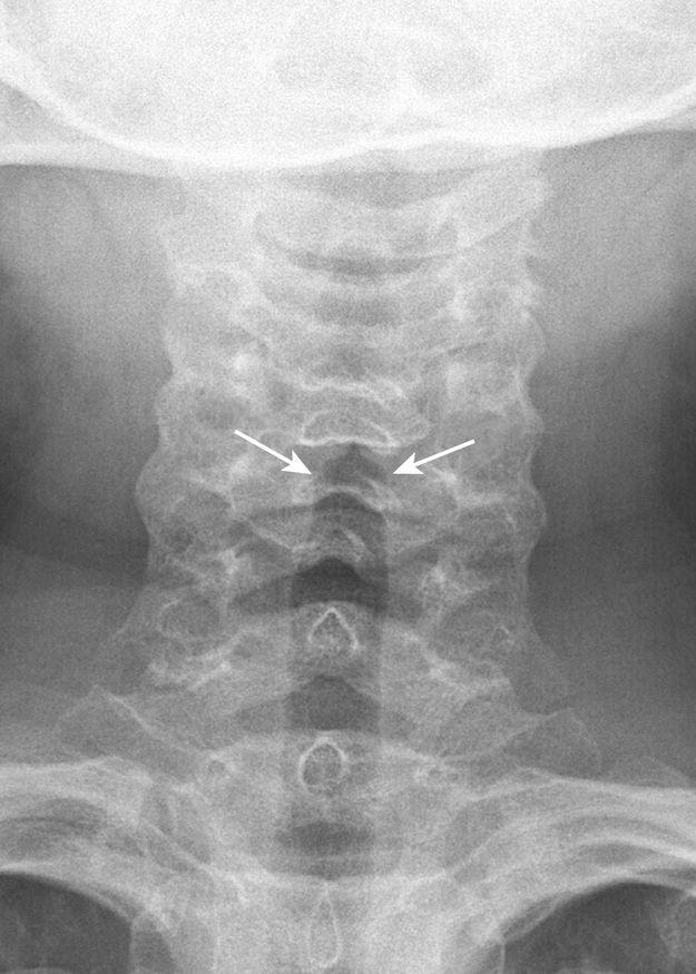
The purpose of obtaining radiographs in a patient with suspected croup is not so much to confirm the diagnosis but rather to exclude other, more serious causes of upper airway obstruction that require intervention However, characteristic radiographic findings that indicate croup are best seen on frontal radiographs With croup, there is loss of the normal shoulders (lateral convexities) of the subglottic trachea secondary to symmetric subglottic edema (Fig 2-1) Normally, the subglottic trachea appears rounded, with “shoulders” that are convexoutward (Fig 2-2) In croup, the subglottic trachea becomes long and thin, with the narrowportion extending more inferiorly than the level of the pyriform sinuses. The appearance has been likened to an inverted V or a church steeple (see Fig. 2-1). The term church steeple can be confusing because some steeples look like croup and some are shaped like the normal subglottic airway (Fig. 2-3). Lateral radiographs may demonstrate a narrowing or loss of definition of the lumen of the subglottic trachea (see Fig 2-1) or hypopharyngeal overdistention With croup, the
epiglottis and aryepiglottic folds appear normal.
Epiglottitis
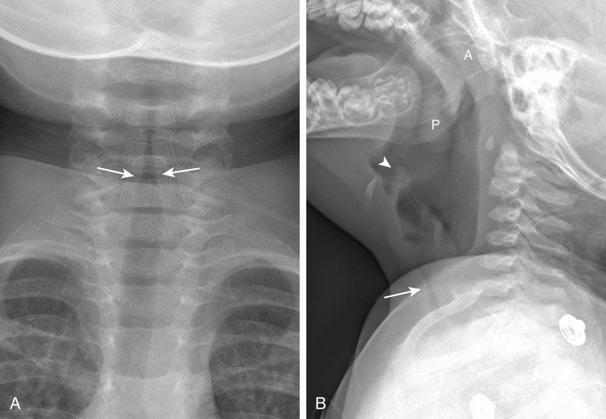
In contrast to croup, epiglottitis is a life-threatening disease that can potentially require emergent intubation. The possibility that a child with epiglottitis might arrive in a deserted radiology department was once a constant source of anxiety for on-call radiology residents. However, most cases of epiglottitis are caused by Haemophilus influenzae and are nowpreventable by immunization (HiB vaccine), so the incidence of epiglottitis has dramatically decreased The causes of epiglottitis are nowalso more heterogeneous Related to this, care of children with epiglottitis is nowmore of a challenge because health care workers are less used to recognizing and treating patients with this disorder Children with epiglottitis are usually toxic appearing and present with an abrupt onset of stridor, dysphagia, fever, restlessness, and an increase in respiratory distress when recumbent. In the pre-HiB vaccine era, the classically described peak age of incidence was 3.5 years. However, since the introduction of the HiB vaccine, there has been a marked increase in the mean age of presentation to 14.6 years. Because of the risk for complete airway obstruction and respiratory failure, no maneuvers should be performed that make the patient uncomfortable If the diagnosis is not made on physical examination, a single lateral radiograph of the neck should be obtained, usually with the patient erect or in whatever position that allows the patient to breathe comfortably Children with epiglottitis should never be made to lie supine against their will to obtain a radiograph because it can result in acute airway obstruction and, potentially, death.
A, Frontal radiograph showing symmetric subglottic narrowing (arrows) with loss of normal shouldering. The narrowing extends more inferiorly than the piriformsinuses. B, Lateral radiograph showing subglottic narrowing (arrow) Note normal-appearing epiglottis (arrowhead) and thin aryepiglottic folds Also note mildly enlarged adenoid (A) and palatine (P) tonsils
▪ FIGURE 2-1 Croup
▪ FIGURE 2-2
Normal frontal radiograph of the airway The subglottic airway demonstrates rounded shoulders (arrows) that are convexoutward.
▪ FIGURE 2-3 Steeple sign.
The term steeple sign can be confusing It is meant to denote the pointed configuration of the subglottic trachea on a frontal radiograph of the airway when subglottic edema has effaced the normally convexlateral shoulders in this region However, some steeples look like croup (white arrows), and some look like a normal subglottic airway (black arrow)
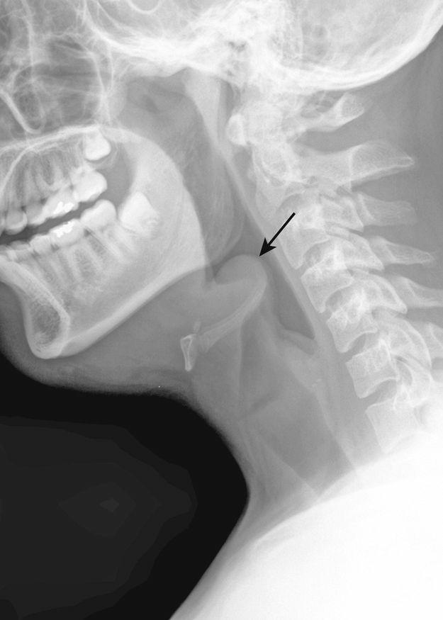
▪ FIGURE 2-4 Epiglottitis
Lateral radiograph showing marked thickening of the epiglottis (arrow). The aryepiglottic folds are also narrowed
With epiglottitis, on the lateral radiograph, there is marked enlargement of the epiglottis. A normal epiglottis typically has a thin appearance with the superior aspect being sharply pointed. The swollen epiglottis has been likened to the appearance of a thumb. With epiglottitis, there is also thickening of the aryepiglottic folds (Figs. 2-4 and 2-5). The aryepiglottic folds are the soft tissues that extend from the epiglottis anterosuperiorly to the arytenoid cartilage posteroinferiorly and normally are convex downward When the aryepiglottic folds become abnormally thickened, they appear convexsuperiorly Symmetric subglottic narrowing, similar to croup, may be seen on frontal radiography (if obtained); do not let that be confusing
▪ FIGURE 2-5 Epiglottitis
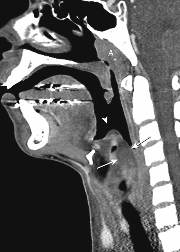
Sagittal CT image showing low-attenuation swelling of the epiglottis (arrowhead). Also note marked thickening and low-attenuation edema of the aryepiglottic folds (arrows)
An obliquely imaged, or so-called omega-shaped, epiglottis may artifactually appear wide because both the left and right sides of the epiglottis are being imaged adjacent to each other This should not be confused with a truly enlarged epiglottis The absence of thickening of the aryepiglottic folds can be helpful in making this differentiation. With an omega-shaped epiglottis (normal variant), often both the left and right walls of the epiglottis are visible.
In current times, related to both the uncommon occurrence of epiglottitis and the frequent reliance on computed tomography (CT) to evaluate more common inflammatory neck conditions (such as retropharyngeal abscess), it is increasingly more common to see and diagnose epiglottitis on CT rather than on radiography Although not classically advocated as a diagnostic tool for epiglottitis (given the risks of laying such patients supine and giving them intravenous [IV] contrast), the findings of epiglottitis are easily identified on CT (see Fig 2-5) Findings include swelling and low-attenuation edema of the epiglottis and aryepiglottic folds associated with inflammatory stranding in adjacent fat.
Exudative Tracheitis
Exudative tracheitis (also known as bacterial tracheitis, membranous croup, or membranous laryngotracheobronchitis) is another uncommon but potentially life-threatening cause of acute upper