s:to
Anterior ethmoid n.
Angular a.
Transverse facial a.
Lateral nasal a.
Superior labial a.
Inferior labial a
Facial a.
Massater m.
1.1 Regional anatomy based on masseter muscle and arterial system of the face.
Temple Eyelid
Nasofacial sulcus Dorsum
Upper cutaneous lip
Mandibular border
Lower cutaneous lip
Chin
Fig. 1.3 Cosmetic units and junction lines.
anticipated in these areas, scars tend to visually disappear when placed within them. Conversely, scars crossing junction lines stand out as noticeable.
Cosmetic units are the areas cordoned off by the junction lines. They share common characteristics of skin color and texture, pore size, elasticity, thickness, presence or absence of hair, and shading. The cheek, temple, chin, and eyelids are their own well-defined units, whereas the nose, cheek,
Hairline
Nasal root
Lateral sidewall
Nasal
dorsum
Ala nasi
Nasal tip
Philtrum
Melo-labial fold
Infra oral crease
Vermillion–lip
Vermlion–cutaneous
junction
Labiomental line
and ear are subdivided into smaller units. The nose in particular has been defined to include the root, dorsum, lateral side walls, paired alae nasi, soft triangles, columella, and tip. Several useful principles of closure have been derived from the conceptualization scheme noted previously. These include:
■ Scars should be oriented within junction lines or within or parallel to the RSTLs.
■ Closures should be confined to a single cosmetic unit when possible.
■ When closure is not possible within a single cosmetic unit, skin should be recruited from adjacent units and scar lines oriented so they lie within the intervening junction lines and within or parallel to the RSTLs.
■ When a defect involves several cosmetic units, consider repairing each unit individually.
■ When most of a cosmetic unit or subunit is missing, con sider sacrificing the remaining portion and replace he entire unit.1
FREE MARGINS: CONCEPT OF TENSION VECTOR OF CLOSURE
Another important concept when performing facial surgery is that of the free margins—the eyebrows, eyelids, lips, and nostril rims. These structures are extremely important, as they offer little resistance to the forces of wound closure and can be easily distorted by excess tension. This can occur from the immediate direct exertion of tension by a side-toside closure or the delayed application of tension as a second intention healing wound or split-thickness graft site contracts. The resulting asymmetries can be both cosmetically unsettling and functionally disabling Ectropion of the
Fig.
Scowl lines
Crow’s feet
Fig. 1.2 Relaxed tension ines of the face.
lower lid can lead to permanent visual problems, while eclabion and lack of a proper oral seal can cause problems with phonation, eating, and drinking, in addition to being aesthetically unpleasing.
Tension vectors of closure can be favorably manipulated by one of several techniques:
■ Offset bias suturing: Each suture is skewed a bit from its anticipated placement 90 degrees to the long axis. The cumulative effect is to favorably alter the tension vector the requisite number of degrees to avoid pulling the free margin in question out of position.
■ Flaps: Tissue rearrangement is designed so the tension vector is favorably disposed and avoids pulling on free margins.
■ Suspension or anchoring suture: Instead of directing buried suture toward the free margin, a suture is placed from the undersurface of the closure down to and anchored to the underlying periosteum or perichondrium of the area. All tension is then directed downward toward these strong, unyielding structures, and away rom the eyelid or eyebrow at risk of being distorted.
■ Full-thickness skin graft: Placement of a full-thickness graft prevents wound contraction and the development of a tension vector of closure in any direction.
Concavities and Convexities: Implications for Second Intention Healing
The face is not a flat structure. Rather, it is composed of undulating well-defined elevations and depressions that need to be taken into consideration when planning closures. Scar lines that traverse from convexity to concavity may contract in an unbecoming manner and may require preplanning with an S-plasty configuration for appropriate closure. However, the major effect of whether an area is flat, concave, or convex determines how well defects resolve when allowed to heal by second intention. Although the size and depth of the defect have some impact on the final healing, defects on concave surfaces such as the temple, the medial canthus, and the alar groove do well, whereas defects on convex surfaces such as the malar eminence the tip of the nose, and the chin generally heal poorly with webbed or elevated scars. Defects on flat surfaces, such as the forehead or cheek, typically respond somewhere in between.
The Aging Face
With time, predictable wrinkles and sagging take place. This is compounded by changes related to overexposure to ultraviolet radiation in sunlight. Up until 30 years of age, people do not have wrinkles at rest (Glogau I). The RSTLs first appear as hyperanimation lines perpendicular to the pull of the underlying muscles of facial expression (Glogau II). Crow’s feet lines and small crinkles under the eyes when smiling are often the first to become noticeable. With time, the elastic tissue and collagen fascicles that traverse the
subcutaneous fat compartment and bind the muscles of facial expression to undersurface of the dermis degenerate and the RSTLs become permanently etched on the face (Glogau III) If the patient has significant photo-damage with deposition of solar elastosis within the papillary dermis, the lines become even more prominent, usually with a pronounced roadmap of lines all over the face (Glogau IV).
Aging has a profound effect upon the facial skeleton, the greatest on the bones of dental origin, the maxilla and mandible. These recede in their anterior proection and height beginning at a relatively early age Regression of the orbital rim creates inferolateral and superomedial enlargement of the orbit causing loss of support for the periorbital soft tissues (Fig. 1.4). Enlargement of the piriform aperture deepens the nasolabial groove, and these changes, previously attributed to gravity, create laxity of restraining fascial tissues and a generalized sagging of the skin that manifests as brow ptosis, dermatochalasis of the upper eyelids, hollowing under the eyes, vertical lines in the preauricular area, deepened melo-labial folds, rhytids of the perioral area, and pronounced jowls. These areas of redun dant and excess, along with the temple and the glabella, constitute the reservoirs of skin available for recruitment for tissue rearrangemens
Generalized lipodystrophy of subcutaneous fat and specific resorption of the facial fat compartments and particularly the buccal fat pad (Figs. 1.5 and 1.6) leads to a volume depletion of the face, resulting in a sunken appearance of the cheeks and a hollowing of the temples.4 What used to be thought of as a simple act of gravity creating a sagging face is now known to be a much more complicated interaction of fat atrophy, bone resorption, dynamic musculature, and changes in collagen and elastin.
Damage also results in splotchy hyperpigmentation and hypopigmentation; vascular changes in the form of elangiectasia of the cheeks and poikiloderma of the neck and
Fig. 1.4 Facial skeletal resorption with arrow ize indicating degree.
Subcutaneous midligament line
Retnacula cutis A´ A B B
Retaining ligament
Fg 15 SMAS, Superficial musculoaponeurotic system.
Dermis
Sub-Q
Fig. 1.6 Retaining ligament.
Tear trought lig.
Upper masseteric cutaneous lig.
rims of the ears; and the signature damage of ultraviolet radiation, the deposition of solar elastotic material in the papillary dermis, giving the skin a yellowish, thickened, and leathery appearance.
An accurate assessment of the aging face is important not only in correctly judging where there is available skin for recruitment in tumor defect repair but also for determining which cosmetic procedure is most applicable for any partcular patient. An upper lid blepharoplasty may only compound severe brow ptosis if that condition is not also addressed. Similarly, it is important to recognize if a resurfacing (ablative laser, chemical peel, etc.), revolumizing (fillers), or tissue tightening procedure (radiofrequency, ultrasounds, etc.) would offer a particular patient the greatest benefit.
Mandibular cutaneous lig.
Mandibular septum
Periosteum/ deep fascia
Bone SMAS
Superior temporal septum
Inferior temporal septum
Orbicularis retaining lig.
Zygomatic cutaneous ligaments
Plastysma auricular fascia
Masseteric cutaneous ligaments
The Musculoaponeurotic System
INTRODUCTION
Humans communicate by use of the muscles of facial expression (the mimetic muscles). By use of this silent mode of interacting, human discourse is enriched by nuance and subtlety. Shades of annoyance, reverie, indifference, skepticism, sarcasm and so on are molded onto the spoken word. Muscles of facial expression are unique in that they are the only muscles to insert into the skin. They do so via fibrous septae that connect the superior portion of the muscle to the undersurface of the dermis and are part of the branching network of fibers known as the retinacular cutis. This is part of a larger complex system of fibrous septa
1.7
Middle forehead compartment
Central forehead compartment
Sup. orbital compartment
Inf. orbital compartment
Nasolabial compartment
Medial cheek compartment
Sup. jowl compartment
Sup. temporal septum
Inf. temporal septum
Lat. temporal-cheek compartment
Orbicularis retaining lig.
Inf jowl compartmen
Mandibular cutaneous lig.
in the subcutaneous layer of the face that connect to the retaining ligaments and contribute to the septa that create the fat compartments of the face. They also insert or interdigitate with the other mimetic muscles. So while the frontalis muscle wrinkles the forehead and raises the eyebrow it helps open the eye widely by partially inserting ino the upper fibers of the orbicularis oculi muscle (Fig 17).
Innervation of the muscles is exclusively y branches of the facial or cranial nerve VII This occurs at the lateral undersurface of the muscle The muscles are most effective and concentrated in the midplane of the face and exert their major effect around the two major orifices of the face—the eyes and the mouth.
The aponeurotic component is made up of retaining ligaments that are strong and deep fibrous attachments that originate from the periosteum or deep fascia and travel perpendicularly through the facial layers to insert in the dermis (Figs. 1.8 and 1.9). These ligaments act as anchor points, retaining and stabilizing the skin and superficial fasca (superficial musculoaponeurotic system [SMAS]) to the underlying deep fascia and skeleton They tend o be more laterally displaced on the face in the SMAS of the cheek and the superficial temporalis fascia of the temple, which has a superior temporal septum and an inferior temporal septum. The orbicular retaining ligament surrounds the orbit connecting medially to the tear trough ligament (Fig. 1.10). The other major components of the aponeurotic system are the zygomatic cutaneous, masseteric, and mandibular ligaments and the galea aponeurotica is spread over the expanse of the skull, connecting the anteriorly displaced frontalis muscle with the occipitalis muscle of the neck.3 These ligaments and fascial components restrict cutaneous mobility and may be necessary to surgically disrupt in order to mobilize tissue for closures. It is also important to recognize that these retaining ligaments often share intimate relationships with branches of the facial nerve, so knowl-
Lat. orbital compartment
Sup. cheek septum
Mandibular septum
Middle cheek compartment
Platysma auicular lig.
Massenteric cutaneous ligaments
Sub orbicularis oculi fat Deep medial
Sub mental
1.8 Fat compartments.
edge of their anatomic relationship is essential to avoid nerve damage during dissection.
MUSCLES ACTING AROUND THE EYELIDS
The frontalis muscle is the primary muscle of the forehead, and its main function is to wrinkle the skin of the forehead and elevate the eyebrow. Accordingly, it has been called the “surprise” muscle. Through its interdigitations with the upper fibers of the orbicularis oculi muscle, it also asssts in opening the eye widely. Injury to the temporalis muscle results in flattening the skin of the forehead brow ptosis,
Fig.
Retaining ligaments and fat compartments of the face.
Buccal
Temporal fat pad
Lateral
Periauicular
Fig.
Skin
Frontalis m.
Corugator supercilli m.
Procerus m.
Orbicularis oculi m.
Fig. 1.9 Muscles of facial expression.
Nasalis
Levator labii superioris alaeque nasi
Levator labii superioris
Subgalea plane
Galea
Zygomaticus minor
Levator labii anguli oris
Sup. temporal septum
Inf. temporal septum
Temporal adhesion
SMAS
Sub-Q
Zygomaticus major
Lat. orbital thickening
Sub-SMAS plane
Zygomatic ligament
Deep fascia
Mandibular ligament
Menals
Risoius
Platysma
Depressor labii inferiorsi
Depressor anguli oris
and the larger transverse head that runs laterally to insert widely into the skin of the eyebrow. It functions mainly to pull the eyebrow medially and slightly downward, and creates the vertical scowl lines of the glabella. Recently the depressor supercilii has been described. It arises just above and deep to the corrugator supercilii and extends vertically superior to also attach into the skin of the medial eyebrow. It appears that there are three muscles that act in concert to depress the medial eyebrow: the most deeply placed depressor supercilii, the middle oblique head of the corrugator supercilii, and the vertically oriented fibers o the medial/superior orbicularis oculi muscle.
Masseteric ligaments
The key to the orbital region is he large, sphincter-like circumferential orbicularis ocul muscle. It has both outer orbital and inner palpebral portions. The orbital portion originates broadly from the superior and inferior bony orbital rims and inserts into the medial and lateral canthal tendons, as well as the superficial temporalis fascia. It also interdigitates with fibers from the frontalis, corrugator supercilii and procerus muscles. The orbital division is under voluntary control and acts to close the eye tightly and depress the eyebrow. Unilateral contraction results in a wink. The palpebral portion over the orbital septum and tarsal plate acts to gently close the eye (voluntary) or blink (involuntary). The muscle is innervated by fibers of the temporal and zygomatic branches of the facial nerve.
Fig. 1.10 Sagittal section of the rtaning ligaments. SMAS, Superficial musculoaponeurotic sysem.3
and accentuation of the effects of dermatochalasis and upper visual field gaze.
The corrugator supercilii muscle has come under intense nterest with widespread popularity of botulinum toxin injections. The muscle originates from the frontal bone of the medial orbit in line with and just above the medial canthal tendon insertion. It has two slips—the oblque head that runs a short distance superiorly to insert into the skin of the medial eyebrow and helps depress the medial brow,
MUSCLES ACTING AROUND THE NOSE
The nose is relatively devoid of musculature when compared with the eyes and lips. The muscles consist mainly of the procerus muscle that extends down from the frontalis vertically onto the upper nose across the root and into the aponeurosis of the nasalis muscle on the nasal dorsum. Contraction, usually in concert with the adjacent lip elevators, “scrunches” up and shortens the nose and causes the transverse RSTLs across the root. The levator labii superioris alaeque nasi arises from the maxilla under the medial orbicularis oculi and descends vertically to the mid-upper
lip, but also sends slips to the lateral ala nasi. In concert with alar fibers of the nasalis muscle, it aids in dilating the nostril with each inspiration. The broad thin nasalis muscle is the intrinsic nasal muscle with alar fibers just discussed, transverse fibers over the dorsum that just tense the skin over the dorsum, and the depressor septi portion that pulls down the septum and aids in deep inspiration.
MUSCLES ACTING AROUND THE MOUTH
As noted earlier, the mouth is the most expressive part of the face. This is due to the myriad number of muscles that not only insert into the overlying skin but also insert and exert motion from every possible direction into the sphincterlike orbicularis oris muscle. Further modulation of fine muscular movement is aided by a structure called the modiolus (hub, Latin), from which the orbicularis originates. Lying about 1 cm lateral to the oral commissure it is a muscular platform-like confluence of several of the muscles inserting into the orbicularis and acting on the orner of the mouth, including the zygomaticus major, levator anguli oris, risorius, depressor anguli oris, buccinator, and platysma muscles. Actions are amplified or muted by finetuning of the synergistic and antagonistic muscles. The action of the orbicularis muscle itself is to draw the lips together and to pucker the lips.
There are four lip elevators. From most medial to lateral, they include the levator labii superioris alaeque nasi, levator labi superioris, zygomaticus major, and zygomaticus minor. They are all important in smiling, sneering, and snarling movements. The upper lip and corner of the mouth are retracted and elevated by the risorius and levator anguli oris muscles. The buccinator muscle, beloved by trumpet players, plays an important role in chewing by keeping food tucked inward between the teeth
The muscles working on he lower lip include the mentalis muscle, which wrinkes the skin of the chin and also helps pouch out and protrude the lower lip (sniveling); the depressor anguli oris, which retracts and depresses the corner of the mouth; and the depressor labii inferioris muscle, which, as the name implies, depresses the lower lip. The depressor anguli oris can be injected with botulinum toxin to improve the down turning of the corner of the mouth that occurs with age. The latter two are important (along with the upper lip elevators and angle retractors) n attaining the wide, happy, toothy grin associated with laughing. The marginal mandibular nerve innervates all three muscles. Finally, the platysma muscle, innervated by the cervical nerve, stretches broadly from the chest upward across the expanse of the neck over the jaw line to blend with the angle of the mouth depressors. Despite being thin in some people, it helps cover and protect the marginal mandibular nerve at the anterior border of the masseter muscle at the jaw line.
Motor Nerves
INTRODUCTION
There are two kinds of muscles of the face: the muscles of facial expression and the skeletal muscles of mastication
(pterygoid, masseter, and temporal muscles). The former are innervaed entirely by the facial (VIIth cranial) nerve, whereas the latter are supplied by motor fibers of the trigeminal (Vth cranial) nerve. The facial nerve has the greatest clinical relevance to dermatologic surgeons and will be the topic of this section.
FACIAL NERVE (CRANIAL NERVE VII)
The facial nerve is a complex structure with an anatomy that varies from patient to patient. The possibility of injuring or severing one of the branches of this nerve is of paramount importance in every procedure performed on the face, whether extirpating a tumor, repairing the defect after removing the tumor, or performing cosmetic “lifting” procedures. The consequences of such a dire event can be cosmetically and functionally devastating for the patient and a source of medical–legal concern for the surgeon. It is important to know the projected pathway and expected depth within the skin of all the motor nerves within the surgical field, so one can not only attempt to avoid them, but also include during the informed consent process a discussion of the potential consequences of injuring them.
After leaving the interior skull at the stylomastoid foramen, the facial nerve classically divides into two major trunks within the substance of the parotid gland, the superior temporofacial and the inferior cervicofacial, which in turn divide into the five major branches: the temporal, zygomatic, buccal, marginal mandibular, and cervical branches (Fig 1.11) In reality, the temporal and marginal mandibular branches are single terminal rami in about 85% of the population; the other branches cross-arborize and have multiple rami, making the muscles by these latter nerves less prone to permanent paresis. Obviously the former are at greater risk for lasting damage if severed or injured during surgery.
ers htts//tme/ebokers //tme/ ers htps://t.me/eb s:/.
TEMPORAL NERVE
The temporal nerve can be roughly projected onto the skin from a line connecting a point 0.5 cm below the tragus of the ear to a point 2 cm above the lateral eyebrow, where it innervates the frontalis muscle. Some fibers go to the upper orbicularis muscle. Like the other branches, it is protected in its initial course by the parotid gland through which it runs. It is most vulnerable at the zygomatic arch and the temple, where it resides deep in superficial temporal fascia below the inferior temporal septum (see Fig. 1.5). Remember that the neurovascular bundle containing the sensory auriculotemporal nerve and superficial temporalis artery and vein are more superficial in the lower subcutaneous fat above the superficial temporal fascia. The temporal nerve is most vulnerable during extirpative surgery involving invasive or recurrent skin tumors that frequently occur in this area. Imprecise undermining in the wrong plane may also damage the nerve. It is prudent to recognize that moor nerves, as myelinated nerves, are also subject to the effects of local anesthesia, and repeat injections as may occur in Mohs micrographic surgery can cause deep, long-lasting nerve blocks. This can cause the unwary surgeon and the patient needless concern for the 10 or more hours it takes nerve function to return. In general, if the surgery has exposed a fascial plane that moves easily in a side-to-side manner to the probing (gloved) finger, the superficial temporal fascia and the temporal nerve are probably intact. If the tissue is an immovable, tightly bound-down glistening membrane, the temporal fascia over the temporal muscle of mastication has been reached, and the nerve has a much higher probability of having been severed.
Damage to the temporal nerve results in paresis of the frontalis, with an ipsilateral inability to wrinkle the forehead, raise the eyebrow, and open the eye widely. The asymmetry is discomforting and functional upper visual field problems may ensue if the resulting brow ptosis is compounded by significant dermatochalasis of the upper lid skin.
ZYGOMATIC AND BUCCAL NERVES
The zygomatic nerve divides into many arborizing rami after leaving the parotid gland, and has interconnections with branches of the buccal nerve. It primarily innervates the orbicularis oculi muscle but also the corrugator supercilii and procerus muscles and upper fibers of the levator labii superioris complex. The zygomaic branches travel deep to the zygomatic ligaments along the zygomatic arch. The buccal nerve shares connections with the zygomatic nerve, but primarily innervates the levator labii superioris, levator labii superioris alaeque nasi, buccinator, zygomaticus major and minor, levator anguli oris, and orbicularis oris muscles.
MARGINAL MANDIBULAR AND CERVICAL NERVES
The marginal mandibular nerve, like the temporal branch is most often a solitary ramus after leaving the parotid gland. It innervates the lip depressors, the risorius muscle, and the mentalis muscles It is particularly prone to injury
because it is relatively superficial and covered by only a variable platysma muscle at the jaw line at the anterior border of the masseter muscle. It may be at, above, or below the aw line between its exit from the parotid and the anterior border of the masseter muscle. Skin cancers as well as deep acne scars occur frequently in these locations, making surgery dangerous for the unwary. The marginal mandibular nerve is just posterior to the mandibular ligament and about 10 mm anterior to the anteior margin of the masseter muscle. Injury to the nerve results in an inability to retract or depress the corner of the mouth when smiling. The unopposed pull from the unaffected side causes the damaged-side lower lip to flatten and rotate inward upon smiling (see Fig. 1.11).
The cervical branch innervates the platysma muscle and is rarely clinical consideration.
Sensory Nerves of the Head and Neck
The major sensory nerves of the head and neck run independently of the motor nerves. In general, they course as part of neurovascular bundles consising of the nerve, an artery, and a vein. Compared with the motor nerves, they are more superficial and hence more prone to surgical injury and/or involvement with invasive skin cancers. Invasive skin cancers can envelop the nerve or travel along it beyond the main tumor mass by perineural invasion. This sometimes results in paresthesia and dysesthesia, but more mportantly in larger subclinical extensions, larger defects, and a higher incidence of recurrences and metastases. On the other hand, injury to a sensory nerve is not as serious as cutting a motor nerve. Damage is usually not permanent, and there is often a full reversal of anesthesia or dysesthesia with time. The latte is dependent upon the distance regeneration has to occur from the sensory ganglion to point of injury. Sensory nerves generally regrow but very slowly. Patients must realize that it may take up to a year for sensation to return, and in some cases regrowth is not complete. The major nerves of the face are the trigeminal (Vth cranial) nerve and the neck branches of the cervical plexus (C2, C3).
With a good grasp of the anatomy of the sensory nerves, one can perform specific nerve blocks (mental, infraorbital, supratrochlear, and supraorbital) or regional blocks, which use combinations of nerve blocks to anesthetize whole areas for surgical procedures, such as on the nose, ear, and forehead/scalp. Unfortunately, much of the cheek is innervated by small terminal branches and requires local anesthesia, often with multiple injection sites.
TRIGEMINAL NERVE (CRANIAL NERVE V)
s://t.me/eookers ://.me
As the nerve of the embryonic first branchial arch, the trigeminal nerve supplies motor fibers to the muscles of mastication, secretory fibers to the lacrimal, parotid, and mucosal glands, and sensory innervation to the face and anterior scalp. It has three main sensory branches originating from the middle cranial fossa-situated trigeminal or gasserian ganglion, which divide the face and scalp both horizontally and vertically (Fig. 112). These nerve
Trigeminal ganglion
Pterygopalatine ganglion
Frontal nerve
Nasociliary nerve
Ophthalmic neve (supraorbital fissure)
• Lacrimal nerve
• Anterior ethmoid nerve
• External nasal branch
anterior ethmoid nerve
Maxillary nerve (foramen rotundrum)
• Nasopalatine nerve
• Infraorbital nerve
• Zygomaticotemporal nerve
• Zygomaticofacial nerve
Mandibular nerve (foramen ovale)
• Mental nerve
• Buccal nerve
Lingual nerve
• Inferior alveolar nerve
• Auriculotemporal nerve
Cervical plexus
• Lesser occipital nerve
• Greater auricular nerve
• Transverse cervical nerve B
Ophthalmic nerve (V1)
• Supraorbital neve (lateral branch)
• Supraorbital nerve (medial branch)
• Supatrochlear nerve
• Infratrochlear nerve
• Lacrimal nerve
• External nasal branch (anterior ethmoidal nerve)
Maxillary nerve (V2)
• Zygomaticotemporal nerve
• Zygomaticofacial nerve
• Infraorbital nerve
Mandibular nerve (V3)
• Auriculotemporal nerve
• Buccal nerve
• Mental nerve
Cervical plexus
• Lesser occipital nerve (C2)
• Great auricular nerve (C2, C3)
• Transverse cervica nerve (C2, C3)
Fig. 1.12 Sensory nerves. (A) Deep. (B) Cutaneous.
divisions have been classically designated as the ophthalmic (V1), maxillary (V2), and mandibular (V3) nerves.
Ophthalmic Nerve (V1)
The ophthalmic nerve (V1) exits the skull at the supraorbital fissure. It has three main branches: the nasociliary, the frontal, and the lacrimal nerves The nasociliary further
divides into the infratrochlear nerve to the medial canthus and the root of the nose, and the external nasal branch of the anterior ethmoidal nerve. The latter reaches the surface of the nose between the nasal bones and the upper lateral cartilage to supply the dorsum, tip, and columella of the nose. Blisters of herpes zoster in its distribution require ophthalmic examination, as the cornea is supplied by a
common nerve origin. The frontal nerve divides into the supratrochlear nerve, which reaches the surface via the supratrochlear notch in the supraorbital rim about 1 cm lateral to midline to supply the medial upper eyelid and forehead/scalp and the supraorbital nerve, which surfaces at the supraorbital foramen about 2.5 cm from midline in the midpupillary line to supply the upper eyelid and the forehead/scalp. Finally, the lacrimal nerve supplies the lateral upper eyelid.
Maxillary Nerve (V2)
The maxillary nerve (V2) emerges from the foramen rotundum and also divides into three main branches. The largest, the infraorbital nerve, exits the infraorbital foramen 0.5 to 1 cm below the infraorbital rim in the midpupillary line to innervate the upper lip, medial cheek, lateral nose, and lower eyelid. The smaller zygomaticofacial supplies the malar eminence, and zygomaticotemporal innervates the temple region.
Mandibular Nerve (V3)
The mandibular nerve (V3) is the largest of the three trigeminal divisions and the only one to carry motor fibers to the muscles of mastication Like the others, it has three main branches: the auriculotemporal, buccal, and mental nerves. The auriculotemporal runs deeply beneath the mandible and reaches the cutaneous surface superior to the parotid gland in company with the superficial temporal artery and vein to supply the lateral ear, temple, and parietal/temporal scalp. The buccal nerve divides into many branches in the region of the buccinator muscle before reaching and supplying the medial cheek. It is not available for nerve block. The mental nerve exits the mental foramen of the alveolar bone in the midpupillary line, along with the mental artery and vein to supply the chin and lower lip.
The infraorbital and mental nerves are prime candidates for nerve block anesthesa These two nerves, along with the infratrochlear, supratrochlear, and supraorbital, can be blocked in various combinations for regional blocks of the nose and forehead/scalp.
Vascular System
The incredibly rich blood supply to the face is responsible for the wide array of surgical procedures that can be carried out there. The vascular system to the face is unique in being supplied by two separate artery complexes, the external and the internal carotid systems (see Fig. 1.5). These systems have rich anastomoses, and here is also an extremely rich cross-anastomosis involving paired bilateral arteries such as the superior labial arteries and the supratrochlear arteries. Furthermore, unlike the blood supply on the trunk and extremities, where perfusion of the surface is via vertically oriented perforating vessels from the underlying skeletal muscles, the final perfusion of the skin of the face is through the horizontally displaced subdermal plexus that lies high in the subcutaneous fat just under the reticular dermis. This allows for wide undermining of side-to-side closures and the seemingly endless number of random flaps (no named artery in pedicle) that have been designed to repair defects of the face. Proper design of axial pattern flaps (ones that
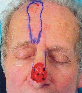
depend on a named artery), such as the paramedian forehead flap, is dependent on knowledge of the location of the major vessels of the face—in this case, the supratrochlear artery
EXTERNAL CAROTID SYSTEM
The external carotid system is the main blood supply to the lower face, temple, and posterior scalp.
The main branch of the external carotid artery to the central face, the facial artery, enters onto the face after exiting the submandibular gland at the anterior border of the masseter muscle at the jaw line (see Fig. 1.1). It runs obliquely superior within the substance of the lip depressor muscle complex toward the commissure of the lip. Within the substance of the orbicularis oris muscle, it first gives off the inferior labial artery that runs medially through the lower lip to meet its pair from the other side. Next, at the level of the upper lip, the similarly disposed superior labial artery is given off and runs transversely through the upper lip to meet its contralateral partner.
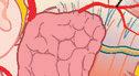
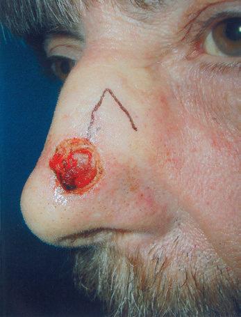
After giving off the superior labial artery, the facial artery becomes known as the angular arery as it makes its way up toward the alar base and consequently upward alongside the nose to anastomose with the dorsal nasal artery, a branch of the ophthalmic artery of the internal carotid system. This anastamotic complex at the medial canthal level is an important vascular pedicle for the dorsal nasal or Rieger flap. Recent anatomic studies revealed that the angular artery has four variations that occur with nearly equal frequency. The illustration (Fig. 1.13) depicts the variations.2 The complex anastomotic vascular arrangement in
Fig. 1.13 Variations of the angular artery. AA, Angular atery FA, facil artery; LNA, lateral nasal artery.
Supraorbital art. and v.
Supratrochlear art. and v.
Nasociliary art. and v.
Dorsal nasal art. and v.
Angular art. and v.
Opthalmic art. and v.
Sentinel v.
Lacimal art. and v.
Infraorbital art. and v.
the periocular area is cause for concern, as many arteries connect directly or indirectly to the retinal and internal carotid arteries and have resulted in blindness and srokes when injected with particulate material such as intradermal fillers (Fig. 1.14).5
Other main branches off the external carotid include the occipital artery that couses posteriorly and superiorly in company with the sensory occipital nerve between the trapezius and sternocleidomastoid muscles, to run just above the occipital muscle to supply the posterior scalp. The postauricular artery arches around the styloid process to a groove between the mastoid process and the external ear to supply the posterior ear and portions of the adjacent scalp above and behind the ear.
After giving off the facial and occipital arteries, the external carotid artery divides into its two terminal branches the superficial temporal artery and the internal maxillary artery. The latter branch is primary interna to he mouth and nose, but after several divisions does supply terminal vessels that exit the infraorbital and mental foramen as same-named arteries, along with their respective veins and sensory nerves. They provide important anastomoses with the facial artery.
The more important division to dermatologic surgeons is the superficial temporal artery. This runs superiorly in front of the ear within the substance of the parotid gland, along with the same-named vein and the auriculotemporal nerve, a sensory branch of the mandibular nerve of the trigeminal (Vth cranial) nerve. As the neurovascular bundle exits the superior pole of the parotid gland, the artery is palpable at the level of the superior attachment of the ear. At this point, it splits into anterior and parietal branches to supply the temple/lateral forehead and parietal scalp, respectively.
Temporal art. Medial temporal v. Ant. and post. deep temporal art.
Zygomatico -temporal art.
Zygomaticofacial art. and v
INTERNAL CAROTID SYSTEM
The main volume of blood from the internal carotid system is dedicated to supplying the brain. A portion is allotted to the face, predominantly to the ophthalmic artery whose terminal branches exit the skull as the supraorbital and supratrochlear arteries, as part of he same-named neurovascular bundles (see Fig. 1.14). They exit their respective foramen and pierce the overlying frontalis muscle to run superiorly. At this point, they are displaced deep in the subcutaneous fat above the frontalis fascia and subsequently over the galea aponeurotica as they course superiorly over the scalp. They supply the forehead and anterior scalp (see Fig 114) As noted earlier, the supratrochlear artery is recruited for the classic and extremely useful axial midline and paramedian forehead flap. Both arteries partake in the rich anastomotic network over the scalp by connecting with branches of the superficial temporalis and occipital arteries. This network is so powerful that traumatic scalping can be repaired by vascular reconnection of even one of these major vessels that supply he salp
The other major branch off the ophthalmic artery is the dorsal nasal branch (in some studies noted as the infratrochlear artery) at the medial canthus. It runs inferiorly along the side of the nose in the medial canthal region to supply the bridge of the nose and to connect with the angular artery of the external carotid system.
It is interesting that above the zygomatic arch, the neurovascular bundles containing the major arteries and veins all course in the deep subcutaneous plane above the fascia or muscles of facial expression. Conversely, below the arch, the vessels are usually within the substance of the mimetic muscles and do not course in the company of the major
Facial art. and v.
Sup. labial art. and v.
Fig. 1.14 Periocular vascular anatomy. (Carruthers JD, Fagien S, Rohrich RJ, Weinkle S, Carruthers A. Blindness caused by cosmetic filler injection: a review of cause and therapy. Plastic and Reconst Surg. 2014;134:1197–1201.)
Superficial temporal a.
Frontal branch
Facial
• Malar node
Zygomaticoorbital a.
Transverse acal a.
Preauricular and parotid
Infra-auricular
Postauricular
• Infraorbital node
Buccinator nodes
Mandibula node
VENOUS SYSTEM
Occipital
Spinal accessory chain
Submental
Submandibular
Superficial cervical node
sensory nerves. Knowledge of the route and depth of the vessels will aid in preserving them during a procedure in the region. On the other hand, when they have to be cut, as in a lip wedge excision, knowing that the labial artery is located in the very posterior portion of the distal orbicularis oris allows the surgeon to locate and clamp it off immediately before it retracts into the substance of the muscle.
The vascular drainage of the face, for he most part, parallels the arterial supply. Generally, he veins have a straighter, less tortuous path than their counterpart arteries (Fig. 1.15).
LYMPHATIC SYSTEM
The advent and widespread use of lymphoscintigraphy have at once confirmed and expanded much of what we know about the lymphatics of the head. The system is certainly much more variable than once believed. The impetus for this interest is that melanoma, squamous cell carcinoma, and Merkel cell carcinoma all tend to metastasize first to primary echelon nodes, and a complete examination for these tumors includes evaluatin of the draining lymph nodes either by manual papation or by histopathologic evaluation, following sentinel lymph node biopsy. Some generalities apply to the lymphatic system of the head and neck. First, the central face and scalp are usually devoid of lymph nodes, except for a few inconsistent, ectopic nodes found above the mimetic muscles in the subcutaneous fat. Generally they are encountered by accident during surgery or found in the pathology specimen. Next, afferent drainage is organized in flow patterns that proceed in a superior to inferior, diagonal direction toward the collecting, primary echelon nodes in the upper neck region These include the submental, submandibular jugulodigastric, and occipital lymph nodes (Fig 116). Drainage from these
Internal jugular chain
superficial nodes then proceeds to the deeper cervical systems in the neck (the spinal accessory internal jugular, and transverse cervical lymph node basins).
The real wild cards in the system are the lymph nodes within the substance of the parotid gland. These nodes are difficult to assess clinically, and even lymphoscintigraphy may prove troublesome due to the proximity of the parotid gland. They drain a wide area, including portions of the anterior scalp, forehead, lateral eyelids, cheeks, nose, and upper lip. Subsequent drainage is to the deeper neck node basins
Special Structures: Lip, Nose, Eyelids, and Ear
LIP
The lips are the guardians of the oral seal that is required for phonation and mastication. On a social level, the marvelously expressive lips convey our nonverbal communication. As such, they are at once the least complicated and the most mobile of the special structures of the face. The two are related; by not having a constricting solid structure such as the bony/cartilaginous frame of the nose or the form-shaping tarsal plate of the eyelid, the lips are quite flexible, stretchable, and elastic. Having only a very thin subcutaneous fat layer in the cutaneous portion also facilitates this ability.
Internally, the lips consist soley of the orbicularis oris muscle and the myriad muscular insertions attached to it. They are covered on the outside by skin, on the inside by a wet mucosa, and at the vermilion by a dry modified mucosa (Fig. 1.17). The submucosa contains many salivary glands on the oral portion
The lips may be divided into vermillion and cutaneous cosmetic units. The cutaneous upper lip is the most complicated section, having a small triangular portion that extends superior and lateral to the alar base and a central subunit, the philtrum. Two convex philtral crests (columns, ridges) define the midline concave philtral bowl. The lower portion of the latter is the beautiful downward-curving Cupid’s
Fig. 1.15 Venous system of the face and corresponding arteries.
Fig 1.6 Lymphatic drainage.
A Surface Anatomy
Philtrum
• Column
• Groove
Cupid’s bow White roll
Tubercle
Collumella
Soft triangle
Alar cartilage
• Lateral crus
• Medial crus
Nasal bone
Lateral catilage
Septal angle
Alar cartilage
B Cross Section of Lip Histology
Cutaneous upper lip Vermilio
Inner lip (skin)
• Lateral crus
• Medial crus
1.18 Structures of the nose.
Labial artery
Orbicularis oris muscle
Epidermis
Hair follicle
Subcutaneous tissue
Sweat gland
Salivary gland
Outer lip (skin)
Fig. 1.17 Structures of the lip and cross section.
bow. The philtrum is a reservoir of extra skin and interdigitating orbicularis muscle that allows for the dramatic stretching and vertical shortening of the upper lip when smiling or opening the mouth wide. In many people, the sharp cutaneous/vermiion junction line has a superimposed white roll. This 1- to 2-mm, glabrous strip of skin is due to an anterior bulging of the orbicularis oris muscle. It can be quite prominent, especially in younger patients, and is an important consideration in placing transverse scar lines
NOSE
The nose is an extremely complex, aesthetically and functionally important midline structure that is all too often the site of invasive skin cancers It is supported by a bony/ cartilaginous infrastructure that features the inflexible paired nasal bones superiorly and the highly mobile paired upper lateral/alar cartilage complex inferiorly (Fig. 1.18). Conversely, the skin over the rigid bony upper portion is highly movable (zone 1), while the skin over the mobile cartilage portion is thick, sebaceous, and bound down (zone 2).
On the surface the nose can be broken down into several cosmetic subunits. These include the root of the nose that extends from medial canthus to medial canthus. The skin lines in this concave unit run transversely across it. Oblt eration of the concave nature of this area during repairs causes the profile to be dramatically altered and the nose to appear very large on the frontal view. The paired lateral sidewalls separate the nose from the cheek and are in turn
separated from each other by the midline dorsum of the nose unit. The heart-shaped convex tip of the nose is the most prominent feature of te nose and is bounded by the paired alae nasi. The columella is the midline structure below the tip separating the nostril apertures and culminating in the upper philtrum of the lip. The RSTLs of the upper nose run obliquely out from the medial canthus down the dorsum and lateral sidewall. They end part-way down, as there are no skin lnes on the lower nose. As noted earlier, they un transversely across the root.
The lower nose is supported by the paired, upper lateral cartilages, which act as extensions of paired nasal bones The paired, lower lateral, or alar cartilages begin as medial crura that form the columella. As they swing back and curve, they form an arch that is the tip of the nose. As the lateral crura diverge obliquely pward and laterally away from the midline vertical nasa septum cartilage, they support the ala nasi. It is important to recognize that the ala nasi are composed of fibro-fatty muscular tissue and are devoid of cartilage. Their ability to flare (and not collapse) with each inspiration is due to the involuntary contraction of the levator labii superioris alaeque nasi and nasalis muscls If these latter muscles have been removed during tumor extirpation, then a cartilage strut must be included in the surgical repair or the nostril will collapse during inspiration.
The upper portion of the nose is innervated by the infratrochlear (V1) of the trigeminal, whereas he anterior branch of the external ethmoid nerve (also V1) supplies the tip. The lateral portions of the nose are innervated by the infraorbital nerve (V2).
EYELIDS
The eyelid is perhaps the most complicated structure on the face and of extreme functional importance. Successful repair of the unique structures that compose the eyelids are contingent on thorough knowledge of the anatomy, as it is so closely related to function. The eyelids guard the globe and orbit and are structured on the orbicularis oculi muscle, whose orbital and palpebral components have been discussed earlier in this chapter. As noted, the orbicularis is
Fig.
A Cross Sectional Anatomy
Whitnall’s check ligament
Fat pad of eyebrow
Orbital fat
Orbital septum
Levator aponeurosis
Orbicularis oculi m
Superior tarsal plate
Inferior tarsal plate
Capsulopalpebral fascia
Orbital septum
Superior palpebral fold
Glands of Zeis
Glands of Moll
B Surface Landmarks
Papilla
Inferior punctum
Inferior canaliculus
Nasolacrimal duct
Inferior meatus
Fig. 1.19 Structures of the eye and lachrimal apparatus.
responsible for closing the lid. The levator palpebrae superioris muscle and aponeurosis is responsible for opening the eye (Fig. 1.19). It is innervated by the oculomotor or third cranial nerve. It arises from the superior orbit and divides into two components: Muller’s muscle, which attaches into the superior margin of the tarsal plate under sympathetic nerve control, and the levator aponeurosis which fuses with
Levator palpebrae superioris m.
Superior rectus m.
Cornea Lens
Inferior rectus m.
Inferior oblique m.
Maxillary sinus
Orbital fat
Orbital septum
Gland of Krause
Muller’s m.
Orbicularis oculi m.
Gland of Wolfring
Levator aponeurosis
Conjunctiva
• Papebral
• Bulbar
Tarsal plate
Meibomian glands
Ciliary muscle of Riolin
Mucocutaneous junction
Lacrima gand
Superior punctum
Superior canaliculus
Fundus
Lacrimal sac
Nasal cavity
Inferior concha
the orbital septum to form the superior palpebral fold about 10 mm above the lid margin and then continues downward to atach to the anterior surface of the tarsal plate. It also sends fibers through the pretarsal orbicularis to insert into the lid skin. This is why the skin in the pretarsal area is bound down, whereas it is loose and eventualy redundant in the preseptal area above the superior palpebral fold. The
space between the orbital septum and the levator aponeurosis contains the orbital fat pads. The skin of the eyelids is the thinnest and most elastic in the body. It contains little subcutaneous fat.
The posterior portion of the eyelid closest to the globe contains the tarsal plate that consists of dense fibrous tissue. This gives form to the lids and also contains the sebaceous Meibomian glands. The conjunctiva covers the posterior eyelids as well as the globe Hair follicles of the eyelashes (cilia), with both sebaceous glands (Zeis) and sweat glands (Moll) exit onto the lid margin below the muscular layer, while the Meibomian glands exit onto the lid closer to the conjunctival surface.
The lacrimal system is important to eye homeostasis (see Fig. 1.19) The lacrimal gland is situated in the postseptal space lateral to the fat pads. The lacrimal collecting system consists of the upper and lower lacrimal puncta at the medial eyelid margins, which open into the lacrimal canaliculi at the medial margin of the tarsi. The upper and ower canaliculi converge under the medial canthal tendon as the common canaliculus, to run horizontally into the nasolacrimal duct on the side of the nose opposite the medial canthus, and drains as the nasolacrimal canal into the nose beneath the inferior turbinae.
EAR
The ear appendage is essentially skin stretched over a rather intricaely molded cartilage infrastructure. It is important to the dermatologic surgical, as it has a high risk for both basal cell (BCC) and squamous cell carcinomas (SCC). Invasive SCC tumors of the helical rim tend to reach the arger lymphatic and venular vessels of perichondrium at a shallower depth than elsewhere on the body, making metastases from this area a real threat.
The outer rim of the ear is the convex helix that extends upward from the lobule o curve around, and ending as the crus of the helix that divides the concha bowl into the upper cymba and the lower cavum (Fig. 1.20). Opposite the cavum and guarding the external auditory meatus is the small protuberance, the tragus. Also superior to the lobule is the convex antihelix. This structure parallels the helix in its vertical dimension and is separated from it by the depression between them, the scapha. The antihelix splits at its superior end into two crura to form a central concavity, the triangular fossa. Only the lobule is not supported by cartilage.
The sensory innervation of the ear is complicated. The anterior surface closest to the cheek as well as the anterior-
Scapha
Helix
superior portion of the external auditory canal is supplied by branches of the auriculotemporal branch of the mandibular division (V3) of the trigeminal nerve. The lower posterior surface as well as the lower anterior helical portion, is innervated by the great auricular nerve (C2, C3), whle he upper posterior portion and the upper vertical porion of the anterior helical area are supplied by the lesser occipital nerve (C2, C3). Variable contributions of the cranial nerves IX and X supply the remaining portions of the external auditory canal. Understanding this innervation pattern is required for regional nerve block of the ear.
References
1. Salasche SJ. Anatomy. In: Rohrer TE, Cook JL, Nguyen TH, et al, eds. Flaps and Grafts in Dermatologic Surgery. Philadelphia: Elsevier; 2007: 1-14.
2. Kim Y, Choi D, Gil Y-C, Hu KS, Tansatit T, Kim HJ. The anatomic origin and course of the angular artery regarding its clinical implications. Dermatol Surg 2014;40(10):1070-1076.
3. Alghoul M, Codner M. Retaining ligaments of the face: review of anatomy and clinical implications. Aesthet Surg J. 2013;33(6):769-782.
4 Rohrich RJ, Pessa JE. The fat compartments of the face: anatomy and clinical implications for cosmetic surgery. Plast Reconstr Surg. 2007; 119(7):2219-2227.
5. Carruthers JD, Fagien S, Rohrich RJ, Weinkle S, Carruthers A Blindness caused by cosmetic filler injection: a review of cause and therapy. Plast Reconstr Surg. 2014;134(6):1197-120
Triangular fossa
Crra of antihelix
Concha
Cymba
Cavum
Antihelix
Crus of helix
Tragus
External acoustic meatus
Antitragus
Lobule
Fig. 1.20 The ear.
ker /t.me/eoo es s://t.me/ebo
Reconstruction of the skin is challenging and rewarding to the dermatologic surgeon. When second intention or side to-side linear closure is not possible, more complex movement of local or distant skin in the form of cutaneous flaps is necessary to repair a surgical defect The basic terminology, physiology, and types of local flaps will be discussed in this chapter.
KEYWORDS flap axial pattern flap random pattern flap advancement flap rotation flap transposition flap interpolation flap
Fig. 2.1 The wound resulting from the extirpation of the umor is the primary defect. The secondary defect is defined as he wound created by cutting, lifting, and sliding the flap to fill the primary defect. Here, the secondary defect will be filled by he second lobe of the flap. The base of the flap is that area that emains attached to the contiguous skin adjacent to the defec
length of the flap, one would simply need to double the width of the flap base, thereby including a sufficient number of vessels in the pedicle to sustain the flap tip. Many felt that there was no ultimate limit to flap length. In 1970 Milton disputed this conventional hypothesis by publishing his research on axial flaps in the pig model. He discovered tha “flaps made under the same conditions of blood supply survive to the same length regardless of widh.”8 Daniel and Williams confirmed Milton’s research on axial flaps and further studied survival of random pattern flaps. Although they concluded that “an increase in width did not result in an increased length of survival,”4 their data showed a trend toward wider pedicles allowing greater flap length survival. Studies by Stell in a pig model and confirmed when extrapolated to humans suggested that viable flap length was not diectly proportional to pedicle width, and that beyond a certain pedicle width, greater recruitment of random vessels cannot support the flap tip (see Fig. 2.2).3 Clinical experience with random flap survival has echoed this survival trend.
Overall, perfusion pressure limits the ultmate length of both axial and random flaps. The greater the perfusion pressure in the flap’s pedicle, the longe the flap can be without undergoing necrosis. Moreover, the greater the perfusion pressure at the flap’s base, the more narrow the pedicle may be. Arteries always have greater perfusion pressures than distal arterioles and capillaries. Accordingly, the longest viable flaps with the narrowest bases (largest length-to-base ratios) are arterial flaps. Therefore a paramedian forehead flap that uses the supratrochlear artery may be at least four times as long as the flap’s pedicle width (at least a 4 : 1 ratio; Fig. 2.3). Musculocutaneous flaps have the next greatest blood supply in the pedicle, followed by fasciocutaneous and random flaps in descending order.
The commonly used random pattern flaps are largely supported by the redundant subdermal vascular plexus in the skin. Fig. 2.4 highlights he innate vasculature of the skin, upon which flap survival depends. The deeper, muscular-based arteries supply the subdermal plexus, which subsequently perfuses the intradermal plexus. The intradermal vasculature alone is usually unable to support tissue viability, ue to low perfusion pressures and blood flow within these distal capillaries.2,6,7 However, the subdermal plexus, found in the mid to superficial subcutaneous fat, contains both arterioles and capillaries with sufficient perfusion pressure to sustain tissue viability following flap movement. This anatomy is critical to understand when undermining (dissecting under the flap and its surrounding area to allow tissue movement) a cutaneous flap. If undermining is performed superficial to the subdermal plexus (i.e., within the dermis), the flap has a significantly increased chance of undergoing tissue ischemia.
Fig. 2.2 Flap perfusion pressures necessary for flap survival. The graph shows how perfusion pressure decreases the greater the distance that the blood must flow from the feeding artery or arteriole (A and B) Show that despite greater recruitment of random arterioles there is a point where perfusion pressure is less than the capillary resistance. At this point of no flow, the flap will necrose.
The perfusion pressures of random cutaneous flaps vary with the flaps’ locations on the body.3,4,6,8–13 As soon as a random pattern flap is incised and raised, there is a significant decrease in the perfusion pressure to the distal, disruped skin.14,15 Fortunately at normal skin temperature, the amount of blood flowing through the facial skin is 10 times greater than that needed to supply the skin’s basic metabolic needs.16–19 Other areas of the body are not as well supplied with this redundancy of blood supply A general rule is that the more distant the surgical flap is located from
the heart, the less the perfusion pressure will be. Hence a flap on the leg should be designed with a smaller flap length-to-pedicle width ratio when compared with a flap on the face.
Subdermal plexus
Intradermal plexus
Fig. 2.4 The support of commonly used random pattern flaps by the redundant subdermal vascular plexus in the skin. The deeper, muscular-based axia arteries (not shown) supply the subdermal plexus, which subsequently perfuses the intradermal plexus. The intradermal vascultue alone is usually unable to support tissue viability because of low perfusion pressures and blood flow within these distal capillaries. The subdermal plexus, found in the mid- to superficial subcutaneous fat, contains both arterioles and capillaries with sufficient perfusion pressure to sustain tissue viability following flap movement Theefore undermining of a random pattern flap must occur below the subdermal plexus.
As previously discussed, there is a point at which no matter how many extra vessels are recruited at the base, the distl perfusion pressure will be less than the critical closing pressure of the arterioles and capillaries (see Fig. 2.2).3,6,8,15,20 When the perfusion pressure is less than this intravascular resistance, the flap receives diminished nutrients and necrosis can ensue. Generally, random cuane ous flaps of the face with a maximum length-to-base ratio of 3 : 1 survive.21–23 On the trunk nd legs the maximum length-to-base ratio is considered to be 2 : 1.12,21,22,24,25 An axial flap on the face may have a 4 : 1 or greater ratio depending on the arterial supply of the pedicle.21,26 Remember that these ratios are only guidelines. Patient-related factors must be taken into account when contemplating the feasibility and design of a flap. In addition to location of the body, locl perfusion pressures can be altered by a variety of factors such as tobacco use, history of radiation to the area, medical comorbidities (i.e., atherosclerosis, high or low blood pressure, arrhythmias), and use of vasoconstrictors. In addition, flap design must take into accoun variations in skin laxity based on location, age, and comorbid conditions.
Flap Physiology
Flap survival is dependent on various factors, including blood flow, angiogenesis and vascularization, edema, wound closure tension, postoperative complications such as hematoma/seromas, and infection. Prior to the first incision, the flap skin is fully vascularized and viable using the definition of normal skin. Once the flap is raised, it is immediately ischemic, since the normal vessels supplying that skin are cut and the flap now depends on decreased circulation from the collateral vessels. A flap is always initially
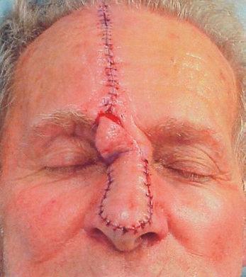
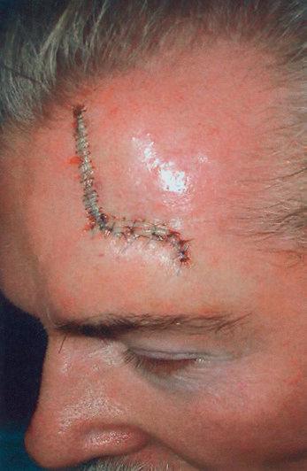
Fig. 2.3 Measuring the flap length to width. By including a large bore artery (the supratrochlear artery) in the flap pedicle, the ratio may be at least as great as 4 : 1 as shown in the preoperative photo (A) and afte suturing (B)
Dermis
Subdermis
viable, since the skin can survive up to 12 to 13 hours of avascularity at 37°F.27–29 An ischemic flap can survive even longer since the blood flow needed to sustain skin is only 2 to 8 cc per 100 g per minute, and normal flow to the skin is 10 times greater than this minimum.16,27 Thus Meyers27 was correct when he commented that a fresh flap is always ischemic but viable.
Once incised and relocated the flap receives its nutrients from both the cutaneous pedicle and the base of the primary defect. Sufficient blood flow through the base of the flap is essential in the initial 24 to 48 hours after the initial flap creation. In both axial and random flaps, blood flow immediately drops as the flap is elevated. For axial flaps, microvascular flow actually increases to a level greater than the preoperative state within 5 hours.15,30 Flow in random pattern flaps, however, starts to improve differentially for up to 4 weeks. Marks used the rat model to show that flow improved on a gradient; flow increased within 14–16 hours to the skin closest to the pedicle, within 24–48 hours o the skin 1 cm distal to the pedicle, and within 96 hours 3 cm distal to the pedicle.15 All sites recovered approximately 30% of their blood flow per day with the proximal most portion recovering full lood flow by the end of 7 days and the most distal tip by the end of 14 days. Until 14 days, recovery of blood flow occurred on a gradient that depended on the distance of the skin from the base of the flap. Microvascular flow grew to higher than preoperative levels from 14 to 21 days, and then gradually returned to baseline durng the fourth week. The opening of collateral vessels appears to allow this sequential recovery of blood flow. However, there appears to be a limit on how fast these collaterals can open.15
This time-dependent opening of collateral vessels may be partially explained by arteriovenous (AV) shunts. These shunts control blood supply to the capillary network that supplies the flap. There ae preAV shunt sphincters under the control of the sympathetic nervous system. When the flap is incised and undermined, local sympathetic nerve fibers are disrupted and release catecholamines. As a result, there is local vasoconstriction for up to 48 hours, by which time the nerve’s supply of norepinephrine is exhausted.27,31 Once sympathetic tone is relaxed, the blood flow to the capillary collaterals is increased to help supply the flap with nutrients.14 However, this effective sympathectomy cannot fully explain why random flaps have a graded flow recovery. Upon incision, the entire flap should have equal catecholamine release and subsequent equal flow recovery.32 Other humoral factors such as prosaglandin release may come into play.33
Local tissue conditions resulting from surgical trauma also decrease flap perfusion and subsequent survival. Following any local injury, the inflammatory cascade releases the powerful vasoconstrictor thromboxane A2.34,35 In addition, free radicals are released, causing direct injury to the flap.3637 Finally, the edema inevitably associated with surgical trauma causes further capillary vessel resistance by increasing manual compression on the skin’s smaller caliber perfusion sources.38 In addition, fluid collection under the flap in the form of a postoperative hematoma or seroma can further decrease blood flow. All of these negative factors decrease perfusion to the flap and can threaten flap survival.
Conversely, the flap may also benefit from surgical trauma. Relative local hypoxemia and increased levels of metabolic by-products induce opening of precapillary sphincters, thereby promoting increased local blood flow.27,31,39 Moreover, adhesion molecules, such as E-selectin are activated following exposure to released coagulation cascade molecules such as endotoxin, interleukin1, and tumor necrosis factor alpha.40 These adhesion molecules recruit molecules including neutrophils to the flap to clear debris and anabolic waste products Finally, ischemic tissue attracts endothelial progenitor cells, which allow for the ingrowth of new vascular channels to supply the flap.41 Flap survival is dependent on the balance of all these factors, ultimately influencing pedicle blood flow.
The nascent flap not only receives nutrients from the pedicle, but it also gains nutrition from the base of the primary defect through angiogenesis, revascularization, and neovascularization.41–44 Within the first two days of flap placement, a fibrin layer develops below the flap and provides a suitable environment for angiogenesis 36 Endothelial cells and macrophages release angiogenic cell factors important in neovascularization, the local growth of new blood vessels into the surgically manipulated skin.41,44–46 Neovascularization is seen as early as 3 days in the rat model,47 and at 4 days in rabbit48 and pig42 models. In staged, pedicled flaps in humans, revascularization adequate for division of the flap pedicle has been demonstrated by the seventh postoperative day.42,49,50 This new vascular growth works in conjunction with the preexisting collateral vesses to nourish the flap.
Tissue edema, wound closure tension, and infection also negatively affect flap blood supply and survival. Although none of these factors can solely lead to necrosis of a well vascularized flap, each can contribute to further ischemia in a marginally perfused flap. Postoperative tissue edema places external force on small capillaries,38 resulting in increased capillary resistance Thus there must be greater perfusion pressure at the pedicle to counter this resistance and ensure flap tip survival. Recent studies in the rat model revealed that significant postoperative edema will not solely cause flap necrosis.51 However, it can be an additive factor, along with high wound closure tensions and/or infection.
Closing wounds under large amounts of tension can place undue vascular stress on the wound edges and tip of the flap. It is typically recommended that one undermine 2 to 4 cm,52 or 50% to 100% of the defect width,53 beyond the wound edge to decrease wound tension. Undermining beyond this distance may be detrimental since there may be unnecessary vascular compromise more bleeding, and greater dead space, all of which can lead to surgical complications. High closure tension leads to dehiscence and wound edge necrosis, but it does not usually lead to entire flap necrosis.54–56 Wound infection can cause partial or complete flap necrosis. With local infection, there is release of toxic free radicals and greater tissue edema.57 Infection can also lead to vessel thrombosis.58 In addition to local tissue destruction and vascular compromise, collagen production and deposition are hindered.59 Therefore flap adhesion to the wound bed and overall tensile strength are affected. Overall, there are many factors involved in the initial period of wound healing that are critical to the flap’s survival. Predictably acceptable flap results depend on
proper flap design, gentle operative technique, and the avoidance of surgical complications.
Flap Biomechanics
In addition to understanding the skin’s vascula supply and physiology, the surgeon must also appreciate the unique biomechanical properties of the skin if flap surgery is to be successful. All materias have characteristic biomechanical properties: stress, strain, creep, and stress relaxation. In regard to skin, stress is the force applied per cross-sectional area, and strain is the change in length divided by the original length of the given tissue, to which a given force is applied. The stress–strain relationship of skin shows that skn unike some other materials, is not truly elastic (Fig. 25). As a small amount of stress (or tension) is placed on the skin, there is a corresponding change in the skin’s length (strain). At a certain point on the stress–strain curve (see zone III in Fig. 2.5), even a large amount of applied force will not result in further incremenal skin stretch.26 This nonelastic property of skin is mainly due to its structural constituents—collagen and elastin. In relaxed skin, collagen is randomly oriented, and elastin is loosely wrapped around and attached to multiple points on the collagen bundles.60–64 When a small amount of force is initially applied to skin, the elastin network is first deformed63,65 and the skin is easily lengthened (see zone I in Fig. 2.5). With sun exposure and intrinsic aging, there is a progressive decrease in the functional elastic fiber network.65 As a result, the stress–strain curve is shifted to the right so that when applied to aged or sun-damaged skin, less appled force results in greater lengthening. As continued force is applied, the collagen fibers begin to reorient parallel to the direction of the force (rapid transition of the curve; see zone II in Fig. 2.5).64 At the point where the elastin and collagen fibers are maximally stretched even a large amount of force will only minimally stretch the skin (see zone III in Fig. 2.5).
Thus the skin is not truly elastic at all levels of applied tension.
Creep refers to the increase in strain seen when skin is under constant stress. When the skin is held at sufficient tension, elastic fibers fragment and collagen fibers will align parallel to the applied force. As a result of this reorganiza tion of the elastic and collagen fiber networks, intertitial fluid will be displaced and can be seen with the naked eye.66 Creep typically begins to occur within several minutes of constant force application. Whie skn demonstrates elastic properties with low loads, skin exhibits “stress relaxation” and creep when larger forces are applied for longer periods of time. Stress relaxation occurs when skin is held under constant tension. In this case the amount of force (stress) required to maintain this tension decreases with time. The skin’s relaxation under stress is closely related to its ability to increase in length when placed under a constant stress (the creep phenomenon). In the case of a cutaneous flap, if held steadily at high tension for 5 to 10 minutes, the skin will lengthen and relax. Surgeons with an understanding of this property may use an intraoperative pulley stitch to take advantage of this mechanical property. As a result of intraoperative creep, a smaller ap can be used to fill a larger defect, but the potental reliance upon secondary motion around the surgical defect must be recognized. When skin is held at a constant tension for several days, stress relaxation occurs. There is a decrease in stress due to an increase in skin cellularity and the permanent stretching of skin components.64 In clinical practice, serial excision and tissue expansion use the principles of creep and stress relaxaion. When a large lesion is partially excised and reconstructed under tension, the skin relaxes and lengthens, allowing further staged excisions.
Fig. 2.5 Stress–strain curve demonstrating the nonlinear properties of skin when stretched (vertical axis). When a small amount of force is ntally applied to skin, the elastin network is first deformed and the skin is easily lengthened (horizontal axis; zone I). As progressive continued force is applied, the collagen fibers begin to reorient parallel o the direction of the force (zone II). At the point where the elastin and collagen fibers are maximally stretched, even a large mout of force will only minimally stretch the skin (zone III) With sun exposure and intrinsic aging, the stress–strain curve is shifted to the right.
Understanding the stress–strain curve is essential for optimal wound closure. When reviewing reconstructive options, it is important to avoid any excessive stress on the defect edges that would force the involved skin into the third part of the stress–strain curve; this involves minimizing tension on the wound closure. Unfavorably high wound closure tensions limit recruitment of skin laxity and can be detrimental to wound healing. Specifically, high tension may lead to wound edge necrosis, wound dehiscence, and cosmeticlly unacceptable scars (i.e., widened, atrophic, or hypertrophic scars). Wound edge tension may be minimized by appropriate undermining; however, care must be taken to understand and minimize the underlying mechanism of stress or restriction. For example, a galeotomy may be used to release galeal fascial restraint and allow for additional movement of the skin and subcutaneous ssue on the scalp; careful dissection of the skin from the underlying orbicularis muscle in the periocular area creates movement of the skin to allow for tension redistribution away from the free margin; and separating the vertical connections within the subcutaneous tissue of the cheek from the underlying superficial muscular aponeurotic system (SMAS) drastically reduces tension on closures and allows for substantial movemen of tissue for closure of large cheek defects.
In addition to understanding the role of tension reduction on wound closure, it is important to identify situations where tension redistribution is required. Specifically, when side-to-side linear closure is not possible due to excessive tension or stress on the primary closure, or when linear
//tm
wound closure abuts a free margin or crosses a cosmetic subunit boundary, the vector of tension may need to be redistributed in the form of a rotation or transposition flaps, discussed later. Overall, by taking advantage of the above mentioned mechanical properties of skin and designing appropriate flaps, the surgeon takes tension off the wound edges, and returns the skin to the first or second zone of the stress–strain curve.64 Understanding these principles, as well as mastering techniques for tension reduction and redistribution, allows the closure of skin flaps under low or moderate tension. As a result, wound edges can survive, and a good cosmetic result can be obtained.
Flaps Defined by Movement
In addition to classifying flaps by their vascularity, flaps are often also categorized by their primary movement directions. The three basic flap movements are advancement, rotation, and transposition. With advancement flaps, the major motion of the flap is largely along a single linear direction toward the primary defect (Fig. 2.6). Rotation flaps are those flaps that are rotated in an arc or curvilinear fashion along a pivot point to fill the adjacent primary defect without crossing any intervening skin (Fig. 2.7). A transposition flap is incised and lifted over intact skin and then placed into the wound (Fig. 2.8). A distinct type of transposition flap is called the interpolation flap. An interpolation flap is a flap, potentially with a predictable, axial vascular supply, that involves two steps or stages. The first stage of the flap is transposing the tissue into the wound with the pedicle overlying the intervening skin. The second stage is separating the flap from its origin and therefore dividing its pedicle.
In addition to classifying flaps by their unidirectional motion, some authors, noting the inherent limitations of such schemata, categorize flaps as sliding or lifting flaps.67,68 Sliding flaps include advancement and rotation flaps, where tissue is pushed into the primary defect. A lifting flap is the typical transposition or interpolation flap, in which tissue must be lifted over normal skin to fill the wound These more general classifications recognize that very few flaps move tissue along a single, predictable path. For example, most rotation flaps also incorporate a ignificant degree of tissue advancement, and vice versa.
As alluded to previously, when discussing the motion of these flaps, the surgeon must understand the tension vector created by the flap’s closure. This vector depends on the primary and secondary motion of the flap. The primary motion of the flap is the tension placed on the flap tissue as i is moved to fill the wound (primary defect) (Fig. 2.9). The secondary motion, created by the movement of the flap into the defect, is the force placed on the tissue surrounding the primary defect (see Fig. 2.9). The combined force created by the primary and secondary motions causes a new tension vector that must be anticipated by the surgeon when deciding which flap can best be used for repair. This final tension vector varies, depending on the flap chosen, and may be appreciated when placing the key stitch. The key stitch is the stitch placed to initially move the flap and accurately place it into the primary defect.69 If not properly planned, the flap’s final tension vector may cause undesirable pulling on free margins such as the eyelid, alar rim, and mouth.
ADVANCEMENT FLAP
The advancement flap has long been used in facial recon str uction. Its first description has been attribued to Celsus
Burow’s triangle was excised here
Fig. 2.6 The lateral forehead skin laxity was used in this unilateral advancement flap. (A) The arrows indicate the direction of the flap movement. (B) The arrow indicates where the Burow’s triangle was removed.
Fig. 2.7 This lateral cheek rotation flap illustrates how the flap is rotated in an arc along a pivot point to fill the adjacent primary defect without crossing any other intervening skin.
Fig. 2.8 A rhombic transposition flap. The flap must be transposed over intact skin to be placed into the defect. The flap must be transposed over intact skin (A) to be placed into the defect (B).
Fig. 29 (A) The primary motion o the flap (yellow arrow). (B) As a result of this motion, tension will be redistributed and the seconday motion tension can be seen on the laterl naal brdge (red arrows).
of ancient Rome, who used the flap for the reconstruction of the cheek and ears.70,71 In the 1800s, French surgeons advocated the use of “lambeau par glissement” (sliding flaps) for facial reconstruction.72 In 1885 Burow modi fied and fur ther described the advantages of this flap in reconstruction.73
The unilateral, bilateral, and V to Y advancement flaps are the most commonly used advancement flaps in dermatologic surgery. The prmary and secondary motions, and thus the resulting tension vectors of these flaps, are usually in a simple linear line along the direction of the flap movement (see Fig. 2.6).73 The key stitch typically brings the leading edge of the flap to the opposite wound edge to close the primary defect. As the tissue is advanced and sutured into place, a standing cone (or cones) develop(s). These standng cone deformities, or dog ears, are caused by the pouching of excess, compressed skin near the flap’s base when tissue is advanced under tension.74
The standing cone may be corrected by either extending the incision line to remove the excess tissue or using an M-plasty (Fig. 2.10A).75,76 By truncating one end of the closure, the M-plasty flap has the advantage of providing a shorter final wound ength. If the wound cannot be extended due to a ree margin or approaching cosmetic subunit boundary, tissue protrusions can be anticipated. As the length of the flap base is longer than the edge of the flap itself, uneven lengths of tissue requiring approximation are created (see Fig. 2.10B). The inequality of tissue lengths can be accounted for by removing a triangle of skin, called a Burow’s triangle,77 from anywhere along the skin surrounding the flap (see Figs. 2.6 and 2.10B). In addition, the unequal lengths can be accounted for by removing a curvi linear segment of skin from the limb of the flap itself, thereby lengthening the overall arm of the flap o equal that of the flap base and redistributing the redundant tissue along the length of the incision (see Fig 2.10C). This is particularly useful on the upper cuaneous lip and eyebrow areas.
When using bilateral advancement flaps to correct a circular defect, the repair may be described as an A-to-T (T-plasty) or an O-to-H flap (H-plasty). These unilateral and bilateral advancement flaps are most commonly used in repairs on the forehead, scalp, and eyelids. The V-to-Y advancement flap78 uses a subcutaneous pedicle that perfuses a triangular flap that advances along a single direction (see Fig. 2.10D). All borders of the flap are incised to the subcutaneous fat, and the resulting island of tissue, now liberated from lateral dermal attachments, s advanced to fill the defect. The pedicle is underneath rather than at the edge of the flap and may include muscle.79–81 Various modifications,82,83 such as parallel release incisions of the pedicle base, allow significant advancement of the flap with predictable flap survival.84 Because the flap’s muscularcontaining, thick pedicle ensures liberal blood flow, this flap is the most likely of all flaps to survive in patients with tobacco use, which can result in decreased perfusion. The flap can be used anywhere on the face, but the authors find this flap to be particularly useful for repairs of the upper cutaneous lip, the alar–cheek sulcus, and the lateral eyebrow, with alopecic defects to help restore medial hairbearing areas. Of note, the V-to-Y advancement flap was previously synonymous with the “island pedicle flap”; however, this terminology now refers exclusively to flaps in which a portion of the donor tissue is deepithelialized and kers
tunneled or passed beneath the skin to a distant recipient site. It is important to differentiate the island pedicle flap from a Vto-Y advancement flap for both clinical documentaton and coding purposes.
ROTATION FLAPS
In 1842 Pancoast described using rotation flaps to close facial wounds.85 Like most random pattern flaps, rotation flaps recruit tissue adjacent to the primary defect. An arc or semicircle flap is incised and pivoted into the wound (Fig. 2.11). Thus the primary motion is rotation. Similar to advancement flaps, the secondary motion of the flap causes a standing cone deformity at the base of the flap. This Burow’s triangle may be excised from anywhere along the arc of the flap from the nonpedicle side (see Fig. 2.11A). Another similarity between rotation and advancement flaps is the placement of the key stitch. This first stitch closes the primary defect.
The surgeon must pay particular attention to the sze of the designed rotation flap. Because it is rotated along an arc, the functional flap length is shorter than the actual length of the flap’s incision see Fig. 2.11B). Hence the planned length must be longer than the diameter of the wound.86 Dzubow recommends that the planned height of the flap be greater than the height of the defect to account for this functional loss of length.87 One advantage of the rotation flap is that its base is typically broad, and therefore the flap’s length-to-width ratio may be as long as 6 : 1.88 This makes the flap particularly useful when closing inelastic areas such as the scalp.
To increase the movement of the rotation flap, a back-cut or Z-plasty may be employed. A back-cut is when an inci sion is placed into the pedicle of the flap to allow greater forward movement (see Fig. 2.11C). Although the back-cut can increase flap mobility, this maneuver cuts into the flap’s base, thereby increasing the risk of tip ischemia. In areas with excellent vascularity, such as the gabella, the back-cut is often incorporated into the rotation flap’s design. Another technique used to increase flap movement and reduce the primary tension vector is the use of a Z-plasty at the tail of the rotation flap (see Fig. 2.11D).
https://t.meboers https://t ers htps://t.me/eokers https://t.me/ ers https/tme/ebokers tts//me/ ers
Rotation flaps are most often used for repairs of the scalp, forehead, cheek, and nose. The Rieger flap,89 or dorsal nasal rotation flap, uses a back-cut in its design to allow closure of small- to medium-sized defects of the distal third of the nose. In 1985 Marchac and Toth modified the flap design by defining an axial pedicle for the flap based on the angular artery.90 This modification allows a greater flap length-topedicle base ratio because of a reliable blood supply. Multiple rotation flaps may be used to repair large defects on the chin, forehead, and nasal tip. When two mirror-image rotation flaps are designed to repair the defect, the closure is called an O-to-Z repair. There are a few specialized forms of bilateral rotation flaps that have also been reported: the Peng flap for the nose and the bilateral vermillion flap on the lip. 92 When three or more rotation flaps are used, it is often referred to as a pinwheel flap.
TRANSPOSITION FLAPS
Transposition flaps were documented in the pre–Common Era. In about 2000 BC, Indians committing adultery were
https://t.me/ebokes https://t.me/ ers ttps://t.me/ebokers https
ers https://t.me/eokers https:/tme kers https:/t.meeookers https://t.me kers ts://tme/eoers tps/. ers