
https://ebookmass.com/product/flaps-and-grafts-in-


https://ebookmass.com/product/flaps-and-grafts-in-
https://ebookmass.com/product/sexual-and-reproductive-health-ofadolescents-with-disabilities-tafadzwa-rugoho/
ebookmass.com

STUART J. SALASCHE, MD and STEPHEN H. MANDY, MD
A working knowledge of anatomy is essential to perform any of the myriad of procedures in dermatologic surgery. Most surgical skills, even the basic ones, such as scar orientation and level of undermining, are predicated on a basic command of anatomy. More complicated tasks, such as nerve block anesthesia, designing tissue flaps and grafts for advanced repair of surgical defects, and the placement of botulinum toxin and fillers, require an even more intimate understanding. Without an indepth working understanding of the pertinent anaomy, there is a real potential for the surgeon to cause damage by cutting into a vital structure or to be so insecure as to not progress in one’s acquisition of sophisticated surgical skills.
Being a keen observer of the surface anatomy of the face enables the surgeon to assess the patient for the vagaries of aging, to identify the reservoirs of redundan skin available for repairs, and to identify optimal placement of scars. It also allows the surgeon to project and visualize the course or location of the deeper vital structures onto the surface.
The masseter muscle is a good starting point. It is the large muscle of mastication that occupies the lateral portion of the cheek below the zygomatic arch (Fig. 1.1). The parotid gland rests on this muscle. At its anterior border on a line drawn from the tragus to the middle of the upper cutaneous lip, the parotid duct can be identified as it dips inward, piercing the buccinator muscle to open into the mouth at the level of the second upper molar Also at the jaw line, just at the anterior border of the lower masseter muscle, the facial artery and vein enter onto the face. Pulsation of the artery is often possible at this location. The superficial temporalis artery pulse can be felt just anterior to the ear at its superior attachment. Just beyond this, it splits into superior and parietal branches.
The midpupillary line is identified with the patient sitting up and gazing straight ahead. Three important openings in the skull can be located. The superior orbital foramen is located at the superior orbital rim, often palpable as a notch along the rim. Through it exits the important supraorbital neurovascular complex. Similarly, the infraorbtal foramen, about 1 cm below the inferior orbtal rim contains the infraorbital neurovascular artery, vein and sensory nerve.
Finally, the mental foramen, located in the alveolar bone of the mandible in the midpupillary line and just below the canine tooth, contains the mental artery, vein, and nerve. All three foramena may be somewhat medial to the midpupillary line The exact location of all three of these orifices is important when performing respective nerve blocks of the sensory nerves exiting them.
The lines and wrinkles that develop with age and sun exposure become an easily recognzabe road map of the face. These wrinkles and creases, first noted as hyperanimation smile lines or frown (scowl) lines, may become permanently etched as elastic tissue degenerates and becomes ineffective in resisting the pull of the underlying muscles of facial expression. These are referred to as the relaxed skin tension lines (RSTLs; Fig. 1.2) and run perpendicular to the exertion of the mimetic muscles below. These lines are often the best choice for the placement of elective scar lines on the face. When they are readily apparent, no problem is posed in designing scar orientation. In younger people, having them animate by grimacing, wrinkling the forehead, smiling, or puckering will usually expose the RSTLs sufficiently to make the correct choce. Similarly, pinching the skin from various directions will also reveal the flow of the RSTLs. Scars not oriented within or parallel to the RSTLs are generally more noticeable, as they do not go with “flow” of the region. This is especially apparent when the patient is smiling or going through some other active form of dynamic emotional expression.
One of the major conceptual advances over the past decade or so in reconstructive and aesthetic surgery is th refine ment and widespread acceptance of the junction lines and cosmetic (aesthetic) units of the face (Fig. 1.3). Cosmetic unit junction lines are the lines on the face at the borders of the cosmetic units. They include the well-defined melolabial fold that separates the cheek from the lip, the mentallabial crease that divides the chin from the cutaneous lower lip, the hairline, and the jaw line. More subtle junction lines separate the cheek from the nose (nasofacial) and lower eyelid from the cheek. The nose has several subunits defined by the alar groove, the dorsal crests, and the nasofacial line. Collectively these are the outlines that caricaturists use along with exaggerated features (broad forehead, wide-set eyes, protruding nose) to rapidly define an individual’s countenance and personality. They are also the best location for camouflaging scars. Since lines and shadows are
Dermatologic, oncological, and aesthetic surgery require an intimate knowledge of the bony structures, muscles, vessels, and nerves of the head and neck. They also require an understanding of their relationship to each other, as well as the tissue planes and supporting structures in which they reside. Preservation of function and appearance is the definitive goal of surgical intervention and depends upon the intimate famiiarity of the structural and functional anatomy. Awareness of the constant evolution in our understanding and variations of anatomic knowledge require continued vigilance to remain proficient.
KEYWORDS
regional anatomy
anatomic variation retaining ligaments fat compartments skeletal aging
Subcutaneous midligament line
Retnacula cutis A´ A B B
Retaining ligament
Fg 15 SMAS, Superficial musculoaponeurotic system.
Dermis
Sub-Q
Fig. 1.6 Retaining ligament.
Tear trought lig.


Upper masseteric cutaneous lig.


rims of the ears; and the signature damage of ultraviolet radiation, the deposition of solar elastotic material in the papillary dermis, giving the skin a yellowish, thickened, and leathery appearance.
An accurate assessment of the aging face is important not only in correctly judging where there is available skin for recruitment in tumor defect repair but also for determining which cosmetic procedure is most applicable for any partcular patient. An upper lid blepharoplasty may only compound severe brow ptosis if that condition is not also addressed. Similarly, it is important to recognize if a resurfacing (ablative laser, chemical peel, etc.), revolumizing (fillers), or tissue tightening procedure (radiofrequency, ultrasounds, etc.) would offer a particular patient the greatest benefit.


Mandibular cutaneous lig.
Mandibular septum
Periosteum/ deep fascia
Bone SMAS
Superior temporal septum
Inferior temporal septum
Orbicularis retaining lig.
Zygomatic cutaneous ligaments
Plastysma auricular fascia
Masseteric cutaneous ligaments
Humans communicate by use of the muscles of facial expression (the mimetic muscles). By use of this silent mode of interacting, human discourse is enriched by nuance and subtlety. Shades of annoyance, reverie, indifference, skepticism, sarcasm and so on are molded onto the spoken word. Muscles of facial expression are unique in that they are the only muscles to insert into the skin. They do so via fibrous septae that connect the superior portion of the muscle to the undersurface of the dermis and are part of the branching network of fibers known as the retinacular cutis. This is part of a larger complex system of fibrous septa
1.7
Middle forehead compartment
Central forehead compartment
Sup. orbital compartment
Inf. orbital compartment
Nasolabial compartment
Medial cheek compartment
Sup. jowl compartment


Sup. temporal septum
Inf. temporal septum
Lat. temporal-cheek compartment
Orbicularis retaining lig.


Inf jowl compartmen
Mandibular cutaneous lig.
in the subcutaneous layer of the face that connect to the retaining ligaments and contribute to the septa that create the fat compartments of the face. They also insert or interdigitate with the other mimetic muscles. So while the frontalis muscle wrinkles the forehead and raises the eyebrow it helps open the eye widely by partially inserting ino the upper fibers of the orbicularis oculi muscle (Fig 17).
Innervation of the muscles is exclusively y branches of the facial or cranial nerve VII This occurs at the lateral undersurface of the muscle The muscles are most effective and concentrated in the midplane of the face and exert their major effect around the two major orifices of the face—the eyes and the mouth.
The aponeurotic component is made up of retaining ligaments that are strong and deep fibrous attachments that originate from the periosteum or deep fascia and travel perpendicularly through the facial layers to insert in the dermis (Figs. 1.8 and 1.9). These ligaments act as anchor points, retaining and stabilizing the skin and superficial fasca (superficial musculoaponeurotic system [SMAS]) to the underlying deep fascia and skeleton They tend o be more laterally displaced on the face in the SMAS of the cheek and the superficial temporalis fascia of the temple, which has a superior temporal septum and an inferior temporal septum. The orbicular retaining ligament surrounds the orbit connecting medially to the tear trough ligament (Fig. 1.10). The other major components of the aponeurotic system are the zygomatic cutaneous, masseteric, and mandibular ligaments and the galea aponeurotica is spread over the expanse of the skull, connecting the anteriorly displaced frontalis muscle with the occipitalis muscle of the neck.3 These ligaments and fascial components restrict cutaneous mobility and may be necessary to surgically disrupt in order to mobilize tissue for closures. It is also important to recognize that these retaining ligaments often share intimate relationships with branches of the facial nerve, so knowl-
Lat. orbital compartment
Sup. cheek septum


Mandibular septum
Middle cheek compartment
Platysma auicular lig.
Massenteric cutaneous ligaments
Sub orbicularis oculi fat Deep medial
Sub mental


1.8 Fat compartments.


edge of their anatomic relationship is essential to avoid nerve damage during dissection.
The frontalis muscle is the primary muscle of the forehead, and its main function is to wrinkle the skin of the forehead and elevate the eyebrow. Accordingly, it has been called the “surprise” muscle. Through its interdigitations with the upper fibers of the orbicularis oculi muscle, it also asssts in opening the eye widely. Injury to the temporalis muscle results in flattening the skin of the forehead brow ptosis,
The temporal nerve can be roughly projected onto the skin from a line connecting a point 0.5 cm below the tragus of the ear to a point 2 cm above the lateral eyebrow, where it innervates the frontalis muscle. Some fibers go to the upper orbicularis muscle. Like the other branches, it is protected in its initial course by the parotid gland through which it runs. It is most vulnerable at the zygomatic arch and the temple, where it resides deep in superficial temporal fascia below the inferior temporal septum (see Fig. 1.5). Remember that the neurovascular bundle containing the sensory auriculotemporal nerve and superficial temporalis artery and vein are more superficial in the lower subcutaneous fat above the superficial temporal fascia. The temporal nerve is most vulnerable during extirpative surgery involving invasive or recurrent skin tumors that frequently occur in this area. Imprecise undermining in the wrong plane may also damage the nerve. It is prudent to recognize that moor nerves, as myelinated nerves, are also subject to the effects of local anesthesia, and repeat injections as may occur in Mohs micrographic surgery can cause deep, long-lasting nerve blocks. This can cause the unwary surgeon and the patient needless concern for the 10 or more hours it takes nerve function to return. In general, if the surgery has exposed a fascial plane that moves easily in a side-to-side manner to the probing (gloved) finger, the superficial temporal fascia and the temporal nerve are probably intact. If the tissue is an immovable, tightly bound-down glistening membrane, the temporal fascia over the temporal muscle of mastication has been reached, and the nerve has a much higher probability of having been severed.
Damage to the temporal nerve results in paresis of the frontalis, with an ipsilateral inability to wrinkle the forehead, raise the eyebrow, and open the eye widely. The asymmetry is discomforting and functional upper visual field problems may ensue if the resulting brow ptosis is compounded by significant dermatochalasis of the upper lid skin.
The zygomatic nerve divides into many arborizing rami after leaving the parotid gland, and has interconnections with branches of the buccal nerve. It primarily innervates the orbicularis oculi muscle but also the corrugator supercilii and procerus muscles and upper fibers of the levator labii superioris complex. The zygomaic branches travel deep to the zygomatic ligaments along the zygomatic arch. The buccal nerve shares connections with the zygomatic nerve, but primarily innervates the levator labii superioris, levator labii superioris alaeque nasi, buccinator, zygomaticus major and minor, levator anguli oris, and orbicularis oris muscles.
The marginal mandibular nerve, like the temporal branch is most often a solitary ramus after leaving the parotid gland. It innervates the lip depressors, the risorius muscle, and the mentalis muscles It is particularly prone to injury
because it is relatively superficial and covered by only a variable platysma muscle at the jaw line at the anterior border of the masseter muscle. It may be at, above, or below the aw line between its exit from the parotid and the anterior border of the masseter muscle. Skin cancers as well as deep acne scars occur frequently in these locations, making surgery dangerous for the unwary. The marginal mandibular nerve is just posterior to the mandibular ligament and about 10 mm anterior to the anteior margin of the masseter muscle. Injury to the nerve results in an inability to retract or depress the corner of the mouth when smiling. The unopposed pull from the unaffected side causes the damaged-side lower lip to flatten and rotate inward upon smiling (see Fig. 1.11).
The cervical branch innervates the platysma muscle and is rarely clinical consideration.
The major sensory nerves of the head and neck run independently of the motor nerves. In general, they course as part of neurovascular bundles consising of the nerve, an artery, and a vein. Compared with the motor nerves, they are more superficial and hence more prone to surgical injury and/or involvement with invasive skin cancers. Invasive skin cancers can envelop the nerve or travel along it beyond the main tumor mass by perineural invasion. This sometimes results in paresthesia and dysesthesia, but more mportantly in larger subclinical extensions, larger defects, and a higher incidence of recurrences and metastases. On the other hand, injury to a sensory nerve is not as serious as cutting a motor nerve. Damage is usually not permanent, and there is often a full reversal of anesthesia or dysesthesia with time. The latte is dependent upon the distance regeneration has to occur from the sensory ganglion to point of injury. Sensory nerves generally regrow but very slowly. Patients must realize that it may take up to a year for sensation to return, and in some cases regrowth is not complete. The major nerves of the face are the trigeminal (Vth cranial) nerve and the neck branches of the cervical plexus (C2, C3).
With a good grasp of the anatomy of the sensory nerves, one can perform specific nerve blocks (mental, infraorbital, supratrochlear, and supraorbital) or regional blocks, which use combinations of nerve blocks to anesthetize whole areas for surgical procedures, such as on the nose, ear, and forehead/scalp. Unfortunately, much of the cheek is innervated by small terminal branches and requires local anesthesia, often with multiple injection sites.
s://t.me/eookers ://.me
As the nerve of the embryonic first branchial arch, the trigeminal nerve supplies motor fibers to the muscles of mastication, secretory fibers to the lacrimal, parotid, and mucosal glands, and sensory innervation to the face and anterior scalp. It has three main sensory branches originating from the middle cranial fossa-situated trigeminal or gasserian ganglion, which divide the face and scalp both horizontally and vertically (Fig. 112). These nerve
Trigeminal ganglion
Pterygopalatine ganglion
Frontal nerve
Nasociliary nerve
Ophthalmic neve (supraorbital fissure)
• Lacrimal nerve
• Anterior ethmoid nerve
• External nasal branch
anterior ethmoid nerve
Maxillary nerve (foramen rotundrum)
• Nasopalatine nerve
• Infraorbital nerve
• Zygomaticotemporal nerve
• Zygomaticofacial nerve
Mandibular nerve (foramen ovale)
• Mental nerve
• Buccal nerve
Lingual nerve
• Inferior alveolar nerve
• Auriculotemporal nerve
Cervical plexus
• Lesser occipital nerve
• Greater auricular nerve
• Transverse cervical nerve B
Ophthalmic nerve (V1)
• Supraorbital neve (lateral branch)
• Supraorbital nerve (medial branch)
• Supatrochlear nerve
• Infratrochlear nerve
• Lacrimal nerve
• External nasal branch (anterior ethmoidal nerve)
Maxillary nerve (V2)
• Zygomaticotemporal nerve
• Zygomaticofacial nerve
• Infraorbital nerve
Mandibular nerve (V3)
• Auriculotemporal nerve
• Buccal nerve
• Mental nerve
Cervical plexus
• Lesser occipital nerve (C2)
• Great auricular nerve (C2, C3)
• Transverse cervica nerve (C2, C3)
Fig. 1.12 Sensory nerves. (A) Deep. (B) Cutaneous.
divisions have been classically designated as the ophthalmic (V1), maxillary (V2), and mandibular (V3) nerves.
Ophthalmic Nerve (V1)
The ophthalmic nerve (V1) exits the skull at the supraorbital fissure. It has three main branches: the nasociliary, the frontal, and the lacrimal nerves The nasociliary further
divides into the infratrochlear nerve to the medial canthus and the root of the nose, and the external nasal branch of the anterior ethmoidal nerve. The latter reaches the surface of the nose between the nasal bones and the upper lateral cartilage to supply the dorsum, tip, and columella of the nose. Blisters of herpes zoster in its distribution require ophthalmic examination, as the cornea is supplied by a
Superficial temporal a.

Frontal branch
Facial
• Malar node


Zygomaticoorbital a.


Transverse acal a.
Preauricular and parotid
Infra-auricular
Postauricular
• Infraorbital node
Buccinator nodes
Mandibula node


Occipital


Spinal accessory chain
Submental
Submandibular
Superficial cervical node


sensory nerves. Knowledge of the route and depth of the vessels will aid in preserving them during a procedure in the region. On the other hand, when they have to be cut, as in a lip wedge excision, knowing that the labial artery is located in the very posterior portion of the distal orbicularis oris allows the surgeon to locate and clamp it off immediately before it retracts into the substance of the muscle.
The vascular drainage of the face, for he most part, parallels the arterial supply. Generally, he veins have a straighter, less tortuous path than their counterpart arteries (Fig. 1.15).
The advent and widespread use of lymphoscintigraphy have at once confirmed and expanded much of what we know about the lymphatics of the head. The system is certainly much more variable than once believed. The impetus for this interest is that melanoma, squamous cell carcinoma, and Merkel cell carcinoma all tend to metastasize first to primary echelon nodes, and a complete examination for these tumors includes evaluatin of the draining lymph nodes either by manual papation or by histopathologic evaluation, following sentinel lymph node biopsy. Some generalities apply to the lymphatic system of the head and neck. First, the central face and scalp are usually devoid of lymph nodes, except for a few inconsistent, ectopic nodes found above the mimetic muscles in the subcutaneous fat. Generally they are encountered by accident during surgery or found in the pathology specimen. Next, afferent drainage is organized in flow patterns that proceed in a superior to inferior, diagonal direction toward the collecting, primary echelon nodes in the upper neck region These include the submental, submandibular jugulodigastric, and occipital lymph nodes (Fig 116). Drainage from these
Internal jugular chain
superficial nodes then proceeds to the deeper cervical systems in the neck (the spinal accessory internal jugular, and transverse cervical lymph node basins).
The real wild cards in the system are the lymph nodes within the substance of the parotid gland. These nodes are difficult to assess clinically, and even lymphoscintigraphy may prove troublesome due to the proximity of the parotid gland. They drain a wide area, including portions of the anterior scalp, forehead, lateral eyelids, cheeks, nose, and upper lip. Subsequent drainage is to the deeper neck node basins
The lips are the guardians of the oral seal that is required for phonation and mastication. On a social level, the marvelously expressive lips convey our nonverbal communication. As such, they are at once the least complicated and the most mobile of the special structures of the face. The two are related; by not having a constricting solid structure such as the bony/cartilaginous frame of the nose or the form-shaping tarsal plate of the eyelid, the lips are quite flexible, stretchable, and elastic. Having only a very thin subcutaneous fat layer in the cutaneous portion also facilitates this ability.
Internally, the lips consist soley of the orbicularis oris muscle and the myriad muscular insertions attached to it. They are covered on the outside by skin, on the inside by a wet mucosa, and at the vermilion by a dry modified mucosa (Fig. 1.17). The submucosa contains many salivary glands on the oral portion
The lips may be divided into vermillion and cutaneous cosmetic units. The cutaneous upper lip is the most complicated section, having a small triangular portion that extends superior and lateral to the alar base and a central subunit, the philtrum. Two convex philtral crests (columns, ridges) define the midline concave philtral bowl. The lower portion of the latter is the beautiful downward-curving Cupid’s
Whitnall’s check ligament
Fat pad of eyebrow
Orbital fat
Orbital septum
Levator aponeurosis
Orbicularis oculi m
Superior tarsal plate
Inferior tarsal plate
Capsulopalpebral fascia
Orbital septum
Superior palpebral fold
Glands of Zeis
Glands of Moll
B Surface Landmarks
Papilla
Inferior punctum
Inferior canaliculus
Nasolacrimal duct
Inferior meatus
Fig. 1.19 Structures of the eye and lachrimal apparatus.
responsible for closing the lid. The levator palpebrae superioris muscle and aponeurosis is responsible for opening the eye (Fig. 1.19). It is innervated by the oculomotor or third cranial nerve. It arises from the superior orbit and divides into two components: Muller’s muscle, which attaches into the superior margin of the tarsal plate under sympathetic nerve control, and the levator aponeurosis which fuses with
Levator palpebrae superioris m.
Superior rectus m.
Cornea Lens
Inferior rectus m.
Inferior oblique m.
Maxillary sinus
Orbital fat
Orbital septum
Gland of Krause
Muller’s m.
Orbicularis oculi m.
Gland of Wolfring
Levator aponeurosis
Conjunctiva
• Papebral
• Bulbar
Tarsal plate
Meibomian glands
Ciliary muscle of Riolin
Mucocutaneous junction
Lacrima gand
Superior punctum
Superior canaliculus
Fundus
Lacrimal sac
Nasal cavity
Inferior concha
the orbital septum to form the superior palpebral fold about 10 mm above the lid margin and then continues downward to atach to the anterior surface of the tarsal plate. It also sends fibers through the pretarsal orbicularis to insert into the lid skin. This is why the skin in the pretarsal area is bound down, whereas it is loose and eventualy redundant in the preseptal area above the superior palpebral fold. The
ABSTRACT
Reconstruction of the skin is challenging and rewarding to the dermatologic surgeon. When second intention or side to-side linear closure is not possible, more complex movement of local or distant skin in the form of cutaneous flaps is necessary to repair a surgical defect The basic terminology, physiology, and types of local flaps will be discussed in this chapter.
KEYWORDS flap axial pattern flap random pattern flap advancement flap rotation flap transposition flap interpolation flap
viable, since the skin can survive up to 12 to 13 hours of avascularity at 37°F.27–29 An ischemic flap can survive even longer since the blood flow needed to sustain skin is only 2 to 8 cc per 100 g per minute, and normal flow to the skin is 10 times greater than this minimum.16,27 Thus Meyers27 was correct when he commented that a fresh flap is always ischemic but viable.
Once incised and relocated the flap receives its nutrients from both the cutaneous pedicle and the base of the primary defect. Sufficient blood flow through the base of the flap is essential in the initial 24 to 48 hours after the initial flap creation. In both axial and random flaps, blood flow immediately drops as the flap is elevated. For axial flaps, microvascular flow actually increases to a level greater than the preoperative state within 5 hours.15,30 Flow in random pattern flaps, however, starts to improve differentially for up to 4 weeks. Marks used the rat model to show that flow improved on a gradient; flow increased within 14–16 hours to the skin closest to the pedicle, within 24–48 hours o the skin 1 cm distal to the pedicle, and within 96 hours 3 cm distal to the pedicle.15 All sites recovered approximately 30% of their blood flow per day with the proximal most portion recovering full lood flow by the end of 7 days and the most distal tip by the end of 14 days. Until 14 days, recovery of blood flow occurred on a gradient that depended on the distance of the skin from the base of the flap. Microvascular flow grew to higher than preoperative levels from 14 to 21 days, and then gradually returned to baseline durng the fourth week. The opening of collateral vessels appears to allow this sequential recovery of blood flow. However, there appears to be a limit on how fast these collaterals can open.15
This time-dependent opening of collateral vessels may be partially explained by arteriovenous (AV) shunts. These shunts control blood supply to the capillary network that supplies the flap. There ae preAV shunt sphincters under the control of the sympathetic nervous system. When the flap is incised and undermined, local sympathetic nerve fibers are disrupted and release catecholamines. As a result, there is local vasoconstriction for up to 48 hours, by which time the nerve’s supply of norepinephrine is exhausted.27,31 Once sympathetic tone is relaxed, the blood flow to the capillary collaterals is increased to help supply the flap with nutrients.14 However, this effective sympathectomy cannot fully explain why random flaps have a graded flow recovery. Upon incision, the entire flap should have equal catecholamine release and subsequent equal flow recovery.32 Other humoral factors such as prosaglandin release may come into play.33
Local tissue conditions resulting from surgical trauma also decrease flap perfusion and subsequent survival. Following any local injury, the inflammatory cascade releases the powerful vasoconstrictor thromboxane A2.34,35 In addition, free radicals are released, causing direct injury to the flap.3637 Finally, the edema inevitably associated with surgical trauma causes further capillary vessel resistance by increasing manual compression on the skin’s smaller caliber perfusion sources.38 In addition, fluid collection under the flap in the form of a postoperative hematoma or seroma can further decrease blood flow. All of these negative factors decrease perfusion to the flap and can threaten flap survival.
Conversely, the flap may also benefit from surgical trauma. Relative local hypoxemia and increased levels of metabolic by-products induce opening of precapillary sphincters, thereby promoting increased local blood flow.27,31,39 Moreover, adhesion molecules, such as E-selectin are activated following exposure to released coagulation cascade molecules such as endotoxin, interleukin1, and tumor necrosis factor alpha.40 These adhesion molecules recruit molecules including neutrophils to the flap to clear debris and anabolic waste products Finally, ischemic tissue attracts endothelial progenitor cells, which allow for the ingrowth of new vascular channels to supply the flap.41 Flap survival is dependent on the balance of all these factors, ultimately influencing pedicle blood flow.
The nascent flap not only receives nutrients from the pedicle, but it also gains nutrition from the base of the primary defect through angiogenesis, revascularization, and neovascularization.41–44 Within the first two days of flap placement, a fibrin layer develops below the flap and provides a suitable environment for angiogenesis 36 Endothelial cells and macrophages release angiogenic cell factors important in neovascularization, the local growth of new blood vessels into the surgically manipulated skin.41,44–46 Neovascularization is seen as early as 3 days in the rat model,47 and at 4 days in rabbit48 and pig42 models. In staged, pedicled flaps in humans, revascularization adequate for division of the flap pedicle has been demonstrated by the seventh postoperative day.42,49,50 This new vascular growth works in conjunction with the preexisting collateral vesses to nourish the flap.
Tissue edema, wound closure tension, and infection also negatively affect flap blood supply and survival. Although none of these factors can solely lead to necrosis of a well vascularized flap, each can contribute to further ischemia in a marginally perfused flap. Postoperative tissue edema places external force on small capillaries,38 resulting in increased capillary resistance Thus there must be greater perfusion pressure at the pedicle to counter this resistance and ensure flap tip survival. Recent studies in the rat model revealed that significant postoperative edema will not solely cause flap necrosis.51 However, it can be an additive factor, along with high wound closure tensions and/or infection.
Closing wounds under large amounts of tension can place undue vascular stress on the wound edges and tip of the flap. It is typically recommended that one undermine 2 to 4 cm,52 or 50% to 100% of the defect width,53 beyond the wound edge to decrease wound tension. Undermining beyond this distance may be detrimental since there may be unnecessary vascular compromise more bleeding, and greater dead space, all of which can lead to surgical complications. High closure tension leads to dehiscence and wound edge necrosis, but it does not usually lead to entire flap necrosis.54–56 Wound infection can cause partial or complete flap necrosis. With local infection, there is release of toxic free radicals and greater tissue edema.57 Infection can also lead to vessel thrombosis.58 In addition to local tissue destruction and vascular compromise, collagen production and deposition are hindered.59 Therefore flap adhesion to the wound bed and overall tensile strength are affected. Overall, there are many factors involved in the initial period of wound healing that are critical to the flap’s survival. Predictably acceptable flap results depend on
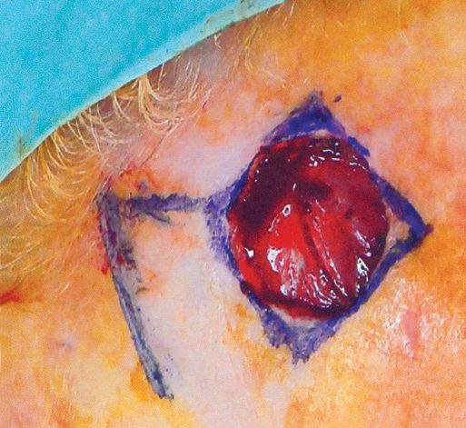
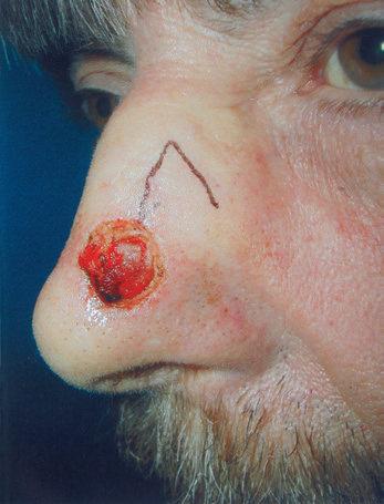
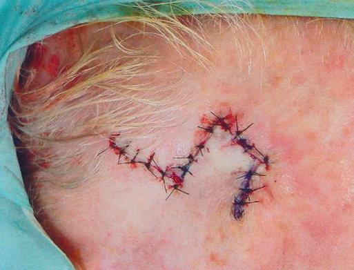
Fig. 2.7 This lateral cheek rotation flap illustrates how the flap is rotated in an arc along a pivot point to fill the adjacent primary defect without crossing any other intervening skin.
of ancient Rome, who used the flap for the reconstruction of the cheek and ears.70,71 In the 1800s, French surgeons advocated the use of “lambeau par glissement” (sliding flaps) for facial reconstruction.72 In 1885 Burow modi fied and fur ther described the advantages of this flap in reconstruction.73
The unilateral, bilateral, and V to Y advancement flaps are the most commonly used advancement flaps in dermatologic surgery. The prmary and secondary motions, and thus the resulting tension vectors of these flaps, are usually in a simple linear line along the direction of the flap movement (see Fig. 2.6).73 The key stitch typically brings the leading edge of the flap to the opposite wound edge to close the primary defect. As the tissue is advanced and sutured into place, a standing cone (or cones) develop(s). These standng cone deformities, or dog ears, are caused by the pouching of excess, compressed skin near the flap’s base when tissue is advanced under tension.74
The standing cone may be corrected by either extending the incision line to remove the excess tissue or using an M-plasty (Fig. 2.10A).75,76 By truncating one end of the closure, the M-plasty flap has the advantage of providing a shorter final wound ength. If the wound cannot be extended due to a ree margin or approaching cosmetic subunit boundary, tissue protrusions can be anticipated. As the length of the flap base is longer than the edge of the flap itself, uneven lengths of tissue requiring approximation are created (see Fig. 2.10B). The inequality of tissue lengths can be accounted for by removing a triangle of skin, called a Burow’s triangle,77 from anywhere along the skin surrounding the flap (see Figs. 2.6 and 2.10B). In addition, the unequal lengths can be accounted for by removing a curvi linear segment of skin from the limb of the flap itself, thereby lengthening the overall arm of the flap o equal that of the flap base and redistributing the redundant tissue along the length of the incision (see Fig 2.10C). This is particularly useful on the upper cuaneous lip and eyebrow areas.
When using bilateral advancement flaps to correct a circular defect, the repair may be described as an A-to-T (T-plasty) or an O-to-H flap (H-plasty). These unilateral and bilateral advancement flaps are most commonly used in repairs on the forehead, scalp, and eyelids. The V-to-Y advancement flap78 uses a subcutaneous pedicle that perfuses a triangular flap that advances along a single direction (see Fig. 2.10D). All borders of the flap are incised to the subcutaneous fat, and the resulting island of tissue, now liberated from lateral dermal attachments, s advanced to fill the defect. The pedicle is underneath rather than at the edge of the flap and may include muscle.79–81 Various modifications,82,83 such as parallel release incisions of the pedicle base, allow significant advancement of the flap with predictable flap survival.84 Because the flap’s muscularcontaining, thick pedicle ensures liberal blood flow, this flap is the most likely of all flaps to survive in patients with tobacco use, which can result in decreased perfusion. The flap can be used anywhere on the face, but the authors find this flap to be particularly useful for repairs of the upper cutaneous lip, the alar–cheek sulcus, and the lateral eyebrow, with alopecic defects to help restore medial hairbearing areas. Of note, the V-to-Y advancement flap was previously synonymous with the “island pedicle flap”; however, this terminology now refers exclusively to flaps in which a portion of the donor tissue is deepithelialized and kers
tunneled or passed beneath the skin to a distant recipient site. It is important to differentiate the island pedicle flap from a Vto-Y advancement flap for both clinical documentaton and coding purposes.
In 1842 Pancoast described using rotation flaps to close facial wounds.85 Like most random pattern flaps, rotation flaps recruit tissue adjacent to the primary defect. An arc or semicircle flap is incised and pivoted into the wound (Fig. 2.11). Thus the primary motion is rotation. Similar to advancement flaps, the secondary motion of the flap causes a standing cone deformity at the base of the flap. This Burow’s triangle may be excised from anywhere along the arc of the flap from the nonpedicle side (see Fig. 2.11A). Another similarity between rotation and advancement flaps is the placement of the key stitch. This first stitch closes the primary defect.
The surgeon must pay particular attention to the sze of the designed rotation flap. Because it is rotated along an arc, the functional flap length is shorter than the actual length of the flap’s incision see Fig. 2.11B). Hence the planned length must be longer than the diameter of the wound.86 Dzubow recommends that the planned height of the flap be greater than the height of the defect to account for this functional loss of length.87 One advantage of the rotation flap is that its base is typically broad, and therefore the flap’s length-to-width ratio may be as long as 6 : 1.88 This makes the flap particularly useful when closing inelastic areas such as the scalp.
To increase the movement of the rotation flap, a back-cut or Z-plasty may be employed. A back-cut is when an inci sion is placed into the pedicle of the flap to allow greater forward movement (see Fig. 2.11C). Although the back-cut can increase flap mobility, this maneuver cuts into the flap’s base, thereby increasing the risk of tip ischemia. In areas with excellent vascularity, such as the gabella, the back-cut is often incorporated into the rotation flap’s design. Another technique used to increase flap movement and reduce the primary tension vector is the use of a Z-plasty at the tail of the rotation flap (see Fig. 2.11D).
https://t.meboers https://t ers htps://t.me/eokers https://t.me/ ers https/tme/ebokers tts//me/ ers
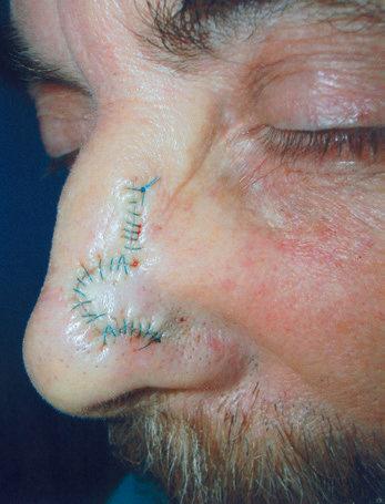
Rotation flaps are most often used for repairs of the scalp, forehead, cheek, and nose. The Rieger flap,89 or dorsal nasal rotation flap, uses a back-cut in its design to allow closure of small- to medium-sized defects of the distal third of the nose. In 1985 Marchac and Toth modified the flap design by defining an axial pedicle for the flap based on the angular artery.90 This modification allows a greater flap length-topedicle base ratio because of a reliable blood supply. Multiple rotation flaps may be used to repair large defects on the chin, forehead, and nasal tip. When two mirror-image rotation flaps are designed to repair the defect, the closure is called an O-to-Z repair. There are a few specialized forms of bilateral rotation flaps that have also been reported: the Peng flap for the nose and the bilateral vermillion flap on the lip. 92 When three or more rotation flaps are used, it is often referred to as a pinwheel flap.
Transposition flaps were documented in the pre–Common Era. In about 2000 BC, Indians committing adultery were
https://t.me/ebokes https://t.me/ ers ttps://t.me/ebokers https
ers https://t.me/eokers https:/tme kers https:/t.meeookers https://t.me kers ts://tme/eoers tps/. ers