Mindfulness intervention for mild cognitive impairment led to attention-related improvements and neuroplastic changes: Results from a 9-month randomized control trial
I dedicate this fourth edition of the “Pink Book” to the younger generations of veterinarians who are at the beginning of their careers and are pursuing their interest and passion in exotic animal medicine and surgery. I have been privileged to meet and work with so many bright and dedicated young veterinarians during my career at the Animal Medical Center, my travels while lecturing, and my work as a journal and book editor. You have made my career journey so worthwhile. I am grateful for all that I have learned from every one of you, and I hope that I have been able to pass on some of my knowledge and experience to you. I thank all of my friends and colleagues who have worked with me on this book for so many editions and for so many years—you have truly made this book what it is. I am grateful for my co-editors of this edition, Connie J. Orcutt and Christoph Mans, who put so much time and effort into this book, and especially Jim Carpenter, who has worked with me on three editions of this book—we could not have done this without you. I thank my children, Zachary and Chelsea Messinger, who are forever the center of my life, and my sister, Marcia Quesenberry, for always being there for me and encouraging me to move forward. Lastly, I thank John Harris for being so patient and loving while I worked on this book and for always reminding me to enjoy life.
Katherine E. Quesenberry
With immense gratitude, I dedicate this book to the professional colleagues I’ve worked with over the years. First among them is Kathy Quesenberry, who introduced me to both exotic pet medicine and medical writing. As a mentor and friend, she has always modeled professionalism, intellectual curiosity, and compassion. I also thank the extremely talented and dedicated clinicians, technicians, and great friends with whom I’ve shared professional and personal successes and struggles; they include my “exotic” colleagues, Jennifer Graham, Flo Tseng, Wendy Emerson, Deborah Kennedy, Jenny Hayes, and Lauren Skeens, as well as the “nonexotic” clinicians at Angell Animal Medical Center, who always pushed the envelope for my unusual patients. Finally, I offer my deep thanks and admiration to the veterinarians who authored these chapters—those searchers who attempted and accomplished things that had not been tried before and then generously shared their discoveries with the rest of us.
Connie J. Orcutt
I would like to dedicate this book to all the kind and generous people who have supported me throughout my career. From the staff at the Avian Exotic Service at the Animal Medical Center in 2005 (welcoming the German veterinary student who just got off the boat and barely spoke any English) to all the great people I met and worked with at many institutions since, including the University of Tennessee, the Ontario Veterinary College, the Vetsuisse Faculty Zurich, the Tai Wai Small Animal and Exotic Hospital, the Milwaukee County Zoo, and University of Wisconsin-Madison—thank you. I particularly would like to thank Kathy Quesenberry for inviting me to be a co-editor of the fourth edition of the “Pink Book,” which has been a great honor. I would like to thank Tom Donnelly and Cyndi Brown for their mentorship, which had a profound impact on my career. Special thanks go to Penny Rudolph for the many opportunities provided over the years. Most importantly, I would like to thank and dedicate this book to my wife for her support while I was editing the “Pink Book” and to my children, without whom this book likely would have been published two years earlier.
Christoph Mans
I wish to acknowledge all of our colleagues who contributed their knowledge and time to this edition of Ferrets, Rabbits, and Rodents: Clinical Medicine and Surgery. I also thank Dr. Kathy Quesenberry for her friendship and for once again inviting me to collaborate on this edition of the Pink Book. I also wish to thank the 42 interns and residents whom I have helped train in zoological/exotic pet medicine at Kansas State University and who have inspired my professional life. I am indebted to Drs. Bonnie Rush, Dean, and Elizabeth Davis, Department Head, for their strong support of me and for my professional/academic growth. I also wish to thank veterinary students Sarah Wilson, Karissa Severud, Elizabeth Loos, and Danielle Windle for their assistance in the preparation of this text. I would like to dedicate this book to my family (wife, Terry; son, Michael; and daughter, Erin, and her family—husband, Steve, and my grandkids, Kylie, Hayden, and Asher) who have supported me as I pursued my passion for zoological and wildlife medicine for the past 45 years.
James W. Carpenter
FERRETS, RABBITS, and RODENTS
CLINICAL MEDICINE and SURGERY
Katherine E. Quesenberry
DVM, MPH, Diplomate ABVP (Avian) Service Head Avian and Exotic Pet Service Chief Medical Officer The Animal Medical Center New York, NY, United States
Connie J. Orcutt
DVM, Diplomate AVBP (Exotic Companion Mammal)
Brookline, MA, United States
Christoph Mans
Dr. med. vet., Diplomate ACZM, Diplomate ECZM (Zoo Health Management) Clinical Associate Professor School of Veterinary Medicine University of Wisconsin-Madison Madison, WI, United States
James W. Carpenter
MS, DVM, Diplomate ACZM Professor, Zoological Medicine Department of Clinical Sciences College of Veterinary Medicine Kansas State University Manhattan, KS, United States
Elsevier
3251 Riverport Lane
St. Louis, Missouri 63043
FERRETS, RABBITS, AND RODENTS: CLINICAL MEDICINE AND SURGERY, FOURTH EDITION
No part of this publication may be reproduced or transmitted in any form or by any means, electronic or mechanical, including photocopying, recording, or any information storage and retrieval system, without permission in writing from the publisher. Details on how to seek permission, further information about the Publisher’s permissions policies and our arrangements with organizations such as the Copyright Clearance Center and the Copyright Licensing Agency, can be found at our website: www.elsevier.com/permissions.
This book and the individual contributions contained in it are protected under copyright by the Publisher (other than as may be noted herein).
Notice
Practitioners and researchers must always rely on their own experience and knowledge in evaluating and using any information, methods, compounds or experiments described herein. Because of rapid advances in the medical sciences, in particular, independent verification of diagnoses and drug dosages should be made. To the fullest extent of the law, no responsibility is assumed by Elsevier, authors, editors or contributors for any injury and/or damage to persons or property as a matter of products liability, negligence or otherwise, or from any use or operation of any methods, products, instructions, or ideas contained in the material herein.
Previous editions copyrighted 2012, 2004, and 1997.
Library of Congress Control Number: 2020933690
Content Strategist: Jennifer Catando
Content Development Specialist: Kathryn DeFrancesco
Thomas N. Tully, Jr., DVM, MS, Diplomate ABVP (Avian), Diplomate ECZM (Avian)
Professor Zoological Medicine
Department of Veterinary Clinical Sciences
Louisiana State University
School of Veterinary Medicine
Baton Rouge, LA, United States
Alexandra van der Woerdt, DVM, MS, Diplomate ACVO, Diplomate ECVO Service Head
Department of Ophthalmology
The Animal Medical Center
New York, NY, United States
Yvonne R.A van Zeeland, DVM, MVR, PhD, Diplomate ECZM (Avian, Small Mammal)
Associate Professor
Division of Zoological Medicine, Department of Clinical Sciences of Companion Animals
Faculty of Veterinary Medicine
Utrecht University Utrecht, Netherlands
Molly Varga, BVetMed, DZooMed Lead Clinician
Department of Exotic Medicine and Surgery
Rutland House Veterinary Referrals
Merseyside, United Kingdom
David Vella, BSc, BVSc, Diplomate ABVP (Exotic Companion Mammal)
Director
Sydney Exotics & Rabbit Vets
North Shore Veterinary Specialist Centre
Sydney, New South Wales, Australia
Raquel M. Walton, VMD, MS, PhD, Diplomate ACVP (Clinical Pathology)
Clinical Pathologist
Center for Animal Referral and Emergency Services
Idexx Laboratories, Inc.
Langhorne, PA, United States
Bruce H. Williams, DVM, Diplomate ACVP
Senior Pathologist
Veterinary Pathology Service
Joint Pathology Center
Silver Spring, MD, United States
Nicole R. Wyre, DVM, Diplomate ABVP (Avian, Exotic Companion Mammal)
Supervising Veterinarian
Zodiac Pet & Exotic Hospital
Fortress Hill, Hong Kong
PREFACE
In this fourth edition of Ferrets, Rabbits, and Rodents: Clinical Medicine and Surgery, we have gathered together an experienced, international team of authors to provide our readers with a comprehensive source of the clinically relevant information on topics concerning the medicine and surgery of common small mammals kept as pets. The knowledge base of small mammal medicine has advanced rapidly with each edition of this book, so much so that it is difficult to capture and filter the most relevant and timely information into one source. As in the previous three editions, our goal remains to present the most relevant information in a succinct format that is easily readable and user friendly. With the hard work and dedication of all four editors, Connie J. Orcutt, Christoph Mans, James W. Carpenter, and myself, as well as the many talented authors who have contributed to this fourth edition of the “Pink Book,” we feel we have accomplished our goal.
This edition continues in the tradition of the previous three editions in presenting separate sections on the medicine and husbandry of ferrets, rabbits, rodents, and other small mammals. In this edition, as in previous editions, we also have included dedicated chapters on sugar gliders and African pygmy hedgehogs, and we have added chapters on degus, prairie dogs, and skunks. We have consolidated a separate section
titled “Surgical Techniques and Dentistry,” which includes specific chapters on soft tissue surgery of ferrets, rabbits, and rodents, as well as chapters on general principles, orthopedics in small mammals, diagnostic and surgical endoscopy, and small mammal dentistry. In the “General Topics” section, we have a new chapter on hematology and biochemistry, which discusses specific information on and interpretation of clinical pathology in small mammals.
As in the previous editions, this book would not have been successful without the expertise and team effort of the editors. We have collaborated in all aspects of this book to bring the pieces together, including author selection, chapters, photos, and editing. We are extremely grateful to our support team at Elsevier, namely Jennifer Catando, Kathryn DeFrancesco, and Karthikeyan Murthy, who have been patient and supremely helpful as we worked through the roadblocks we encountered. Their professionalism and expertise has been invaluable. As with the previous three editions, we are confident that the format, presentation, information, and reliability of this fourth edition of the “Pink Book” will continue to set it apart as the standard in this subspecialty of veterinary medicine.
Katherine E. Quesenberry
SECTION I Ferrets
1 Basic Anatomy, Physiology, and Husbandry of Ferrets, 1
Lauren V. Powers and David Perpiñán
2 Basic Approach to Veterinary Care of Ferrets, 13
Katherine E. Quesenberry and Ricardo de Matos
3 Gastrointestinal Diseases of Ferrets, 27
Heidi L. Hoefer
4 Disorders of the Urinary and Reproductive Systems in Ferrets, 39
Nicola Di Girolamo and Minh Huynh
5 Cardiovascular and Other Diseases of Ferrets, 55
James K. Morrisey and Rebecca L. Malakoff
6 Respiratory Diseases of Ferrets, 71
David Perpiñán
7 Endocrine Diseases of Ferrets, 77
Nico J. Schoemaker and Yvonne R.A. van Zeeland
8 Neoplasia in Ferrets, 92
Bruce H. Williams and Nicole R. Wyre
9 Dermatologic Diseases of Ferrets, 109
Dario d’Ovidio and Domenico Santoro
10 Musculoskeletal and Neurologic Diseases, 117
Minh Huynh and Stéphanie Piazza
SECTION II Rabbits
11 Basic Anatomy, Physiology, and Husbandry of Rabbits, 131
Thomas M. Donnelly and David Vella
12 Basic Approach to Veterinary Care of Rabbits, 150
João Brandão, Jennifer Graham, and Katherine E. Quesenberry
13 Gastrointestinal Physiology and Nutrition of Rabbits, 162
Susan M. Smith
14 Gastrointestinal Diseases of Rabbits, 174
Barbara L. Oglesbee and Brigitte Lord
15 Respiratory Disease, 188
Angela M. Lennox and Elisabetta Mancinelli
16 Disorders of the Urinary and Reproductive Systems, 201
Nicola Di Girolamo and Paolo Selleri
17 Dermatologic Diseases of Rabbits, 220
Molly Varga and Susan Paterson
18 Neurologic and Musculoskeletal Diseases, 233
Peter G. Fisher, Frank Künzel, and Helena Rylander
19 Cardiovascular Disease, 250
Connie J. Orcutt and Rebecca L. Malakoff
20 Lymphoreticular Disorders, Thymoma, and Other Neoplastic Diseases, 258
Katherine E. Quesenberry, Anthony A. Pilny, and Rachel S. St-Vincent
SECTION III Rodents
21 Guinea Pigs, 270
Charly Pignon and Joerg Mayer
22 Chinchillas, 298
Christoph Mans and Thomas M. Donnelly
23 Degus, 323
Vladimir Jekl
24 Prairie Dogs, 334
David Eshar and Sara M. Gardhouse
25 Rats and Mice, 345
Jennifer Frohlich
26 Hamsters and Gerbils, 368
Yasutsugu Miwa and Joerg Mayer
SECTION IV Other Small Mammals
27 Sugar Gliders, 385
Cathy Johnson-Delaney
28 African Pygmy Hedgehogs, 401
Grayson A. Doss and James W. Carpenter
29 Skunks, 416
Livia Benato and Dario d’Ovidio
SECTION V Surgical Techniques and Dentistry
30 General Principles of Surgical Techniques, 426
R. Avery Bennett Jr.
31 Soft Tissue Surgery: Ferrets, 432
Vladimir Jekl and Sue Casale
32 Soft Tissue Surgery: Rabbits, 446
David Sanchez-Migallon Guzman, Zoltan Szabo, and Michele A. Steffey
33 Soft Tissue Surgery: Rodents, 467
Zoltan Szabo
34 Orthopedics in Small Mammals, 483
David Sanchez-Migallon Guzman and Amy S. Kapatkin
35 Exotic Mammal Diagnostic and Surgical Endoscopy, 498
Stephen J. Divers and Izidora Sladakovic
36 Small Mammal Dentistry, 514
Angela M. Lennox, Vittorio Capello, and Loic Frederic Legendre
SECTION VI General Topics
37 Anesthesia, Analgesia, and Sedation of Small Mammals, 536
Michelle G. Hawkins and Peter J. Pascoe
38 Diagnostic Imaging, 559
Alison L. Tarbell and Anthony J. Fischetti
39 Hematology and Biochemistry of Small Mammals, 569
Andrea Siegel and Raquel M. Walton
40 Ophthalmologic Diseases of Small Mammals, 583
Alexandra van der Woerdt
41 Emergency and Critical Care of Small Mammals, 595
Jay N. Gladden and Angela M. Lennox
42 Zoonotic Diseases Associated With Small Mammals, 609
Mark A. Mitchell and Thomas N. Tully, Jr.
Appendix: Formulary, 620
James K. Morrisey and James W. Carpenter Index, 631
SECTION I Ferrets
1
Basic Anatomy, Physiology, and Husbandry of Ferrets
Lauren V. Powers, DVM, Diplomate ABVP (Avian, Exotic Companion Mammal) and David Perpiñán, DVM, MSc, PhD, Diplomate ECZM (Herpetology)
OUTLINE
Natural History and Domestication, 1
Uses, 2
Anatomy and Physiology, 2
Integument, 2
Coat, 2
Skin and Associated Glands, 3
Anal Glands, 3
Gastrointestinal System, 3
Teeth and Salivary Glands, 3
Esophagus, Stomach, and Intestines, 3
Liver, Gallbladder, and Pancreas, 6
Urogenital System, 7
Kidneys, Ureters, and Urinary Bladder, 7
Male Reproductive Tract, 7
Female Reproductive Tract, 7
Cardiovascular and Lymphatic Systems, 7
Heart and Major Blood Vessels, 7
Lymphatic Structures, 7
NATURAL HISTORY AND DOMESTICATION
The domestic ferret (Mustela putorius furo) belongs to the family Mustelidae, the largest family within the mammalian Order Carnivora. Along with ferrets, the Genus Mustela includes polecats, mink, weasels, and ermines (also called stoats).
The domestic ferret is most likely a direct descendant of the European polecat (M. putorius) but may also be descended from the steppe (or Siberian) polecat (M. eversmannii).8,16,31,52 The domestic ferret is also closely related to the black-footed ferret (M. nigripes) but is likely not a direct descendant.
Free-ranging ferrets and polecats are found throughout Europe, Asia, and North America. Black-footed ferrets nearly became extinct due to habitat destruction and the deliberate
Respiratory System, 8
Endocrine System, 8
Adrenal Glands, 8
Thyroid and Parathyroid Glands, 8
Musculoskeletal System, 9
Neurologic System and Special Senses, 9
Brain and Spinal Cord, 9
Special Senses, 9
Physiology and Reproduction, 9
Life Expectancy and Physiology, 9
Body Size and Seasonal Weight Variation, 9
Reproduction, 9 Behavior, 10
Husbandry, 10
Housing, 10
Environmental Enrichment, 10 Nutrition, 10
depopulation of prairie dogs (Cynomys species), their main food source.25,36 Captive breeding and reintroduction programs for the black-footed ferret have reestablished populations in some areas of North America; however, the species continues to be listed as endangered under the US Endangered Species Act. Ferrets likely have been domesticated for more than 2000 years, probably first being domesticated in southern Europe. 8,11,16,25,31 They may have been introduced into the United Kingdom by the Romans or Normans.8,11,25 In the late 1800s, domestic ferrets were intentionally introduced into New Zealand to depopulate feral colonies of European rabbits ( Oryctolagus cuniculus ). 25 Because of the lack of predatory species, feral populations of ferrets were established and are still present, raising concerns for the spread of infectious
diseases (e.g., Mycobacterium bovis in livestock and zoonotic diseases, such as rabies), and preying on native bird populations. 16,25 Similarly, ferrets were released in Australia in the 1800s to depopulate feral colonies of introduced rabbits; however, predators prevented the establishment of feral populations of ferrets. 25 The domestic ferret was introduced into the United States approximately 300 years ago, most likely for pest depopulation and hunting.16,25,31 There are currently no established feral populations of domestic ferrets in North America.31
USES
In the early 1900s, tens of thousands of ferrets were bred in the United States to help rid granaries, barns, and warehouses of rodents.16 Ferrets were also used for pest control on ships.11,16,25 In some areas of the world, ferrets are still used for rodent and rabbit control.
Ferrets have long been bred to hunt European rabbits, and ferrets are still used to hunt in some areas of the world.16,25,31 In the past, domestic ferrets were used to hunt rabbits in the United States; however, most states now prohibit this practice, primarily to protect native rabbit species.16,25
Ferrets were farmed for fur in North America in the early 1900s and even earlier in Europe.16 Ferret fur farming was popular in New Zealand as late as the 1980s and still exists in a few areas of Northern Europe.16,25 Ferret fur has also been used in artists’ paint brushes.25
“Ferret-legging” is a type of English pub game in which two ferrets are placed into each competitors’ trousers before the leg openings and waist are securely closed. The competitor who withstands the presence of the ferrets the longest is the winner.16
Because of their elongated, narrow body and willingness to travel through long, narrow tunnels, domestic ferrets have been used to string cable and wire through long stretches of conduit for the oil, aviation, and telephone industries.16
Ferrets have been used in biomedical research since the early 1900s, when they were used to study human influenza and other viral diseases, and are now widely used for experimental studies in numerous biomedical fields.16,19,31 Ferrets are popular as research models because of their small size, high fecundity, biologic similarities to humans, and susceptibility to many human pathogens and diseases.16,19,23,31,43
Ferrets are popular as companion animals worldwide.25 Domestic ferrets bred for today’s pet market are docile, curious, intelligent, playful, and interactive with humans.6,11,17 Their small size and relatively quiet nature makes them popular pets for smaller homes. Because of large-scale commercial breeding (eg, Marshall Farms Group Ltd., North Rose, NY), ferrets are readily available in pet stores in the United States. Commercially bred ferrets typically undergo surgical sterilization and anal sacculectomy and receive their initial canine distemper vaccination before shipment to pet stores. Ferrets are also available from small-scale commercial breeders and hobby breeders, but they may not have undergone surgical sterilization or anal sacculectomy before purchase.
Ferrets were banned in many states until the availability of a rabies vaccine licensed by the US Department of Agriculture for use in this species.25 Ferret ownership is still prohibited or restricted in some cities and states, such as New York City and California, because of concerns regarding rabies exposure, attacks on people (particularly infants), and establishment of feral populations.16,17 Such concerns are now less warranted due to the availability of a licensed rabies vaccine and to genetic selection and routine surgical alteration of ferrets. In the European Union, only domestic ferrets, cats, and dogs are eligible for pet passports and free travel among member countries, provided they are microchipped and current on rabies vaccination. Although keeping ferrets is popular in other parts of the world, ownership is restricted or prohibited in some areas to protect native wildlife.
ANATOMY AND PHYSIOLOGY
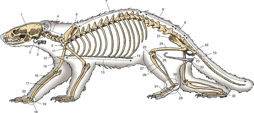
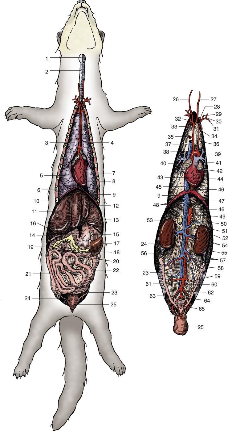
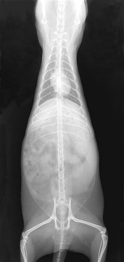
The basic anatomy and physiology of the domestic ferret is similar to that of other carnivores. The following is a brief review of clinically relevant features. Skeletal anatomy is depicted in Fig. 1.1, visceral anatomy in Fig. 1.2, and normal radiographic anatomy in Figs. 1.3 and 1.4. Selected physiologic values are detailed in Table 1.1. The reader is directed to other publications containing more extensive reviews of ferret anatomy and physiology.10,12,15,23,48
Integument Coat
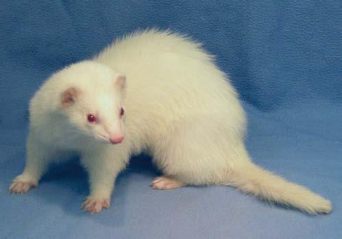
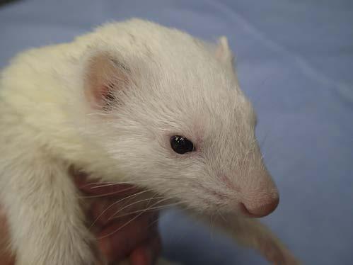
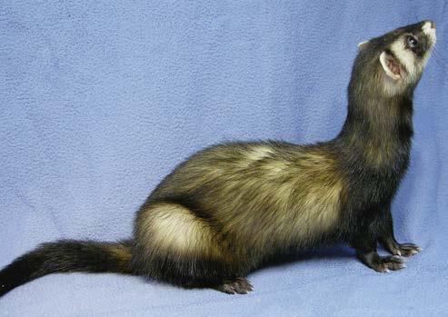
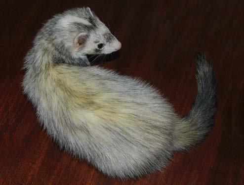
The pelage of the domestic ferret consists of long, coarse guard hairs and a fine, white to yellow undercoat, which results in excellent insulation.10,26 There are no specific breeds of domestic ferrets; instead, ferrets are often classified according to the color and pattern of their coat (Fig. 1.5).26 The two predominant varieties of domestic ferret are the fitch (also known as sable, or wild-type) and the albino.19 Some of the color standards recognized by the American Ferret Association are sable (warm, deep brown), black, black sable, champagne, and albino (white guard hairs and unpigmented eyes). Some standardized patterns for white markings include mitt (white paws), panda (nearly completely white head), and blaze (white blaze from the forehead down the back of the neck). Other recognized color patterns include solid, standard, and point (with a distinct color difference between the color of the body and the points). The dark-eyed white is a combination of any solid white ferret with pigmented eyes. The color and pattern of the coat and mask can change over time. Ferrets living outdoors tend to be darker in color.26 Ferrets undergo a heavy shed in the spring and fall along with seasonal weight changes.12 The coat may be shorter in summer months and longer in the fall, and lighter in color in the winter and darker in the fall.12,26 Some intact females (jills) in estrus can become dramatically alopecic and completely lose the undercoat, exposing bare patches of skin.12 Sexually altered ferrets of either sex have a less dramatic molt and color change. Warn clients that shaved fur may not regrow for weeks to months, and that before erupting, the fur may cause the skin to appear bluish. Regrown fur may have a different color or texture than surrounding fur.
Fig. 1.1 Skeletal Anatomy of a Ferret 1, Calvaria; 2, hyoid apparatus; 3, larynx; 4, seven cervical vertebrae; 5, clavicle; 6, scapula; 7, 15 thoracic vertebrae; 8, five lumbar vertebrae; 9, three sacral vertebrae; 10, 18 caudal vertebrae; 11, first rib; 12, manubrium; 13, sternum; 14, xiphoid process; 15, humerus; 16, radius; 17, ulna; 18, carpal bones; 19, accessory carpal bone; 20, metacarpal bones; 21, ilium; 22, ischium; 23, pubis; 24, femur; 25, patella; 26, fabella; 27, tibia; 28, fibula; 29, tarsal bones; 30, calcaneus; 31, metatarsal bones; 32, talus; 33, os penis. (Adapted from An NQ, Evans HE. Anatomy of the ferret. In: Fox JG, ed. Biology and Diseases of the Ferret. Philadelphia: Lea & Febiger; 1988:14.)
Skin and Associated Glands
Ferrets have very active sebaceous glands that produce a strong musky odor.19 During the breeding season, intact ferrets have increased sebaceous secretions, resulting in a more intense odor, yellow to orange discoloration of the undercoat, and oily fur.19 Ferrets lack sweat glands, making them very susceptible to heat prostration.20,33
Anal Glands
Ferrets have a well-developed pair of anal glands, which produce a serous yellow liquid with a strong odor. Frightened or threatened ferrets can express their anal glands but cannot project the fluid over long distances.19,20 The anal glands typically measure 10 mm × 5 mm, and the ducts open into the anal canal at about 4 o’clock and 8 o’clock positions.26 External anal sphincter muscle encloses each duct.10,20,26 Although most of a ferret’s odor arises from its sebaceous glands and not its anal glands,19 domestic ferrets raised at large commercial breeding facilities in the United States are routinely descented between 5 and 6 weeks of age. This practice is being increasingly questioned on ethical grounds. Therefore, in other countries, and increasingly in the United States with small-scale breeders, ferrets may be descented later or not at all.
Gastrointestinal System
Teeth and Salivary Glands
Ferret dentition is typical of carnivores, with long, curved canine teeth and shearing and crushing premolars and molars (See also Chapter 36). Permanent teeth erupt between 50 and 74 days of age.31 Ferrets have 34 permanent teeth, and the dental formula
of the adult ferret is 2(I33 C11 P33 M12).19,20,26,27 Note that ferrets have three rather than four premolars, and the maxillary carnassial tooth is likely the fourth premolar.10 Incisors and canine teeth each have a single root. The premolars have one to two roots each, except for the carnassial tooth, which has three roots. The maxillary first molar and mandibular first molar have three roots, and the tiny mandibular second molar has only one root.10,34 Supernumerary incisors are common.4,19,26,34 Ferrets have five major pairs of salivary glands: the parotid, mandibular, sublingual, molar, and zygomatic glands.10,20,26
Esophagus, Stomach, and Intestines
Similar to the dog, the muscle of the ferret esophagus is striated along the entire length cranial to the diaphragm.15,44 There is no true gastroesophageal sphincter, and ferrets are readily able to vomit.20,26,27,38 Although domestic ferrets are popular experimental research models for the study of emesis, ferrets with gastrointestinal (GI) obstruction rarely vomit.26 The ferret’s simple monogastric stomach is similar in shape to that of the dog, consisting of the cardia, fundus, body, and pylorus.10,19,26,31,38 The stomach contacts the diaphragm and left liver lobes cranially, the ascending colon dorsally, and the spleen and left pancreatic limb caudally.38 The stomach is joined to the spleen by the gastrosplenic ligament and is separated from the papillary process of the caudate liver lobe by the lesser omentum.10,38 A full stomach is readily palpable and displaces the intestines to the right.26,27,38
The small intestine is comparatively short; therefore the adult ferret has a comparatively short GI transit time of 3 to 4 hours.5,10,20,27,38,40 The duodenum consists of three
Fig. 1.2 (A) Ventral aspect of the viscera of a ferret in situ. (B) Anatomy of the viscera and most important blood vessels as seen after removal of the lungs, liver, and gastrointestinal tract. 1, Larynx; 2, trachea; 3, right cranial lobe of lung; 4, left cranial lobe of lung; 5, right middle lobe of lung; 6, right caudal lobe of lung; 7, left caudal lobe of lung; 8, heart; 9, diaphragm; 10, quadrate lobe of liver; 11, right medial lobe of liver; 12, left medial lobe of liver; 13, left lateral lobe of liver; 14, right lateral lobe of liver; 15, stomach; 16, right kidney; 17, spleen; 18, pancreas; 19, duodenum; 20, transverse colon; 21, jejunoileum; 22, descending colon; 23, uterus; 24, ureter; 25, urinary bladder; 26, right common carotid artery; 27, left common carotid artery; 28, vertebral artery; 29, costocervical artery; 30, superficial cervical artery; 31, axillary artery; 32, right subclavian artery; 33, right internal thoracic artery; 34, left internal thoracic artery; 35, branch to thymus; 36, left subclavian artery; 37, brachiocephalic (innominate) artery; 38, cranial vena cava; 39, aortic arch; 40, right atrium; 41, pulmonary trunk; 42, left atrium; 43, right ventricle; 44, left ventricle; 45, caudal vena cava; 46, aorta; 47, esophagus; 48, hepatic veins; 49, celiac artery; 50, cranial mesenteric artery; 51, left adrenolumbar vein; 52, left adrenal gland; 53, right adrenal gland; 54, left renal artery and vein; 55, left kidney; 56, suspensory ligament of ovary; 57, left ovarian artery and vein; 58, left ovary; 59, left deep circumflex iliac artery and vein; 60, caudal mesenteric artery; 61, broad ligament of uterus; 62, left external iliac artery; 63, right common iliac vein; 64, left internal iliac artery; 65, rectum (Adapted from An NQ, Evans HE. Anatomy of the ferret. In: Fox JG, ed. Biology and Diseases of the Ferret. Philadelphia: Lea & Febiger; 1988:14.)
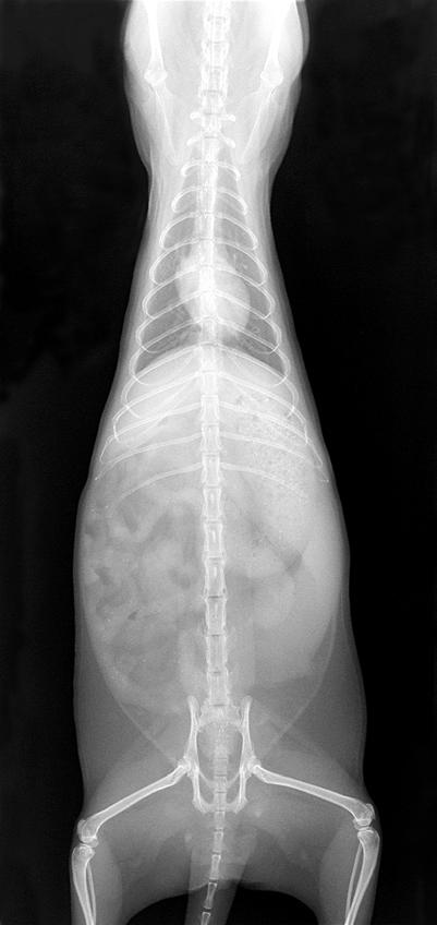
Fig. 1.3 (A) Ventrodorsal radiograph of a 1-year-old, spayed female ferret. Note normal positioning of thoracic and abdominal viscera. (B) Same radiograph as (A): 1, trachea (endotracheal tube within lumen); 2, lung; 3, cranial mediastinum; 4, left primary bronchus; 5, heart; 6, liver; 7, stomach; 8, spleen; 9, left kidney; 10, urinary bladder; 11, right primary bronchus; 12, small intestine; 13, right kidney. (Silverman S, Tell LA. Radiology of Rodents, Rabbits, and Ferrets: An Atlas of Normal Anatomy and Positioning. St. Louis: Elsevier Saunders; 2005:233.)
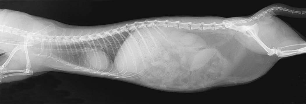
Fig. 1.4 (A) Lateral (right lateral recumbency) radiograph of a 1-year-old, spayed female ferret. Note normal positioning of thoracic and abdominal viscera. (B) Same radiograph as (A): 1, trachea (endotracheal tube within lumen); 2, lung; 3, pulmonary vasculature; 4, bronchus; 5, pulmonary vein; 6, stomach; 7, kidney; 8, spleen; 9, colon; 10, intrathoracic adipose tissue; 11, heart; 12, liver; 13, small intestine; 14, urinary bladder. (Silverman S, Tell LA. Radiology of Rodents, Rabbits, and Ferrets: An Atlas of Normal Anatomy and Positioning. St. Louis: Elsevier Saunders; 2005:232.)
portions: the shorter, sigmoid-shaped cranial portion; the descending portion, in contact with the right kidney caudally; and the ascending portion.38 A hairpin turn separates the descending and ascending portions. The mesoduodenum encloses the right limb of the pancreas and a portion of the lesser omentum.10 The jejunum and ileum are macroscopically indistinguishable, creating a jejunoileum.20,38 The ferret lacks a cecum, ileocecal valve, and appendix; therefore the ileocolic junction is indistinct and is typically defined as the region where the ileojejunal and colic arteries join.20,27,38 The jejunoileal mucosa is flat, whereas the colonic mucosa forms longitudinal folds.38 The large intestine consists of the colon (with ascending, transverse, and descending regions), rectum, and anus.10,38
Liver, Gallbladder, and Pancreas
The ferret’s liver is relatively large and consists of six lobes: left lateral, left medial, right lateral, right medial, quadrate, and caudate.10,20,26,27 The gallbladder lies between the quadrate and right medial lobes and measures approximately 2 cm by 1 cm.10,20,26 Although variable in pattern, the cystic duct usually joins the left, right, and central hepatic ducts to form the common bile duct.10
The pancreas is V-shaped and divided into right and left lobes connected by a body that lies close to the pylorus and is contained within the mesoduodenum.10 The left lobe extends along the dorsal caudal stomach and medial to the spleen, and the right lobe follows the descending duodenum. Ducts from the left and right lobes connect to form the common pancreatic
TABLE
1.1 Selected Biologic Values for the Domestic Ferret15,19,20,26, 45
Parameter
Life expectancy
Rectal temperature
Heart rate
Blood volume
Noninvasive blood pressure (high-definition oscillometry, n = 63; sedated with 0.2 mg/ kg butorphanol and 0.2 mg/kg midazolam)45
Respiratory rate
Tidal volume
Stomach capacity
Gastrointestinal transit time
Urinary output
Bladder capacity
Urine pH
Puberty
Reproductive life span
Gestation
Litter size
Birth weight
Eyes and ears open
Weaning age
Water intake
Maintenance fluid needs
Maintenance caloric needs
‒1.2 kg
5‒11 years
Mean 101.8°F [38.8°C] (Range 100°‒104°F [37.8°‒40°C])
200‒400 beats/min
Male 60 mL
Female 40 mL
Systolic: 95‒155 mmHg
Diastolic: 51‒87 mmHg
Mean: 69‒109 mmHg
33‒36 breaths/min
10‒11 mL/kg
50 mL/kg when distended
2.5‒3.6 hours (meat); liquids may reach rectum within 1 hours
1 mL/h (range 0.33‒5.8 mL/h)
Approximately 5 mL/kg (higher under increased pressure)
6.5‒7.5
Male 9 months (as early as 23 wk with photoperiod manipulation)
Female 8‒12 months (as early as 16 wk with photoperiod manipulation)
Male Throughout life
Female 2‒5 years
41 days (range 39‒42 days)
8 kits (range 1‒18 kits)
6‒12 g
28‒34 days
6‒8 weeks
75‒100 mL/d
Unknown, estimated at 60 mL/ kg/day
200‒300 kcal/kg/day
duct that joins the bile duct before opening into the duodenum about 3 cm caudal to the cranial duodenal flexure at the major duodenal papilla.10,38 The minor duodenal papilla is often absent in ferrets.38
Urogenital System
Kidneys, Ureters, and Urinary Bladder
The ferret’s kidneys are retroperitoneal and average 2.4 to 3.0 cm in length, 1.20 to 1.35 cm in width, and 1.10 to 1.35 cm in thickness.10 The cranial margin of the right kidney sits in a fossa of the caudate lobe of the liver.10 The ureters pass from the renal pelvises and extend caudally along the ventral aspect of the psoas muscle, entering the dorsolateral urinary bladder just caudal to its neck.10 The urinary bladder sits ventrally in the abdomen just cranial to the pelvic inlet. Although the bladder is small, it can easily hold 10 mL of urine at low pressure.20,48
Male Reproductive Tract
The reproductive tract of the intact male (hob) ferret resembles that of the dog, with a palpable os penis.19,20 Unlike the dog, however, the tip of the os penis is J-shaped, making urethral catheterization somewhat challenging (see Chapter 2).20 The preputial opening lies just caudal to the umbilical area along the ventral abdomen. The scrotum lies just caudal to the os penis.10 The prostate gland is the single accessory reproductive gland in male ferrets. It completely surrounds the proximal urethra and measures approximately 1.5 cm by 0.6 cm.10,21 Each ductus deferens opens into the urethra at the level of the prostate gland.10
Female Reproductive Tract
The reproductive tract of the jill closely resembles that of other carnivores, containing two long uterine horns, a short uterine body, and a single cervix.10,20,41 Paired ovaries are located just caudal to the kidneys. Each ovary attaches to the abdominal wall by the suspensory ligament cranially and to the uterine horn by the proper ligament caudally. The uterus is suspended by the broad and round ligaments. The urethra opens into the vaginal floor.10 The vulva is located in the perineum ventral to the anus. In the nonestrous jill, the urogenital opening appears as a small slit. During estrus or with adrenocortical disease, the vulva can swell considerably and resemble a pink doughnut. Both female and male ferrets have three to five pairs of nipples.19,20
Cardiovascular and Lymphatic Systems
Heart and Major Blood Vessels
The ferret’s heart lies in the caudal thoracic cavity, between the sixth and eighth ribs, with the apex to the left.20,26,27 The cardiodiaphragmatic ligament can contain varying amounts of fat,20 causing the cardiac silhouette to lift dorsally from the sternum on a lateral radiographic projection. A single brachiocephalic artery exits the aorta just proximal to the left subclavian artery; at the level of the thoracic inlet, it divides into the right and left common carotid arteries and the right subclavian artery.10,27 This anatomic variation is believed to aid cerebral blood flow during extreme head and neck rotations.51
Lymphatic Structures
The thymus is located in the cranial mediastinum and can vary in size with age.10 The mediastinum is believed to be complete in ferrets, and it contains mediastinal lymph nodes.20 The palatine tonsil is a flattened, ovoid structure that can be seen lateral to
the ventral sulcus of the soft palate during a thorough oral examination.10,20 The mandibular lymph node lies just rostral to the mandibular salivary gland and can easily be confused with this structure. The abdominal cavity has several major lymph nodes, including a prominent, palpable node at the root of the mesentery that can be mistaken for a mass or foreign body on exam.10,37
The spleen, which normally measures 5.1 cm × 1.8 cm, lies in the left hypogastric region and parallels the greater curvature of the stomach.10 Splenic size reportedly increases slightly as ferrets age.47 When enlarged, the spleen can extend diagonally from the upper left to the lower right abdomen, crossing the midline. Splenic size can increase dramatically with use of certain anesthetics from splenic sequestration of blood; this may result in a reduced hematocrit.30 Splenomegaly is common in ferrets and is associated with benign extramedullary hematopoiesis of undetermined origin, as well as numerous disease conditions.20,30 Use caution when palpating an enlarged spleen, as iatrogenic splenic rupture has been reported in ferrets.49
Respiratory System
The lungs are comparatively long in ferrets and have a filling capacity of about three times the predicted value for body size.19,27,46 The right lung is divided into cranial, middle, caudal,
and accessory lobes, whereas the left lung has a cranial and caudal lobe.10,19,20,26, 27 Pinpoint yellow foci on the lung surface are typically foci of alveolar histiocytosis, the significance of which is unknown.19 The trachea is wide and very long, bifurcating at the fifth intercostal space.20
Endocrine System
Adrenal Glands
An adrenal gland is embedded in fat and covered by peritoneum near each kidney.10 Each gland lies ventral to the ipsilateral adrenolumbar artery. The right gland lies in close apposition to the caudal vena cava and is draped by the caudate lobe of the liver. Adrenal gland length in female ferrets is 5.0 to 10.0 mm for both glands, whereas in males, measurements are 7.0 to 10.5 mm for the left gland and 7.5 to 13.5 mm for the right. Blood is supplied to the adrenal glands from the renal artery, with branches arising directly from the aorta, as well as the right adrenolumbar artery for the right adrenal gland.18 Accessory adrenal tissue can occasionally be found.10,20
Thyroid and Parathyroid Glands
The thyroid gland is located ventrally along the neck, with each lobe positioned lateral to the trachea between the third
Fig. 1.5 Examples of Various Colors and Patterns of the Domestic Ferret (A) Albino. (B) Sable. (C) Darkeyed white. (D) Blaze. (Photography subjects for Fig. 1.5A–B provided by J. Ball. Photography subjects for Fig. 1.5C–D provided by P. Ogle.)
and eleventh tracheal rings.10,20 The parathyroid glands are small pinkish structures that lie along the medial surface of the cranial thyroid, contacting the fourth to fifth tracheal ring.10,20 The glands may be paired or occasionally single on either side.10
Musculoskeletal System
The ferret’s skeleton is lightweight but very flexible and strong.10,27 The vertebral formula is C7, T15 (14), L6 (5 or 7), S3, Cd18.10,20,27 The thorax is comparatively large with a narrow thoracic inlet and small first ribs.10,26,27 There are 15 pairs of ribs (occasionally 14), the first 10 of which are attached to the sternum and the last 5 comprising the costal arch.10,27 The spine is very flexible, facilitating 180-degree turns in a narrow passage. Despite having short legs, ferrets can climb remarkably well. Each foot has five digits with nonretractable claws.19
Neurologic System and Special Senses
Brain and Spinal Cord
The ferret brain has a typical mammalian design, which resembles that of other carnivores.23 The cerebral cortex is folded into several gyri and sulci, unlike many rodent species used as research models.23 The ferret’s spinal cord and peripheral nerves are similar to those of other carnivores.20 The cauda equina begins at about the level of the last lumbar vertebra.20
Special Senses
Vision. The ferret eye appears to be less well developed than in other carnivores, including other mustelids such as mink.50 The ferret is adapted to nocturnal living, and its eyesight is relatively poor compared to its olfactory and auditory senses.27,50 However, the ferret is a skilled hunter and can track rapid object movements in the range of 25 to 45 cm/sec. Ferrets appear to track moving objects with movements of the head rather than pronounced ocular movements.11,20 The eyes are widely spaced, allowing for a field of view of about 270 degrees.50 The eye has a prominent third eyelid, proportionately large cornea, horizontal elliptical pupillary opening, and large, spherical lens.27,32,50 Dorsal and ventral nasolacrimal puncta are present, although the dorsal punctum is smaller.20 The retina is similar to that of the dog, and pigmented ferrets have a well-defined tapetum lucidum.50 Ferrets have scotopic (dim-light) vision, and the retina is primarily comprised of rods.50 Ferrets are believed to have limited color discrimination at best.20,27,32 Albino ferrets have impaired motion perception and contrast sensitivity.31 Hearing. The structure of the middle and inner ear is similar to that in the dog, although the ferret lacks a distinct tubular ear canal.27 Auditory function in the ferret is similar to that in the cat, although the auditory response may be more primitive.27,35 The hearing range is about 20 Hz to 44 kHz.50 Ferrets with white markings, such as pandas and blazes, are prone to deafness associated with a congenital Waardenburg-like syndrome.39
Taste and Olfaction. Ferrets rely extensively on their sense of smell.27 Domestic ferrets appear to develop their olfactory and taste preferences for food items during the first few months of life, which may explain why diet changes in pet adult ferrets can be quite challenging.2,11 Feeding kits a variety of foods during
their first 6 months of life may help broaden dietary selectivity later in life.11
PHYSIOLOGY AND REPRODUCTION
Life Expectancy and Physiology
Ferrets typically live about 6 to 8 years but can occasionally live as long as 11 to 12 years.12,31 Normal physiologic values of the domestic ferret are presented in Table 1.1
Body Size and Seasonal Weight
Variation
Domestic ferret kits weigh approximately 6 to 12 g at birth and around 300 to 450 g at weaning.12 Ferrets typically reach adult size by 6 months of age.12 Normal adult weights are 1 to 2 kg for hobs and 0.6 to 1 kg for jills.27,29,41 If ferrets are surgically altered before weaning, females (sprites) tend to become comparatively larger and males (gibs) comparatively smaller, with weights of 0.8 to 1.2 kg for the two sexes. Gibs lack the pronounced muscular neck and shoulders characteristic of hobs. Ferrets tend to gain weight as winter approaches and lose weight in the spring, with seasonal weight fluctuation approaching 40% in some individuals.12,19,20 This weight variability is far less pronounced in surgically altered animals and those living indoors under altered photoperiods.
REPRODUCTION
Jills become sexually mature at 8 to 12 months of age, usually during the first spring after birth, whereas hobs reach puberty at about 9 months of age.12,19,28,41 Ferrets are seasonally polyestrous, requiring alternating periods of long and short days for a normal reproductive cycle.12,19,28 In both sexes, fertility increases as the days get longer. Spermatogenic activity in the hob occurs from December to July, during which testicles enlarge.12 If not bred, jills may remain in persistent estrus from late March into early August, although this depends on the geographic area.12,28,41 To an inexperienced observer, copulation appears violent, with the hob biting and dragging the jill by the neck.19,41 A receptive jill will remain limp and not fight back. For successful mating, it is suggested to wait until the jill has been in estrus for 10 days before placing her with the hob. The jill and hob can be left together for up to 48 hours or can be bred for shorter periods on 2 consecutive days.28,37 Jills are induced ovulators, requiring neck restraint and intromission, and ovulation generally occurs 30 to 36 hours after copulation.12,19,28,41 If ovulation is not induced mechanically or chemically, the jill may remain in estrus until the photoperiod changes. Prolonged estrus carries the risk of severe anemia and thrombocytopenia because of bone marrow suppression caused by persistent hyperestrogenism.27,42 In one report, 55% of jills with prolonged estrus were thrombocytopenic, and the mortality rate reached about 40%.42
Gestation length in the ferret is 39 to 42 days. In the absence of fertilization, a pseudopregnancy of 40 to 42 days may result.12,19,20,28 The typical litter size is 8 to 18 kits, although litters of 20 kits have been recorded.26 Kits are born blind and deaf with a thin coat of white fur.12,19,28 Jills raise the kits alone. Kits
begin eating soft food by 21 days of age, often before their eyes open, and are generally weaned by 6 to 8 weeks of age.12,19,27
BEHAVIOR
Domestic ferrets are highly social and often enjoy playing, exploring, and sleeping with other ferrets. Their play is often very rough and can resemble true aggression. Ferrets tend not to form social hierarchies, although they may fight with each other, especially when introduced to a new ferret.6,7,11 They are highly inquisitive and show little fear of heights or open spaces.11
Ferrets typically engage with humans and do not normally show fear of people or unfamiliar objects.8,11 They generally do not bite unless they are in pain or fear or are overstimulated or poorly socialized.6,11 However, ferrets are natural predators, so keep them away from rodents, birds, and other small animals.7
Domestic ferrets generally adapt to diurnal living and normally sleep 12 to 16 hours a day.6,11 They often sleep quite soundly and require several minutes to awaken, particularly older ferrets and those with hypoglycemia associated with insulinoma.6,11 Ferrets are usually quiet but can produce vocalizations such as the “dook,” “buck-a-buck,” or “chuckling,” the most common sound.6,7,11,44 The low- or high-pitched sound generally signifies excitement or pleasure. Other sounds include hissing (a sign of displeasure), screaming (a sign of fear or pain), and barking, or chirping.6,7,11 The “weasel war dance,” signifying pleasure and excitement, consists of a ferret jumping in the air and moving quickly from side to side or flipping or rolling, frequently “dooking.”6,7,11,44 Tail piloerection, or “bottle brush tail,” can occur when a ferret is afraid, angry, or excited. It can also appear during vaccine-induced anaphylaxis.11
Ferrets are easily litter box trained and defecate frequently. They often prefer to eliminate in corners and may not use a litter box if it is not perfectly clean.7,11,47 Ferrets also enjoy digging and caching objects and food in dark, enclosed spaces.7,11
HUSBANDRY
The following discussion of husbandry is a brief overview of the keeping of ferrets as pets. Details on the care of ferrets under research conditions are published elsewhere. 13,24,31,47
Housing
Most domestic ferrets kept as pets are housed indoors, although some are allowed time outdoors, and some hunting ferrets are kept outdoors.24 Although outdoor housing may allow enrichment opportunities and exposure to natural light, ferrets housed outdoors are at increased risk of escape, injury, predation, exposure to temperature extremes, and infectious diseases, such as rabies and heartworm disease. 13,17,24 Ferrets are adapted to temperate climates; therefore avoid temperatures above 86°F (30°C).24 Provide a heated shelter in climates where temperatures drop below freezing. Detailed plans for outdoor enclosures for ferrets are published elsewhere.24
Wire cages provide good ventilation and are comparatively easy to clean, but bar spacing must be less than 1 inch (2.5 cm) to prevent escape. Wooden cages are challenging to keep clean
but can be lined with waterproofing such as vinyl or linoleum. Do not house ferrets in glass aquariums, which do not allow for proper ventilation.13,47
Provide ferrets with dark areas for sleeping and retreat.47 Access to a dark, enclosed area can reduce stress in a hospitalized ferret. Use cloth sacks and tubes, pillow cases, and towels for this purpose.17 Slings, hammocks, and cage shelves are also popular.17 If a ferret ingests fabric, use cardboard, plastic, or wooden boxes. Although many ferrets prefer sleeping together, provide at least one sleep area for each ferret in multi-ferret enclosures.17
Ferrets require access to litter boxes in the main enclosure and all play areas. Because ferrets often back into corners to eliminate, plastic corner litter boxes with tall rear sides are useful. Ferrets occasionally ingest litter, so use pelleted paper instead of clay or clumping litters.
Ferrets allowed to explore the home environment risk injury, escape, electrocution, foreign body ingestion, and poisoning, so monitor them closely and “ferret proof” all areas.17 Ferrets can escape through small holes and ventilation ducts. They love to chew rubber and foam and enjoy burrowing in mattresses and furniture, so cover undersides of couches, chairs, and mattresses with wire mesh or a thin sheet of wood.17 Remove plastic, foam, and rubber items—e.g., soft dog and cat toys, elastic bands, headphones, ear buds, erasers, and athletic shoes—from the environment. Also, avoid using reclining and rocking chairs around exploring ferrets.
Environmental Enrichment
Ferrets benefit from regular time out of the primary enclosure to play and explore. They enjoy burrowing and playing in tunnels, tubes, and dryer hoses.31,47 Boxes filled with sterile soil or sand or other materials can stimulate natural digging behaviors.31,46 Ferrets also enjoy climbing stairs and ramps, and some ferrets even enjoy playing in water.47
Popular enrichment objects include boxes, paper bags, crumpled paper balls, hard plastic or polyvinyl chloride pipes, fabric tubes, and rigid plastic toys. Boxes can be filled with biodegradable packing peanuts, rice, crumpled paper balls, or small solid plastic balls.31 Ferrets especially like toys that move or make noise. Rotate enrichment items to avoid habituation.47 Applying food scents or safe essential oil scents on toys can transform them into “new” items.
Wild mustelids spend a large percentage of their time exploring and foraging.47 Provide a variety of foods and delivery methods as enrichment for domestic ferrets. Hide food items in objects and place them in different locations. Some ferret owners even feed whole prey diets, in part, to allow for natural feeding behavior.47
Positive-reinforcement training may strengthen the ferret–human bond and provide the ferret with mental stimulation. 17 Food can serve as a training reward if the ferret accepts treats.
NUTRITION
As obligate carnivores, ferrets naturally eat whole, small prey animals, including small to medium-sized mammals, birds,
eggs, amphibians, reptiles, crustaceans, fish, worms, and insects.14,17,31 Polecats cache their kill in the den, eating small frequent meals rather than gorging.3 Ferrets have minimal gut flora, so they cannot digest fiber or efficiently metabolize carbohydrates. In general, they tolerate antibiotic administration without gastrointestinal upset.3,17 Remember to expose juvenile ferrets to a variety of foods to promote dietary flexibility as adults.
The ferret’s exact nutritional requirements are unknown, so most formulations rely on requirements for mink or cats.12,31 A whole-prey or balanced fresh or freeze-dried carnivore diet is most appropriate. If an owner does not want to feed a 100% whole-prey diet, consider giving an occasional whole mouse or chick as environmental enrichment. Stools of ferrets on a whole-prey diets are firm and have a low volume and odor, whereas ferrets eating dry kibble produce formed stools that are soft and voluminous and may contain undigested grain.
Nevertheless, the most common diet fed to pet ferrets is dry kibble. Kibble is harder than natural food items, and ferret teeth wear down after long periods of consuming kibble. The wear pattern can even be exacerbated when ferrets selectively chew kibble in specific areas of the mouth.9 However, commercial soft diets or diets based on lean meat can predispose ferrets to periodontitis.1,22,34
With any ferret diet, ensure that crude protein levels are 30% to 35% and fat content is 15% to 20%.51 A diet of 35% protein and more than 20% fat is recommended for reproduction and growth.3,12 Protein should consist of high-quality meat sources instead of grains or other vegetable matter such as pea protein. High levels of plant proteins can result in urolithiasis and do not meet a ferret’s amino acid requirements.3,14 Although it is not known whether taurine is an essential amino acid for ferrets, commercial diets are supplemented with taurine and vitamins to account for losses during processing.14 A good-quality cat food that meets protein and fat requirements for ferrets can generally suffice for maintenance, but it may not be adequate for demanding life stages.14
Fat supplements containing omega-3 oils, fish oils, or meat fat should only be used in very small amounts or given as occasional treats or rewards. Foods to supplement kibble include fresh human-grade raw organ or muscle meat and raw egg. Although cooking meat or eggs may not be necessary if they are fresh and suitable for human consumption, raw or undercooked foods may carry Salmonella species, Campylobacter jejuni, or Escherichia coli. Adding a small amount of high-quality canned cat food can provide variety. Dairy products can be used as fat and protein supplements but may cause soft stools in some ferrets. Many ferrets like sweets, a fact that pet food companies may exploit by producing sugar-coated grain treats. Avoid these products, which may predispose to, or at least exacerbate, insulinoma. Similarly, although ferrets enjoy fruits, give them only occasionally and in very small amounts or not at all.
A good dietary strategy is to offer a variety of food items throughout a ferret’s life, including a minimum of weekly wholeprey foods, daily high-quality ferret kibble, and small amounts
of high-quality canned cat food or other meat-based treats fed two or more times a week. Offer dry kibble two or three times a day, and vary the times and locations of feeding. Provide water at all times in either sipper bottles or heavy crock-type bowls.31 Adult domestic ferrets drink approximately 75 to 100 mL of water daily.31 Many ferrets love to play in water, so use bowls that are not easily overturned.31 Do not add supplements to a ferret’s water supply.
Intact ferrets on a natural photoperiod normally increase their food intake by 30% in the winter and reduce their intake in the spring.22 This pattern is less marked in neutered ferrets or ferrets on artificial photoperiods. Do not fast ferrets for more than 3 hours. Ferrets older than 3 years are prone to develop insulinoma, and a longer fast could result in a serious hypoglycemic episode.
REFERENCES
1. Antonelli TS, Leischner CL, Ososky JJ, et al. The effect of captivity on the oral health of the critically endangered black-footed ferret (Mustela nigripes). Can J Zool. 2016;94(1):15–22.
2. Apfelbach R. Olfactory sign stimulus for prey selection in polecats (Putorius putorius L.). Z Tierpsychol. 1973;33(3):270–273.
3. Bell JA. Ferret nutrition. Vet Clin North Am Exot Anim Pract 1999;2(1):169–192.
4. Berkovitz BKB. Supernumerary deciduous incisors and the order of eruption of the incisor teeth in the albino ferret. J Zool. 1968;155(4):445–449.
5. Bleavins MR, Aulerich RJ. Feed consumption and food passage time in mink (Mustela vison) and European ferrets (Mustela putorius furo). Lab Anim Sci. 1981;31(3):268–269.
6. Boyce SW , Zingg B M , Lightfoot T L . Behavior of Mustela putorius furo. Vet Clin North Am Exot Anim Pract . 2001;4(3):697–712.
8. Church B. Ferret-polecat domestication: genetic, taxonomic and phylogenetic relationships. In: Lewington JH, ed. Ferret Husbandry, Medicine and Surgery. 2nd ed. Philadelphia, PA: Saunders; 2007:122–150.
9. Church RR. The impact of diet on the dentition of the domesticated ferret. Exot DVM. 2007;9:30–39.
10. Evans HE, An NQ. Anatomy of the ferret. In: Fox JG, Marini RP, eds. Biology and Diseases of the Ferret. 3rd ed. Ames, IA: John Wiley & Sons; 2014:23–67.
11. Fisher P. Ferret behavior. In: Bradley Bays T, Lightfoot TL, Mayer J, eds. Exotic Pet Behavior: Birds, Reptiles, and Small Mammals. St. Louis, MO: Saunders Elsevier; 2006:163–205.
12. Fox JG, Bell JA, Broome R. Growth and reproduction. In: Fox JG, Marini RP, eds. Biology and Diseases of the Ferret. 3rd ed. Ames, IA: John Wiley & Sons; 2014:187–209.
13. Fox JG, Broome R. Housing and management. In: Fox JG, Marini RP, eds. Biology and Diseases of the Ferret. 3rd ed. Ames, IA: John Wiley & Sons; 2014:145–155.
14. Fox JG, Schultz CS, Vester Boler BM. Nutrition of the ferret. In: Fox JG, Marini RP, eds. Biology and Diseases of the Ferret. 3rd ed. Ames, IA: John Wiley & Sons; 2014:123–143.
15. Fox JG. Normal clinical and biologic parameters. In: Fox JG, Marini RP, eds. Biology and Diseases of the Ferret. 3rd ed. Ames, IA: John Wiley & Sons; 2014:157–185.
16. Fox JG. Taxonomy, history, and use. In: Fox JG, Marini RP, eds. Biology and Diseases of the Ferret. 3rd ed. Ames, IA: John Wiley & Sons; 2014:5–22.
17. Harris LM. Ferret wellness management and environmental enrichment. Vet Clin North Am Exot Anim Pract. 2015;18(2):233–244.
18. Holmes RL. The adrenal glands of the ferret, Mustela putorius J Anat. 1961;95(Pt 3):325–336.
19. Hrapkiewicz K, Colby L, Denison P Clinical Laboratory Animal Medicine: An Introduction. 4th ed. Ames, IA: Wiley-Blackwell; 2013:298–336.
20. Ivey E, Morrisey J. Ferrets: examination and preventive medicine. Vet Clin North Am Exot Anim Pract. 1999;2(2):471–494.
21. Jacob S, Poddar S. Morphology and histochemistry of the ferret prostate. Acta Anat (Basel). 1986;125(4):268–273.
22. Johnson-Delaney CA. Nutrition. In: Johnson-Delaney CA, ed. Ferret Medicine and Surgery. Boca Raton, FL: CRC Press; 2017:47–64.
23. Kroenke CD, Mills BD, Olavarria JF, Neil JJ. Neuroanatomy of the ferret brain with focus on the cerebral cortex. In: Fox JG, Marini RP, eds. Biology and Diseases of the Ferret. 3rd ed. Ames, IA: John Wiley & Sons; 2014:69–80.
24. Lewington JH. Accommodation. In: Lewington JH, ed. Ferret Husbandry, Medicine and Surgery. 2nd ed. Philadelphia: WB Saunders; 2007:34–56.
25. Lewington JH. Classification, history and current status of ferrets. In: Lewington JH, ed. Ferret Husbandry, Medicine and Surgery 2nd ed. Philadelphia, PA: Saunders; 2007:3–14.
26. Lewington JH. External features and anatomy profile. In: Lewington JH, ed. Ferret Husbandry, Medicine and Surgery. 2nd ed. Philadelphia, PA: Saunders; 2007:15–33.
27. Lewington JH. Ferrets. In: O’Malley B, ed. Clinical Anatomy and Physiology of Exotic Species: Structure and Function of Mammals, Birds, Reptiles and Amphibians. Philadelphia: WB Saunders; 2005:237–261.
28. Lindeberg H. Reproduction of the female ferret (Mustela putorius furo). Reprod Domest Anim. 2008;43(suppl 2):150–156.
29. MacDonald D The Velvet Claw: A Natural History of the Carnivores. London: BBC Pubns; 1993.
30. Mayer J, Erdman SE, Fox JG. Diseases of the hematopoietic system. In: Fox JG, Marini RP, eds. Biology and Diseases of the Ferret. 3rd ed. Ames, IA: John Wiley & Sons; 2014:311–334.
31. Mayer J, Marini RP, Fox JG. Biology and diseases of ferrets. In: Fox JG, Anderson LC, Otto G, et al., eds. Laboratory Animal Medicine. 3rd ed. San Diego, CA: Academic Press; 2015:578–622.
32. Miller PE. Ferret ophthalmology. Semin Avian Exot Pet Med. 1997;6(3):146–151.
33. Moody KD, Bowman TA, Lang CM. Laboratory management of the ferret for biomedical research. Lab Anim Sci. 1985;35(3):272–279.
34. Nemec A, Zadravec M, Račnik J. Oral and dental diseases in a population of domestic ferrets (Mustela putorius furo). J Small Anim Pract. 2016;57(10):553–560.
35. Nodal FR, King AJ. Hearing and auditory function in ferrets. In: Fox JG, Marini RP, eds. Biology and Diseases of the Ferret. 3rd ed. Ames, IA: John Wiley & Sons; 2014:685–710.
36. Parker SP, ed. Grzimek’s Encyclopedia of Mammals. Vol. 3. New York, NY: McGraw-Hill; 1990:388–449.
37. Paul-Murphy J, O’Brien RT, Spaeth A, et al. Ultrasonography and fine needle aspirate cytology of the mesenteric lymph node in normal domestic ferrets (Mustela putorius furo). Vet Radiol Ultrasound. 1999;40(3):308–310.
38. Pignon C, Huynh M, Husnik R, et al. Flexible gastrointestinal endoscopy in ferrets (Mustela putorius furo). Vet Clin North Am Exot Anim Pract. 2015;18(3):369–400.
39. Piazza S, Abitbol M, Gnirs K, et al. Prevalence of deafness and association with coat variations in client-owned ferrets. J Am Vet Med Assoc. 2014;244(9):1047–1052.
40. Poddar S, Murgatroyd L. Morphological and histological study of the gastro-intestinal tract of the ferret. Acta Anat. 1976;96(3):321–334.
41. Purcell K, Brown SA Essentials of Ferrets: A Guide for Practitioners. 2nd ed. Lakewood, CO: Amer Animal Hospital Assn; 1999.
42. Sherrill A, Gorham J. Bone marrow hypoplasia associated with estrus in ferrets. Lab Anim Sci. 1985;35(3):280–286.
43. Sun X, Sui H, Fisher JT, et al. Disease phenotype of a ferret CFTR-knockout model of cystic fibrosis. J Clin Invest. 2010;120(9):3149–3160.
44. Talbot S, Freire R, Wassens S. Effect of captivity and management on behaviour of the domestic ferret (Mustela putorius furo). Appl Anim Behav Sci. 2014;151:94–101.
45. van Zeeland YRA, Wilde AC, Bosman IH, et al. Non-invasive blood pressure measurement in ferrets (Mustela putorius furo) with high-definition oscillometry. Vet J. 2017;228:53–62.
46. Vinegar A, Sinnett EE, Kosch PC, Miller ML. Pulmonary physiology of the ferret and its potential as a model for inhalation toxicology. Lab Anim Sci. 1985;35(3):246–250.
47. Vinke CM, Schoemaker NJ. The welfare of ferrets (Mustela putorius furo T): A review on the housing and management of pet ferrets. Appl Anim Behav Sci. 2012;139(3-4):155–168.
48. Whary MT. Physiology of the ferret. In: Fox JG, Marini RP, eds. Biology and Diseases of the Ferret. 3rd ed. Ames, IA: John Wiley & Sons; 2014:81–122.
49. Williams BH. Splenic rupture following palpation in a ferret. Exot DVM. 2001;3(4):7–8.
50. Williams DL. The ferret eye. In: Williams DL, ed. Ophthalmology of Exotic Pets. West Sussex, UK: John Wiley & Sons; 2012:73–85.
51. Willis LS, Barrow MV. The ferret (Mustela putorius furo L.) as a laboratory animal. Lab Anim Sci. 1971;21(5):712–716.
52. Zeuner FE A History of Domesticated Animals. New York, NY: Harper & Row; 1963.
2
Basic Approach to Veterinary Care of Ferrets
Katherine E. Quesenberry, DVM, MPH, Diplomate ABVP (Avian) and Ricardo de Matos, LMV, MSc, Diplomate ABVP (Avian), Diplomate ECZM (Avian, Small Mammal)
OUTLINE
Restraint and Physical Examination, 13
Restraint, 13
Physical Examination, 13
Preventive Medicine, 14
Vaccinations, 15
Canine Distemper, 15
Rabies, 15
Vaccine-Associated Adverse Events, 16
Parasites, 16
Endoparasites, 16
Ectoparasites, 17
Hospitalization, 17
Clinical and Treatment Techniques, 17
Venipuncture, 17
The approach to preventative medicine and basic veterinary care in ferrets is very similar to that used in dogs and cats. Special equipment needs are minimal, and pet ferrets can be easily incorporated into a general small animal practice. However, there are unique aspects of handling, restraint, and clinical and treatment techniques used in ferrets. For procedures not discussed here, modify techniques used in other small animals by using instrumentation appropriate for the ferret’s small size and choosing appropriate sedation or anesthesia to facilitate the procedure while minimizing stress or discomfort in the ferret.
RESTRAINT AND PHYSICAL EXAMINATION
Restraint
Most ferrets are docile and can be easily examined without assistance. However, an assistant is usually needed when taking the rectal temperature, when administering injections or oral medications, or if an animal tends to bite. Young ferrets often nip, and nursing females and ferrets that are handled infrequently may bite. Ferrets often bite without warning. Therefore always ask the owner if the ferret bites before handling it and take precautions accordingly. Obtain the rabies vaccination history before physical examination. Be aware that rabies protocols for animal bites from vaccinated and unvaccinated ferrets differ by locale. Ferrets that are prone to biting and are not currently vaccinated for rabies may need to be sedated for procedures requiring restraint.
Reference Intervals, 19
Intravenous Catheters, 20
Fluid Therapy, 21
Antibiotic and Drug Therapy, 21
Pain Management, 21
Nutritional Support, 22
Urine Collection and Urinalysis, 22
Urinary Catheterization, 22
Blood Pressure Monitoring, 24
Bone Marrow Collection, 24
Blood Transfusion, 24
Splenic Aspiration, 25
Cerebrospinal Fluid Tap, 25
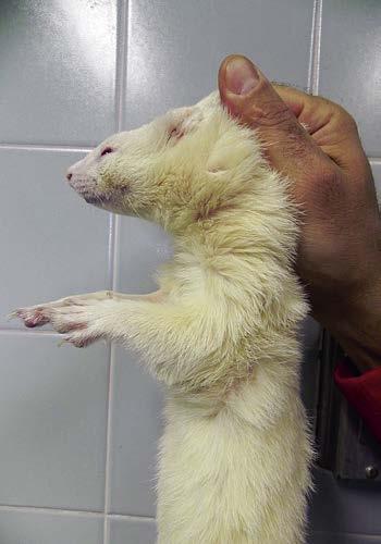
Depending on the ferret’s disposition, several basic manual restraint methods can be used for examination. For tractable animals, lightly restrain the ferret on the examination table. Examine the mucous membranes, oral cavity, head, and skin. Then pick the ferret up and support its body with one hand while using the other hand to auscultate the thorax and palpate the abdomen. For an active animal or one that bites, scruff the ferret and suspend it with all four legs off the table (Fig. 2.1). Most ferrets become relaxed with this hold, and the veterinarian can examine the oral cavity, head, and body; palpate the abdomen; vaccinate; and clean the ears. However, even scruffing may not work for fractious animals. To restrain a ferret for a procedure, hold it firmly by the scruff of its neck and around the hips without pulling the legs back. Most ferrets struggle if their legs are extended by pulling on the feet. Some animals can be distracted during a procedure by feeding a meat-based canned food or a small amount of a supplement such as FerreTone (8-in-1 Pet Products, Islandia, NY) by syringe. For very fractious or anxious animals or for procedures requiring lengthy restraint, light sedation or anesthesia may be indicated (see Chapter 37).
Physical Examination
Most ferrets strenuously object to having their temperature taken with a rectal thermometer, and a temperature taken after a ferret struggles may be artificially high. Therefore measure the rectal temperature early in the physical examination with a
flexible digital thermometer that is well lubricated. The normal rectal temperature of a ferret is 100.5°F to 102.5°F (38.0°C to 39.2°C); however, a mean of 102°F (38.8°C), with a wider range of 100°F to 104°F (37.8°C to 40.0°C), is also reported.22
Physical examination of a ferret can be performed quickly and efficiently if a few simple guidelines are followed. Observe the attitude of the animal. Ferrets may sleep in the carrier in the veterinary office; however, once awakened for the examination, a ferret should be alert and active. Assess hydration by observing the skin turgor of the eyelids, tenting of the skin at the back of the neck, and moistness of the oral mucous membranes. Note that skin turgor can be difficult to evaluate in a cachectic animal. Estimate the capillary refill time by digitally pressing on the gingiva.
Examine the eyes, nose, ears, and facial symmetry. Cataracts can develop in both juvenile and adult animals. Retinal degeneration occurs in ferrets and may be indicated by abnormal pupil dilation. Inspect for nasal discharge, and ask the owner about any history of sneezing or coughing. The ears may have a brown waxy discharge, but excessive brown exudate may indicate infestation with ear mites. Observe the facial symmetry. Although uncommon, salivary mucoceles occur in ferrets and present as a unilateral swelling on the side of the face, usually in the cheek or temporal area (see Chapter 3).
The teeth should be clean and the gingiva pink. Dental tartar is common in pet ferrets, possibly related to feeding kibble instead of natural prey.13 Plaque buildup may be exacerbated by feeding a diet with a high mineral content.26 Remove excess dental tartar by prophylactic techniques used in dogs and cats, and recommend measures to prevent tartar buildup. A pet toothpaste can be used to decrease the rate of calculus formation.26,28,38 Gingivitis is a common sequela of excessive dental tartar. Ferrets often break off the tip of one or both canine teeth, and the broken tooth may appear dark. However, ferrets rarely exhibit sensitivity
associated with a fractured canine. If the ferret exhibits sensitivity when the tip of the canine is probed, recommend a root canal or extraction, depending on the degree of tooth damage (see Chapter 36). Bruxism often indicates gastrointestinal discomfort.
Palpate the submandibular, axillary, popliteal, and inguinal lymph nodes. Nodes should be soft and may sometimes feel enlarged in overweight animals because of surrounding fat. Any firmness or asymmetry warrants fine-needle aspiration or biopsy. If two or more nodes are enlarged and firm, a diagnostic workup is indicated.
Auscultate the heart and lungs in a quiet room. Ferrets have a rapid heart rate (180 to 250 beats/min) and often a pronounced sinus arrhythmia. If a ferret is excited and has a very rapid heart rate, subtle murmurs may be missed. Valvular disease, cardiomyopathy, and congestive heart failure are seen in ferrets, and any murmur or abnormal heart rhythm should be investigated further (see Chapter 5). The ferret’s normal respiratory rate is 33 to 36 breaths/min (see Chapter 6).
Palpate the abdomen while either scruffing the ferret or supporting it around the thorax with one hand. This allows the abdominal organs to displace downward, facilitating palpation. If the history is consistent with an intestinal foreign body or urinary blockage, palpate gently to avoid causing iatrogenic injury, such as a ruptured bladder. Palpate the cranial abdomen, paying attention to the presence of gas or any firm, irregularly shaped material in the stomach area, especially in ferrets with a history of vomiting, melena, or chronic weight loss. The spleen is often enlarged, which may or may not be significant, depending on other clinical findings (see Chapter 5). A very enlarged spleen may indicate systemic disease or, very rarely, idiopathic hypersplenism, and further diagnostic workup is warranted.
Examine the genital area, observing the size of the vulva in females. Vulvar enlargement in a spayed female is consistent with either adrenal disease or an ovarian remnant; the latter is rare. Examine the preputial area and size of the testicles of male ferrets; preputial and testicular tumors are sometimes seen.
Check the fur for evidence of alopecia. Tail tip alopecia is common and may be an early sign of adrenal disease. Symmetric, bilateral alopecia or thinning of the fur that begins at the tail base and progresses cranially is a common finding in ferrets with adrenal disease. Examine the skin on the back and neck for evidence of scratching. Pruritus is common with adrenal disease and also may indicate ectoparasites (e.g., fleas or Sarcoptes scabiei). Palpate and visually examine the skin thoroughly for masses. Mast cell tumors are common and are variable in size. Often, the fur around a mast cell tumor is matted with dried blood from the animal’s scratching. Other types of skin tumors, such as sebaceous adenomas and basal cell tumors, are also common (see Chapter 9). Perform an excisional biopsy of any lump found on the skin.
PREVENTIVE MEDICINE
Young, recently purchased ferrets need serial canine distemper vaccinations until they are 14 weeks of age.3 Rabies vaccines should be given annually beginning at 3 months of age.14 Ferrets should be examined annually until they are 4 to 5 years
Fig. 2.1 Restrain an active ferret by scruffing the loose skin on the back of the neck. The ferret will relax and allow you to palpate the abdomen or administer a vaccine.