Knowledge Capitalism And State Theory: A “Space-Time” Approach Explaining Development Outcomes In The Global Economy 1st Edition Edition Carlos Manuel Sanchez Ramirez
No part of this publication may be reproduced or transmitted in any form or by any means, electronic or mechanical, including photocopying, recording, or any information storage and retrieval system, without permission in writing from the publisher. Details on how to seek permission, further information about the Publisher’s permissions policies and our arrangements with organizations such as the Copyright Clearance Center and the Copyright Licensing Agency, can be found at our website: www.elsevier.com/permissions.
This book and the individual contributions contained in it are protected under copyright by the Publisher (other than as may be noted herein).
Chapter 1.8 Endoscopic Approaches to Frontal and Maxillary Sinus Fractures: Copyright of images is retained by Bradford A. Woodworth, MD.
Notices
Practitioners and researchers must always rely on their own experience and knowledge in evaluating and using any information, methods, compounds or experiments described herein. Because of rapid advances in the medical sciences, in particular, independent verification of diagnoses and drug dosages should be made. To the fullest extent of the law, no responsibility is assumed by Elsevier, authors, editors or contributors for any injury and/or damage to persons or property as a matter of products liability, negligence or otherwise, or from any use or operation of any methods, products, instructions, or ideas contained in the material herein.
Although all advertising material is expected to conform to ethical (medical) standards, inclusion in this publication does not constitute a guarantee or endorsement of the quality or the value of such product or the claims made of it by its manufacturer.
ISBN: 978-0-323-49755-8
E-ISBN: 978-0-323-50960-2
Content Strategist: Belinda Kuhn
Content Development Specialist: Sharon Nash
Project Manager: Anne Collett
Design: Ryan Cook
Illustration Manager: Amy Faith Heyden
Illustrator: MPS North America LLC
Marketing Manager: Claire McKenzie
Printed in China
Video 1.2.1 Systematic method for reading a craniofacial CT scan.
Krystal Archer-Arroyo
Video 1.3.1 Radiological evaluation of a right posttraumatic orbital deformity.
Rüdiger M. Zimmerer, Nils-Claudius Gellrich
Video 1.3.2 Intraoperative real-time navigation with the Brainlab Curve System (Brainlab, Feldkirchen, Germany).
Rüdiger M. Zimmerer, Nils-Claudius Gellrich
Video 1.3.3 Intraoperative 3D imaging with the Ziehm Vision RFD C-Arm (Ziehm Imaging, Nürnberg, Germany). Positioning and laser-guided adjustment of the device in relation to the patient’s anatomy with fluoroscopy. Afterwards, 3D CBCT data sets of the facial skeleton can be generated in a single 165 degree rotation.
Rüdiger M. Zimmerer, Nils-Claudius Gellrich
Video 1.3.4 Intraoperative data fusion. Using the Brainlab Curve system (Brainlab, Feldkirchen, Germany) the intraoperatively generated posttherapeutic 3D CBCT data set can be fused with the preoperative virtual plan.
Rüdiger M. Zimmerer, Nils-Claudius Gellrich
Video 1.5.1 Selection of donor facial nerve branches, cross-facial nerve grafting, and masseter to facial nerve transfer.
Shai M. Rozen
Video 1.5.2 Identification of masseter nerve and gracilis muscle harvest and facial inset.
Shai M. Rozen
Video 1.9.1 Repair of orbital floor fracture with Medpor titanium implant via retroseptal transconjunctival approach.
Amir H. Dorafshar
VIDEO CONTENTS
Video 1.11.1 Medial canthal “tug” indicating a firm stop and intact medial canthal tendon.
Craig Birgfeld
Video 1.11.2 Orbital spanning plate.
Craig Birgfeld
Video 1.12.1 Orbitozygomaticomaxillary complex fractures surgical techniques.
Alan S. Herford, Meagan Miller, Daniel Cantu
Video 1.18.1 Splinting of teeth. Armamentarium: local anesthetic, cheek retractors, dental etch (30%–40% phosphoric acid), dental prime and bonding agents, curing light, 26-gauge wire.
Alan S. Herford, Meagan Miller, Daniel Cantu
Video 1.18.2 Reduction and fixation of dentoalveolar fractures. Armamentarium: local anesthetic, cheek retractors, Erich arch bars, 26-gauge wires.
Matthew E. Lawler, Corbett A. Haas, Zachary S. Peacock
Video 2.3.1 Limited right eye upward gaze in setting of trapdoor fracture sparing entrapment of inferior rectus muscle. Resolution of globe dismotility in 24 hours without surgical intervention.
Jonathan Y. Lee, Jesse A. Goldstein, Joseph E. Losee
Video 3.2.1 Bone splitting technique: autograft split calvarial cranioplasty vs. bone particulate.
Christian J. Vercler
Video 3.12.1 Computer-designed lag screw fixation implant and computer-designed implant with drain port.
Jeffrey Lee, Chad Gordon, Michael J. Yaremchuk
“PRECISION MEDICINE” AND FACIAL INJURIES, 2018
In the past 30 years, much progress has occurred in the treatment of facial injuries with regard to classification, treatment options, timing, and technique of reductions of both the bone and soft tissue. Treatment planning options are numerous, and patient-specific implants and computerized plans are used more frequently. Facial injuries now benefit from refined classifications, which incorporate outcome data, which then cycle refinement of the classification and treatment options, producing a recurring cycle of continuous treatment improvements.
Since all disease occurs in patterns, recognition of the patterns of facial injuries allows the use of specific algorithms for management of the various categories, which include minimal, intermediate, and severe injuries to each anatomical portion of the facial skeleton. The recurring process of diagnosis, treatment, and outcome analysis yields progressive changes and improvements to both the taxonomy and thereby the future treatment algorithms, perpetuating the recurring cycle of improvement.1,2
Practically, division of the face by functional parts creates an anatomical treatment algorithm organized on “CT-based fracture classification”3 determined by (1) anatomical area of the face and (2) the energy or “comminution and displacement” of the particular anatomical part injured.
The papers cited3–8 propose a comprehensive treatment organization for both the bone and soft tissue. Functionally, there are four areas of the facial skeleton:
(I) Frontal bone (supraorbital and frontal sinus areas)
(II) Upper midface (zygomas and nasoethmoid)
(III) Lower midface and occlusion
(IV) Mandible (horizontal and vertical sections)
These papers1–15 cover exposure, reduction, and fixation for each degree of bone injury (low energy – mild; middle energy – moderate; high energy – severe) in each anatomical area (zygoma, horizontal mandible, nasoethmoid, etc.), and then deal with how to improve the quality of the soft tissue in terms of the original injury, timing of fracture repair, and repair and replacement of soft tissue onto the anatomically restored facial skeleton.
Within each of the four areas, each section may be classified as minimally, moderately or severely displaced, and therefore treated with (a) no or (b) minimal open reduction, (c) a standard open reduction, or (d) an extended open reduction. The latter is reserved for the most comminuted and displaced fractures requiring multiple and complete exposures for alignment and fixation at all buttress articulations of a particular anatomical area.
For the zygomatic portion of a panfacial fracture, it could be so minimally displaced that no reduction would be necessary. More frequently, the standard minimally displaced zygoma (incomplete or “greensticked” at the zygomaticofrontal suture) could be approached by inferior alone approaches at the zygomatic buttress and inferior orbital rim. For the standard displacement, a more complete set of anterior incisions, the “anterior alone” approaches of the lower midface gingival buccal sulcus and upper/lower eyelid incisions, allows complete exposure and fixation of all anterior zygoma buttress articulations. The severely comminuted zygoma would require the anterior complete exposure plus the zygomatic arch (anterior and posterior exposures) via a coronal incision for reduction and fixation at each zygomatic buttress and within the orbit laterally and inferiorly.
Similarly, for the nasoethmoid area,5 the simplest fractures would be undisplaced or “greensticked“ at the articulation with the internal angular process of the frontal bone, and therefore would, like the “greensticked” zygoma, be amenable to inferior alone approaches where the infraorbital rim and pyriform aperture fractures are aligned and stabilized through only a gingival buccal sulcus incision, perhaps supplementing it with the eyelid for the next degree of severity. With increasing displacement and comminution of the nasoethmoidal orbital area, the lower eyelid and coronal approaches would have to be added to permit more complete exposure and perhaps also provide for detachment and reattachment of the medial canthal ligament in extreme cases.
The approaches and degree of fracture displacements would be summed over all the anatomical areas of fracture in the patient, permitting a comprehensive treatment plan for all areas to be developed for what is necessary in each area, and then an order of treatment is developed for the entire case, linking and sequencing the anatomical areas by a plan which combines segmental reductions. In this way, the treatment is the least (but still maximally effective) for each anatomical area according to the procedures necessary for each component of the fracture. Such approaches were first described in an article about “CT-based fracture treatment”.3
Generally, my preference, if no neurosurgical urgency is present, is to begin with stabilization of the occlusion by reduction of the split palate and mandible alveolar fractures, and then to address the vertical and horizontal mandible. Or if one needs to begin in the frontal bone because of simultaneous brain injury, the frontal bone is reduced after defunctionalizing the frontal sinus; perhaps first using wires for temporary positioning, and then proceeding to rigid fixation after the multiple areas of reduction required are confirmed as accurate. Then, the upper midface could be reduced and stabilized, then completing the reduction of the lower face, then linking the upper and lower face at the Le Fort I level.
Several comments about the soft tissue bear repeating:
1. Every facial fracture has an injury to the soft tissue, and it may be minimal, moderate or severe. How the soft tissue responds (and therefore the ultimate quality and the position of the soft tissue) depends on when and what you do to the bone, and whether or not the soft tissue can respond by healing and remodeling over a precisely reconstructed bony facial skeleton. The early bone reduction and the repair of the incisions and refixation of the soft tissue to a reduced skeleton become the method of “treatment” of the soft tissue, the “soft tissue reduction”.
2. Timing and facial fracture management: Since the soft tissue has an initial injury, it makes good sense to confine the incisions and dissection and repair to this initial injury period, rather than to create a second soft tissue injury with the definitive reduction in the vulnerable period of soft tissue healing 1–2 weeks after the initial soft tissue trauma.
While many isolated, simple fractures may be operated on at any time after the injury with little compromise in the ultimate soft tissue quality, true high-energy facial injuries begin to develop soft tissue scar and contracture immediately from the time of the initial injury, and the soft tissue becomes stiffened, thicker, discolored, and less pliable with each day of initial healing.
Soft tissue contracture and stiffness develop in the shape of the underlying displaced bone fracture. Making reduction incisions and dissecting in the “vulnerable period” 1–3 weeks after the initial injury creates a second set of soft tissue injuries from the surgery, further
damaging the soft tissue, whereas confining the incisions to the initial injury period isolates the two soft tissue injuries to a single reaction where the soft tissue reacts to a single insult, yielding the best result one could achieve in terms of soft tissue quality and position.
3. Dehiscence and displacement of soft tissue: The layered closure of incisions prevents soft tissue dehiscence. Reattaching the repaired soft tissue to the facial skeleton places the soft tissue correctly over the reconstructed and realigned facial skeleton, allowing the soft tissue to heal in the correct position and with the correct contour. The shape and position of the bone are the pattern for the remodeling and repair by internal scar of the soft tissue injury. In some cases, such as the nasoethmoidal orbital area, placing soft tissue bolsters over the soft tissue pressing it against the maxilla and the lateral nose keeps the tissue aligned to the repaired facial skeleton over the complex contours and angles of the central upper midface area, prevents hematoma (minimizing excess fibrosis and thickness of soft tissue), and keeps the soft tissue stretched to length over the entire curving surface of the anatomically reduced bone. These several considerations create a comprehensive plan for treatment of any facial injury, both for the bone and for the soft tissue. Of course, immediate bone grafting and microvascular flap transfer may be added where necessary to replace critical missing areas of the facial skeleton and soft tissue. Mismatched cutaneous soft tissue islands from microvascular transfer may be removed secondarily by serial excision or standard facial cutaneous flap reconstruction, covering the mismatched soft tissue added with a contoured area of soft tissue with matching cutaneous color.
So read on, enjoy, and benefit from the mastery of experienced practitioners who are ready to assist you with these difficult problems.
Paul N. Manson MD
Distinguished Service Professor of Plastic Surgery Johns Hopkins School of Medicine Professor of Surgery, The University of Maryland R Adams Cowley Shock Trauma Unit
REFERENCES
1. Manson P, Clark N, Robertson B, et al. Subunit principles in midface fractures: the importance of sagittal buttresses, soft tissue reductions and
sequencing treatment of segmental fractures. Plast Reconstr Surg 1999;103:1287–1306.
2. Manson P, Markowitz B, Mirvis S, et al. Toward CT-based facial fracture treatment. Plast Reconstr Surg. 1990;85:202–212.
3. Markowitz B, Manson P, Sargent L, et al. Management of the medial canthal tendon in nasoethmoid orbital fractures: The importance of the central fragment in treatment and classification. Plast Reconstr Surg 1991;87:843–853.
4. Crawley W, Clark N, Azman P, Manson P. The edentulous Le Fort fracture. J Craniofacial Surg. 1997;8:298–308.
5. Hendrickson M, Clark N, Manson P. Sagittal fractures of the maxilla: classification and treatment. Plast Reconstr Surg. 1998;101:319–332.
6. Rodriguez ED, Stanwix MG, Nam AJ, et al. Twenty-six-year experience treating frontal sinus fractures: a novel algorithm based on anatomical fracture pattern and failure of conventional techniques. Plast Reconstr Surg. 2008;122(6):1850–1866.
7. Manson PN, Stanwix M, Yaremchuk M, et al. Frontobasilar fractures: anatomy, classification and clinical significance. Plast Reconstr Surg 2009;124:2096–2106.
8. Manson P, Iliff N. Management of blowout fractures of the orbital floor: early repair of selected injuries. Surv Ophthalmol. 1991;35: 280–291.
9. Lee R, Gamble B, Manson P. Facial lacerations: patterns, associated injuries and the McFontz classification system. Plast Reconstr Surg 1995;99:1544–1554.
10. Lee RH, Gamble B, Manson P, et al. Patterns of facial lacerations from blunt trauma. Plast Reconstr Surg. 1997;99:1544–1554.
11. Clark N, Birely B, Manson PN, et al. High-energy ballistic and avulsive facial injuries: classification, patterns, and an algorithm for primary reconstruction. Plast Reconstr Surg. 1996;98:583–601.
12. Robertson B, Manson P. The importance of serial debridement and “a second look” procedures in high-energy ballistic and avulsive facial injuries. Oper Tech Plast Reconstr Surg. 1998;5(3):236–246.
13. Rodriguez E, Martin M, Bluebond-Langner R, et al. Microsurgical reconstruction of post-traumatic high-energy maxillary defects: establishing the effectiveness of early reconstruction. Plast Reconstr Surg 2007;120(7):103S–117S.
14. Rodriguez E, Bluebond-Langner R, Park J, et al. Preservation of contour in periorbital & midfacial craniofacial microsurgery: reconstruction of the soft tissue elements and skeletal buttresses. Plast Reconstr Surg 2008;121(5):1738–1747.
15. Fisher M, Dorafshar A, Bojovic B, et al. The evolution of critical concepts in aesthetic craniofacial microsurgical reconstruction. Plast Reconstr Surg 2012;130(2):389–398.
PREFACE
“If I have seen further it is by standing on the shoulders of Giants.”
Isaac Newton, 1675
The idea for the genesis of this textbook originated from being invited to co-author a book chapter on facial trauma with Drs. Eduardo Rodriguez and Paul Manson in the fourth edition of Neligan’s Plastic Surgery textbook. In helping to co-author this book chapter I realized that to provide a comprehensive text on the subject area in the limited number of words was impossible; we therefore decided to expand the book chapter into a textbook. The textbook initially set out to cover primary traumatic repair, but as we conceived the book chapters, we thought that no textbook on facial trauma surgery would be complete without describing delayed posttraumatic reconstruction.
This textbook has been based on the principles and concepts of craniofacial surgery for the care of patients with facial traumatic injuries that were originally described and taught by Dr. Paul Manson, and later expanded upon by Dr. Eduardo Rodriguez to include microsurgical applications to craniofacial reconstruction over the last 40 years at the R Adams Cowley Shock Trauma Center at the University of Maryland Medical Center. I have also tried to add my own perspective and insights, which I have gained over the last decade treating patients with traumatic facial injuries at the R Adams Cowley Shock Trauma Center and at the Johns Hopkins Hospital.
We decided to ask internationally recognized authors across the disciplines of Plastic and Reconstructive Surgery, Oral & Maxillofacial Surgery, Otolaryngology and Facial Plastic Surgery, Oculoplastic Surgery and Neurological Surgery to contribute their perspectives in
their respective expert areas in the treatment of patients with craniofacial traumatic injuries. Each book chapter has a concluding “Expert Commentary” by Dr. Paul Manson offering his viewpoint on the subject. Several book chapters also include evidence-based summaries in areas of controversy and video attachments to supplement and clarify surgical technique. We have also included chapters on virtual surgical planning, 3D printing, intraoperative surgical navigation, and the roles of microsurgery and facial transplantation in the treatment of facial traumatic injuries to provide an advanced and modern approach.
This book is focused towards the young reconstructive surgeon or surgical trainee wishing to understand basic principles and concepts of primary traumatic facial injury repair and secondary facial reconstruction. It is designed to be of value to all the subspecialties of reconstructive surgery and we hope to improve the delivery of traumatic facial injury care to patient populations not only in the United States but around the world.
Amir H. Dorafshar MD, FACS, FAAP
John W. Curtin, MD Chair of Plastic and Reconstructive Surgery Professor of Surgery and Neurological Surgery Division of Plastic and Reconstructive Surgery Rush University Medical Center, Chicago, IL, USA
Adjunct Professor Department of Plastic and Reconstructive Surgery Johns Hopkins University School of Medicine Baltimore, MD, USA
Bizhan Aarabi MD, FRCSC
Professor
Department of Neurosurgery
University of Maryland Medical Center and R Adams Cowley Shock Trauma Center Baltimore, MD, USA
Resident Department of Oral and Maxillofacial Surgery
University of Maryland Baltimore, MD, USA
Majd Al Mardini DDS, MBA, FRCD(C)
Director of Ocular and Maxillofacial Prosthetics Unit
Department of Dentistry and Maxillofacial Prosthetics
University Health Network, Princess Margaret Cancer Center Toronto, ON, Canada
Brian Alpert DDS, FACD, FICD, FACS
Professor of Oral and Maxillofacial Surgery
University of Louisville School of Dentistry; Chief, Oral and Maxillofacial Surgery and Dentistry
University of Louisville Hospital Louisville, KY, USA
Oleh Antonyshyn MD, FRCSC
Associate Professor
Department of Plastic and Reconstructive Surgery
University of Toronto Toronto, ON, Canada
Krystal Archer-Arroyo MD
Assistant Professor
Department of Radiology and Nuclear Medicine
University of Maryland School of Medicine Baltimore, MD, USA
Said C Azoury MD
Resident Physician
Division of Plastic Surgery
Department of Surgery University of Pennsylvania Health System Philadelphia, PA, USA
LIST OF CONTRIBUTORS
Craig Birgfeld MD, FACS
Associate Professor
Department of Surgery, Division of Plastic Surgery
University of Washington Seattle, WA USA
Kofi D.O. Boahene MD
Associate Professor
Department of Otolaryngology – Head and Neck Surgery
Johns Hopkins University School of Medicine Baltimore, MD, USA
Colin M. Brady MD
Craniofacial Fellow
Division of Plastic and Maxillofacial Surgery
Department of Surgery
University of Southern California
Los Angeles, CA, USA
Steven R. Buchman MD
M. Haskell Newman Professor in Plastic Surgery
Professor of Neurosurgery, University of Michigan Medical School; Chief, Pediatric Plastic Surgery, CS Mott Children’s Hospital; Director, Craniofacial Anomalies Program, University of Michigan Medical Center
University of Michigan Medical Center
Ann Arbor, MI, USA
Patrick Byrne MD, MBA
Professor and Director
Division of Facial Plastic and Reconstructive Surgery
Department of Otolaryngology – Head and Neck Surgery
Johns Hopkins School of Medicine Baltimore, MD, USA
Daniel Cantu DDS
Philip J. Boyne and Peter Geistlich Research Fellow
Department of Oral and Maxillofacial Surgery
Loma Linda University Loma Linda, CA, USA
John P. Carey MD
Professor and Division Chief for Otology, Neurotology and Skull Base Surgery
Department of Otolaryngology – Head and Neck Surgery
Johns Hopkins University School of Medicine Baltimore, MD, USA
Edward H. Davidson MD, MA (Cantab), MBBS
Assistant Professor
Department of Plastic Surgery and Reconstructive Surgery
Montefiore Medical Center
Albert Einstein College of Medicine
Bronx, NY, USA
Kristopher M. Day MD
Plastic Surgery Resident
Department of Plastic Surgery
University of Tennessee Chattanooga Chattanooga, TN, USA
A. Lee Dellon MD, PhD
Professor of Plastic Surgery and Professor of Neurosurgery
Johns Hopkins University Baltimore, MD, USA
Sarah W. DeParis MD
Oculoplastic Surgeon
Department of Ophthalmology
The Permanente Medical Group San Rafael, CA, USA
J. Rodrigo Diaz-Siso MD
Postdoctoral Research Fellow
Hansjörg Wyss Department of Plastic Surgery
New York University Langone Medical Center
New York, NY, USA
Amir H. Dorafshar, MD, FACS, FAAP
John W. Curtin, MD Chair of Plastic and Reconstructive Surgery
Professor of Surgery and Neurological Surgery
Division of Plastic and Reconstructive Surgery
Rush University Medical Center
Chicago, IL, USA
Adjunct Professor
Department of Plastic and Reconstructive Surgery
Johns Hopkins University School of Medicine
Baltimore, MD, USA
Edward Ellis III, DDS, MS
Professor and Chair
Department of Oral and Maxillofacial Surgery
University of Texas Health Science Center at San Antonio San Antonio, TX, USA
Jeffrey Fialkov MD, MSc FRCSC
Associate Professor
Department of Plastic and Reconstructive Surgery
University of Toronto Toronto, ON, Canada
Robert L. Flint DMD, MD
Assistant Professor of Oral and Maxillofacial Surgery
University of Louisville School of Dentistry Louisville, KY, USA
Christopher R. Forrest MD, MSc, FRCSC, FACS
Chief, Plastic and Reconstructive Surgery, Medical Director
The Centre for Craniofacial Care and Research
The Hospital for Sick Children Toronto, ON, Canada; Chair/Professor
Division of Plastic and Reconstructive Surgery
University of Toronto Department of Surgery Toronto, ON, Canada
Nils-Claudius Gellrich MD, DDS
Professor and Chair
Department of Oral and Maxillofacial Surgery
Hannover Medical School Hannover, Germany
Dane J. Genther MD
Professional Staff
Division of Facial Plastic and Reconstructive Surgery
Department of Otolaryngology – Head and Neck Surgery
Head and Neck Institute
Cleveland Clinic Cleveland, OH, USA
Jesse A. Goldstein MD
Assistant Professor of Pediatric Plastic Surgery
Department of Plastic Surgery
Children’s Hospital of Pittsburgh
University of Pittsburgh Medical Center Pittsburgh, PA, USA
Chad R. Gordon DO, FACS
Director, Neuroplastic and Reconstructive Surgery
Fellowship Director, Neuroplastic & Reconstructive Surgery (Plastic Surgery)
Co-Director, Multidisciplinary Adult Cranioplasty Center
Associate Professor of Plastic Surgery and Neurosurgery
Department of Plastic and Reconstructive Surgery
Johns Hopkins University School of Medicine
Michael Grant MD, PhD, FACS
Professor and Chief
Trauma Plastics
R Adams Cowley Shock Trauma Center
University of Maryland School of Medicine Baltimore, MD, USA
Joseph S. Gruss MD
Professor
Division of Plastic Surgery
Seattle Children’s Hospital
University of Washington Seattle, WA, USA
Corbett A. Haas DDS MD
Resident in Oral and Maxillofacial Surgery
Department of Oral and Maxillofacial Surgery
Harvard School of Dental Medicine/ Harvard Medical School
Massachusetts General Hospital Boston, MA, USA
Alan S. Herford DDS, MD, FACS
Professor and Chair
Department of Oral and Maxillofacial Surgery
Loma Linda University
Loma Linda, CA, USA
Larry H. Hollier MD, FACS, FAAP Surgeon in Chief
Texas Children’s Hospital
S. Baron Hardy Endowed Chair
Professor of Plastic Surgery, Orthopedic Surgery, and Pediatrics
Chief of Plastic Surgery
Baylor College of Medicine Houston, TX, USA
Richard A. Hopper MD
Professor
Division of Plastic Surgery
Seattle Children’s Hospital
University of Washington Seattle, WA, USA
Matthew G. Huddle MD
Resident Physician
Department of Otolaryngology – Head and Neck Surgery
Johns Hopkins School of Medicine
Baltimore, MD, USA
Lewis C. Jones DMD, MD
Adjunct
Assistant Professor of Oral and Maxillofacial Surgery
University of Louisville School of Dentistry Louisville, KY, USA
Bartlomiej Kachniarz MD, MBA
Resident Physician
Plastic and Reconstructive Surgery
Johns Hopkins University School of Medicine
Baltimore, MD, USA
Leslie Kim MD, MPH
Assistant Professor and Director
Division of Facial Plastic and Reconstructive Surgery
Department of Otolaryngology – Head and Neck Surgery
The Ohio State University School of Medicine Columbus, OH, USA
George M. Kushner DMD, MD, FACS
Professor of Oral and Maxillofacial Surgery and Residency Program Director
University of Louisville School of Dentistry Louisville, KY, USA
Matthew E. Lawler DMD MD
Resident in Oral and Maxillofacial Surgery
Department of Oral and Maxillofacial Surgery
Harvard School of Dental Medicine/ Harvard Medical School
Massachusetts General Hospital Boston, MA, USA
Andrew Lee MD
Resident Physician
Division of Otology, Neurotology and Skull
Base Surgery
Department of Otolaryngology – Head and Neck Surgery
Johns Hopkins University School of Medicine
Baltimore, MD, USA
Jeffrey Lee MD
Craniofacial Reconstructive and Aesthetic Surgery Fellow
Division of Plastic and Reconstructive Surgery
Massachusetts General Hospital Boston, MA, USA
Jonathan Y. Lee MD, MPH
Craniofacial and Pediatric Plastic Surgery Fellow
Department of Plastic Surgery
Children’s Hospital of Pittsburgh University of Pittsburgh Medical Center Pittsburgh, PA, USA
Fan Liang MD
Assistant Professor
Trauma Plastics
R Adams Cowley Shock Trauma Center
University of Maryland School of Medicine Baltimore, MD, USA
Joseph Lopez MD, MBA
Resident
Department of Plastic and Reconstructive Surgery
Johns Hopkins Medical Institution Baltimore, MD, USA
Joseph E. Losee MD
Ross H. Musgrave Professor of Pediatric Plastic Surgery
Department of Plastic Surgery
Children’s Hospital of Pittsburgh University of Pittsburgh Medical Center Pittsburgh, PA, USA
Matthew R. Louis MD Resident
Department of Plastic and Reconstructive Surgery
Johns Hopkins Hospital Baltimore, MD
Alexandra Macmillan MA (Cantab.), MBBS
Research Fellow
Department of Plastic and Reconstructive Surgery
Johns Hopkins Medical Institution Baltimore, MD, USA
Paul N. Manson MD
Distinguished Service Professor
Department of Plastic, Reconstructive and Maxillofacial Surgery
Johns Hopkins University School of Medicine; Professor of Surgery
R Adams Cowley Shock Trauma Center
University of Maryland School of Medicine
Baltimore, MD, USA
Meagan Miller DDS Resident
Department of Oral and Maxillofacial Surgery
Loma Linda University Loma Linda, CA, USA
Shannath L. Merbs MD, PhD, FACS Professor
Departments of Ophthalmology and Oncology
Johns Hopkins University School of Medicine
Baltimore, MD, USA
Stuart E. Mirvis MD, FACR
Retired Professor, Department of Radiology and Nuclear Medicine
University of Maryland School of Medicine
Baltimore, MD, USA
Corey M. Mossop MD
Interim Chief – Neurosurgery Service and Instructor
Department of Surgery
Tripler Army Medical Center and the Uniformed Services University of the Health Sciences
Honolulu, HI and Bethesda, MD, USA
Gerhard S. Mundinger MD
Assistant Professor, Plastic and Reconstructive Surgery
Assistant Professor, Department of Cell Biology and Anatomy
Craniofacial Center Director
Director of Pediatric Plastic Surgery
Children’s Hospital of New Orleans
Louisiana State University Health Sciences Center
New Orleans, LA, USA
Arthur J. Nam MD, MS
Assistant Professor
Division of Plastic, Reconstructive and Maxillofacial Surgery
R Adams Cowley Shock Trauma Center
University of Maryland School of Medicine
Baltimore, MD, USA
Lauren T. Odono DDS
Oral Maxillofacial Surgery Resident
Division of Plastic and Maxillofacial Surgery
Department of Surgery
University of Southern California
Los Angeles, CA, USA
Devin O’Brien-Coon MD, MSE
Assistant Professor
Department of Plastic and Reconstructive Surgery
Johns Hopkins University School of Medicine Baltimore, MD, USA
Ira D. Papel MD
Professor
Division of Facial Plastic and Reconstructive Surgery
Department of Otolaryngology – Head and Neck Surgery
Johns Hopkins University School of Medicine
Facial Plastic Surgicenter Baltimore, MD, USA
Zachary S. Peacock DMD, MD, FACS
Assistant Professor
Department of Oral and Maxillofacial Surgery
Harvard School of Dental Medicine/ Harvard Medical School
Massachusetts General Hospital Boston, MA, USA
Daniel Perez DDS
Associate Professor
Department of Oral and Maxillofacial Surgery
University of Texas Health Science Center at San Antonio San Antonio, TX, USA
Christian Petropolis MD, FRCSC
Assistant Professor
Department of Plastic and Reconstructive Surgery
University of Manitoba Winnipeg, MB, Canada
David B. Powers MD, DMD, FACS, FRCS (Ed)
Associate Professor of Surgery
Director, Duke Craniomaxillofacial Trauma Program
Duke University Medical Center Durham, NC, USA
Andrew M. Read-Fuller MD, DDS
Clinical Assistant Professor, Oral and Maxillofacial Surgery
Texas A&M University College of Dentistry and Baylor University Medical Center Dallas, TX, USA
Richard J. Redett MD
Professor
Department of Plastic and Reconstructive Surgery
Johns Hopkins University School of Medicine Baltimore, MD, USA
Likith V. Reddy MD, DDS, FACS
Clinical Professor and Program Director
Department of Oral and Maxillofacial Surgery
Texas A&M University College of Dentistry and Baylor University Medical Center; Clinical Professor, Surgery
Texas A&M University College of Medicine Dallas, TX, USA
Sashank Reddy MD, PhD
Resident
Department of Plastic and Reconstructive Surgery
Johns Hopkins Medical Institution Baltimore, MD, USA
Douglas D. Reh MD
Associate Professor, Otolaryngology – Head and Neck Surgery
Department of Otolaryngology – Head and Neck Surgery
Johns Hopkins University School of Medicine Baltimore, MD, USA
Isabel Robinson BA
Medical Student
Hansjörg Wyss Department of Plastic Surgery
New York University Langone Medical Center
New York, NY, USA
Eduardo D. Rodriguez MD, DDS
Helen L. Kimmel Professor of Reconstructive Plastic Surgery
Hansjörg Wyss Department of Plastic Surgery
New York University Langone Medical Center
New York, NY, USA
Christopher R. Roxbury MD
Resident
Department of Otolaryngology – Head and Neck Surgery
Johns Hopkins University School of Medicine
Baltimore, MD, USA
Shai M. Rozen MD, FACS
Professor of Plastic and Reconstructive Surgery
Director of Microsurgery
Director of The Facial Palsy Program
Director of Clinical Research
Department of Plastic and Reconstructive Surgery
UT Southwestern Medical Center Dallas, TX, USA
Larry Sargent MD, FACS, FAAP
Board-Certified Plastic and Craniofacial Surgeon
Sargent Plastic Surgery
Salt Lake City, UT, USA
Tatyana A. Shamliyan MD, MS
Senior Director
Evidence-Based Medicine Quality Assurance
Elsevier Inc. Philadelphia, PA, USA
David A. Shaye MD Instructor
Department of Otolaryngology
Harvard Medical School
Massachusetts Eye & Ear Boston, MA, USA
Ghassan G. Sinada DDS, MBA
Director, Oral Oncology and Maxillofacial Prosthodontics
Milton J. Dance Jr. Head and Neck Cancer Center
Greater Baltimore Medical Center Baltimore, MD, USA
Ryan M. Smith MD
Assistant Professor, Facial Plastic Surgery and Reconstruction
Department of Otorhinolaryngology – Head and Neck Surgery
Rush University Medical Center Chicago, IL, USA
Mark W. Stalder MD
Assistant Professor of Clinical Surgery
Division of Plastic and Reconstructive Surgery
Louisiana State University Health Sciences Center
New Orleans, LA, USA
E. Bradley Strong MD Professor
Department of Otolaryngology
University of California at Davis Sacramento, CA, USA
Tufts University School of Dental Medicine Boston, MA, USA
Jeffrey G. Trost Jr. MD
Resident
Division of Plastic and Reconstructive Surgery
Baylor College of Medicine Houston, TX, USA
Anthony P. Tufaro DDS, MD, FACS Professor of Surgery, Plastic Surgery & Surgical Oncology
Department of Surgery
University of Oklahoma Health Science Center
Oklahoma City, OK, USA
Mark Urata, MD, DDS, FACS, FAAP
Audrey Skirball Kenis Endowed Chair and Chief
Division of Plastic and Reconstructive Surgery
Chair, Keck School of Medicine of USC
Division of Oral and Maxillofacial Surgery
Ostrow School of Dentistry of USC
Los Angeles, CA, USA
Christian J. Vercler MD, MA, FACS, FAAP
Associate Professor
Section of Plastic Surgery
Department of Surgery
University of Michigan Medical Center Ann Arbor, MI, USA
Gary Warburton DDS, MD, FDSRCS, FACS
Associate Professor
Program Director & Division Chief Oral & Maxillofacial Surgery
University of Maryland Baltimore, MD, USA
Heather M. Weinreich, MD MPH
Assistant Professor
Department of Otolaryngology – Head and Neck Surgery
University of Illinois Chicago, IL, USA
Tyler Wildey MD, DDS
Past Resident, Oral and Maxillofacial Surgery
Texas A&M University College of Dentistry and Baylor University Medical Center Dallas, TX, USA
S. Anthony Wolfe MD
Chief, Division of Plastic Surgery
Miami Children’s Health System Miami, FL, USA
Bradford A. Woodworth MD
James J. Hicks Professor of Otolaryngology
Department of Otolaryngology – Head and Neck Surgery
University of Alabama at Birmingham Birmingham, AL, USA
Robin Yang DDS, MD
Chief Resident
Department of Plastic and Reconstructive Surgery
Johns Hopkins University Baltimore, MD, USA
Michael J. Yaremchuk MD
Clinical Professor of Surgery, Harvard Medical School
Chief of Craniofacial Surgery, Massachusetts General Hospital Boston, MA, USA
Elizabeth Zellner MD
Craniofacial Fellow
The Centre for Craniofacial Care and Research
Division of Plastic and Reconstructive Surgery
The Hospital for Sick Children Toronto, ON, Canada
Rüdiger M. Zimmerer MD, DDS
Assistant Professor and Consultant
Department of Oral and Maxillofacial Surgery
Hannover Medical School
Hannover, Germany
ACKNOWLEDGMENTS
While there are too many people to name individually in this acknowledgment section, I would like to mention the enormous gratitude I have for the author contributors to this book who have come together from various subspecialties to give their unique perspectives on facial trauma surgery for this multidisciplinary comprehensive textbook. I greatly appreciate the sacrifice of their time taken away from their families, friends and practices to make this book a reality. Shianne Pietrowksi, my administrative assistant, who helped with the formatting and re-typing of several book chapters, deserves special recognition for her humor and determination to help me get through the multiple revisions of each chapter. The team at Elsevier, particularly Sharon, Belinda, Elaine and Tatyana, whom I have worked with individually and collectively through various phases of the book, have been excellent throughout. Lastly, I would like to thank my close family and friends for your patience with me for the many nights and weekends that I have not been present or with you. Your encouragement and support have been instrumental in my determination to complete this book.
Amir H. Dorafshar January 2019
First and foremost, I would like to thank God for providing me the opportunity to share the knowledge of my fellow authors with you. Next, to my parents who sacrificed their lives for me to succeed; to my mentors who helped inspire me; to my trainees who have made me a better teacher; and to my patients who have taught me humility.
Assessment of the Patient With Traumatic Facial Injury
Arthur J. Nam, Edward H. Davidson, Paul N. Manson
BACKGROUND
Incidence
of Facial Trauma in the United States and Worldwide
The spectrum of facial trauma includes soft tissue and bone, and ranges from the simple to the complex. Epidemiology varies with local and global demographic factors and reflects a complex interplay of influences, including those related to the environment, economics, age, gender, and mechanism of injury. Any understanding of the incidence of facial trauma is further confounded by the presumptive underreporting and treatment of minor injuries. As a result, the plethora of data is often conflicting. Nonetheless, the incidence of facial fractures presenting to the emergency room is approximately 500,000 per year in the United States, with nasal fractures likely the most common, followed by mandible fractures.1 These commonly occur in males more than females, are most frequent in the second and third decades of life, and are most frequently the result of altercations, assaults, falls, work or home accidents, and motor vehicle or motorcycle collisions (MVCs). While many studies cite mandible fractures as being more common than nasal fractures, this has been attributed to a sampling bias favoring inpatient admissions or requirement for in-hospital treatment rather than capturing all emergency room presentations. Several older studies show higher rates of injury from MVCs, prior to the mandatory implementation of airbags and restraining devices.2 Despite this, MVCs remain the most important cause of facial trauma all over the world. Global trends also reflect an increase in the male/female injury ratio in countries where the social custom is for women to be more confined to the home.3 Associated soft tissue trauma is the most common concomitant injury, occurring in approximately 30% of facial fractures. Concomitant fractures of the skull, upper limbs, and associated areas are estimated to occur in around 25% of facial fractures; these include intracranial injuries in 12%–45.5%, and associated cervical spine injury with facial injury in up to 9.7%.1,4 One must therefore always exclude brain and cervical spine injuries in the patient with facial injuries as trauma is often a geographic injury to the head and neck. Missed injuries of the spine, extremities, and pelvis are also frequent (10%) and are easily missed.
Patterns of Facial Trauma and Causes
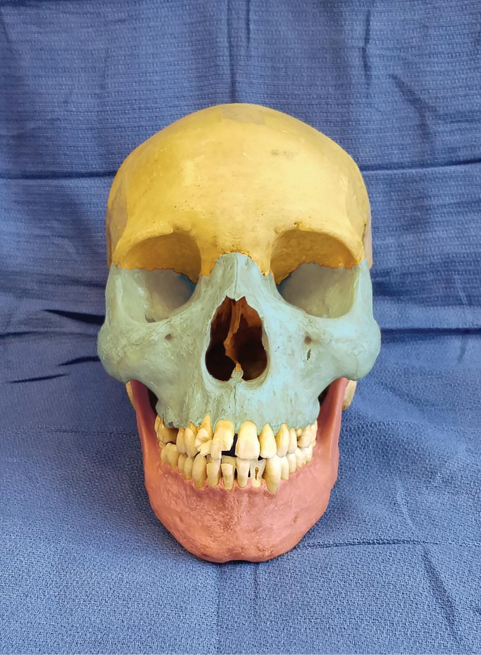
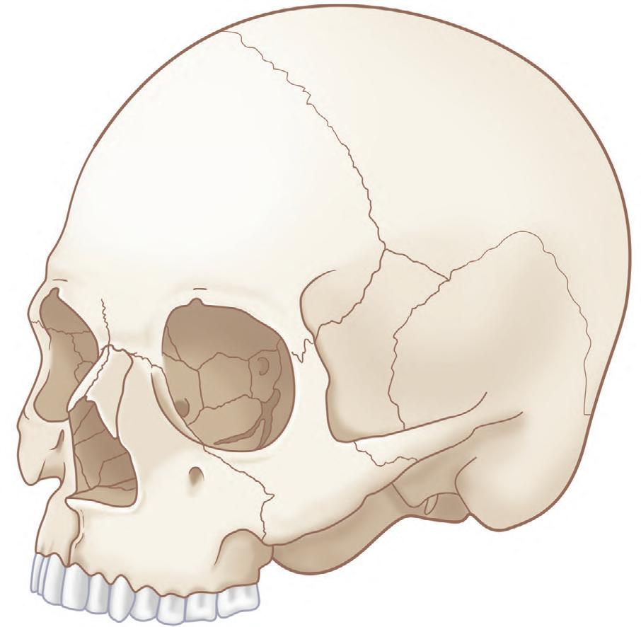
Patterns of facial injuries may be subdivided into soft tissue, bony skeleton, and/or dentoalveolar trauma. Descriptions can also be made based on location in facial thirds: the upper third (including the frontal bone, frontal sinuses, and orbital roofs), middle third (including the orbit, nose, malar region, and maxilla), and lower third (including the
mandible and its dentition) (Fig. 1.1.1). Blunt trauma can result in relatively predictable fracture patterns due to the presence of facial buttresses and resultant functional skeletal units (Fig. 1.1.2).5 Injury to the upper third may reveal frontal sinus fractures, which require the determination of whether injury affects the inner table, outer table, or both, degree of displacement, and the presence of nasofrontal outflow tract obstruction.6 Midface fracture patterns may include the characteristic Le Fort fracture patterns, but are more frequently asymmetric and more extensive on the side of the force application, or orbitozygomaticomaxillary complex (OZMC), orbital, nasal and naso-orbitoethmoid (NOE) fractures in isolation or in combination. Lower third facial fractures, i.e., those of the mandible, also demonstrate reproducible patterns. The most prevalent site of mandibular fracture reported in the literature is variable, though mandibular angle and condyle are the most frequently cited.7 Based on the mandibular “ring” concept, mandibular fractures have conventionally been thought to involve at least two sites, however, unifocal mandibular fractures commonly occur. Common multifocal patterns include mandibular body and contralateral angle/ramus/condyle, angle and contralateral parasymphysis, symphysis/ parasymphysis, and bilateral condyle. True panfacial fractures involve all thirds of the face simultaneously and are less involved on the contralateral side, both in terms of fracture extent and comminution.8 These fracture patterns differ depending on mechanism, with most panfacial fractures usually resulting from MVCs and, less commonly, from gunshot wounds (GSWs). Sports injuries typically are isolated to the mandible or upper midface, and assaults are predictive of isolated mandible, midface or zygoma fractures. MVCs and GSWs each predict a higher severity of injury than assaults, falls, or sports injuries.9,10
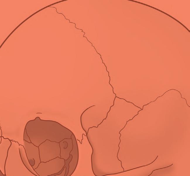
Patterns of facial trauma in the pediatric population differ from those in adults. Facial fractures are relatively less common in children due to parental supervision as well as intrinsic anatomical factors such as larger fat pads, decreased pneumatization of sinuses, increased skeletal flexibility secondary to more malleable bone stock, and compliant sutures. The large cranium partially shields the rest of the face from injury. Atypical craniofacial fracture patterns precede the Le Fort patterns seen in adulthood and as an incompletely pneumatized frontal sinus transmits energy directly from the site of impact to the supraorbital foramen and then to the orbit, superior NOE and anterior maxillary wall or zygoma.11,12 Younger patients are at higher risk of dentoalveolar trauma, including crown fractures (the most common injury), luxations, avulsions, subluxations, root fractures, and intrusions, with approximately one-third occurring in those younger than 10 years of age. These most often result from activities of daily living, play, MVCs, and sports.13
The relative incidence of fracture patterns is debated; mandible fractures are often cited as the most common pediatric facial fracture, accounting for 20%–50% of all pediatric facial fractures.14–17 Anatomical distribution varies with age; isolated condylar fracture incidence decreases, while body and angle fractures increase.18 Others have reported that nasal fractures comprise up to 50% of pediatric facial fractures, but usually escape hospital registries.14,19 Nasal and maxillary fractures were the most common osseous injuries among infants in the US National Trauma Databank, while mandible fractures were more common in older teenagers, with mandible fractures being the overall most common facial fracture.20 Nasal fractures are likely often underreported, with many treated as an outpatient or not treated and thus not reported.21 One review found 54% of fractures to occur in the skull, one-third in the upper and middle thirds of the face, and the remainder in the lower third.22 Another group stratified patients by dental maturity – primary, mixed, and permanent dentition – and concluded orbital fracture was the most common fracture type for all age groups combined. This group showed activities of daily living as the most common cause of injury in 0- to 5-year-olds, MVCs, sports, and play in ages 6–11, and violence, sports, and MVCs as the most common causes of injury in 12- to 18-year-olds.23
Similarly, fracture patterns, demographics, and mechanisms of injury differ significantly between geriatric and nongeriatric adult craniofacial trauma patients. Falls are more frequent causes of fractures in geriatric patients whereas assaults, MVCs, and pedestrians struck were significantly more frequent causes in the nongeriatric adult population. Mandible fractures and panfacial fractures are more common in the nongeriatric population while higher incidences of orbital floor, maxillary, and condylar fractures are more common in geriatric patients and are dependent on geriatric age status, rather than mechanism of injury alone.24,25 The most common cause of soft tissue injury has been shown to be falls and therefore older people seem excessively prone to this injury.
Penetrating soft tissue trauma, including that resulting from bites, stab wounds, gunshot, and other ballistic injuries, can result in injury configurations and fracture patterns that fall outside the usual predictable blunt trauma patterns. Distribution of soft tissue injury is frequently concentrated in a “T-shaped” area that includes the forehead, nose, lips, and chin as well as commonly affecting the lateral brows, malar eminences, forehead, and occiput.26 Patterns may be described by facial
Fig. 1.1.2 Transverse (blue), vertical (red), and sagittal (green) buttresses of the facial skeleton. (From Prein J, Ehrenfeld M, Manson PN, editors. Principles of internal fixation of the craniomaxillofacial skeleton: trauma and orthognathic surgery. AO Foundation, Thieme; 2012, Fig. 1.3.1-5, p.24.)
aesthetic units27 or in reference to bony landmarks. Depth and structures suspected to be injured are also important considerations, especially the globe, eyelids, canthi, lacrimal system, lips, nose, facial nerve, and parotid duct. Ballistic injuries including GSWs are further uniquely characterized by blast vasospasm.28 These may be further complicated by a combination of blunt, penetrating, and burn injuries.29
These patterns of injury and interplay with epidemiology, mechanism, and patient demographics emphasize the importance of a meticulous history and a sequential and directed physical examination as central to the evaluation of a patient with facial trauma. This process collects information that predicts the injury pattern, which is further documented by examination and radiographs to serve as a basis for diagnosis and management.
SURGICAL ANATOMY
Facial Soft Tissue Anatomy
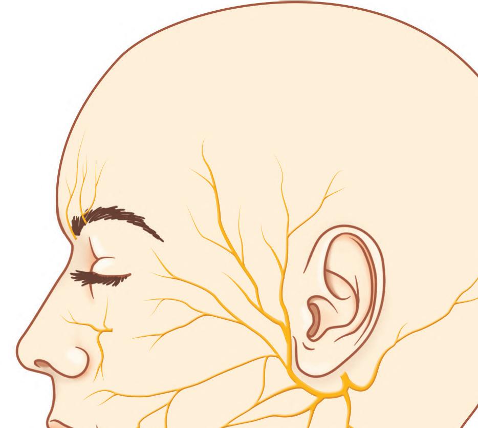
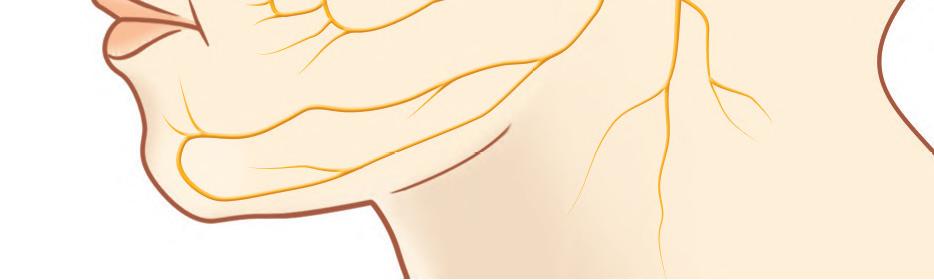
Knowledge of the functional anatomy of the face is fundamental to the understanding of facial trauma management. Important soft tissue considerations are the exact locations of the unique structures of the face and their relations to aesthetic units, layers of the face, and surface landmarks of deep structures such as the lacrimal system, Stenson’s duct, and neurovascular networks, including facial nerve “danger zones”30 (Fig. 1.1.3). The eyelids, nose, ears, and lips are unique structures of the face. A detailed description of their component tissues is beyond the scope of this chapter but an appreciation of this detailed anatomy is vital for reapproximation, repair, and reconstruction following injury and is found in individual sections dealing with regional areas. Similarly,
aesthetic facial subunits and the lines of skin tension (Langer’s lines) can help guide soft tissue repair strategy. The tenet from tumor surgery that loss of more than 50% of a subunit requires consideration of excision of the remainder of that subunit and reconstruction of it as a whole should be considered, but may not be as valid in youth or in cutaneous facial injury. The soft tissue planes of the face from superficial to deep are skin, subcutaneous fat, superficial musculoaponeurotic system (SMAS), containing mimetic muscles, deep fascia and fat compartments, and periosteum. The lacrimal system is the apparatus that produces tears and manages their transfer and drainage; it extends from the lacrimal gland in the lateral upper eyelid, over the corneal surface to the lacrimal canaliculi, through the lacrimal sac and the nasolacrimal duct to the inferior meatus of the nose. Injuries in the region of the eyelids, medial canthus, and upper lateral posterior nasal region should raise suspicion of potential injury to these structures. Stenson’s duct is the conduit for saliva from the parotid gland to the mouth, emerging at a papilla adjacent to the second maxillary molar. An evaluation of Stenson’s duct integrity is important in deep lacerations of the central cheek (especially those that occur near a reference line from the tragus to the lateral oral commissure). Sialocele, fistulae, and infection follow duct or gland injury. The trigeminal nerve branches that supply sensory innervation to the face may be injured as they exit bony foramina approximately in line with the mid-pupil, the supraorbital foramen/ notch, infraorbital foramen, and mental foramen for the three principal sensory branches of V1, V2, and V3 respectively. The blood supply to the face is exceedingly robust and focal injury of single vessels rarely results in clinically significant tissue ischemia in otherwise virgin tissue due to efficient and profuse collateral supply. Injury to the facial artery and its branches such as the labial arteries of the lips, the superficial temporal artery, angular vessels and its other branches can cause significant bleeding and, if untreated, near exsanguination. Identification of foci of blood loss, along with ability to control epistaxis, are essential for control of bleeding from facial injuries as discussed further below. Facial nerve deficit can be one of the most devastating ramifications of an injury to the face. While the facial nerve can be injured anywhere along its course, there are “danger zones” where it is particularly vulnerable to injury; the temporal branch along Pitanguy’s line (from 0.5 cm below the tragus to 1.5 cm above the lateral eyebrow), the zygomatic and buccal branches around Zuker’s point (halfway point along a reference line from the helical root to the oral commissure),31 and the marginal mandibular branch overlying the inferior mandibular body border. Nerve injuries medial to the lateral canthus are less clinically significant due to arborization of the nerve, but any cut nerve seen should be repaired. For those more proximal branches, operative exploration and repair within 72 hours optimizes recovery of motor function.
Facial Skeletal Anatomy
Fig. 1.1.3 Facial anatomic zones in which major facial nerve branches are susceptible to injury. Zone 1 – great auricular nerve; Zone 2 –temporal branch of VII; Zone 3 – marginal mandibular branch of VII; Zone 4 – zygomatic and buccal branches of VII; Zone 5 – supraorbital and supratrochlear nerves of V1; Zone 6 – infraorbital nerve of V2; Zone 7 – mental nerve of V3. (Modified from Holzman NL, Doherty ST, Seckel BR. Facial nerve danger zones, Fig. 7-1. Plastic Surgery Key. https:// plasticsurgerykey.com/facial-nerve-danger-zones/.)
The structural support of the facial skeleton may be organized by the description of the facial buttresses. Nasofrontal/nasomaxillary, zygomatic, and pterygomaxillary buttresses are the major structures of vertical support of the maxilla; mandibular, maxillary–palatal, zygomatic, and frontal buttress are responsible for anteroposterior projection; and the orbital buttress has both vertical and horizontal components (see Fig. 1.1.2). These are the supporting pillars of the facial skeleton; alignment and stabilization following injury literally provides the bony foundation for the restoration of facial form and support.5
Adult and Primary Dentition and Nomenclature
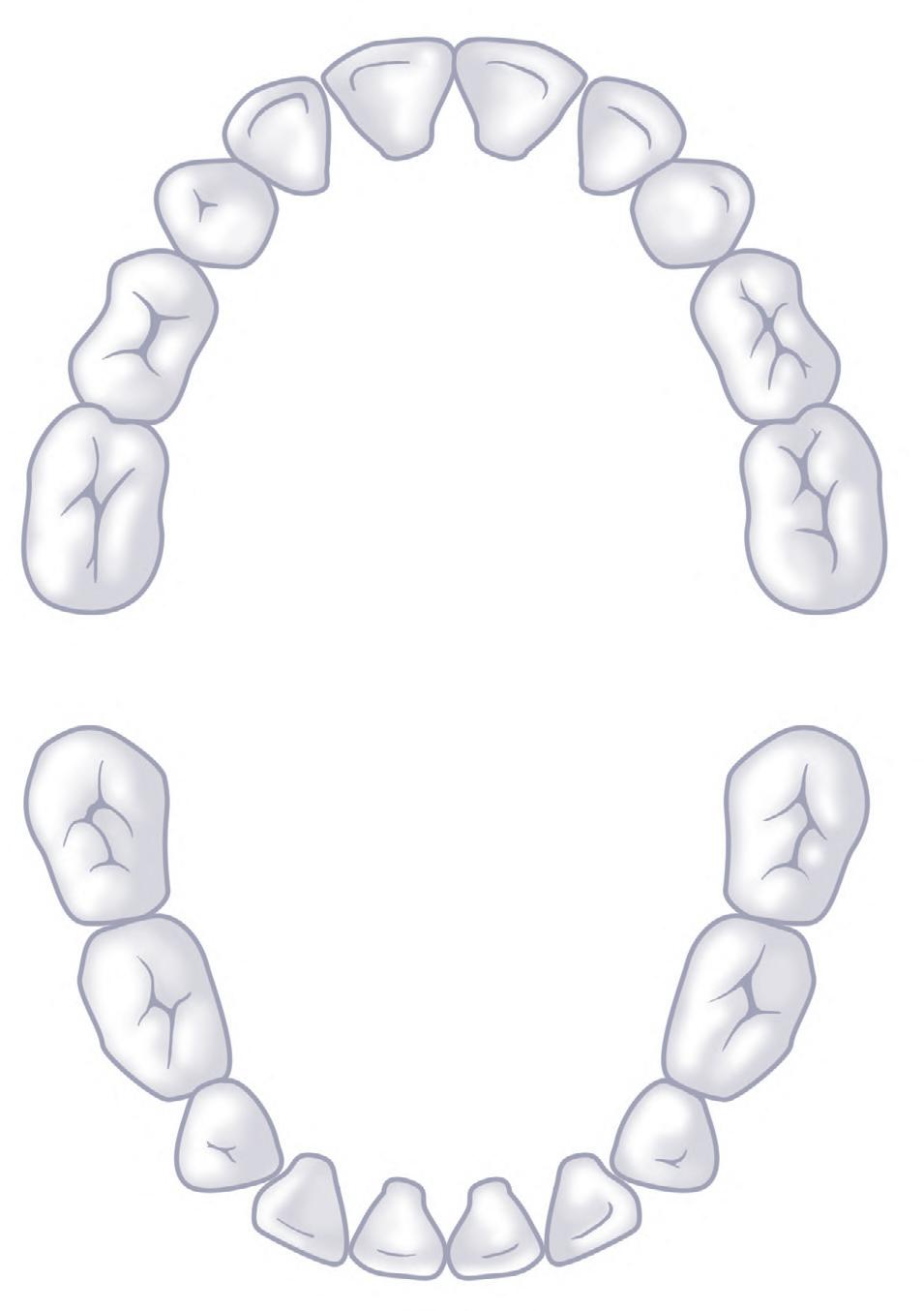
Assessment of dental trauma requires fluent knowledge of the nomenclature of dentition, both adult (permanent) and primary (deciduous) teeth. There are 20 primary teeth denoted by letters A to T proceeding from upper right second molar (A), to upper left second molar (J),
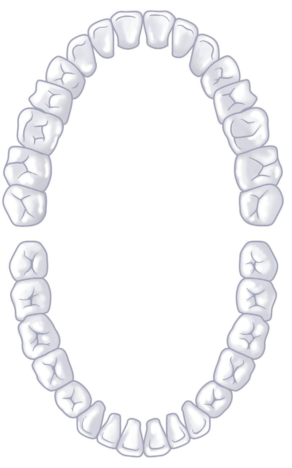
then lower left second molar (K) to lower right second molar (T) (Fig. 1.1.4). Similarly, permanent teeth are denoted by numbers 1–32 from upper right third molar (1) to upper left third molar (16) and then lower left third molar (17) to lower right third molar (32) (Fig. 1.1.5).
The mandibular first molar is the first permanent tooth to erupt, typically at 6 years, followed by incisors at age 6–9, then canines between 9 and 12 years, first premolars at 10–11, second premolars at 11–12, second molars at 11–13, and finally third molars around 17–21 years of age. There is specific anatomical terminology for orientation when referencing teeth, namely mesial (towards midline) and distal (away from midline) in reference to the dental arch, as well as lingual (towards tongue)/palatal (towards palate) versus buccal (towards cheek), labial (toward the lip) (Fig. 1.1.6).
The practical application of the anatomy of the soft tissue, osseous, and dentoalveolar structures enables accurate diagnosis and nomenclature of facial injury, forms the basis for surgical exposure of the craniofacial skeleton, and as such is the cornerstone of fracture management. Incisions and exposures must be designed to respect aesthetic units, navigating the various layers of the face without injury to vital structures, preserving nerves and vessels to expose underlying fractures and skeletal buttresses.
CLASSIFICATION
There is no widely adopted classification of facial trauma that encompasses all of the soft tissue, dentoalveolar, and bony injuries. Rather, certain injury patterns do have well described and accepted classification systems, such as Le Fort fractures, NOE fractures, and dentoalveolar fractures, and each will be discussed below. Facial injuries are usually classified descriptively and largely by pattern. Soft tissue injuries may Upperteeth
Central incisor
Lateral incisor
Canine (cuspid)
First molar
Second molar
months
months
months
months
Lowerteeth
Second molar
months
First molar
Canine (cuspid)
Lateral incisor
Central incisor
months
months
months
be classified by descriptions of their mechanism (sharp, crush, ballistic, blunt, burn), laceration and/or avulsion, location, orientation (vertical, transverse, oblique), and depth. In general, fractures can be classified by pattern: location, displacement, comminution, and whether they are open/closed to the skin. Most commonly, facial fractures are classified as being upper, middle or lower third (i.e., mandible) fractures or a combination; a true “panfacial” fracture has components involving upper, middle, and lower thirds simultaneously.
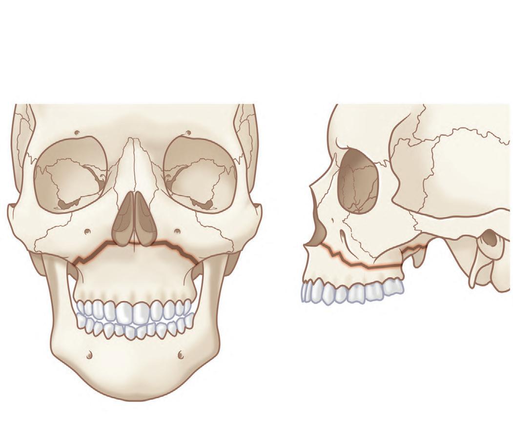
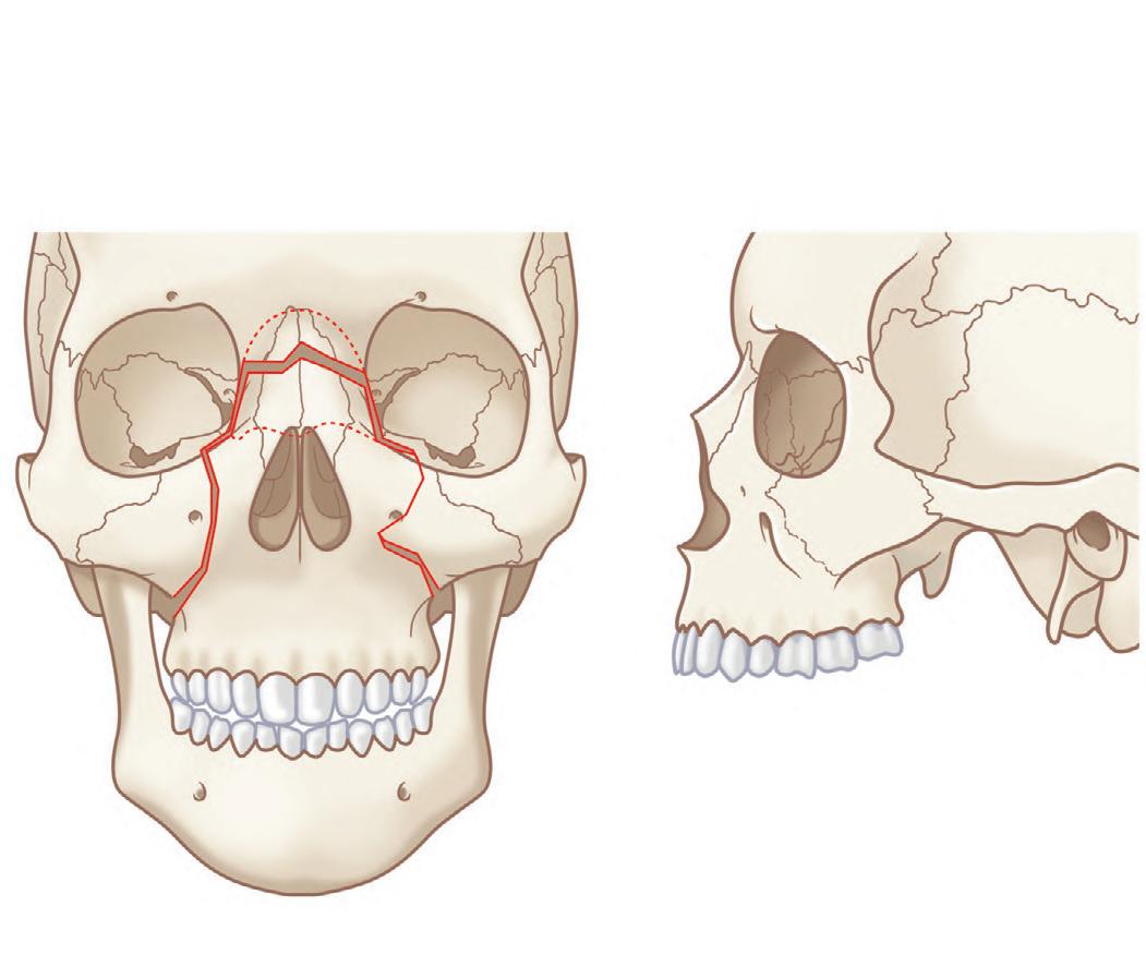
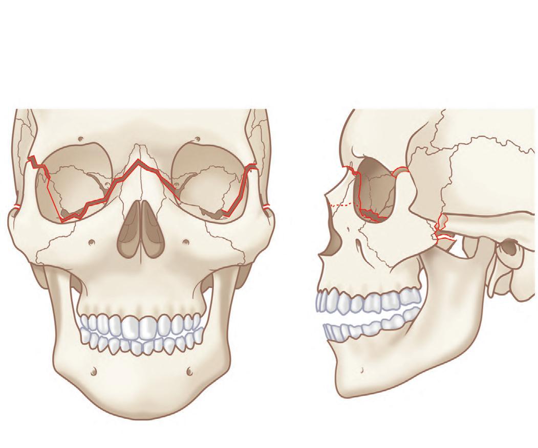
The Le Fort classification is a widely adopted and historical description of midface fracture patterns and fracture line locations in the maxilla with strong historical roots to original French cadaver experiments.32,33 The hallmark of Le Fort fractures is traumatic pterygomaxillary separation, which signifies fractures of the pterygoid plates. Le Fort Type I fractures involve the lateral and medial walls of the maxillary sinus, propagating posteriorly above the alveolar process from the pyriform aperture (Fig. 1.1.7). Le Fort Type II fractures extend through the inferior orbital rim and orbital floor and the maxillary sinuses, and across the nose either high or low, forming a pyramidal shape of varying heights (Fig. 1.1.8). Le Fort Type III fractures extend horizontally from the nasofrontal suture to the frontozygomatic suture, through the orbits, and transect the zygomatic arches (Fig. 1.1.9). Le Fort I, II, and III fractures are conceptualized as a “floating palate,” “floating maxilla,” and “craniofacial dysfunction,” respectively.34,35 Most Le Fort fractures are usually bilateral, but asymmetric due to the asymmetric forces creating the fracture. It is thus common to have a higher-level fracture (i.e., Le Fort III) on the side of force application, and a lower-level fracture (i.e., Le Fort II) on the contralateral side. Lesser Le Fort segments usually exist within the overall Le Fort fracture pattern, reflecting comminution. Accurate bilateral description of the fracture pattern is critical for planning of the open reduction. The fracture pattern is defined by stating
Fig. 1.1.4 Primary teeth nomenclature and eruption chart.
the highest level of Le Fort fracture on each side up to (and including) the frontal bone and the nature of the fragment that includes the maxil lary dentition (i.e., dentoalveolar fracture, split palate). fracture pattern is thus precisely defined and guides the surgeon where open reduction should be performed.
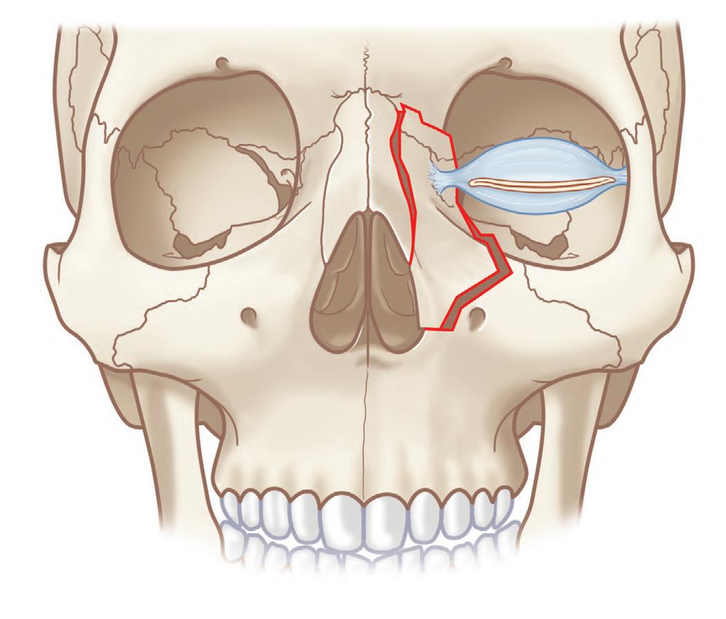
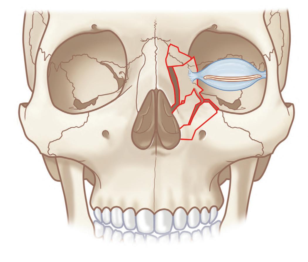
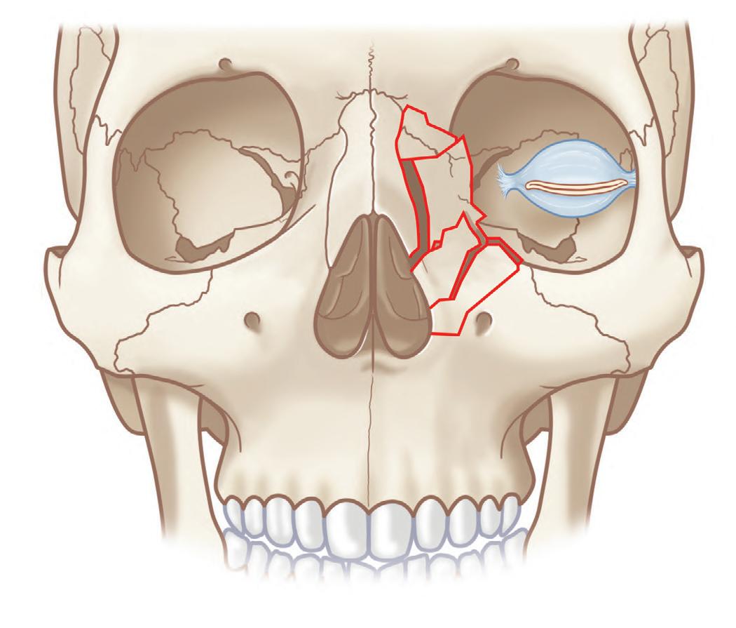
Markowitz and colleagues classified NOE fractures based on comminution and its impact on the medial canthal tendon-bearing bone fragment (“central fragment” of the NOE fracture)37 (Fig. 1.1.10). Type I fractures are characterized by a single noncomminuted “central” fragment without medial canthal tendon detachment/avulsion. The simplest fractures are “greensticked,” or incomplete superiorly and displaced inferiorly, at the inferior orbital rim and pyriform aperture. Complete Type I fractures have a complete fracture at all buttresses including the nasofrontal suture. Type II fractures are characterized by a comminuted central fragment without medial canthal tendon disruption, and no fractures extending underneath the insertion of the medial canthal ligament. Type III fractures are characterized by a severely comminuted central fragment with fractures extending under the medial canthal tendon insertion and avulsion of the medial canthal ligament. Type I require open reduction internal fixation (ORIF) with junctional miniplates, Types II and III also require transnasal wiring and peripheral miniplate fixation.
Dentoalveolar injuries can be classified as tooth fractures, injuries of the periodontal apparatus, and/or injuries to supporting bone tissues.38 There are numerous classification systems, such as that by Andreasen 39 and Garcia-Godoy.40 Most are based on the World Health Organization Classification (I = Fracture of enamel of tooth, II = Fracture of crown without pulpal involvement, III = Fracture of crown with pulpal involveFracture of root of tooth, V = Fracture of crown and root
Fig. 1.1.5 Permanent (adult) teeth chart.
Buccal
Lingual
Mesial
Distal
Fig. 1.1.6 Orientation terminology in reference to the dental arch.
Fig. 1.1.7 Le Fort I fracture is located transversally above the dental apices and separates the dentoalveolar process, the hard palate, and the pterygoid processes, resulting in “floating palate.” (From Prein J, Ehrenfeld M, Manson PN, editors. Principles of internal fixation of the
Fig. 1.1.8 Le Fort II fracture forms a pyramidal shape, resulting in “floating maxilla.” There may be high and low variations as it crosses the nasal bridge: high at frontal bone (blue arrow), and low under the nasal bone (black arrow). (From Prein J, Ehrenfeld M, Manson PN, editors Principles of internal fixation of the craniomaxillofacial skeleton: trauma and orthognathic surgery. AO Foundation, Thieme; 2012, Fig. 3.2-2a–b, p.194.)
of tooth, VI = Fracture of tooth unspecified, VII = Luxation of tooth, VIII = Intrusion or extrusion of tooth, IX = Avulsion of tooth, X = Other injuries, including laceration of oral soft tissues).41
CLINICAL PRESENTATION
The spectrum of facial trauma encompasses the superficial skin laceration to the panfacial fracture with overlying composite soft tissue injury, and everything in between. Strategies of evaluation should therefore obviously be tailored to the severity of the clinical presentation, but must always be thorough and complete. ATLS guidelines should always be adhered to for evaluation of airway, bleeding, and circulation. Screening for life-threatening injuries, bleeding, brain injury, and cervical spine assessment should precede facial trauma evaluation. Assuming the airway is patent, the patient is breathing adequately, and is hemodynamically stable, it is then prudent not to delay assessment of the face for soft tissue and bone injuries. Indeed, facial injuries may threaten
Fig. 1.1.9 Le Fort III fracture extends horizontally from the nasofrontal suture to the frontozygomatic suture and zygomatic arches, resulting in “craniofacial dissociation.” (From Prein J, Ehrenfeld M, Manson PN, editors. Principles of internal fixation of the craniomaxillofacial skeleton: trauma and orthognathic surgery. AO Foundation, Thieme; 2012, Fig. 3.2-3a–b, p.194.)
the airway and may be the source of massive bleeding, demanding emergent evaluation/control of hemorrhage in such cases. Finally, a directed, complete history and physical examination of the face is performed.
History
Pertinent information from the patient’s clinical history includes mechanism of injury (blunt, sharp, dog bite, assault, MVC, etc.), previous injuries/trauma to the face, comorbidities, medications (especially anticoagulants and antiplatelet medications), and allergies.
Physical Examination
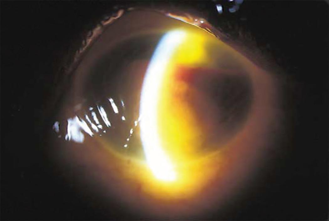
There is no universal convention for the sequencing of a thorough facial examination but the examination should progress in an orderly fashion and be complete; one may elect to work cephalad to caudad or vice-versa. Once the sequential examination is complete, second examinations are conducted in each anatomical area. One must be systematic, and complete; with experience, the entire exam is completed first and then a specific detailed exam can be more targeted toward focal, obvious injuries. For example, working from cephalad to caudad, one may proceed through inspection, palpation, and special tests as follows. Inspect for ecchymosis/edema/hematoma/lacerations throughout (including scalp, ears, and under the chin), pupillary reaction to light, and assessment of visual acuity/double vision; globe position (hypoglobus/enophthalmos/ exophthalmos) and globe rupture, subconjunctival hemorrhage, chemosis (swelling of the conjunctiva), hyphema (blood in the anterior chamber, as sign of globe injury) (Fig. 1.1.11), telecanthus, rounding of eyelid commissure; note asymmetries, deviations (e.g. of the nose), and drainage. A cranial nerve exam can then be performed (typically olfactory examination of CN I is deferred) but testing of extraocular movements, pupil response, and visual acuity, assessment for diplopia and inspection of globe are mandatory in any periorbital trauma or generalized injury (a detailed discussion of the eye examination is presented later in this chapter). Examination of sensation in the three principal distributions of CN V should be performed in the frontal, maxillary, and mandibular regions. CN VII facial motor function can then be assessed with instruction to “raise eyebrows, squeeze eyes closed, puff out cheeks, smile and show teeth.” A crude hearing (VIII) and balance assessment can be performed by basic questioning. Gag reflex can be assessed (IX) and instruction to “shrug” shoulders (XI), turning
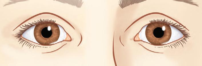
Fig. 1.1.10 Markowitz and colleagues’ classification of NOE fractures. (A) Type I: Single central fragment bearing the medial canthal ligament. (B) Type II: Comminuted central fragment with medial canthal ligament attached to a bone fragment. (C) Type III: Comminuted central fragment with detached medial canthal ligament. (From Prein J, Ehrenfeld M, Manson PN, editors. Principles of internal fixation of the craniomaxillofacial skeleton: trauma and orthognathic surgery. AO Foundation, Thieme; 2012, Fig. 3.5-3a–c, p.236.)
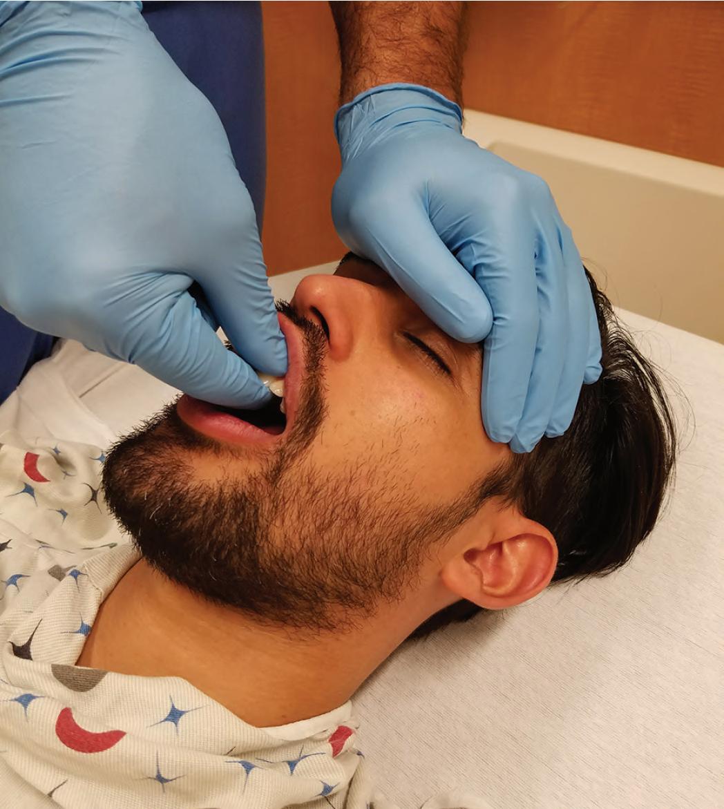
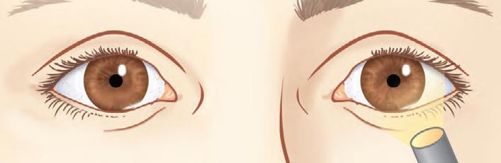
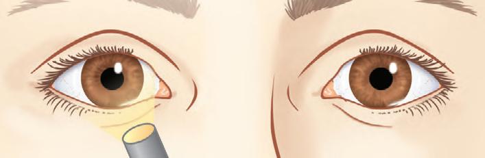
of the head from side to side, and protrusive movement of the tongue (XII) completes the cranial nerve examination. Sequential palpation of all bony surfaces is performed; first the scalp, for hematoma/depression/ skull fractures, then the frontal process of the zygoma, lateral and medial orbital rims, nasal bones, body of zygoma for tenderness and step-offs (bilateral examination helps identify differences), and zygomatic arches. The nose is examined for deviation, crepitus, lacerations, and contour. Flattening of the nasal bridge and an upturned nasal tip indicate frontal impact nasal or nasoethmoid fractures. Assessment for any maxillary mobility can be performed to detect movement of the maxillary dental arch while stabilizing the head with the second hand (Fig. 1.1.12). A test for lateral mobility of the dentition reflects split palate or palatoalveolar fractures. Intraoral exam should include inspection for ecchymosis, lacerations, edema, state of dentition, fractured, missing teeth or bleeding gums (including counting of teeth), presence of any malocclusion or trismus, and a bimanual mandible exam to test stability (Fig. 1.1.13). The mandible should be ranged and the temporal mandibular joints assessed for stability and tenderness.
1.1.12 Assessment for maxillary mobility while stabilizing the head.
For any nasal injury, intranasal examination should be performed with a nasal speculum to detect intranasal lacerations or septal hematoma. Clear nasal or bloody but watery drainage should create suspicion for CSF leak, which can be tested by beta-2 transferrin assay or with the “halo” or “ring” test (created by collecting a drop of the draining fluid on tissue paper – CSF moves further from the center than blood by capillary action) creating a “double ring” sign of clear fluid surrounding an inner blood ring. Similarly, for ear trauma/bleeding otoscopy
Fig. 1.1.11 Slit-lamp photograph of eye demonstrating total hyphema.
Fig.
is indicated to examine for hemotympanum (which may be indicative of ear canal lacerations from temporomandibular joint, mandibular condyle or skull base trauma) and otorrhea. Battle’s sign is a hematoma of the mastoid from a basal skull fracture.
For suspected injury to Stenson’s duct, the duct can be cannulated with a size 22 plastic angiocath sleeve and flushed (Fig. 1.1.14). One technique for this is to dilate the papilla of the duct on the buccal mucosa at the level of the second maxillary molar with a lacrimal dilator then probe (Fig. 1.1.14), with serial dilation as necessary, until an angiocath sleeve can be passed to cannulate the duct. Once passed, the cannulated duct can be irrigated with normal saline, and any drainage of fluid from the facial laceration indicates a duct injury. Most commonly, duct transection is accompanied by buccal branch facial nerve palsy.
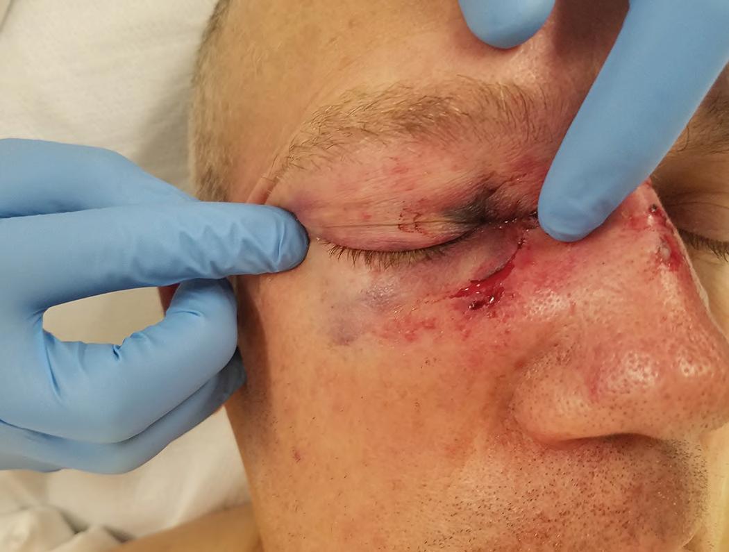
For any periorbital or orbital trauma, examination of the eye should be more extensive than that previously described as part of the cranial nerve screening examination. Any telecanthus or rounding of eyelid commissure indicates canthal detachment which can be confirmed by the eyelid traction test, which assesses the status of the medial canthal tendon’s attachment to the bone. Grasping the eyelid and pulling it displays abnormal canthal mobility in canthal avulsion. A bimanual examination can move the “central fragment” of an NOE fracture between a clamp placed under the canthus intranasally (not the nasal bone) and a palpating finger placed directly over the canthus externally (Fig. 1.1.15). Testing of visual acuity (Snellen® pocket card), diplopia, pupillary light reflex, and any limitation of extraocular movements may be supplemented with forced duction testing (Fig. 1.1.16). Presence of Marcus Gunn pupil or relative afferent pupillary defect (RAPD) (Fig. 1.1.17) may be suggestive of injury to the optic nerve or retina, and ophthalmological consultation should be obtained. In a RAPD, a patient’s affected eye has decreased pupillary response to light, and will dilate when a
bright light is swung from the unaffected eye to the affected eye. The affected eye may sense the light and produce pupillary sphincter constriction to some degree, albeit reduced. In suspected nasolacrimal duct injury, Jones testing is performed. For the Jones I test, a drop of fluorescein is placed on the conjunctiva and if detected in the nose within
Fig. 1.1.13 Bimanual examination of mandible.
Fig. 1.1.14 Cannulating the Stenson’s duct with lacrimal probe. The transection of the duct is clear with the probe visible within the intraoral laceration. The duct was repaired with 8-0 nylon interrupted sutures under loupe magnification.
Fig. 1.1.15 Lid retraction test
5 minutes, the test is said to be positive and indicative of a patent duct. A Jones II test follows a negative Jones I test. Remaining excess fluorescein is irrigated with saline and if detected in the nose Jones II test is positive and there is a functional/partial obstruction; if no saline appears in the nose there is complete obstruction. Partial stenosis of the canaliculi can respond to prolonged intubation/stenting of the lacrimal drainage system, otherwise dacrocystorhinostomy (DCR) is necessary. In the context of orbital and periorbital trauma, consultation to ophthalmology can be considered for suspected globe injury, optic nerve
dysfunction (normal globe with absent vision, direct optic nerve injury or indirect optic nerve injury, e.g. deceleration injuries), compartment syndrome (the orbit feels firm, visual loss, decreased extra-ocular motility, proptosis from hemorrhage/edema, guitar pick sign on CT scan, in which the back of the globe is no longer round), or for a confirmatory preoperative visual evaluation/test.
RADIOLOGICAL EVALUATION
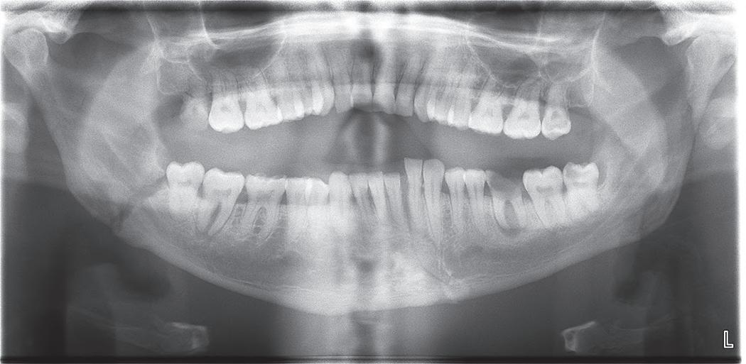
The initial diagnostic imaging of choice for evaluating facial injuries is computed tomography (CT). In addition, many patients have concomitant head injuries requiring head and brain CT scan, which can be obtained at the same time as the CT of facial bones. Plain facial radiographs provide little information and are not worthwhile. CT scans in several planes and with 3-dimensional reconstructions provide precise anatomic identification and quantification of facial fractures which can be viewed in axial, coronal, and sagittal 2-dimensional images. Threedimensional reconstruction of facial bones augments the information obtained, but does not replace 2-dimensional images. 3D images provide spatial relationships which aid in planning complex repairs (Fig. 1.1.18). Panoramic radiographs (orthopantomograms) are occasionally necessary and show the entire mandible, including the condyles, dentoalveolar bone, dentition, and the location/path of the inferior alveolar nerves (Fig. 1.1.19). The benefits of panoramic radiographs include ease of obtaining in dental offices and they are less expensive than the CT images, however, 2D representation of a 3D object will carry inherent limitations in accurately portraying the displacement, extent, and angulation of the fractures. They also have the disadvantage of blurring the symphysis and they require a standing patient for the study. For these reasons, CT is the imaging modality of choice by most practitioners for evaluating facial trauma patients.
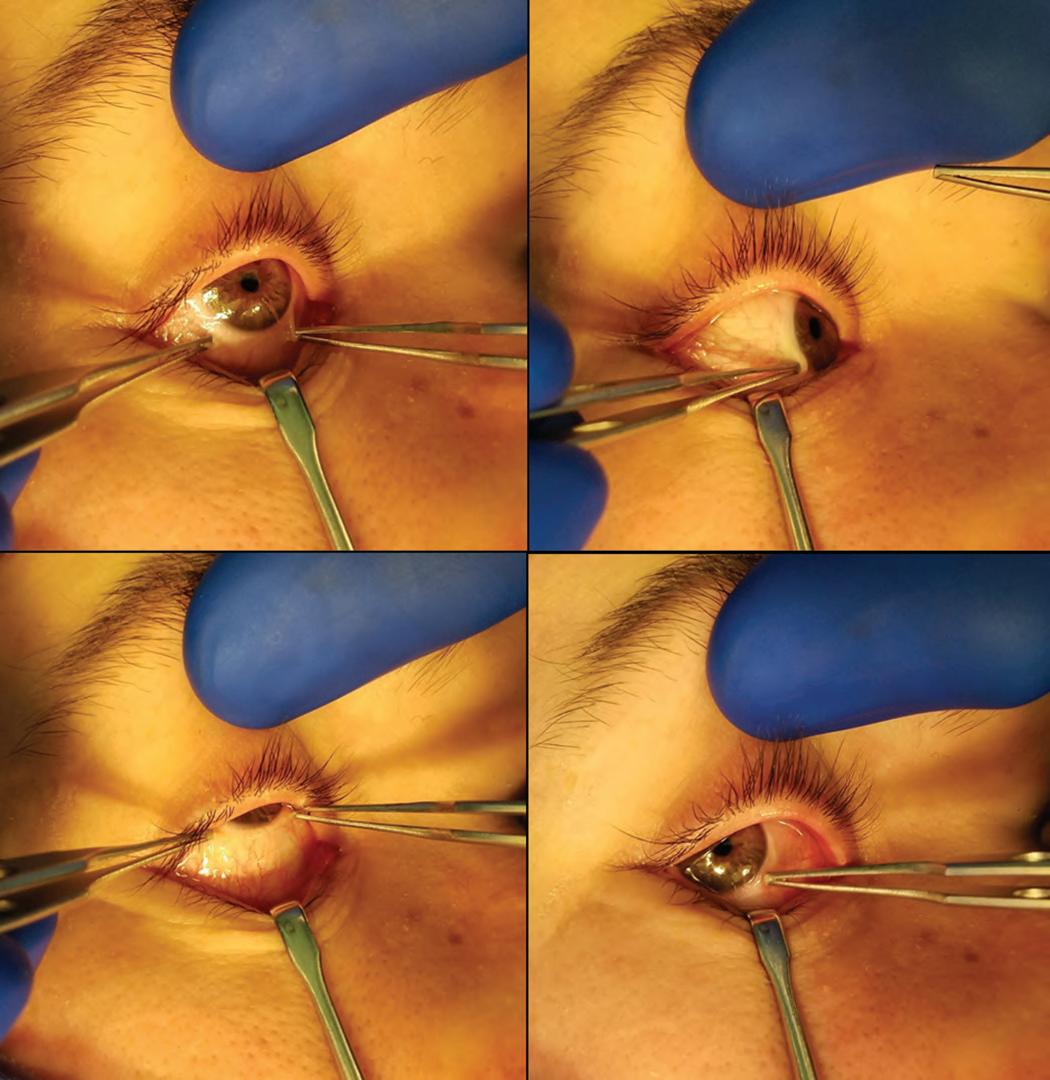
Fig. 1.1.16 Forced duction test of the right globe. (A) Bulbar conjunctiva is grasped with a toothed pick-up and forced (B) medially, (C) superiorly, and (D) laterally.
Normal response to light
Positive RAPD of right eye
Fig. 1.1.17 Marcus Gunn or relative afferent pupillary defect (RAPD).
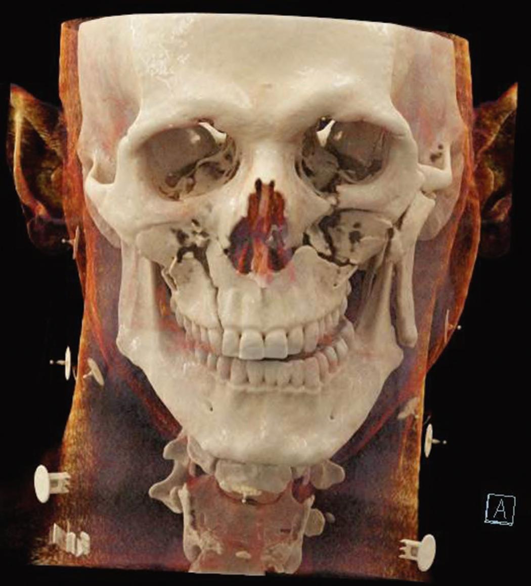
Fig. 1.1.18 Three-dimensional CT reconstruction of facial bones with multilevel fractures.
SURGICAL TREATMENTS
Initial Considerations
Evaluation of facial injury is followed with synchronous initiation of treatment to control bleeding as well as presurgical interventions that may include wiring of teeth or fractures, application of arch bars, manual reduction of grossly displaced fractures, and cleansing/irrigation of wounds, removal of gross contamination, and initiation of tetanus protection, antibiotic, and analgesic therapy. Repair of soft tissue injuries may commence at any time.
Substantial bleeding can occur from soft tissue injuries, especially scalp lacerations and injuries to superficial temporal and facial arteries. Large bleeding scalp lacerations typically require debridement and closure with combinations of sutures possibly over a drain to limit blood loss and fluid accumulation. Suture ligation of vessels and laceration closure both achieve control of bleeding vessels. Facial nerve branches should be avoided in clamps and ligatures. A low threshold for formal intraoperative exploration of these injuries best prevents low-quality wound repairs and recurrent wound problems, including hematomas, and best ensures meticulous hemostasis and ideal tissue approximation. Sharp but minimal debridement of irregular lacerations at the marginal zone of contusion ensures healthy, surgically created wound edges for optimal healing. Problem areas for debridement include the eyelids, lips, distal nose, nostril rims, ear, and eyebrow.
Midface fractures can result in significant epistaxis requiring hemostatic control by anterior–posterior nasal packing (usually posteriorly with nasal balloons/Foley catheters) and anteriorly (with nasal packing). Open fractures causing hemorrhage can also be covered with sterile dressings after cleansing; debridement dressings can be impregnated with hemostatic adjuncts but are of secondary importance to definitive control. Definitive treatment of bleeding may benefit from operative intervention or interventional radiology-guided embolization. Manually repositioning grossly displaced fractures and application of maxillary reduction/rest with intermaxillary fixation (IMF) can also help with bleeding. Patients transferred to the radiology suite must be monitored, observed, and properly resuscitated and stabilized prior to transfer out of the trauma bay.
It is also appropriate in the emergency room setting to close lacerations (after adequate debridement and irrigation). Debridement must be complete but purposefully conservative. Heavily contaminated wounds and those with significant tissue loss will likely warrant formal operating room management. All foreign material, road tattoo, and particles must be removed completely. Secondary procedures to remove foreign material are largely ineffective. Septal and auricular hematomas should be promptly incised, drained, and dressed with an intranasal Doyle
splint or soft lubricated compression dressings, as failure to recognize and manage these results in septal necrosis and perforations, and skin and/or cartilage necrosis or “cauliflower ear.”
Patients With Concomitant Neurological, Cardiopulmonary, or Extremity Trauma
Facial injuries do not necessarily occur in isolation, and practitioners should be cognizant of issues regarding management of patients with simultaneous neurological, cardiopulmonary or extremity trauma. Although there are injuries that require immediate operative intervention, such as entrapped muscle in orbital fractures and blindness, in the absence of active bleeding from major named vessels in the face, most facial trauma is not a life-threatening situation; however, the benefits of definitive prompt management of facial injuries have been underemphasized in the literature.
Partially avulsed or flail segments of fractured facial bones should be stabilized in the ER by replacement into proper position, perhaps wired, and then definitively managed in the operating room. A dental “bridle wire” for mandible fractures may be used in the emergency room for initial control of positioning flail mandibular fragments. Maxillomandibular fixation using intermaxillary fixation screws or arch bars can be placed for temporary stabilization of mandible or palate fractures at the bedside, or at the time of treatment of other injuries such as craniotomy or repair of open lower extremity/pelvic fractures. The possibilities for treatment of facial injuries in the operating room while other definitive or life-saving treatments are occurring have also been understated in the literature. Consideration should be given to tetanus vaccination, initiation of antibiotics, especially for soft tissue wounds caused by burns or bites and fractures violating sinuses and the dentition.
All facial interventions as outlined above should be performed in compliance with general trauma guidelines and in the context of working together as a multidisciplinary team with other trauma care subspecialists and healthcare providers achieving communication and consensus for optimal management of each individual patient’s care needs.
Various Surgical Exposures of Craniofacial Skeleton
Exposure of Upper Third and Upper Midface (Frontal Sinus, Le Fort II and III)
The coronal incision allows the wide exposure necessary to access the frontal bone, upper NOE, medial/superior/lateral orbital walls, zygoma, the zygomatic arch and temporomandibular joint. The coronal incision location can be adjusted based on the hairline pattern (possibly using a zig-zag in the hair for camouflage), and inferior/lateral extension will be necessary to expose the zygomatic arch/temporal mandibular joint areas (Fig. 1.1.20). The coronal incision may be extended retroauricularly as well as preauricularly. The infraorbital rim and orbital floor can be accessed through local incisions via subciliary, midtarsal, or transconjunctival approaches with or without lateral canthotomy (Fig. 1.1.21). The extent and pattern of the fracture, lid anatomy/laxity, and presence of concomitant lacerations will influence the choice of eyelid incision. Exposure of the upper lateral orbital rim (i.e., zygomaticofrontal suture area) can be achieved through local incisions such as the lateral portion of the upper blepharoplasty incision (1–2 cm only), or the transconjunctival incision with lateral cantholysis and superior dissection (Fig. 1.1.22). Considerable postoperative swelling accompanies the latter approach.
Exposure of Lower Midface (Le Fort I and Palate)
The standard approach for exposure of the maxilla is the intraoral vestibular incision around the maxillary arch 8–10 mm away from the
Fig. 1.1.19 Panoramic radiograph of right angle and left parasymphyseal fractures. Dentoalveolar diseases involving the right second molar and left first molar are visualized.
AFig. 1.1.20(A) Coronal approach: wide area of exposure (pink) is possible through coronal incision. (From Prein J, Ehrenfeld M, Manson PN, editors. Principles of internal fixation of the craniomaxillofacial skeleton: trauma and orthognathic surgery. AO Foundation, Thieme; 2012, Fig. 3.4-4, p.227.)
BFig. 1.1.20(B) Coronal approach: various modifications of coronal incision. (From Prein J, Ehrenfeld M, Manson PN, editors. Principles of internal fixation of the craniomaxillofacial skeleton: trauma and orthognathic surgery. AO Foundation, Thieme; 2012, Fig. 3.2-5b–c, p.196.)
attached gingival mucosa (Fig. 1.1.23), leaving a myomucosal cuff for multilayered suture-layered closure. The central area over the incisors frequently may be preserved, which assists alignment and closure. Layered closure of the muscle and mucosa is recommended and sutures besides chromic have more secure staying power and may have to be removed in the postoperative period.
Exposure of Lower Face (Mandible)
Typically, intraoral incisions are used for exposure of symphysis, parasymphysis, body and angle of the mandible. However, under certain circumstances transcutaneous incisions will allow improved exposure for complicated fracture reduction and fixation. Extraoral incisions are preferred for comminuted fractures. The intraoral vestibular incision is typically located on the mobile mucosa 8–10 mm from the attached
gingival mucosa (Fig. 1.1.24). Again, this provides a healthy myomucosal cuff for incision closure at the end of the procedure. Layered muscle repair of the mentalis/buccinators incisions for intraoral closure is essential, and if omitted can result in lip or cheek ectropion. Transcutaneous incisions may be considered if there are existing facial lacerations in proximity to the fractures, or when significant comminution or loss of bone are present requiring access to the inferior border of the mandible for placement of a load-bearing reconstruction plate (Fig. 1.1.25A). Cephalad retraction of the facial vessels (which often need to be ligated) protects the marginal branch of the facial nerve (Fig. 1.1.25B). Transcutaneous approaches offer the best exposure to the condyle and ascending ramus (i.e., posterior mandible); although intraoral ramal incisions are preferred by some but are generally acknowledged to be more challenging (Fig. 1.1.26).