No part of this publication may be reproduced or transmitted in any form or by any means, electronic or mechanical, including photocopying, recording, or any information storage and retrieval system, without permission in writing from the publisher. Details on how to seek permission, further information about the Publisher’s permissions policies and our arrangements with organizations such as the Copyright Clearance Center and the Copyright Licensing Agency, can be found at our website: www.elsevier.com/permissions.
This book and the individual contributions contained in it are protected under copyright by the Publisher (other than as may be noted herein).
Notices
Knowledge and best practice in this field are constantly changing. As new research and experience broaden our understanding, changes in research methods, professional practices, or medical treatment may become necessary.
Practitioners and researchers must always rely on their own experience and knowledge in evaluating and using any information, methods, compounds, or experiments described herein. In using such information or methods they should be mindful of their own safety and the safety of others, including parties for whom they have a professional responsibility.
To the fullest extent of the law, neither the Publisher nor the authors, contributors, or editors, assume any liability for any injury and/or damage to persons or property as a matter of products liability, negligence or otherwise, or from any use or operation of any methods, products, instructions, or ideas contained in the material herein.
Library of Congress Cataloging-in-Publication Data
A catalog record for this book is available from the Library of Congress
British Library Cataloguing-in-Publication Data
A catalogue record for this book is available from the British Library
ISBN 978-0-12-816053-4
For information on all Academic Press publications visit our website at https://www.elsevier.com/books-and-journals
Numbers in parentheses indicate the pages on which the authors’ contributions begin.
Divya Aickara (239), Department of Dermatology and Cutaneous Surgery, University of Miami, Miami, FL, United States
Riccardo Alessandro (1,81), Department of Biomedicine, Neuroscience and Advanced Diagnostics, University of Palermo, Palermo, Italy
Shamila D. Alipoor (383), Molecular Medicine Department, Institute of Medical Biotechnology, National Institute of Genetic Engineering and Biotechnology (NIGEB), Tehran, Iran
Ramaroson Andriantsitohaina (343), SOPAM, U1063, INSERM, UNIV ANGERS, SFR ICAT, Bat IRIS-IBS; Angers University Hospital, Angers, France
Jun Araya (307), Division of Respiratory Diseases, Department of Internal Medicine, The Jikei University School of Medicine, Tokyo, Japan
Koji Asano (101), Department of Urology, Jikei University School of Medicine, Tokyo, Japan
Evangelos Badiavas (239), Department of Dermatology and Cutaneous Surgery, University of Miami, Miami, FL, United States
Matthew A. Bailey (257), University/BHF Centre for Cardiovascular Science, Queen’s Medical Research Institute, University of Edinburgh, Edinburgh, United Kingdom
Scott Bonner (285), University of Oxford, Department of Paediatrics, Oxford, United Kingdom
Han Chen (23), Microscopy Imaging Facility, Penn State Hershey Medical Center, Hershey, PA, United States
Yong Cheng (123), Department of Biological Sciences, Eck Institute for Global Health, Center for Rare and Neglected Diseases, University of Notre Dame, Notre Dame, IN, United States
Raul Coimbra (325), Division of Trauma, Surgical Critical Care, Burns and Acute Care Surgery, Department of Surgery, University of California San Diego, San Diego, CA, United States
Alice Conigliaro (1), Department of Biomedicine, Neuroscience and Advanced Diagnostics, University of Palermo, Palermo, Italy
Denis Corbeil (39), Biotechnology Center and Center for Molecular and Cellular Bioengineering, Technische Universität Dresden, Dresden, Germany
Chiara Corrado (1), Department of Biomedicine, Neuroscience and Advanced Diagnostics, University of Palermo, Palermo, Italy
Todd W. Costantini (325), Division of Trauma, Surgical Critical Care, Burns and Acute Care Surgery, Department of Surgery, University of California San Diego, San Diego, CA, United States
Dragos Cretoiu (199), Department of Cell and Molecular Biology and Histology, Carol Davila University of Medicine and Pharmacy; Alessandrescu-Rusescu National Institute of Mother and Child Health, Fetal Medicine Excellence Research Center, Bucharest, Romania
Sanda Maria Cretoiu (199), Department of Cell and Molecular Biology and Histology, Carol Davila University of Medicine and Pharmacy, Bucharest, Romania
André Cronemberger-Andrade (179), Laboratory of Cellular Immunology and Biochemistry of Fungi and Protozoa, Department of Pharmaceutical Sciences, Federal University of São Paulo (UNIFESP), Diadema, SP, Brazil
James W. Dear (257), University/BHF Centre for Cardiovascular Science, Queen’s Medical Research Institute, University of Edinburgh, Edinburgh, United Kingdom
Alexandru Florian Deftu (199), Department of Anatomy, Animal Physiology and Biophysics, Faculty of Biology, University of Bucharest; Life, Environmental and Earth Sciences Division, Research Institute of the University of Bucharest (ICUB), Bucharest, Romania
Antonia Teona Deftu (199), Department of Anatomy, Animal Physiology and Biophysics, Faculty of Biology, University of Bucharest; Life, Environmental and Earth Sciences Division, Research Institute of the University of Bucharest (ICUB), Bucharest, Romania
Shin Egawa (101), Department of Urology, Jikei University School of Medicine, Tokyo, Japan
Brian P. Eliceiri (325), Division of Trauma, Surgical Critical Care, Burns and Acute Care Surgery, Department of Surgery, University of California San Diego, San Diego, CA, United States
Simona Fontana (1), Department of Biomedicine, Neuroscience and Advanced Diagnostics, University of Palermo, Palermo, Italy
Yu Fujita (307), Division of Molecular and Cellular Medicine, National Cancer Center Research Institute; Division of Respiratory Diseases, Department of Internal Medicine, The Jikei University School of Medicine, Tokyo, Japan
Andrew F. Hill (285), La Trobe University, La Trobe Institute for Molecular Science, Melbourne, VIC, Australia
Robert W. Hunter (257), University/BHF Centre for Cardiovascular Science, Queen’s Medical Research Institute, University of Edinburgh, Edinburgh, United Kingdom
Tsukasa Kadota (307), Division of Molecular and Cellular Medicine, National Cancer Center Research Institute; Division of Respiratory Diseases, Department of Internal Medicine, The Jikei University School of Medicine, Tokyo, Japan
Ju-Seop Kang (467), Department of Pharmacology & Clinical Pharmacology Lab, College of Medicine, Hanyang University, Seoul, South Korea
Nobuyoshi Kosaka (101,307,433), Division of Molecular and Cellular Medicine, National Cancer Center Research Institute; Department of Molecular and Cellular Medicine, Institute of Medical Science; Department of Translational Research for Extracellular Vesicles, Institute of Medical Science, Tokyo Medical University, Tokyo, Japan
Kazuyoshi Kuwano (307), Division of Respiratory Diseases, Department of Internal Medicine, The Jikei University School of Medicine, Tokyo, Japan
Soazig Le Lay (343), SOPAM, U1063, INSERM, UNIV ANGERS, SFR ICAT, Bat IRIS-IBS, Angers, France
Aurelio Lorico (39), College of Medicine, Touro University Nevada, Henderson, NV, United States; Mediterranean Institute of Oncology, Viagrande, Italy
Imre Mäger (285), University of Oxford, Department of Paediatrics, Oxford, United Kingdom
M. Carmen Martinez (343), SOPAM, U1063, INSERM, UNIV ANGERS, SFR ICAT, Bat IRIS-IBS; Angers University Hospital, Angers, France
Jeffrey D. McBride (239), Department of Dermatology and Cutaneous Surgery, University of Miami, Miami, FL, United States
Esmaeil Mortaz (383), Clinical Tuberculosis and Epidemiology Research Center, National Research Institute of Tuberculosis and Lung Diseases (NRITLD); Department of Immunology, Faculty of Medicine, Shahid Beheshti University of Medical Sciences, Tehran, Iran
Soumyalekshmi Nair (357), Exosome Biology Laboratory, Centre for Clinical Diagnostics, UQ centre for Clinical Research, Royal Brisbane and Women's Hospital, The University of Queensland, St Lucia, QLD, Australia
Denis Noble (487), Department of Physiology, Anatomy & Genetics, University of Oxford, Oxford, United Kingdom
Takahiro Ochiya (101,307,433), Division of Molecular and Cellular Medicine, National Cancer Center Research Institute; Department of Molecular and Cellular Medicine, Institute of Medical Science, Tokyo Medical University, Tokyo, Japan
Siew-Wai Pang (147), Department of Medical Sciences, School of Healthcare and Medical Sciences, Sunway University, Petaling Jaya, Malaysia
Beatrice Mihaela Radu (199), Department of Anatomy, Animal Physiology and Biophysics, Faculty of Biology, University of Bucharest; Life, Environmental and Earth Sciences Division, Research Institute of the University of Bucharest (ICUB), Bucharest, Romania
Stefania Raimondo (81), Department of Biomedicine, Neuroscience and Advanced Diagnostics, University of Palermo, Palermo, Italy
Laura Saieva (81), Department of Biomedicine, Neuroscience and Advanced Diagnostics, University of Palermo, Palermo, Italy
Carlos Salomon (357), Exosome Biology Laboratory, Centre for Clinical Diagnostics, UQ centre for Clinical Research, Royal Brisbane and Women's Hospital, The University of Queensland, St Lucia, QLD, Australia; Department of Clinical Biochemistry and Immunology, University of Concepción, Concepción, Chile; Department of Obstetrics and Gynecology, Ochsner Baptist Hospital, New Orleans, LA, United States
Jeffery S. Schorey (123), Department of Biological Sciences, Eck Institute for Global Health, Center for Rare and Neglected Diseases, University of Notre Dame, Notre Dame, IN, United States
Jeffrey M. Sundstrom (23,415), Department of Ophthalmology, Penn State College of Medicine; Department of Ophthalmology, Penn State Hershey Medical Center, Hershey, PA, United States
Sin-Yeang Teow (147), Department of Medical Sciences, School of Healthcare and Medical Sciences, Sunway University, Petaling Jaya, Malaysia
Ana Claudia Torrecilhas (179), Laboratory of Cellular Immunology and Biochemistry of Fungi and Protozoa, Department of Pharmaceutical Sciences, Federal University of São Paulo (UNIFESP), Diadema, SP, Brazil
Fumihiko Urabe (101), Division of Molecular and Cellular Medicine, National Cancer Center Research Institute; Department of Urology, Jikei University School of Medicine, Tokyo, Japan
Sarah R. Weber (23,415), Department of Ophthalmology, Penn State College of Medicine; Department of Ophthalmology, Penn State Hershey Medical Center, Hershey, PA, United States
Eduard Willms (285), University of Oxford, Department of Paediatrics, Oxford, United Kingdom
Matthew J.A. Wood (285), University of Oxford, Department of Paediatrics, Oxford, United Kingdom
Patricia Xander (179), Laboratory of Cellular Immunology and Biochemistry of Fungi and Protozoa, Department of Pharmaceutical Sciences, Federal University of São Paulo (UNIFESP), Diadema, SP, Brazil
Junjie Xiao (199), Department of Cardiology, The First Affiliated Hospital of Nanjing Medical University, Nanjing; Cardiac Regeneration and Ageing Lab, Experimental Center of Life Sciences, School of Life Science, Shanghai University, Shanghai, People’s Republic of China
Zhongdang Xiao (433), State Key Laboratory of Bioelectronics, School of Biological Science and Medical Engineering, Southeast University, Nanjing, China
Yuanjun Zhao (23,415), Department of Ophthalmology, Penn State College of Medicine; Department of Ophthalmology, Penn State Hershey Medical Center, Hershey, PA, United States
Mi Zhou (23,415), Department of Ophthalmology, Penn State College of Medicine; Department of Ophthalmology, Penn State Hershey Medical Center, Hershey, PA, United States
Yueyuan Zhou (433), Division of Molecular and Cellular Medicine, National Cancer Center Research Institute, Tokyo, Japan; State Key Laboratory of Bioelectronics, School of Biological Science and Medical Engineering, Southeast University, Nanjing, China
Editor biography
Lawrence R. Edelstein
President and Founder, Medimark Corporation, Del Mar, CA, United States
Lawrence R. Edelstein, Ph.D. is a neuroscientist and pharmaceutical industry consultant with research interests in multisensory convergence/integration (claustrum) and intercellular communication (exosomes, telocytes). His interest in exosomes was fueled by a theme issue he guest-edited with John Smythies and Denis Noble entitled “Epigenetic information-processing mechanisms in the brain” (2014, https: //royalsocietypublishing.org/toc/rstb/369/1652). That undertaking proved to be the impetus for a series of peer-reviewed articles in which he and his colleagues theorized as to the whys and wherefores of the multifunctional roles played by seemingly omnipresent and phyla-agnostic exosomes. In addition, Dr. Edelstein is co-editor (along with John Smythies and Vilayanur S. Ramachandran, M.B.B.S., Ph.D., Hon. F.R.C.P.) of the book Claustrum - Structural, Functional and Clinical Neuroscience (2014, www.elsevier.com/books/the-claustrum/smythies/978-0-12-404566-8).
John Raymond Smythies
John Raymond Smythies, M.B. B.Chir., M.D., F.R.C.P., F.R.C. Psych. (1922–2019) was the Director of the Integrative Neuroscience Program in the Department of Psychology at the University of California San Diego. As a pre-eminent neuropsychiatrist and neuroscientist he made significant contributions to both these disciplines. Together with Humphry Osmond he developed the first biochemical theory of schizophrenia - the transmethylation hypothesis. This has recently come back into focus following the finding that DNA methylation is abnormal in schizophrenia. He made extensive contributions to knowledge in a number of fields including the neuropharmacology of psychedelic drugs; the functional neuroanatomy of synapses with particular regard to the role of synaptic plasticity, endocytosis and redox factors; the role in the brain of orthoquinone metabolites of catecholamines; and, in particular, theories of brain-consciousness relations. More recently he developed foundational hypotheses and theories specific to the function of exosomes, telocytes and the claustrum, and on epigenetic processes in information processing in the brain. Professor Smythies served as President of the International Society of Psychoneuroendocrinology from 1970–1974, Consultant to the World Health Organization from 1963–1968, and Editor of the International Review of Neurobiology from 1958–1991. He was
elected a member of the Athenaeum in 1968. He held the positions of Professor Emeritus and the Charles Byron Ireland Professor of Psychiatric Research at the University of Alabama Medical Center at Birmingham, Visiting Scholar at the Center for Brain and Cognition, University of California San Diego, and Senior Research Fellow at the Institute of Neurology, University College London. He published over 240 scientific papers and sixteen books. https://en.wikipedia. org/wiki/John_Raymond_Smythies
Peter J. Quesenberry
Paul Calabresi Professor of Oncology, Professor of Medicine, The Warren Alpert Medical School of Brown University, Providence, RI, USA
Peter J. Quesenberry, M.D., is the Paul Calabresi Professor of Oncology at The Warren Alpert Medical School of Brown University. He received his medical degree from the University of Virginia, completed residency at University Hospital and Boston City Hospital in Boston, MA, and completed a Hematology/ Oncology Fellowship at St. Elizabeth's Hospital.
Professor Quesenberry is a leading investigator in stem cell biology and extracellular vesicle research. He was President of the International Society of Hematology, editor of the journal Experimental Hematology from 1990–1998 and the leukocyte editor for the Year Book of Hematology from 1987–1998. More recently he is a co-editor-in-chief for the Journal of Extracellular Vesicles.
Denis Noble
Emeritus Professor of Cardiovascular Physiology, Department of Physiology, Anatomy, and Genetics, University of Oxford, Oxford, UK
Denis Noble, C.B.E., Ph.D., F.R.S. is a British biologist who held the Burdon Sanderson Chair of Cardiovascular Physiology at the University of Oxford from 1984 to 2004 and was appointed Professor Emeritus and co-Director of Computational Physiology. He is one of the pioneers of systems biology.
Professor Noble developed the first viable mathematical model of the working heart in 1960 using his discovery, with his supervisor Otto Hutter, of two of the main cardiac potassium ion channels. These discoveries were published in Nature (1960) and The Journal of Physiology (1962). The work was later developed with Dick Tsien, Dario DiFrancesco, Don Hilgemann and others to become the canonical models on which more than 100 cardiac cell models are based today.
He was elected President of the International Union of Physiological Sciences (IUPS) at its Congress in Kyoto in 2009 and was re-elected for a second term at the 2013 Congress in Birmingham, UK.
He is the author of the first popular book on systems biology, The Music of Life, and his most recent lectures concern the implications for evolutionary biology.
Professor Noble has published more than 500 papers and 11 books. https:// en.wikipedia.org/wiki/Denis_Noble
Preface
Nothing speaks more clearly to the envisaged merits of a major clinical and scientific discovery than the rapidly increasing number of journal articles and their citation in a brief period of time, followed apace by the launch of biopharma start-ups founded on such, each competing to fill their respective pipelines with internally developed and in-licensed compounds. Case in point, exosomes, a seemingly omnipresent and phyla-agnostic extracellular vesicle of endosomal origin which has quickly come to the fore in the context of intercellular communication, with payloads of miRNA, mRNA, lncRNA, and transcription factors at the ready. Arguably, the most compelling aspect of exosomes, and conceivably their raison d'être, is their role in transgenerational epigenetics, the topic of our closing chapter.
My interest in exosomes was sparked by a theme issue I guest-edited with my fellow editors John Smythies and Denis Noble entitled “Epigenetic information-processing mechanisms in the brain” (https://royalsocietypublishing. org/toc/rstb/369/1652). This proved to be the impetus for a series of peerreviewed articles in which my colleagues and I theorized as to the whys and wherefores of the multifunctional role of exosomes in most if not all living organisms. Over 2 years in the making, we are so very pleased to see this undertaking come to fruition: Exosomes—A Clinical Compendium.
Armed with a modicum of publishing experience as a neuroscientist, author, editor and founding editor-in-chief of a scientific journal, I was quick to note that there was a multidisciplinary component missing from the extant narrative. The time was ripe for an exosome book with a rather daunting objective—to bottle lightning by inviting contributions from a global cohort of peer-acknowledged expert clinicians and researchers across a wide range of medical disciplines, affording each an equal voice at the table. Exosomes—A Clinical Compendium serves to provide readers with a broad and timely overview of exosomes in health and disease. Within its 21 chapters, our authors have summarized the most recent laboratory and clinical findings, thereby illuminating the path forward for prospective investigative efforts.
To the exosome novitiate I say, welcome to what I consider to be the leading edge of diagnostic, therapeutic and theragnostic research. Have at it! To those for whom exosomes have played and continue to play an integral role in their
xxiv Preface
clinical and research endeavors and, in turn, helping to advance the field, I say thank you for your unwavering efforts as without them this book would have withered on the vine.
Lawrence Edelstein, Ph.D.
Acknowledgments
Early in the evolution of this book it became apparent that the study of extracellular vesicles in general—and of exosomes in particular—has been accelerating at a remarkable pace. Nowhere has this been more evident than in the context of their clinical implications across the spectrum of medical disciplines. First and foremost we are deeply indebted to our authors, each a peeracknowledged expert in their respective fields of endeavor. We are also grateful for the steadfast guidance and assistance provided by our colleagues at Elsevier, most notably Mica Haley, Tracy Tufaga, Swapna Praveen, Mohana Natarajan, Jyotsna Gopichandran, André Wolff, and Jaclyn Truesdell. Lastly, were it not for a series of theory/hypothesis articles by our late co-editor John Smythies on the raison d'être of exosomes and telocytes in reference to neural coding, repair and cognate mechanisms, it is fair to say that this book would not have seen the light of day.
Special acknowledgment
In memory of a gentleman scientist and rara avis of the highest order—John Raymond Smythies, M.B. B. Chir., M.D., F.R.C. Psych., F.R.C.P. (https:// en.wikipedia.org/wiki/John_Raymond_Smythies).
Apropos of an early British maritime phrase oft-used by John, who was a Surgeon-Lieutenant with the Royal Naval Volunteer Reserve from 1946–49 aboard H.M.S. Porlock Bay, “All is shipshape and Bristol fashion!” Mission accomplished.
Chapter 1
Exosome basic mechanisms
Alice Conigliaro, Chiara Corrado, Simona Fontana, Riccardo Alessandro
Department
of Biomedicine, Neuroscience and Advanced Diagnostics, University of Palermo, Palermo, Italy
1 Exosomes biogenesis and release
Exosomes biogenesis is correlated to intraluminal vesicles (ILVs) formation and starts with a first invagination of the plasmatic membrane, leading to the formation of early endosomes, low-density vesicles very close to the inner membrane surface.
Different materials can be internalized by endocytosis and by receptor mediated-endocytosis and early endosomes are responsible of their sorting; after receptor-ligand interaction on the cell surface, ligand is separated from its receptor, located inside the early endosome and transferred by endosomal transport vesicles to the late endosome. The receptor, instead, can be recycled on the membrane surface or partially degraded via late endosomes-lysosomes pathway, in order to turn off the signal and reduce the receptor concentration on the cell surface.
During their path, mediated by microtubules, inside the cell, early endosomes go through different modifications; they can fuse each other or with vesicles containing acid hydrolases, thus forming intermediate structure, called multivesicular bodies (MVB) and containing ILVs. The formation of ILVs is the second event of membrane invagination; in this case, it involves endosomal membrane.
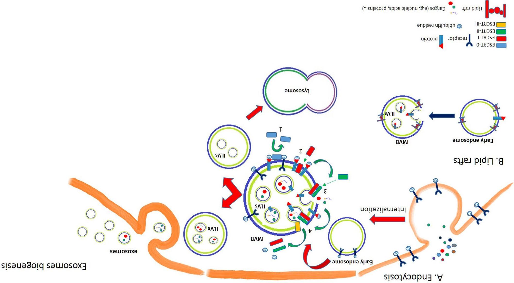
MVBs originate by two different mechanisms: first, the Endosomal Sorting Complex Request for Transport (ESCRT) is responsible of the MVB formation; alternatively, the MVBs can originate from endosomes that contain specific domains on their membrane, known as lipid rafts (Fig. 1).
At this point, MVB, containing ILVs, can follow two different fates. MVB can fuse with other MVBs or with late endosomes and receive vesicles originating from transGolgi and containing lysosomal enzymes. In this case, during the maturation process, the pH becomes more acid thus activating the hydrolases that digest molecules inside MVB and allow the transformation in lysosomes, in eukaryotic cells. This pathway allows a rapid and consistent turnover
of transmembrane proteins and lipids. Alternatively, MVBs migrate toward the membrane, fuse with the plasma membrane and release ILVs outside of the cell, as exosomes. Exosomes are, therefore, the only secreted vesicles of endolysosomal origin, generated from internal membranes.
2 Endosomal sorting complex request for transport (ESCRT) and its role in exosome biogenesis
The role of ESCRT in exosome biogenesis emerges by the evidence that numerous ESCRT proteins have been discovered in exosomes preparations from different cell types or biological fluids, for this reason, many components are acknowledged in the scientific community as exosomal markers.
ESCRT pathway is, by now, a well-described mechanism to explain ILVs and MVB formation. ESCRT is composed of five multimeric cytosolic complexes: ESCRT 0, I, II, III, and Vps4 [1] (Fig. 1).
Specific ubiquitinated proteins, contained in early endosomes, are recognized, in their ubiquitination site, by ESCRT-0 that places them in specific endosomes' areas enriched in phosphatidyl inositol monophosphate (PI3P).
ESCRT-0 is composed of two subunits, called HRS (hepatocyte growth factor regulated tyrosine kinase substrate) and STAM1/2 (signal transducing adaptor molecule 1/2), which bind one to each other and are able to recognize ubiquitin and PI3P enriched domains. This allows recruiting ESCRT-I, a heteromeric complex composed of Tsg101 (tumor susceptibility gene 101), Vps28, Vps37 and Mvb12 (multi-vesicular body 12).
ESCRT-I takes the place of ESCRT-0 and recruits ESCRTII, composed of four subunits: Vps22-EAP30, Vps36-EAP45 and two subunits of Vps25-EAP20.
ESCRTI-II can start the endosome's membrane invagination thus allowing internalization of different molecules/cargo, such as nucleic acids or proteins.
ESCRT-III is another heterotetrameric complex (Vps20-CHMP6; Snf7CHMP4; Vps24-CHMP3; Vps2-CHMP2); activation and its subsequent recruitment to the endosome is mediated by the binding of the ESCRT- II subunit Vps25 with Vps20. ESCRT-III has the role to detach the ubiquitin residues from the protein and to allow the complete invagination of the membrane (membrane budding), thus generating the ILVs. Cargo deubiquitination is mediated also by accessory subunits recruited by ESCRT-III, such as Bro1/Alix (BCK1-like resistance to osmotic shock protein-1/apoptosis linked gene 2 interacting protein X) that, after binding to Snf7, recruits the enzyme Doa4 (degradation of alpha 4), to complete the cargo deubiquitination [2]. Finally, others adaptor proteins assist Vps4-ESCRT III interaction and allow the Vps4 ATPase activity, required for the final membrane budding and scission, for ESCRT subunits removal and for recycling and cargo delivery.
In summary, the ESCRT complex controls the entire process from ILV budding to the selection of cargo, the membrane remodeling and the incorporation of ILVs into MVB. An interesting study of RNAi, on 23 components of ESCRT
FIG. 1 Exosomes biogenesis. Exosomes biogenesis can occur according different mechanisms. (A) Endocytosis: after receptor-ligand interaction on the cell surface, ligand is separated from its receptor and located inside the early endosome. The receptor, instead, can be recycled on the membrane surface or partially degraded via late endosomes-lysosomes pathway. Early endosome subsequently forms multivesicular bodies (MVB) containing intraluminal vesicles (ILVs). The endosomal sorting complex request for transport (ESCRT) is responsible of the MVB formation. (1) ESCRT0 binds ubiquitin residues thus allowing the recruitment of ESCRTI, which finally undermines ESCRT0 (2). (3) ESCRTII starts endosome's membrane invagination and binds ESCRTIII (4), that detaches the ubiquitin residues from the protein and allows the complete invagination of the membrane (membrane budding), thus generating the ILV. MVBs migrate toward the membrane, fuse with the plasma membrane and ILVs are released as exosomes. Alternatively, MVB can fuse with other MVBs or with late endosomes, receive from transGolgi a set of vesicles containing lysosomal enzymes that allow the transformation in lysosomes. (B) Lipid rafts. MVBs can also originate from endosomes that contain specific domain on their membrane, known as lipid rafts.
complex, allowed clarifying that only few of them are essential in exosome biogenesis. Among these subunits, in fact, Hrs, Tsg101 and STAM1 simultaneous silencing (ESCRT0/I complex) is able to decrease exosome secretion; on the contrary, inhibition of CHMP4C, VPS4B, VTA1 and Alix (ESCRT III complex) increases the same process of exosomes secretion [3]. Recent investigations, demonstrated, in opposition to the role of VPS4 just stated, that its inhibition decreases exosomes release [4]. In addition, Alix, through its interaction with several ESCRT proteins, such as Tsg101 or CHMP4, is involved in protein composition/cargo loading, ILVs' budding and its incorporation into MVBs.
Numerous proteins collaborate with ESCRT complexes in all the steps of exosome biogenesis, from endosomal budding to ILV's formation.
Syntenins are soluble proteins that act as intracellular adaptors, through their PDZ domains, recruiting syndecans, membrane proteins carrying heparan sulfate chains (HS). Syndecans, via HS, bind numerous ligands such as adhesion molecules and growth factors thus allowing them to interact with their receptors and assist the endocytosis process. Recent evidences demonstrated that Alix binds the N-terminal of syntenins and connects syndecans with the ESCRT machinery. Therefore, this heterotrimeric complex is involved not only in exosomal sorting and cargo, as discussed later, but also in endosomal budding and exosomes biogenesis [5, 6]
Related to late endosome trafficking and syntenin-exosomes production, the role of a GTP binding protein is emerged: ADP-ribosylation factor 6 (ARF6). ARF6 is able to activate phosphatidyl-inositol (4)-phosphate 5-kinase (PIPK), involved in PIP2 synthesis. The PIP2 synthesis supports the recruitment of syntenin-syndecan from perinuclear compartment to the plasma membrane [7].
Another kinase, upstream of ARF6, regulates syntenin/syndecan activity: the oncoprotein Src. Src acts on endosomal trafficking by phosphorylation of syntenin/syndecan tyrosine residues thus stimulating endosomal budding and the biogenesis of specific syntenin-dependent exosomes [8].
Heat shock proteins (Hsps) are chaperones generally involved in the control of protein aggregation and folding. These proteins were initially studied for their role in this intracellular pathway, subsequent observations indicated their presence outside the cell. Now it is well known that Hsps are proteins secreted by a non-classical pathway and, most of them, by exosomes. Interestingly, Hsps are also involved in exosomes biogenesis by collaborating and interacting with ESCRTs proteins [9].
3 ESCRT-independent mechanism of exosomes biogenesis
Simultaneous depletion of critical ESCRT proteins demonstrated that MVB biogenesis and ILV formation is also ensured through ESCRT independent mechanisms, thus suggesting that the regulation of this process is more complex than expected. The lack of ESCRT complex, indeed, induces a deep morphological
modification of the MVBs that appear enlarged, even if early and late endosome differentiation is maintained.
The existence of ILVs in this circumstance, although different in number and size, suggests that their formation could occur via an ESCRT-independent endocytic sorting mechanisms and allow to hypothesize that both mechanisms of exosomes biogenesis could co- exist and function together, in higher eukaryotes. Cell type and cellular homeostasis could be the elements that drive different subpopulation of exosomes through one of the two mechanisms of biogenesis.
Cells mutated for ESCRTs are still able to form ILVs due also to the lipid composition of their membranes. It is well known that the biophysical characteristics of single membrane lipid, such as the size of head group, length and saturation of acyl chains, are fundamental for membrane curvature.
Endosomes with particular areas enriched in cholesterol and sphingolipids, the lipid rafts, curve inward and may determine the MVBs formation, through the help of pH gradient across the membrane. In this case and without the assistance of ESCRTs, endosomes membrane invagination is due to the synthesis, mediated by phospholipases, of ceramides from sphingolipids. Ceramides alone, due to their cone shape, or in association with cholesterol generate particular domains that favor membrane deformation and ILV budding.
Moreover, ceramide induces exosomal biogenesis through its conversion into sphingosine 1-phosphate (S1P), which binds its receptor on the membrane of MVBs [10].
Neutral Sphingomyelinases (SMases), enzymes involved in sphingomyelin conversion in ceramide, are preferentially located in Golgi-ER but also in plasma membrane thus participating into exosome biogenesis. Inhibition of SMases, in specific cell types, reduces exosomal release of certain proteins thus demonstrating that ceramide is essential to generate microdomains that favor membrane budding [11].
Others lipid modifying enzymes, phospholipase D2 (PLD2) and diacylglycerol kinase α (DGKα), are involved in exosomes biogenesis through the production of phosphatidic acid (PA) that favors, as well as ceramide, membrane invagination [7, 12].
On the other hand, sphingomyelin has a high affinity for cholesterol in the membrane; its hydrolysis increases cholesterol migration from the PM to intracellular membrane and consequently membrane fluidity. It is well known, in fact, that cholesterol molecules cluster in lipid domains thus affecting plasma membrane lipid order and finally vesicle shedding.
Numerous proteins mediate the ESCRT independent mechanism of exosomes biogenesis. Tetraspanins, such as CD9, CD63 and CD81, are transmembrane proteins originally identified in B lymphocytes and generally involved in cell fusion, migration and cell adhesion [13]. Furthermore, tetraspanins are abundant in exosomes and, for this reason, commonly recognized as exosomes markers. In particular, tetraspanins have four transmembrane domains through
which they interact with many others proteins, cholesterol and gangliosides, thus generating the TEM domain (tetraspanin-enriched domain). TEM domain can affect, finally, membrane bending and actin polymerization. In addition, tetraspanins mediate cargo sorting and ILV formation, for example CD9 participates in plasma membrane fusion, while CD63, similarly to syndecans, interacts with the PDZ domain of syntenin [14].
4 MVBs transport to the membrane and exosomes release
The mechanism, through which MVBs decide, instead of fuse with lysosomes, to move up to the plasma membrane for exosome release, is not well understood.
However, it is clear that exosomes release outside the cell in the extracellular microenvironment is due to specific protein-protein and protein-lipid interactions during the MVBs fusion with the plasma membrane.
Proteins involved in this event of membrane fusion are, certainly, the SNAREs proteins, tethering factors as well as many small GTPases.
SNARE proteins are generically interested in vesicles fusion to the target membrane that occurs through the formation of a complex of three-four subunits, one R-SNARE in the vesicle and two-three Q-SNAREs in the target membrane. Overexpression of the R-SNARE VAMP7 (vesicle associated membrane protein 7) causes the formation of enlarged MVBs in the cell periphery thus impairing exosomes release [15]. In line with this, knockdown of another R-SNARE, YKT6, reduces the level of Tsg101 secreted in exosomes [16].
Microtubules and cytoskeleton proteins, such as actin and its binding proteins, mediate MVBs transport to the membrane. The role of the actin binding protein cortactin in MVBs trafficking is demonstrated by knockdown or overexpression experiments where cortactin decrease/increase exosomes release [17]
RAB family of small GTPases participates in endosomal traffic and it has been recently involved also in membrane trafficking, vesicles transport along cytoskeleton, MVBs docking to plasma membrane and exosomes release. The main role of RAB27 and RAB35 in docking of MVBs at the plasma membrane has been demonstrated in numerous studies [18–20]. Interestingly, down regulation of RAB7, involved in late endosome traffic, as well as overexpression of RAB5, responsible of large endosomes formation, blocks the release of SDC/ syntenin contacting exosomes [5, 21]. Other groups of small GTPases are involved in exosomes release, such as Rho/Rac/cdc42 family [22].
Tetraspanins, previously cited for their role in exosomes biogenesis, are also involved in ESCRT independent exosomes release. Tetraspanins can be found in TEM domain at the plasma membrane but they are able also to interact with the cytoskeleton through other proteins thus affecting their release via exosomes.
We mentioned above that membrane lipids are important for membrane curvature thus modulating MVBs formation; moreover, lipids play a key role in MVBs fusion to the plasma membrane thus increasing exosomes secretion.
Addition of an ether lipid precursor that increases cellular lipids, is able to increase exosomal release [23, 24]; in line with this experimental observation, addition of cholesterol increases typical exosomal proteins, such as Alix or CD63 [23, 24]. On the contrary, metabolic inhibition of cholesterol synthesis, in another cell model, increased secretion of several exosomal proteins [25].
Exosomes release is affected also by other components as calcium, or by different mechanisms. Calcium is able to interfere with the activity of enzymes that regulate plasma membrane symmetry such as translocases or lipid scramblases. Translocases, that allow the inversion of phosphatidylserine and phosphatidylethanolamine from the outer to the inner layer of plasma membrane or lipid scramblases, that promote lipids movement across the membrane, affect exosomes release.
Treatment with a calcium ionophore, that increases intracellular level of calcium, augments exosomes secretion [26]. Moreover, the calcium sensor proteins synaptotagmins, generically implicated in vesicular transport, are able to regulate exosomes secretion [27]
Recent evidences identify ISGylation as a novel ubiquitin-like modification that is able to control exosomes release through promoting protein aggregation and enhancing MVB degradation by the autophagosome-lysosome compartment. In particular, ISGylation of TSG101 and, consequently, its degradation is sufficient to affect exosomes secretion [28].
Cellular stresses such as irradiation, chemotherapy oxidative stress or hypoxia are signals to increase exosomes release. Interestingly, recent evidence from stress condition allowed to identify new actors as putative “balance needle” in the MVB fate, sorting them to exosomes or endolysosomal pathway. An example could be the extracellular small heat shock protein αB-Crystallin (αBC). Exosomes released under oxidative stress condition are enriched of αBC; interestingly Gangalum and collaborators demonstrated that αBC inhibition results in an increased expression of the lysosome marker LAMP1 and of the late endosome marker RAB7 indicating the activation of endo-lysosomal pathway [29]. These data allow us to hypothesize a pivotal role of αBC in exosomes release.
5 Basal composition and cargo
The numerous -omics studies performed in the last decade have clearly demonstrated that exosomes contain and transport multiple types of biological macromolecules that maintain their whole activity when delivered to target cells. This bioactive cargo, including nucleic acids (both DNA and all types of RNAs) lipids, and soluble or membrane-bound proteins, is strictly related to the type and functional state of the producing cells although it is not an identical subset of their contents. Growing evidence of last years clearly indicates that the internalization of macromolecules into exosomes is not a random process but the biological and molecular mechanisms driving this process are still far from being fully understood [30]. Since the functional properties of exosomes as mediators
of cell-to-cell communication and their capability to modify the behavior of target cells is specifically related to their cargo, the deep characterization of their molecular components as well as the understanding of the pathways leading to internalization process currently represent crucial aims in the field of exosome research [31]. Moreover, since exosome's cargo and biomolecule internalization machinery are specifically related to disease states, exosomes are widely considered as promising potential source for the discovery of novel biomarkers.
In the following sections, a detailed insight of exosome molecular composition will be given and the known mechanisms related to their incorporation into vesicles are described.
Proteins: Exosomes contain a complex set of proteins (cytosolic, nuclear, mitochondrial, ribosomal and membrane-bound proteins) derived from the parent cell. Data from multiple proteomics studies have clearly demonstrated that among these exosome proteins some are irrespective of their cell origin and can therefore be considered “exosomal markers”, while others define a unique exosome signature specifically related to the producing cell, determining the exosome properties and activities.
Specifically, among the vesicle-specific proteins often used as markers there are cytosolic proteins such as 14-3-3 proteins and specific heat stress proteins (HSPs) as well as several proteins related to the biogenesis process of EVs such as tetraspanins (CD9, CD63, CD81), lectins, GTPases, major histocompatibility complex (MHC) molecules and proteins of ESCRT complexes (Alix and TSG101) [32, 33].
Beyond to contain these well-maintained proteins, exosomes contain a discrete subset of proteins specifically related to the phenotype of originating cell, through which exosomes are able to differentially reprogram the properties of proximal and distal recipient cells. Accumulating evidence derived from studies on tumor-derived exosomes (TDEs) indicates that depending on their protein content these nanovesicles have a peculiar role in regulating cell survival, tumor progression, metastasis and chemoresistance [31]. A well-known study form Lyden's group showed that changes in integrin composition differentially drive TDEs to a tissue-specific colonization inducing an organ-specific pre-metastatic niche formation [34].
SWATH-based quantitative proteomic analysis highlighted that exosomes released by metastatic colon cancer cells are significant enriched in several cytoskeletal-associated proteins as well as in proteins related to RhoA/ROCK signaling, such as RacGAP1 and thrombin, when compared to those released by less aggressive tumor cells. It has been demonstrated that metastatic tumor cell derived exosomes are able to spread the malignant properties in tumor microenvironment, affecting both the tumor cell plasticity and endothelial cell behavior, and this ability is specifically related to their protein signature. RacGAP1 and thrombin have indeed identified as key mediators of the effects induced in target cells by metastatic exosomes [35].
Several studies have also evidenced that treatment of cancer cells with antitumor compounds can alter the basal protein composition of TDEs reverting
their pro-tumor actions. Taverna et al. reported that exosomes released by Chronic Myeloid Leukemia cells (curcumin/CML-exos) were significantly modified in their protein cargo after treatment with curcumin, a plant-derived compound well known for its anticancer effects. In particular, curcumin/CMLexos were depleted in pro-angiogenic proteins and enriched in proteins with anti-angiogenic activity in comparison to exosomes released by no-treated CML cells. These changes determined the loss of CML-exosome's ability to promote the angiogenic phenotype and to alter the endothelial barrier organization [36].
Although accumulating studies have provided many details on protein composition of exosomes clearly indicating that is cell type dependent and that can also be influenced by different cellular conditions or treatments, the mechanisms of protein loading are not yet fully understood. The most described and characterized system involved in exosomal protein sorting is that mediated by ESCRT family members, but growing evidence suggests that several ESCRT-independent pathways can be also involved [37]. Moreover, the enrichment of selected sets of proteins in exosomes suggests that their sorting can be driven by specific mechanisms. Many proteins detected in exosomes have post-translational modifications (PTMs), such as glycosylation, phosphorylation, ubiquitination or SUMOylation. This observation suggests that PTMs can confer specific properties to proteins playing a critical role in regulation of protein sorting into exosomes [38]. Moreover, among the ESCRT-independent mechanisms of proteins loading into exosomes, those mediated by lipid raft and ceramide have been also described. Interestingly it has been reported that some proteins lose their exosome localization following the disruption of lipid rafts (chemokins, αB-crystallin, stem cell surface markers) or when metabolic pathway of ceramide is blocked (CD63) [37].
Nucleic acids: Exosomes contain different types of nucleic acids, such as single-stranded (ssDNA) and double-stranded DNA (dsDNA), mitochondrial DNA (mtDNA), mRNA, micro RNA (miRNA) and long non-coding RNA (lncRNA) [31].
DNA: The presence of DNA molecules in exosomes (ExoDNA) has been widely reported [39]. DNA, both mitochondrial and genomic, was found in exosomes isolated from cell culture supernatants as well as in human and mouse biological fluids and DNAse treatment has revealed that ds-DNA (unlike ssDNA and RNAs) is mostly found inside exosomes rather than outside [40–42]. The ability of exosomes to mediate the horizontal transfer of DNA has been supported by the detection in normal recipient human neutrophils of ExoDNA containing the BCR/ABL hybrid gene derived from K562 cells [43]. Even if the physiological significance of DNA exosome-mediated transfer is not yet fully clear, there is evidence showing that in recipient cells exoDNA can localize to nucleus where is transcripted [44]. Moreover, it has been recently proposed that secretion of genomic DNA fragments via exosome can play an important role in maintaining cellular homeostasis by avoid the cytoplasmic accumulation of DNA that can elicit cellular senescence or apoptosis [45]. The presence
of genomic DNA in exosomes reflecting the mutational status of parental tumor cells for genes as P53, KRAS and EGFR strongly supports the diagnostic value of exoDNA and its potential role in clinic [40, 42, 46]. Although the presence of DNA in exosomes is well documented and its characteristics have been widely described, numerous doubts remain about the mechanisms leading to DNA loading into exosomes. Since it has reported that based on their originating cell exosomes can contain distinct types of DNA (e.g. mtDNA was found in exosomes derived from astrocytes and glioblastoma but not in others) [42, 47], it has been hypothesized that DNA packaging into exosomes could be dynamically regulated by cell type-specific mechanisms. On the other side, the presence inside exosomes of DNA fragments equally distributed over the whole genome without bias for specific regions could indicate that DNA sorting in exosomes is a random process [40, 42].
RNAs: All cell types release exosomes enriched both in coding mRNAs and in non-coding RNAs such as micro RNAs (miRNAs), long noncoding RNAs (lncRNAs), ribosomal RNA (rRNA) and circular RNAs (circRNAs) [37, 48] Over the last few years several interesting data is emerging about the selective package of RNA molecules within exosomes [48–50]. Due to the growing interest in miRNAs as key regulators of gene expression able to drive cell phenotype, particular attention is now focused on the sorting mechanisms of these small RNAs. The evidence that miRNA profiles of secreted exosomes are distinct from those of the originating cells has strongly indicated that their sorting into exosomes cannot occur randomly [51, 52].
To date several pathways and molecules are reported to be involved in miRNAs sorting in exosomes but many aspects of this complex system remain to be further explored.
Data from current research indicates that miRNA incorporation into exosomes can be guided by specific sequences present in certain miRNAs and by their interaction with some enzymes or other proteins [31, 53]. RNA-binding proteins are among the proteins mainly involved in regulating exosomal miRNA content. Y-box protein I (YBX1), appears to be required for the sorting of specific miRNA in exosomes as well as the ubiquitous heterogeneous nuclear ribonucleoprotein A2B1 (hnRNPA2B1) binding the miRNAs EXO-motif (GGAG) or protein SYNCRIP that is a crucial component of exosomal miRNA sorting machinery in hepatocytes [31, 37, 54, 55].
Beside the RNA-binding proteins, other proteins have been reported to have a critical role in regulating miRNA packaging in exosomes. Knock out experiments for proteins as Argonaute 2 (Ago2), Alix and Neutral Sphingomyelinase 2 (nSMase 2) showed their direct involvement in regulation of exosomal miRNA levels [29]. Post-transcriptional modifications have been also described to drive the sorting of miRNA into EVs. Koppers-Lalic et al. demonstrated that non-templated nucleotide additions are associated with enrichment of miRNAs in EVs (3′-urydilation) or retention within the cell of origin (3′-adenylation) [56]. Moreover, it has been reported that miRNA is retained in cytoplasm when
expression levels of its target transcript are high, showing that mRNA-miRNA interaction can modulate incorporation into EVs [57]. The role of raft-like regions of MVB as a target for miRNAs has been also suggested [58]. Finally, Melo et al. have reported that breast cancer associated exosomes contain premiRNAs associated with the RISC-Loading Complex thus showing the cellindependent capacity to process precursor miRNAs into mature miRNAs [59].
Packaging of miRNAs into exosomes physically protects them from enzymatic degradation, assuring their effective horizontal transfer to other cells where they can induce the activation of different physiological and pathological processes. Many studies carried out in the last decade have focused on the role of exosomal miRNA in modulating tumor microenvironment. Several exosomal miRNAs as miR-9, miR-105 or miR-21 have been widely described for their significant function in regulating tumor proliferation, vascularization, immune system activity, metastasis and other biological characteristics supporting cancer progression [31, 53].
In addition to transport miRNAs, exosomes carry a broad range of lncRNAs known to modulate gene expression by translational inhibition or by acting as competitive endogenous RNA [31]
As the other biomolecules, lncRNAs are probably selectively sorted in exosomes, since it has been observed that some of them are enriched in exosomes, while others are less present. Also in this case, the mechanisms determining the exosomal lncRNA content are not well known. Specific proteins seem work as lncRNAs carriers driving their internalization into exosomes, but not clear data is still available [53]. Exosome-derived long noncoding RNAs have shown to be involved in the regulation of several steps of tumor progression [53].
Finally, an interesting aspect to consider is that exosomes, for their extreme stability in blood, urine, and other body fluids of patients all body, provide a consistent source for detecting miRNA and lncRNAs as disease biomarkers. Numerous studies have reported interesting data concerning different amount and composition of exosomal miRNAs and lncRNAs between patients with disease and healthy individuals suggesting that circulating exosomal miRNA and lncRNAs may be used as liquid biopsies and noninvasive biomarkers for the early detection, diagnosis, and clinical management of patients [48, 53–55].
Lipids: A complete dataset of exosomal lipids has not yet available, but is clear that the lipid bilayer of exosomes has an exclusive lipid composition with respect to the parental cells. It is characterized by selective enrichment in sphingomyelins, cholesterol, phosphatidylserine, phosphatidylcholine, phosphatidylethanolamine and ganglioside GM3 [31]. The exosomal lipids are asymmetrically distributed on the two side of the bilayer and they are organized to form lipid raft-like domains that seem have a specific role in exosome structure and formation [31, 60]
It has been also observed that exosomes from hepatocarcinoma cells (Huh7) and Mesenchymal Stem Cells are enriched in cardiolipins, while glioblastoma cells (U87) exosomes are enriched in sphingomyelins, indicating that exosomal
lipid composition can depend on the cell type of origin [61]. Exosomes are also enriched in ceramide, involved in the budding of exosomes into the lumen of multivesicular bodies and it has reported that the inhibition of the synthesis of neutral sphingomyelin, a ceramide precursor, induces a significant decrease of exosome release. Vesicular lipids are not essential for exosome biogenesis, release and interaction with target cells, but they have to be also considered as bioactive components able to regulate pathophysiological pathways. Already in 2002 Kim and colleagues showed that extracellular membrane vesicles from tumor cells promote angiogenesis via sphingomyelin they transport [62]. Exosomes have been also described as intercellular signalosomes carrying GTP-activatable phospholipases and prostaglandins from activated to resting cells [63]
Recently, lipidomic studies performed on urinary exosomes collected from prostate cancer patients and healthy volunteers have provided preliminary but interesting results supporting the use of exosomal lipids as biomarkers [60].
Further characterization of exosome composition and cargo sorting will allow to better understand the biological relevance of these natural nanocarriers and will provide new knowledge for developing innovative diagnostic and therapeutic strategies.
6 Mechanisms of action
6.1
Exosome mechanism of action
Firstly, identified as garbage disposal, the high number of scientific publications about exosomes revealed their pivotal role in regulating physiological and pathological states by transferring information in a horizontal way. Macromolecules and bioactive compounds, packaged inside these phospholipidic spheres, are transferred from exosome producing cells to compliant receiving cells, being these near or far along the body. Exosomes are listed among the strategies adopted by cells to communicate each other showing a remarkable role in homeostasis maintenance so that their study has been defined as “new endocrinology” [63]. Moreover, the identification of these vesicles in different body fluids including semen, blood, urine, cerebrospinal fluid and milk, corroborates this definition.
A growing number of research groups are currently engaged into a deeper investigation of exosomes' world in order to understand their communication strategies and take advantages from this delivery system.
Unfortunately, the final effects of exosomes following the interaction with receiving cells are not easy predictable, since many factors affect cells/exosomes interaction.
First, exosomes are selective couriers that bind preferentially with specific cell types which compliance is required. An interesting study, aiming to investigate the correlation between exosomes and cancer metastases, demonstrated that among tissue-resident stromal cells only few of them uptake tumor
exosomes and that this ability depends on exosomes integrins [33]. A proteomic approach was adopted to characterize exosomes released from tumors that, although different for histological origin, shared the preferential site of metastases. Thanks to this study, Hoshino et al. demonstrated that TDEs, driven by their integrins (ITGs), chose metastatic site targeting specific organs and selecting stromal cells inside the tissues. Exosomes exposing ITGα6β4 and ITGα6β1, in fact, were found internalized by S100A4-positive cells, while the expression of ITGαvβ5 allowed the exosome uptake by F4/80+ macrophages [33].
Exosomes, once reached the correct site and recognized the acceptor cytotypes, can adopt different strategies to transform receiving cells, depending on the molecular interactors that physically mediate cell-exosome contacts. Activation of cell receptors and downstream signaling pathways, membranes fusion, exosomes internalization and nuclear translocation are among the molecular mechanisms by which exosomes deliver their messages
Proceeding from the outside, the first step of an exosome-receiving cell interaction could be the ligand/receptor binding.
Ligands exposed on exosome's surface, binding specific membrane receptors on receiving cells, can activate them turning on signal transduction pathways.
The best-studied example is the exosome-mediated cell death, this is a strategy used by several cancer cells to promote immune tolerance. Tumor derived exosomes, through the surface expression of death signals, as the PD-L1 (programmed death-ligand 1) or Fas Ligand, promote a systemically suppression of the immune system by inducing apoptosis in receiving T cells and NK cells [64, 65]. Also in physiological condition exosomes cooperate with APC (antigen presenting cell) as mediators of immune regulatory signals; an interesting paper demonstrated, for example, that dendritic cells of immunized mice release in the plasma MHCII+/FasL+ exosomes that are able to suppress immune response in Ag-specific manner through Fas activation [66]. In line with collected data, engineered exosomes exposing TRAIL are now proposed as strategy for the delivery of pro-apoptotic signals to tumor cells [67].
Decorated with cell membrane ligands and receptors, exosomes take part to processes commonly mediated by cell-cell contacts such as development, organogenesis and tissue homeostasis [68]. Endothelial cells, for example, produce exosomes exposing the Notch ligand Dll4, that, once on the surface of receiving cells, promote Notch-cleavage and activation, finally resulting in cells vessel branching and increase of vessel density in vitro. Moreover, as the vesicles remain linked to the activated receptor, these are transported inside the cell together with receptor during the internalization phase [69]. In addition, also secreted proteins of both Hedgehog and WNT families, were found able to promote morphogenetic signals, directly loaded in the exosome bilayer or carried as exosomeassociated proteins, while exosomes with a membrane-associated TGF-β1 have been found able to activate TGF-β receptor in receiving cells [70, 71].
Others example of exosomes that activate signal transduction can be found among tumor derived exosomes such as amphiregulin (AREG) expressing