Disablement in the Physically Active Scale
Specifically designed for use with active patients, for this scale, the patient responds to 16 questions in four domains: impairments, functional limitations, disability, and overall quality of life. Scores range from 0 to 64, with higher scores representing increased perceived disablement.4
Minimal Clinically Important Difference (in athletic patients with acute injuries): 9 points5
REFERENCES
1. Michener, LA: Patient- and clinician-rated outcome measures for clinical decision making in rehabilitation. J Sport Rehabil, 20:37, 2011.
2. Valovich McLeod, TC, and Register-Mihalik, JK: Clinical outcomes assessment for the management of sport-related concussion. J Sport Rehabil, 20:46, 2011.
3. Kamper, SK, Maher, CG, and Mackay, G: Global rating of change scales: a review of strengths and weaknesses and considerations for design. J Man Manip Ther, 17:163, 2009.
4. Vela, LI, and Denegar, C: Transient disablement in the physically active with musculoskeletal injuries, part 1: a descriptive model. J Athl Train, 45:615, 2010.
5. Vela, LI, and Denegar, C: The disablement in the Physically Active Scale, part II: the psychometric properties of an outcomes scale for musculoskeletal injuries. J Athl Train, 45:630, 2010.
Minimal Detectable Change: 0.45 point
Minimally Clinically Important Change: 23
Box 1-1
International Classification of Functioning, Disability, and Health—cont’d
While the examination process in this text focuses on the identification of impairments and activity limitations, doing this without understanding the resulting participation restriction leads to ineffective treatment. Likewise, not all impairments result in activity limitations. For example, a patient may have decreased ROM in a joint without impacting the ability to perform daily activities. In this example, a treatment approach that focuses on impairment-level treatment (increasing the ROM) will have limited impact on the patient’s quality of life.
The following illustrates the primary components of the ICF model using a sprained knee ligament as the health condition:
Definition
Health Condition
Body Structures and Functions
Interruption or interference of normal bodily processes or structures
Structural, physiological, mental, or emotional impairments
Activity Limitation
Participation Restriction
Restriction or lack of ability to perform a simple task that has meaning for the patient
How the impairment(s) impacts the patient’s ability to perform a task
An inability or limitation to perform in life situations
Examples of Assessment Techniques
Imaging
Lab work
History
Pain questionnaires
Instrumented testing
Joint play
Manual muscle tests
Stress tests
Selective tissue tests
Observation during a functional task such as walking or reaching that is necessary and/or important for the patient
Measurement/Find ing
Ligament disruption
Increased laxity with firm end-feel
Pain at rest = 3.0/10
Pain at worst = 7.5/10
that might influence recovery. For example, patients have a wide range of pain tolerance, apprehension, fear, and desire to return to activity. Each of these components will impact treatment and recovery, further illustrating the need to consider the patient’s unique values and circumstances in the examination process.
Regional Interdependence
The concept of regional interdependence can be illustrated by walking without bending your knee. The resulting gait requires functional (movement) adaptations at the foot, ankle, hip, and lumbar spine. When prolonged aberrant motion occurs at one joint, potentially injurious stresses may be applied to the proximal and distal segments along the kinematic chain.
Historically, orthopedic diagnosis and the subsequent interventions have been derived using the biomedical model that focuses on the underlying pathology. The concept of regional interdependence recognizes that impairments in body regions other than the symptomatic one
Question the patient regarding life impact. Which desired activities is the patient unable to do?
Inability to go up stairs
Unable to participate in football practice (but only if patient wants to participate in football)
can contribute to the patient’s complaints (see Chapter 6, p. 146).1
The Role ofThe Uninjured Paired Structure
The uninjured (opposite) body part provides an immediate reference point for comparison with the injured segment. In the case of an injured extremity, the patient may use the uninjured limb to demonstrate the mechanism of injury (MOI) or the movements that produce pain (Table 1-1). A portion of the history-taking process should be used to identify prior injury of the uninjured limb that could influence the bilateral comparison.
Although the role and importance of the uninjured body part are clear, where it fits into the evaluation process is not. One strategy is to perform each task on the uninjured body part before involving the injured side. The rationale for this technique is that the patient’s apprehension will decrease if the evaluation is performed first on the uninjured side. The other school of thought suggests
Table
Segment
History
1-1 Role
of the Noninjured Limb in the Examination Process
Relevance
Past medical history: Establishes preinjury health baseline and identifies conditions that can influence the current problem
History of present condition: Replicates the mechanism of injury, primary complaint(s), activity limitations, and participation restrictions
Functional Assessment
Inspection
Palpation
Joint and Muscle Function Assessment
Joint Stability Tests
Selective Tissue Tests
Neurological Test
Vascular Screening
Provides information regarding how the condition impacts the patient’s ability to perform relevant tasks; may be influenced by arm dominance
Provides a reference for symmetry, alignment, and color of the superficial tissues
Provides a reference for symmetry of bones, alignment, tissue temperature, tissue density, or other deformity as well as the presence of increased tenderness
Provides a reference to identify impairments relating to available ROM, strength, and pain with movement
Provides a reference for end-feel, hypermobility or hypomobility, and pain
Provides a reference for pathology of individual ligaments, joint capsules, and musculotendinous units, as well as the body’s organs
Provides a reference for sensory, reflex, and motor function
Provides a reference for blood circulation to and from the involved extremity
that testing the uninvolved limb first may increase the patient’s apprehension and cause muscle guarding. This text assumes that the noninjured body part will be evaluated first; however, the urgency of some acute injuries, such as joint dislocations, makes comparison with the noninjured limb irrelevant.
Clinical Assessment
The term “assessment” describes the broad array of techniques used to obtain information regarding the patient’s condition and the impact of the condition on the patient’s life, including physical activity. Compared with acute evaluations, clinical assessments are performed in a relatively controlled environment. In the clinical setting, the clinician has luxuries that are not available at an athletic venue, including evaluation tools (e.g., tape measures, goniometers), medical records, and, perhaps most importantly, time.
An injury evaluation normally includes physical contact between the patient and the clinician. At times, the physical contact may involve areas of the patient’s body— such as the pelvic region or the chest in female patients— that call for the utmost in discretion. Regardless of the area of physical contact or the sex of the patient and clinician, the patient must always give informed consent for the clinician to perform the evaluation. Patients who are younger than 18 years old or who have a cognitive impairment that would preclude an informed consent must have their needs represented by a guardian if at all possible. A patient suffering a medical emergency may not be able to give consent for treatment. In this case, a clinician’s
duty to provide emergency medical care overrides obta ining consent.
History
The most informative portion of an examination is the patient’s history. Identifying the MOI, appreciating the influence of any underlying medical conditions, and understanding the impact of the condition on the patient’s life are examples of information obtained in this component. This process involves asking relevant questions, active listening, and note-taking.2 Although “History” is identified as a discrete step in the diagnostic process, circumstances later in the examination may necessitate that the history be revisited, either to obtain more information or to confirm other findings.
The remainder of the examination refines the information derived from the history. The history provides information about the structures involved, the extent of the tissue damage, and the resulting activity limitations and participation restrictions. When examining acute conditions, identifying the MOI is vital to understanding the forces placed on certain structures. For chronic conditions, a determination of changes in training routines, equipment, or posture will help narrow the diagnostic possibilities and directly influence the intervention strategy.2
Obtaining a medical history relies on the ability to communicate with the patient. The quality, depth, and breadth
Muscle guarding Voluntarily or involuntarily assuming a posture to protect an injured body area, often through muscular spasm
Examination Map: Overview of the key elements of the examination model used throughout this text.
PAST MEDICAL HISTORY
Establish general information
- age, activities, occupation, limb dominance
Establish prior history of injury to area
- When (in years, months, days)?
- Number of episodes?
- Seen by physician or other healthcare provider?
- Immobilization? If so, how long?
- Surgery? Type?
- Limitation in activity? Duration?
- Residual complaints? (Full recovery?)
- Is this a similar injury? How is it different?
Establish general health status (medications, mental status, chronic or acute diseases, etc.)
HISTORY OF THE PRESENT CONDITION
Establish chief complaint
- What is the patient’s level of function? What are the participation restrictions?
- What is the primary problem and resulting activity limitations with regard to activities of daily living (ADLs) and/or sport?
- What is the duration of the current problem?
- Mechanism of injury?
- Self-initiated treatment (ice, rest, continue to participate) and its effectiveness
Establish pain information
- Pain location, type, and pattern: does it change?
- What increases and decreases pain?
- Pattern relative to sport-specific demands
Establish changes in demands of activity and/or occupation
- Changes in activity?
- New activity pattern?
- New equipment?
- ADLs?
Other relevant information
- Pain/other symptoms anywhere else?
- Altered sensation?
- Crepitus, locking, or catching?
FUNCTIONAL ASSESSMENT
What functional limitations does the patient demonstrate?
What impairments cause the functional limitations?
Which are most problematic?
INSPECTION*
Obvious deformity
Swelling and discoloration
General posture
Scars, open wounds, cuts, or abrasions
PALPATION*
Areas of point tenderness
Change in tissue density (scarring, spasm, swelling, calcification)
Deformity
Temperature change
Texture
JOINT AND MUSCLE FUNCTION ASSESSMENT*
Active range of motion
- Evaluate for ease of movement, pain, available ranges (quantified via goniometry)
Manual muscle tests
- Evaluate for pain and weakness
Passive range of motion
- Evaluate for difference from active ROM, pain, end-feel, available range (quantified via goniometry)
JOINT STABILITY TESTS*
Stress testing
- Evaluate for increased pain and/or increased or decreased laxity relative to opposite side
Joint play
- Evaluate for increased pain and/or increased or decreased mobility relative to opposite side
*Compare bilaterally of information gained from the patient’s responses will correspond to the clinician’s communication skills. Sociocultural differences between the clinician and patient may create an unrecognized communication barrier that can negatively influence the rest of the evaluation. An awareness of these differences can facilitate communication and improve patient care (Box 1-2).
SELECTIVE TISSUE TESTS*
Provocation testing
- Stress increases pain/symptoms and/or indicates instability
Alleviation testing
- Application of force decreases pain or symptoms
NEUROLOGICAL ASSESSMENT*
Sensory
- Assess spinal nerve root and peripheral nerve sensory function
Motor
- Determine spinal nerve root and peripheral motor nerve function
Reflex
- Assess spinal level reflex function
VASCULAR ASSESSMENT*
Capillary refill
- Assess for adequate perfusion
Distal pulses
Assess for adequate blood supply
DIFFERENTIAL DIAGNOSIS
Include all diagnoses that have not been excluded by the differential diagnosis process. Ideally, the clinical diagnosis is obtained by ruling out all of the potential differential diagnoses.
DISPOSITION
rognosis
- Predict probable short- and long-term outcome of the intervention
Intervention
- Identify treatment goals (such as return to activity) based on identified impairments, activity limitations, and participation restrictions
Open-ended inquiries are useful during the history-taking process because they encourage the patient to describe the nature of the complaint in detail. Asking questions that can be answered “yes” or “no” limits the amount of information that can be deduced from the patient’s response. Consider the different responses to, “Does your shoulder hurt when you raise your arm?” versus, “Tell me about what makes
■ General medical health: What is the patient’s general health status and what, if any, comorbidities are present? Athletes are often assumed to be in prime physical health. Unfortunately, this is not always correct. Prior physical examinations, including preparticipation and annual physical examinations, may reveal congenital abnormalities or diseases that could affect the evaluation and treatment of the injury.
The use of medications and other medical treatments now allows individuals to compete with conditions that once would have excluded them from competition. Conditions such as cystic fibrosis, asthma, human immunodeficiency virus (HIV), spastic colitis, Crohn disease, renal disease, hypertension, and undescended testicles may not preclude strenuous physical activity. A prudent examination involves questions regarding the existence of any underlying medical conditions and review of any existing medical records.
The signs and symptoms of certain tumors and other systemic pathologies may masquerade as overuse injuries, strains, sprains, and other inflammatory conditions.6,7 For example, testicular cancer may clinically appear to be a chronic adductor injury. Patients who present with apparent musculoskeletal injuries, but are lacking a relevant history to explain the symptoms or have symptoms that fail to resolve within a typical time frame, must be promptly referred to a physician (Table 1-2). Many of these referral alerts may be first recognized during the acute (on-field) examination (see Chapter 2).
■ Relevant illnesses and lab work: Chronic systemic illnesses or laboratory findings that can affect injury management and influence the healing process should be noted at the time of examination. For example, people with diabetes often have associated sensory and vascular deficits that delay healing and alter pain perception.
■ Medications: What prescription or over-the-counter medications, supplements, and/or herbal remedies is the individual taking? Certain medications impede tissue healing and may interact with any medications used to treat the current condition (Table 1-3).
■ Smoking: Cigarette smoking is associated with a decreased tolerance for exercise, an increased risk for low back pain and musculoskeletal disorders, and an increased risk for cardiovascular disease. In addition, smoking is associated with delayed fracture and wound healing.14
■ Family medical history: Some conditions have a hereditary component, such as osteochondral defects, anterior cruciate ligament tears, or foot abnormalities. A family history of cardiac abnormalities and cardiacrelated sudden death are strong predictors for hypertrophic cardiomyopathy, myocardial infarction, and other heart-related conditions.15-17 The strongest indicator of an athlete’s predisposition to sudden death is a family history of cardiovascular-related sudden
Table 1-2
Referral Alerts
Finding
Chest pain
Dizziness
Shortness of breath
Unexplained pain in the left arm
Unexplained swelling of the ankles/legs
Unexplained weight gain
Unexplained weight loss
Moles or other acute skin growths
Slow-to-heal skin lesions
Blood in the stool
Unremitting night pain
Blood in the urine
Pain in the flank following the course of the ureter
Low back pain associated with the above
Loss of balance/coordination
Loss of consciousness
Bilateral hyperreflexia
Acute hyporeflexia
Inability to produce voluntary muscle contractions
Unexplained general muscular weakness
Bowel or bladder dysfunction
Fever, chills, and/or night sweats
Insidious joint or bone pain
Possible Active Pathology or Condition
Congestive heart failure
Myocardial infarction
Splenic rupture Cancer
Kidney stones
Kidney/bladder infections
Neurological involvement
Amenorrhea
Severe dysmenorrhea
Systemic disease or infection
Ankylosing spondylitis (spine)
Rheumatoid arthritis
Lyme disease
Osteomyelitis
Osteosarcoma
Septic arthritis
Gout
Pregnancy
Ectopic pregnancy
death. The preparticipation examination’s medical history questionnaire must identify any family history of cardiac-related sudden death and any such history warrants full examination by a cardiologist.
Comorbidities Disorder(s) unrelated to the condition for which the patient seeks assistance
Sign An observable condition that indicates the existence of a disease or injury
Sudden death Unexpected and instantaneous death occurring within 1 hour of the onset of symptoms; most often used to describe death caused secondary to cardiac failure
Table 1-3 Potential Medication Effects on Musculoskeletal Healing
Medication (or medication family)
Beta-blockers
Corticosteroid
Cox-2 Inhibitor
Nonsteroidal Anti-Inflammatory Drugs (NSAIDs)
Salicylate
Anticoagulant
History of the Present Condition
Generic Name (trade name) Example
Metoprolol (Lopressor®)
Propranolol (Inderal®)
Atenolol (Tenormin®)
Methylprednisolone (Medrol®)
Dexamethasone (Decadron®)
Celecoxib (Celebrex®)
Ibuprofen (Motrin®)
Diclofenac (Voltaren®)
Aspirin
Warfarin (Coumadin®)
The following information should be obtained during the history-taking process:
■ Mechanism of the injury: How did the injury occur? The description of the MOI helps to identify the involved structures and the forces placed on them (see Chapter 4). Was the trauma caused by a single traumatic force (macrotrauma), or was it the accumulation of repeated forces (microtrauma), resulting in an insidious onset of the symptoms? For example, “I got hit on the outside of my knee” describes a mechanism that produces compressive forces laterally and tensile forces medially.
For athletic injuries, practice and game videos can be used to help identify the MOI. These films may allow the medical team to actually view the mechanism and circumstances surrounding the injury.
■ Relevant sounds or sensations at the time of injury: What sensations were experienced? Did the patient or bystanders hear any sounds, such as a “pop” that could be associated with a tearing ligament or a “crack” associated with bone fracturing? Determining the relationship between true physical dysfunction and the reported sensations is useful. For example, true “giving way” or instability would involve the subluxation of a joint (see Chapter 4). The physical sensation of a joint’s giving way, but without true joint subluxation, indicates pain inhibition or weakness of the surrounding muscles.
■ Onset and duration of symptoms: When did this problem begin? With acute macrotrauma, the signs and symptoms tend to present themselves immediately. The signs and symptoms associated with chronic or insidious microtrauma, such as overuse syndromes, tend to progressively worsen with time and continued stresses. In the early stages, patients with overuse syndromes complain of pain associated with fatigue
Potential Negative Effect
Decreased tolerance to exercise coupled with reduced perceived exertion
Prolonged use: Muscle weakness, loss of muscle mass, tendon rupture, osteoporosis, aseptic necrosis of femoral and humeral heads, spontaneous fractures8
Possible inhibition of soft tissue and bone healing9
The effect of NSAIDs on bone healing is unclear. Some studies indicate a delay,10-12 but a systematic review indicated that there was no detrimental effect on bone healing.13
Prolonged bleeding times
Prolonged bleeding times
and after activity. As the condition progresses, pain is also described at the onset of activity and then progresses to pain of a constant nature.
■ Pain: Because of changes in physiology following an injury, acute injuries often have a localized “stinging” type pain. A few hours later, the pain becomes more diffuse and may be described as “burning” or “aching.” Valuable outcome measures to gauge a patient’s progress, the location, type, and severity of pain should be quantified and documented whenever possible (Box 1-3).
• Location of pain: Ask the patient to point to the area of pain. In many cases, the location of the pain correlates with the damaged tissue. Conversely, the patient may be experiencing referred or radicular pain in a region without tissue damage. Often, following an acute injury, the patient is able to use one finger to isolate the area of pain and is more likely to isolate the involved structure or structures. As time passes following the injury, pain becomes more diffuse, and the patient tends to identify the painful area by sweeping the hand over a general area.
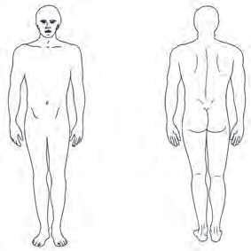
• Referred pain: Referred pain, or pain at a site other than the actual location of trauma, can mislead the patient and the clinician as to the actual location of the pathology. Resulting when the central nervous system (CNS) misinterprets the location and source of the painful stimulus, referred pain patterns can indicate internal injury, such as when damage to the spleen results in left shoulder pain (Fig. 1-1). Musculoskeletal injury can also cause
Insidious Of gradual onset; with respect to symptoms of an injury or disease having no apparent cause
Overuse syndrome Injury caused by accumulated microtraumatic stress placed on a structure or body area
Box 1-3
Pain Rating Scales
Visual Analog Scale (VAS)
Pain as bad as it could be No pain
Using a 10-cm line, the patient is asked to mark the point that represents the current intensity of pain. The VAS value is then calculated by measuring the distance in centimeters from the right edge of the line.
Numeric Rating Scale (NRS)
Pain as bad as it could be No pain 012345678910
The patient is asked to circle the number from 0 (no pain) to 10 (worst pain imaginable) that best describes the current level of pain. Only whole numbers are used with this scale. The VAS and NRS are common outcome measures that are used to quantify the amount of pain that a patient is experiencing over time. They are also useful for measuring pain before and after treatment.
McGill Pain Questionnaire
A. Where is your pain?
Using the drawing on the right, please mark the area(s) where you feel pain. Mark an “E” if the source of the pain is external or “I” if it is internal.. If the source of the pain is both internal, please mark “B”.
B. Pain rating index
Many different words can be used to describe pain. From the list below, please circle those words that best describe the pain you are currently experiencing. Use only one word from each category. You do not need to mark a word in every – Only mark those words that most accurately describe your pain.
1. Flickering Quivering Pulsing Throbbing Beating Pounding
6. Tugging Pulling Wrenching
11. Tiring Exhausting
16. Annoying Troublesome Miserable Intense Unbearable
2. Jumping Flashing Shooting
7. Hot Burning Scalding Searing
3. Pricking Boring Drilling Stabbing
8. Tingling Itchy Smarting Stinging
4. Sharp Cutting Lacerating
9. Dull Sore Hurting Aching Heavy
12. Sickening Suffocating 13. Fearful Fightful Terrifying 14. Punishing Grueling Cruel Vicious Killing
17. Spreading Radiating Penetrating Piercing
18. Tight Numb Drawing Squeezing Tearing
19. Cool Cold Freezing
5. Pinching Pressing Gnawing Cramping Crushing
10. Tender Taut Rasping
15. Wretched Blinding
20. Nagging Nauseating Agonizing Dreadful Torturing
Pain assessment instruments such as the McGill Pain Questionnaire are often used for patients who have complex pain problems. In Par t A, the patient identifies the area(s) of pain and whether the pain is deep or superficial. Part B provides descriptors that the patient uses to determine the intensity and nature of the pain. A visual analog or numeric rating scale is often included as a part of the outcome measure.
Figures from Starkey, C. Therapeutic Modalities (ed 4). Philadelphia: FA Davis, 2013, pp. 49-50.