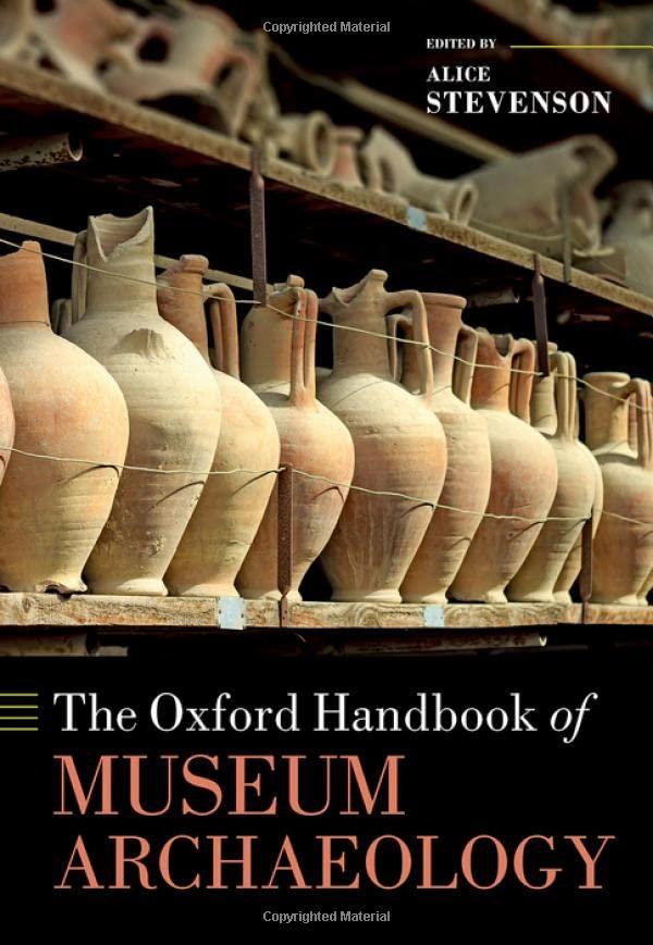
The Oxford Handbook of Museum Archaeology Alice Stevenson
https://ebookmass.com/product/the-oxford-handbook-of-museumarchaeology-alice-stevenson/
ebookmass.com
1600 John F. Kennedy Blvd.
Ste 1800 Philadelphia, PA 19103-2899
ESSENTIALS OF PHYSICAL MEDICINE AND REHABILITATION: MUSCULOSKELETAL DISORDERS, PAIN, AND REHABILITATION, FOURTH EDITION
Copyright © 2019 by Elsevier, Inc. All rights reserved. Mayo retains copyright © to Mayo Copyrighted Drawings. All rights reserved.
ISBN: 978-0-323-54947-9
No part of this publication may be reproduced or transmitted in any form or by any means, electronic or mechanical, including photocopying, recording, or any information storage and retrieval system, without permission in writing from the publisher. Details on how to seek permission, further information about the Publisher’s permissions policies and our arrangements with organizations such as the Copyright Clearance Center and the Copyright Licensing Agency, can be found at our website: www.elsevier.com/permissions
This book and the individual contributions contained in it are protected under copyright by the Publisher (other than as may be noted herein).
Notices
Knowledge and best practice in this field are constantly changing. As new research and experience broaden our understanding, changes in research methods, professional practices, or medical treatment may become necessary.
Practitioners and researchers must always rely on their own experience and knowledge in evaluating and using any information, methods, compounds, or experiments described herein. In using such information or methods they should be mindful of their own safety and the safety of others, including parties for whom they have a professional responsibility.
With respect to any drug or pharmaceutical products identified, readers are advised to check the most current information provided (i) on procedures featured or (ii) by the manufacturer of each product to be administered, to verify the recommended dose or formula, the method and duration of administration, and contraindications. It is the responsibility of practitioners, relying on their own experience and knowledge of their patients, to make diagnoses, to determine dosages and the best treatment for each individual patient, and to take all appropriate safety precautions.
To the fullest extent of the law, neither the Publisher nor the authors, contributors, or editors, assume any liability for any injury and/or damage to persons or property as a matter of products liability, negligence or otherwise, or from any use or operation of any methods, products, instructions, or ideas contained in the material herein.
Previous editions copyrighted 2015, 2008, and 2002.
Library of Congress Control Number: 2018944804
Senior Content Strategist: Kristine Jones
Senior Content Development Specialist: Joanie Milnes
Publishing Services Manager: Julie Eddy
Senior Project Manager: Cindy Thoms
Book Designer: Renee Duenow
T. MARK CAMPBELL, MD, MSC, FRCPC
Clinician Investigator, Physical Medicine and Rehabilitation, Elisabeth Bruyère, Ottawa, Ontario, Canada
Joint Contractures
ALEXIOS G. CARAYANNOPOULOS, DO, MPH
Assistant Professor of Neurosurgery, Brown University, Medical Director Comprehensive Spine Center, Division Director Pain and Rehabilitation Medicine, Department of Neurosurgery, Rhode Island Hospital and Newport Hospital, Providence, Rhode Island Thoracic Sprain or Strain
GREGORY T. CARTER, MD, MS
Chief Medical Officer, Physiatry, St. Luke’s Rehabilitation Institute; Clinical Professor, Biomedical Sciences, Elson S. Floyd College of Medicine, Washington State University, Spokane, Washington; Clinical Faculty, MEDEX, University of Washington School of Medicine, Seattle, Washington Motor Neuron Disease
ISABEL CHAN, MD
Assistant Clinical Professor, Physical Medicine and Rehabilitation, University of Texas Southwestern, Dallas, Texas Pelvic Pain
SOPHIA CHAN, DPT
Medical Student, MS-IV, University of New England College of Osteopathic Medicine, Biddeford, Maine
Coccydynia
Postherpetic Neuralgia
ERIC T. CHEN, MD, MS
Physician, Rehabilitation Medicine, University of Washington, Seattle, Washington Adhesive Capsulitis
AMANDA CHEUNG, BSC, MBT
Faculty of Medicine and Dentistry, University of Alberta, Edmonton, Alberta, Canada Pressure Ulcers
ANDREA CHEVILLE, MD, MSCE
Professor, Department of Physical Medicine and Rehabilitation, Mayo Clinic, Rochester, Minnesota Cancer-Related Fatigue
KELVIN CHEW, MBBCH, MSPMED
Senior Consultant, Sports Medicine Department, Changi General Hospital, Singapore Greater Trochanteric Pain Syndrome
SALLAYA CHINRATANALAB, MD
Assistant Professor of Medicine, Division of Rheumatology and Immunology, Department of Internal Medicine, Vanderbilt University Medical Center, Nashville, Tennessee
Rheumatoid Arthritis
Systemic Lupus Erythematosus
ELLIA CIAMMAICHELLA, DO, JD
Resident Physician, Physical Medicine and Rehabilitation, McGovern Medical School at UT Health in Houston, Houston, Texas
Neural Tube Defects
JOHN CIANCA, MD
Adjunct Associate Professor, Physical Medicine and Rehabilitation, Baylor College of Medicine; Adjunct Associate Professor, Physical Medicine and Rehabilitation, University of Texas Medical Branch, Houston, Texas
Hamstring Strain
DANIEL MICHAEL CLINCHOT, MD
Vice Dean for Education, Chair, Biomedical Education and Anatomy, The Ohio State University, Columbus, Ohio
Femoral Neuropathy
Lateral Femoral Cutaneous Neuropathy
RICARDO E. COLBERG, MD, RMSK
Sports Medicine Physician, Physical Medicine and Rehabilitation, Andrews Sports Medicine and Orthopaedic Center, Birmingham, Alabama
Hip Adductor Strain
EARL J. CRAIG, MD
Clinical Assistant Professor, Department of Physical Medicine and Rehabilitation; Clinical Assistant Professor, Department of Medicine, Indiana University School of Medicine, Indianapolis, Indiana
Femoral Neuropathy
Lateral Femoral Cutaneous Neuropathy
LISANNE C. CRUZ, MD, MSC
Rehabilitation Medicine, Icahn SOM at Mount Sinai, New York, New York
Compartment Syndrome of the Leg
SARA CUCCURULLO, MD
Clinical Professor and Chairman, Residency Program Director, Department of Physical Medicine and Rehabilitation, Rutgers Robert Wood Johnson Medical School, Piscataway, New Jersey; Vice President and Medical Director, JFK Johnson Rehabilitation Institute, Edison, New Jersey
Abdominal Wall Pain
CHRISTIAN M. CUSTODIO, MD
Associate Attending Physiatrist, Rehabilitation Medicine Service, Memorial Sloan Kettering Cancer Center; Associate Clinical Professor, Department of Rehabilitation Medicine, Weill Cornell Medicine, New York, New York
Chemotherapy-Induced Peripheral Neuropathy
ALAN M. DAVIS, MD, PhD
Associate Professor, Physical Medicine and Rehabilitation, University of Utah School of Medicine, Salt Lake City, Utah
Cardiac Rehabilitation
STEPHAN M. ESSER, MD
Southeast Orthopedic Specialists, Jacksonville, Florida
Chronic Ankle Instability
AVITAL FAST, MD
Chief, Rehabilitation Services, Tel Aviv Medical Center, Tel Aviv, Israel
Cervical Spondylotic Myelopathy
Cervical Degenerative Disease
JONATHAN T. FINNOFF, DO
Professor, Department of Physical Medicine and Rehabilitation, Mayo Clinic, Rochester, Minnesota
Suprascapular Neuropathy
Hip Labral Tears
DAVID R. FORBUSH, MD
Assistant Professor of Physical Medicine and Rehabilitation, University of Alabama at Birmingham School of Medicine, Birmingham, Alabama
Total Knee Arthroplasty
PATRICK M. FOYE, MD
Interim Chair and Professor, Physical Medicine and Rehabilitation; Director, Coccyx Pain Center, Rutgers New Jersey Medical School, Newark, New Jersey
Hip Osteoarthritis
MICHAEL FREDERICSON, MD
Professor, Orthopedics and Sports Medicine, Director, Physical Medicine and Rehabilitation, Sports Medicine Fellowship Director, Primary Care, Sports Medicine, Team Physician, Stanford Intercollegiate Athletics, Stanford University, Redwood City, California
Greater Trochanteric Pain Syndrome
Knee Chondral Injuries
JOEL E. FRONTERA, MD
Associate Professor, Vice Chair for Education and Residency Program Director, Department of Physical Medicine and Rehabilitation, McGovern Medical School at the University of Texas Health Science Center, Houston, Texas
Spasticity
WALTER R. FRONTERA, MD, PhD, MA (Hon.), FRCP
Professor, Physical Medicine, Rehabilitation and Sports Medicine, Physiology and Biophysics, University of Puerto Rico School of Medicine, San Juan, Puerto Rico
Cervical Facet Arthropathy
CHAN GAO, MD, PhD
Resident, Department of Physical Medicine and Rehabilitation, Vanderbilt University Medical Center, Nashville, Tennessee
Rotator Cuff Tendinopathy
Rotator Cuff Tear
YOUHANS GHEBRENDRIAS, MD
Assistant Clinical Professor, Physical Medicine and Rehabilitation, University of California Irvine, Orange, California
Myofascial Pain Syndrome
MEL B. GLENN, MD
Associate Professor, Department of Physical Medicine and Rehabilitation, Harvard Medical School, Boston, Massachusetts; Chief, Brain Injury Division, Department of Physical Medicine and Rehabilitation, Spaulding Rehabilitation Hospital, Charlestown, Massachusetts; Medical Director, NeuroRehabilitation (Massachusetts), Braintree, Massachusetts; Medical Director, Community Rehab Care, Watertown, Massachusetts
Postconcussion Symptoms
JENOJ S. GNANA, MD
Department of Physical Medicine and Rehabilitation, Rutgers New Jersey Medical School, Newark, New Jersey
Hip Osteoarthritis
PETER GONZALEZ, MD
Private Practice, Orthopaedic Institute of Central Jersey, Toms River, New Jersey
Iliotibial Band Syndrome
THOMAS E. GROOMES, MD
Associate Professor, Physical Medicine and Rehabilitation, Vanderbilt University Medical Center, Nashville, Tennessee
Total Knee Arthroplasty
Heterotopic Ossification
DAWN M. GROSSER, MD
Orthopaedic Surgeon, South Texas Bone and Joint, Corpus Christi, Texas
Ankle Arthritis
Bunion and Bunionette
Hallux Rigidus
Posterior Tibial Tendon Dysfunction
JONATHAN S. HALPERIN, MD
Chief, Physical Medicine and Rehabilitation, Sharp Rees
Stealy Medical Group, San Diego, California
Quadriceps Tendinopathy
ALEX HAN, BA
Medical Student, Physical Medicine and Rehabilitation, Brown University, Providence, Rhode Island
Thoracic Sprain or Strain
JOSEPH A. HANAK, MD
Clinical Instructor, Physical Medicine and Rehabilitation, Spaulding Rehabilitation Hospital, Charlestown, Massachusetts
Tietze Syndrome
TONI J. HANSON, MD
Assistant Professor, Physical Medicine and Rehabilitation, Mayo Clinic, Rochester, Minnesota
Thoracic Compression Fracture
DAVID E. HARTIGAN, MD
Assistant Professor, Orthopedic Surgery, Mayo Clinic Arizona, Phoenix, Arizona
Labral Tears of the Shoulder
LEI LIN, MD, PhD
Clinical Associate Professor, Physical Medicine and Rehabilitation, Rutgers-Robert Wood Johnson Medical School, Edison, New Jersey
Thoracic Outlet Syndrome
KARL-AUGUST LINDGREN, MD, PhD
ORTON Rehabilitation Centre, Helsinki, Finland Thoracic Outlet Syndrome
UMAR MAHMOOD, MD
Kure Pain Management, Stevensville, Maryland Lumbar Spondylolysis and Spondylolisthesis
JUSTIN L. MAKOVICKA, MD
Orthopedic Surgery Resident, Mayo Clinic Arizona, Phoenix, Arizona
Labral Tears of the Shoulder
STEVEN A. MAKOVITCH, DO
Clinical Instructor, Department of Physical Medicine and Rehabilitation, Harvard Medical School, VA Boston Healthcare, Spaulding Rehabilitation Hospital, Boston, Massachusetts Kienböck Disease
VARTGEZ K. MANSOURIAN, MD
Assistant Professor, Physical Medicine and Rehabilitation, Vanderbilt University School of Medicine; Medical Director, Stroke Rehabilitation Program, Vanderbilt Stallworth Rehabilitation Hospital, Nashville, Tennessee Stroke in Young Adults
BEN MARSHALL, DO
Assistant Professor, Physical Medicine and Rehabilitation, University of Colorado, Aurora, Colorado Collateral Ligament Sprain Meniscal Injuries
JENNIFER N. YACUB MARTIN, MD
Assistant Professor, Department of Physical Medicine and Rehabilitation, Clement J. Zablocki VA Medical Center and Medical College of Wisconsin, Milwaukee, Wisconsin
Upper Limb Amputations
Diabetic Foot and Peripheral Arterial Disease
KOICHIRO MATSUO, DDS, PhD
Professor and Chair, Department of Dentistry and Oral-Maxillofacial Surgery, Fujita Health University, School of Medicine, Toyoake, Aichi, Japan Dysphagia
JUAN JOSE MAYA, MD
Department of Internal Medicine, Division of Rheumatology, Mayo Clinic Florida, Jacksonville, Florida
Ankylosing Spondylitis
A. SIMONE MAYBIN, MD
Department of Physical Medicine and Rehabilitation, Vanderbilt University Medical Center, Nashville, Tennessee
Lumbar Facet Arthropathy
DONALD MCGEARY, PhD, ABPP
Associate Professor, Department of Psychiatry, Clinical Assistant Professor, Department of Family and Community Medicine, ReACH Scholar, University of Texas Health Science Center at San Antonio, San Antonio, Texas Headaches
KELLY C. MCINNIS, DO
Instructor, Physical Medicine and Rehabilitation, Harvard Medical School; Clinical Associate, Physical Medicine and Rehabilitation, Massachusetts General Hospital, Boston, Massachusetts
Repetitive Strain Injuries
PETER MELVIN MCINTOSH, MD
Assistant Professor, College of Medicine, Mayo Clinic, Rochester, Minnesota; Consultant, Department of Physical Medicine and Rehabilitation, Mayo Clinic Florida, Jacksonville, Florida
Scapular Winging
Adhesive Capsulitis of the Hip
ALEC L. MELEGER, MD
Assistant Professor of Physical Medicine and Rehabilitation, Harvard Medical School, Boston, Massachusetts; Associate Director, Spine Center, Newton-Wellesley Hospital, Newton, Massachusetts
Cervical Spinal Stenosis
WILLIAM F. MICHEO, MD
Professor and Chair, Sports Medicine Fellowship Director, Department of Physical Medicine, Rehabilitation, and Sports Medicine, University of Puerto Rico, San Juan, Puerto Rico
Glenohumeral Instability
Anterior Cruciate Ligament Sprain
PAOLO MIMBELLA, MD, MSC
McGovern Medical School—UTHealth, Department of Physical Medicine and Rehabilitation; Academic Chief Resident, Baylor/University of Texas, Houston, Texas
Hamstring Strain
GERARDO MIRANDA-COMAS, MD
Assistant Professor, Rehabilitation Medicine, Sports Medicine Fellowship Director, Icahn School of Medicine at Mount Sinai, New York, New York
Glenohumeral Instability
DANIEL P. MONTERO, MD, CAQSM
Instructor, Orthopedics, Mayo Clinic Florida, Jacksonville, Florida
Hammer Toe
FRANCISCO H. SANTIAGO, MD
Attending Physician, Physical Medicine and Rehabilitation, Bronx-Lebanon Hospital, Bronx, New York
Median Neuropathy
Ulnar Neuropathy (Wrist)
DANIELLE SARNO, MD
Instructor, Department of Physical Medicine and Rehabilitation, Harvard Medical School, Boston, Massachusetts; Physiatrist, Interventional Pain Management, Department of Neurosurgery, Brigham and Women’s Hospital, Boston, Massachusetts Lumbar Spinal Stenosis
ROBERT J. SCARDINA, DPM
Chief and Residency Program Director, Podiatry Service, Massachusetts General Hospital, Boston, Massachusetts
Metatarsalgia
BYRON J. SCHNEIDER, MD
Assistant Professor, Physical Medicine and Rehabilitation, Vanderbilt University Medical Center, Nashville, Tennessee
Lumbar Facet Arthropathy
JEFFREY C. SCHNEIDER, MD
Medical Director, Trauma, Burn and Orthopedic Program, Spaulding Rehabilitation Hospital; Assistant Professor, Physical Medicine and Rehabilitation, Harvard Medical School, Boston, Massachusetts
Burns
FERNANDO SEPÚLVEDA, MD
Assistant Professor, Department of Physical Medicine, Rehabilitation, and Sports Medicine, University of Puerto Rico School of Medicine, San Juan, Puerto Rico
Anterior Cruciate Ligament Sprain
JOHN SERGENT, MD
Professor of Medicine, Division of Rheumatology and Immunology, Department of Medicine, Vanderbilt University Medical Center, Nashville, Tennessee
Rheumatoid Arthritis Systemic Lupus Erythematosus
DANA SESLIJA, MD, MS
Adjunct Professor, Department of Physical Medicine and Rehabilitation, Schulich School of Medicine and Dentistry, Windsor, Ontario, Canada
Fibular (Peroneal) Neuropathy
Tibial Neuropathy (Tarsal Tunnel Syndrome)
VIVIAN P. SHAH, MD
Department of Physical Medicine and Rehabilitation, Rutgers New Jersey Medical School, Newark, New Jersey
Hip Osteoarthritis
JYOTI SHARMA, MD
Associate, Orthopaedic Surgery Department, Geisinger Health System, Danville, Pennsylvania
Wrist Osteoarthritis
Wrist Rheumatoid Arthritis
NUTAN SHARMA, MD, PhD
Associate Professor, Neurology, Harvard Medical School, Cambridge, Massachusetts; Associate Neurologist, Neurology, Massachusetts General Hospital, Boston, Massachusetts; Associate Neurologist, Neurology, Brigham and Women’s Hospital, Boston, Massachusetts Parkinson Disease
ALEXANDER SHENG, MD
Assistant Professor, Sports and Spine, Shirley Ryan AbilityLab, Chicago, Illinois
Posterior Cruciate Ligament Sprain
GLENN G. SHI, MD
Assistant Professor, Orthopedic Surgery, Mayo Clinic, Jacksonville, Florida
Hammer Toe
Morton’s Neuroma Plantar Fasciitis
JULIE K. SILVER, MD
Associate Professor, Department of Physical Medicine and Rehabilitation, Harvard Medical School; Attending Physician, Spaulding Rehabilitation Hospital; Clinical Associate, Massachusetts General Hospital; Associate in Physiatry, Brigham and Women’s Hospital, Boston, Massachusetts Trigger Finger
CHLOE SLOCUM, MD, MPH
Attending Physician, Department of Physical Medicine and Rehabilitation, Spinal Cord Injury Division, Harvard Medical School/Spaulding Rehabilitation Hospital, Boston, Massachusetts
Post-Thoracotomy Pain Syndrome
DAVID M. SLOVIK, MD
Associate Professor of Medicine, Harvard Medical School; Chief, Division of Endocrinology, Newton-Wellesley Hospital, Newton, Massachusetts; Physician, Endocrine Unit, Massachusetts General Hospital, Boston, Massachusetts
Osteoporosis
SOL M. ABREU SOSA, MD
Assistant Professor, Physical Medicine and Rehabilitation, Rush Medical College, Chicago, Illinois
Ulnar Collateral Ligament Sprain Stress Fractures
KURT SPINDLER, MD
Department of Orthopedic Surgery, Cleveland Clinic, Cleveland, Ohio
Knee Chondral Injuries
LAUREN SPLITTGERBER, MD
Resident Physician, Physical Medicine and Rehabilitation, McGaw Medical Center of Northwestern University/ Shirley Ryan AbilityLab, Chicago, Illinois
Posterior Cruciate Ligament Sprain
ARIANA VORA, MD
Instructor, Physical Medicine and Rehabilitation, Harvard Medical School; Staff Physiatrist, Physical Medicine and Rehabilitation, Massachusetts General Hospital; Staff Physiatrist, Physical Medicine and Rehabilitation, Spaulding Rehabilitation Hospital, Boston, Massachusetts
Coccydynia
Postherpetic Neuralgia
MICHAEL C. WAINBERG, MD, MSC
Senior Associate Consultant, Physical Medicine and Rehabilitation, Mayo Clinic, Rochester, Minnesota
Trigger Finger
ROGER WANG, DO
Schwab Rehabilitation Hospital, University of Chicago, Chicago, Illinois
Piriformis Syndrome
JAY M. WEISS, MD
Medical Director, Long Island Physical Medicine and Rehabilitation, Syosset, New York
Lateral Epicondylitis
Medial Epicondylitis
Ulnar Neuropathy (Elbow)
LYN D. WEISS, MD
Chairman and Program Director, Physical Medicine and Rehabilitation, Nassau University Medical Center, East Meadow, New York
Lateral Epicondylitis
Medial Epicondylitis
Radial Neuropathy
Ulnar Neuropathy (Elbow)
SARAH A. WELCH, DO, MA
Resident Physician, Physical Medicine and Rehabilitation, Vanderbilt University Medical Center, Nashville, Tennessee
Cervical Facet Arthropathy
DAVID WEXLER, MD, FRCS(TR&ORTH)
Attending, Orthopedics, Maine General Medical Center, Augusta, Maine
Ankle Arthritis
Bunion and Bunionette
Hallux Rigidus
Posterior Tibial Tendon Dysfunction
J. MICHAEL WIETING, DO, MEd
Associate Dean of Clinical Medicine and Professor of Physical Medicine and Rehabilitation, Lincoln Memorial University-DeBusk College of Osteopathic Medicine, Harrogate, Michigan; Clinical Professor, Department of Physical Medicine and Rehabilitation, Michigan State University-College of Osteopathic Medicine, East Lansing, Michigan
Quadriceps Contusion
ALLEN NEIL WILKINS, MD
Assistant Clinical Professor, Department of Rehabilitation and Regenerative Medicine, Columbia University College of Physicians and Surgeons; Medical Director, New York Rehabilitation Medicine, New York, New York
Foot and Ankle Bursitis
AARON JAY YANG, MD
Assistant Professor, Physical Medicine and Rehabilitation, Vanderbilt University Medical Center, Nashville, Tennessee
Cervical Facet Arthropathy
FABIO ZAINA, MD
Italian Scientific Spine Institute, Milan, Italy
Scoliosis and Kyphosis
MEIJUAN ZHAO, MD
Assistant Professor, Physical Medicine and Rehabilitation, Harvard Medical School; Staff Physiatrist, Physical Medicine and Rehabilitation, Massachusetts General Hospital, Spaulding Rehabilitation Hospital, Boston, Massachusetts
Median Neuropathy (Carpal Tunnel Syndrome)
We dedicate this book to our mentors, teachers, colleagues, and students, who have encouraged us to pursue academic careers with their enthusiasm for knowledge and learning; to our patients, who often are our greatest teachers; and to our families, who support us and provide the foundation for our pursuits.
Walter R. Frontera, MD, PhD, MA (Hon.), FRCP
Julie K. Silver, MD
Thomas D. Rizzo, Jr., MD
Synonyms
Cervical radiculitis
Degeneration of cervical intervertebral disc
Cervical spondylosis without myelopathy
Cervical pain
ICD-10 Codes
M47.812 Cervical spondylosis without myelopathy or radiculopathy
M48.02 Spinal stenosis in cervical region
M48.03 Spinal stenosis in cervicothoracic region
M50.30 Degeneration of cervical disc
M50.32 Degeneration of mid-cervical region
M50.33 Degeneration of cervicothoracic region
M54.2 Cervical pain
M54.12 Cervical radiculitis
M54.13 Cervicothoracic radiculitis
Definition
Cervical spondylotic myelopathy (CSM) is a frequently encountered entity in middle-aged and elderly patients. The condition affects both men and women. Progressive degeneration of the cervical spine involves the discs, facet joints, joints of Luschka, ligamenta flava, and laminae, leading to gradual encroachment on the spinal canal and spinal cord compromise. CSM has a fairly typical clinical presentation and frequently a progressive and disabling course. As a consequence of aging, the spinal column goes through a cascade of degenerative changes that tend to
Cervical Spondylotic Myelopathy
Avital Fast, MD
Israel Dudkiewicz, MD
affect selective regions of the spine. The cervical spine is affected in most adults, most frequently at the C4-C7 region.1,2 Degeneration of the intervertebral discs triggers a cascade of biochemical and biomechanical changes, leading to decreased disc height, among other changes. As a result, abnormal load distribution in the motion segments causes cervical spondylosis (i.e., facet arthropathy) and neural foraminal narrowing. Disc degeneration also leads to the development of herniations (soft discs), disc calcification, posteriorly directed bone ridges (hard discs), hypertrophy of the facets and the uncinate joints, and ligamenta flava thickening. On occasion, more frequently in Asians but not infrequently in white individuals, the posterior longitudinal ligament and the ligamenta flava ossify.2,3 These degenerative changes narrow the dimensions and change the shape of the cervical spinal canal. In normal adults the anteroposterior diameter of the subaxial cervical spinal canal measures 17 to 18 mm, whereas the spinal cord diameter in the same dimension is approximately 10 mm. Severe CSM gradually decreases the space available for the cord and brings about cord compression in the anterior-posterior axis. Cord compression usually occurs at the discal levels.4-6
The encroaching structures may also compress the anterior spinal artery, resulting in spinal cord ischemia that usually involves several cord segments beyond the actual compression site. Spinal cord changes in the form of demyelination, gliosis, myelomalacia, and eventually severe atrophy may develop.2,4,7-9 Dynamic instability, which can be diagnosed in flexion or extension lateral x-ray views, further complicates matters. Disc degeneration leads to laxity of the supporting ligaments, bringing about anterolisthesis or retrolisthesis in flexion and extension, respectively. This may further compromise the spinal cord and intensify the presenting symptoms.2,4
Symptoms
CSM develops gradually during a lengthy period of months to years. Not infrequently, the patient is unaware of any
functional compromise, and the first person to notice that something is amiss may be a close family member. Although pain appears rather early in cervical radiculopathy and alerts the patient to the presence of a problem, this is usually not the case in CSM. A long history of neck discomfort and intermittent pain may frequently be obtained, but these are not prominent at the time of CSM presentation.
Most patients have a combination of upper motor neuron symptoms in the lower extremities and lower motor neuron symptoms in the upper extremities.4 Patients frequently present with gait dysfunction resulting from a combination of factors, including ataxia due to impaired joint proprioception, hypertonicity, weakness, muscle control deficiencies, and unexplained falls.
Studies have demonstrated that severely myelopathic patients display abnormalities of deep sensation, including vibration and joint position sense, which is attributed to compression of the posterior columns.10,11 Paresthesias and numbness may be frequently mentioned. Compression of the pyramidal and extrapyramidal tracts can lead to spasticity, weakness, and abnormal muscle contractions. These sensory and motor deficits result in an unstable gait. Patients may complain of stiffness in the lower extremities or plain weakness manifesting as foot dragging and tripping.5 Symptoms related to the upper extremities are mostly the result of fine motor coordination deficits. At times, the symptoms in the upper extremities are much more severe than those related to the lower extremities, attesting to central cord compromise.4 Most patients do not have urinary symptoms. However, urinary symptoms (i.e., incontinence) may occasionally develop in patients with long-standing myelopathy.12 As CSM develops in middle-aged and elderly patients, the urinary symptoms may be attributed to aging, comorbidities, and cord compression. Bowel incontinence is rare.
Physical Examination
Because of sensory ataxia, the patient may be observed walking with a wide-based gait. Some resort to a cane to increase the base of support and to enhance safety during ambulation. Patients with severe gait dysfunction frequently require a walker and cannot ambulate without one. Many patients lose the ability to tandem walk. The Romberg test result may become positive. Examination of the lower extremities may reveal muscle atrophy, increased muscle tone, abnormal reflexes—clonus or upgoing toes (Babinski sign)—and abnormalities of position and vibration sense. Muscle fasciculations may be observed. The foot tapping test (number of sole tappings while the heel maintains contact with the floor in 10 seconds) is an easy and useful quantitative tool for lower extremity function in these patients.13
In the upper extremities, weakness and atrophy of the small muscles of the hands may be noted. The patient may have difficulties in fine motor coordination (e.g., unbuttoning the shirt or picking a coin off the table). The patient frequently displays difficulty in performing repetitive opening and closing of the fist. In normal individuals, 20 to 30 repetitions can be performed in 10 seconds.
Weakness can occasionally be documented in more proximal muscles and may appear symmetrically. Fasciculations
may appear in the wasted muscles. Hypesthesia, paresthesia, or anesthesia may be documented. On occasion, the sensory findings in the hands are in a glove distribution. As in the lower extremities, the vibration and joint position senses may be disturbed. Hyporeflexia or hyperreflexia may be found. The Hoffmann response may become positive and can be facilitated in early myelopathy by cervical extension.14 In some patients, severe atrophy of all the hand intrinsic muscles is observed.1,5,15-17
The neck range of motion may be limited in all directions. Many patients cannot extend the neck beyond neutral and may feel electric-like sensation radiating down the torso on neck flexion, known as the Lhermitte sign. Often, when a patient stands against the wall, the back of the head stays an inch to several inches away, and the patient is unable to push the head backward to bring it to touch the wall.
Functional Limitations
Patients with CSM have difficulties with activities of daily living. Patients may have difficulties inserting keys, picking up coins, buttoning a shirt, or manipulating small objects. Handwriting may deteriorate. Patients may drop things from the hands and occasionally can complain of numbness affecting the fingers or the palms, mimicking peripheral neuropathy.2,5,16,18,19 They may have problems dressing and undressing. When weakness is a predominant feature, they will be unable to carry heavy objects. Unassisted ambulation may become difficult. The gait is slowed and becomes inefficient. In late stages of CSM, patients may become almost totally disabled and require assistance with most activities of daily living.
Diagnostic Studies
Plain radiographs usually reveal multilevel degenerative disc disease with cervical spondylosis. Dynamic studies (flexion and extension views) may reveal segmental instability with anterolisthesis on flexion and retrolisthesis on extension. In patients with ossification of the posterior longitudinal ligament, the ossified ligament may be detected on lateral plain films. The Torg-Pavlov ratio may help diagnose congenital spinal stenosis. This ratio can be obtained on plain films by dividing the anteroposterior diameter of the vertebral body by the anteroposterior diameter of the spinal canal at that level. The canal diameter can be measured from the posterior wall of the vertebra to the spinolaminar line.20 A ratio of 0.8 or less is indicative of spinal stenosis (Fig. 1.1).21
Magnetic resonance imaging, the study of choice, provides critical information about the extent of stenosis and the condition of the compressed spinal cord. Sagittal and axial cuts clearly show the offending structures (discs, spurs, thickened ligamenta flava) and the cord shape and help to quantify the amount of cord compression. Cord signal changes provide critical information about the extent of cord damage and the prognosis (Fig. 1.2). Increased cord signal on T2-weighted images is abnormal and points to the presence of edema, demyelination, myelomalacia, or gliosis. Decreased cord signal on T1-weighted images may also be observed. Occasionally the increased signal appears as two white dots in T2-weighted images. This is referred to as snake eye appearance (Fig. 1.3). However, these cord signal changes are of
References
1. Heller J. The syndromes of degenerative cervical disease. Orthop Clin North Am. 1992;23:381–394.
2. Nouri A, Tetreault L, Singh A, et al. Degenerative cervical myelopathy. Spine. 2015;40:E675–E693.
3. Machino M, Yukawa Y, Imagama S, et al. Age related and degenerative changes in the osseous anatomy, alignment, and range of motion of the cervical spine. Spine. 2016;41:476–482.
4. Rao R. Neck pain, cervical radiculopathy, and cervical myelopathy. Pathophysiology, natural history, and clinical evaluation. J Bone Joint Surg Am. 2002;84:1872–1881.
5. Law MD, Bernhardt M, White AA III. Evaluation and management of cervical spondylotic myelopathy. Instr Course Lect. 1995;44:99–110.
6. Truumees E, Herkowitz HN. Cervical spondylotic myelopathy and radiculopathy. Instr Course Lect. 2000;49:339–360.
7. Beattie MS, Manley BT. Tight squeeze, slow burn: inflammation and the aetiology of cervical myelopathy. Brain. 2011;134:1259–1263.
8. Breig A, Turnbull I, Hassler O. Effects of mechanical stresses on the spinal cord in cervical spondylosis. J Neurosurg. 1966;25:45–56.
9. Doppman JL. The mechanism of ischemia in anteroposterior compression of the spinal cord. Invest Radiol. 1975;10:543–551.
10. Takayama H, Muratsu H, Doita M, et al. Impaired joint proprioception in patients with cervical myelopathy. Spine (Phila Pa 1976) 2004;30:83–86.
11. Okuda T, Ochi M, Tanaka N, et al. Knee joint position sense in compressive myelopathy. Spine (Phila Pa 1976). 2006;31:459–462.
12. Misawa T, Kamimura M, Kinoshita T, et al. Neurogenic bladder in patients with cervical compressive myelopathy. J Spinal Disord Tech 2005;18:315–320.
13. Numasawa T, Ono A, Wada K, et al. Simple foot tapping test as a quantitative objective assessment of cervical myelopathy. Spine (Phila Pa 1976) 2012;37:108–113.
14. Rhee JM, Heflin JA, Hamasaki T, et al. Prevalence of physical signs in cervical myelopathy: a prospective, controlled study. Spine. 2009;34:890–895.
15. Grijalva RA, Hsu FPK, Wycliffe HD, et al. Hoffmann sign: clinical correlation of neurological imaging findings in the cervical spine and brain. Spine. 2015;40:475–479.
16. Nemani VM, Kim HJ, Piaskulkaew, et al. Correlation of cord signal change with physical examination findings in patients with cervical myelopathy. Spine. 2014;40:6–10.
17. Edwards CC, Riew D, Anderson PA, et al. Cervical myelopathy: current diagnostic and treatment strategies. Spine J. 2003;3:68–81.
18. Ono K, Ebara S, Fiji T, et al. Myelopathy hand. New clinical signs of cervical cord damage. J Bone Joint Surg Br. 1987;69:215–219.
19. Ebara S, Yonenobu K, Fujiwara K, et al. Myelopathy hand characterized by muscle wasting. A different type of myelopathy hand in patients with cervical spondylosis. Spine (Phila Pa 1976). 1988;13:785–791.
20. Yu M, Tang Y, Liu Z, et al. The morphological and clinical significance of developmental cervical stenosis. Eur Spine J. 2015;24:1583–1589.
21. Taha AMS, Shue J, Lebl D, et al. Considerations for prophylactic surgery in asymptomatic severe cervical stenosis. HSSJ. 2015;11:31–35.
22. Banaszek A, Bladowska J, Szewczyk P, et al. Usefulness of diffusion tensor MR imaging in the assessment of intramedullary changes of the cervical spinal cord in different stages of degenerative spine disease. Eur Spine J. 2014;23:1523–1530.
23. Rajasekaran S, Kanna RM, Chittode VS, et al. Efficacy of diffusion tensor imaging indices in assessing postoperative neural recovery in cervical spondylotic myelopathy. Spine. 2017;42:8–13.
24. Olney RK, Lewis RA, Putnam TD, Campellone JV Jr. Consensus criteria for the diagnosis of multifocal motor neuropathy. Muscle Nerve 2003;27:117–121.
25. Lauryssen C, Riew KD, Wang JC. Severe cervical stenosis: operative treatment or continued conservative care. SpineLine. 2006:1–25.
26. Simpson AK, Rhee A. Laminoplasty: a review of the evidence and detailed technical guide. Semin Spine Surg. 2014;26:141–147.
27. Chen GD, Lu Q, Sun JJ. Effect and prognostic factors of laminoplasty for cervical myelopathy with an occupying ratio greater than 50%. Spine. 2016;41:378–383.
28. Fehlings M, Smith JS, Kopjar B, et al. Perioperative and delayed complications associated with surgical treatment of cervical spondylotic myelopathy based patients from the AOSpine North America cervical spondylotic myelopathy study. J Neurosurg Spine. 2012;16:425–432.
29. Zhu Y, Zhang B, Liu H, et al. Cervical disc arthroplasty versus anterior cervical discectomy and fusion for incidence of symptomatic adjacent segment disease. Spine. 2016;41:1493–1502.
30. Park MS, Ju YS, Moon SH, et al. Reoperation rates after anterior cervical discectomy and fusion for cervical spondylotic radiculopathy and myelopathy. Spine. 2016;41L:1593–1599.
31. Guzman JZ, Baird EO, Fields AC, et al. C5 nerve root palsy following decompression of the cervical spine. Bone Joint J. 2014;96-B:950–955.


