University of Alabama at Birmingham, Birmingham, AL, United States
Rangasamy Ramanathan, MD, FAAP
Professor of Pediatrics at the Keck School of Medicine and Division Chief, Director of Neonatal Respiratory therapy program, Program director, Neonatal Perinatal Fellowship Program, University of Southern California, Los Angeles, CA, United States
P. Syamasundar Rao, MD, FAAP, FACC, FSCAI
Professor of Pediatrics, Division of Pediatric Cardiology, UT Health McGovern Medical School, Houston, TX, United States
Maura Helena Ferrari Resende, MD
Clinical Fellow, The Hospital for Sick Children, Toronto, ON, Canada
Rakesh Sahni, MD
Professor of Pediatrics, Columbia University College of Physicians and Surgeons, New York-Presbyterian
Morgan Stanley Children’s Hospital, Columbia University Medical Center, New York, NY, United States
Mitali Sahni, MD
Fellow in Neonatal-Perinatal Medicine, St. Christopher’s Hospital for Children, Drexel University College of Medicine, Philadelphia, PA, United States
Marwa al Sayyed, MBBS, MSc
Neonatal Registrar, NMC speciality Hospital, Dubai, United Arab Emirates
Bernard Schoonakker, MRCPCH
Nottingham University Hospitals NHS Trust, Nottingham, United Kingdom
Craig Smith, MRCPCH
Nottingham University Hospitals NHS Trust, Nottingham, United Kingdom
Augusto Sola, MD
Director Medico Ejecutivo, SIBEN, Por los recién nacidos
Howard Stein, MD, FAAP
Promedica Toledo Children’s Hospital, University of Toledo Health Science Campus, Toledo, OH, United States
RoseMary S. Stocks, MD, PharmD
Department of Otolaryngology, Head and Neck Surgery, University of Tennessee Health Sciences Center, Memphis, TN, United States
Section Head and Service Chief, Neonatology, Professor of Pediatrics, Texas Children’s Hospital, Baylor College of Medicine, Houston, TX, United States
Ru-Jeng Teng, MD
Children’s Research Institute, Medical College of Wisconsin, Children’s Hospital of Wisconsin, Milwaukee, WI, United States
Vikrum A. Thimmappa, MD Department of Otolaryngology, Head and Neck Surgery, University of Tennessee Health Sciences Center, Memphis, TN, United States
Jerome W. Thompson, MD, MBA Department of Otolaryngology, Head and Neck Surgery, University of Tennessee Health Sciences Center, Memphis, TN, United States
David A. Todd, FIMLS, MSc, PhD, MBBS
Centenary Hospital, Canberra, ACT, Australia
Kirtikumar Upadhyay, MD, FAAP
University of Tennessee Health Science Center, Memphis, TN, United States
Karunakar Vadlamudi, MD
Stollery Children’s Hospital, University of Alberta, Edmonton, AB, Canada
Payam Vali, MD
University of California, Davis; UC Davis Children’s Hospital, Sacramento, CA, United States
Máximo Vento, MD, PhD
University and Polytechnic Hospital La Fe Valencia, València, Spain
Sudeep Verma, MD, FNB
KIMS-Institute of cardiac Sciences, Secunderabad-Hyderabad, Telangana, India
Dharmapuri Vidyasagar, MD, FAAP, FCCM, PhD(Hon)
Professor Emeritus Pediatrics, Division of Neonatology, University of Illinois at Chicago, Chicago, IL, United States
Koert de Waal, PhD, FRACP
John Hunter Children’s Hospital, Newcastle, NSW, Australia
Mark F. Weems, MD, FAAP
University of Tennessee Health Science Center, Memphis, TN, United States
Jen-Tien Wung, MD
Professor of Pediatrics, Columbia
University College of Physicians and Surgeons, Columbia University Medical Center, New York, NY, United States
Hakam Yaseen, MD, CES (Paed), DUN (Neonat) [France], CCST (UK), FRCPCH [UK]
University of Sharjah, Medical Director (CMO), University Hospital Sharjah (UHS), Sharjah, United Arab Emirates
Page left intentionally blank
Since the late 1960s, there has been considerable debate about the best way to provide respiratory support for preterm infants with RDS. Early attempts to ventilate infants met with limited success and survivors often suffered from chronic lung disease. In the early 1970s, Gregory et al. reported success in using CPAP to care for preterm infants with RDS; however, despite its simplicity, there was little interest in using that technology. As ventilators increased in sophistication (and complexity), noninvasive ventilation was viewed as a modality that could supplement invasive ventilation, but not as a primary mode. Furthermore, the randomized clinical trials of surfactant suggested that most premature infants with RDS should be intubated and administered surfactant. The pendulum began to swing back toward noninvasive ventilation in the last decade as randomized clinical trials demonstrated that early application of CPAP was better than routinely intubating infants and given surfactant. In 2018, the choices for respiratory support are even greater. Not only are there newer generation of ventilators, but the choices for noninvasive support commonly include nasal
Foreword
intermittent positive pressure ventilation and high-flow nasal cannula. This textbook, Essentials of Neonatal Ventilation, edited by Rajiv, Satyan, and Vidysagar, offers clinicians a complete source for the latest developments in respiratory care of critically ill newborn infants. This book is a unique addition because of its comprehensive nature and practical approach to respiratory care. The authors for each chapter are leaders in their fields. It is noteworthy that the book also addresses complications of mechanical ventilation (e.g., bronchopulmonary dysplasia) and includes sections on common neonatal problems, ECMO and nursing care. The editors should be congratulated on assembling such a wonderful book.
Richard A. Polin, MD William T. Speck, Professor of Pediatrics, College of Physicians and Surgeons, Columbia University, New York, NY, United States Director, Division of Neonatology, Morgan Stanley Children’s Hospital of New York-Presbyterian, New York, NY, United States
Page left intentionally blank
The evolution of assisted ventilation in newborn intensive care has made a unique paradigm shift. Noninvasive ventilation, a significant milestone in the 1970s, has made a comeback in the current decade. Newer methods of synchronization, gentle ventilation, and permissive hypercapnia using both invasive and noninvasive modes are the standard of care in neonatal intensive care today. This book is a Herculean attempt to standardize and optimize ventilatory care at the bedside. Each chapter is written by international experts in the field, hoping to ignite a path to the successful resolution of the pulmonary dysfunction, without lung and brain morbidity. Technologies of promise of the future are incorporated, and noninvasive monitoring and assessment are given significant emphasis. The neonatal intensivist is currently exposed to a huge arena of everevolving technologies. The bedside practitioner will find this book helpful in knowing the benefits and limitations of these technologies and support neonatal gas exchange without compromising neurodevelopmental outcome.
More advanced technology is not always better. Simple techniques such as nasal CPAP with noninvasive
Preface
monitoring have great outcomes in preterm and term infants with lung injury. This book gives great emphasis to this basic technology.
The chapters are designed to evolve from the basics to applied physiology and graduate through the assisted ventilation technologies. A section on cardiac issues in respiratory care, nutritional support, and ancillary care is deliberately magnified for the intensivist to manage accurately and objectively a critical neonate with respiratory distress.
This book with E-Book facilities of videos on critical chapters supplemented by lecture presentations would prove to be a handy and reliable bedside companion for all NICUs all over the world. The presentations and illustrations are provided to assist in education of a new generation of neonatal providers. We gratefully acknowledge the authors for contributing to these chapters, and providing videos and illustrations to enhance the book.
Rajiv gratefully acknowledges the didactic teaching of his fellow teacher Dr. Elizabeth John, whose extreme sensitivities to the adjustment of CPAP up or down still ring a bell in his ears. This singular caution to optimize continuous positive airway pressure or positive end-expiratory pressure laid the foundation of his strategy in any critical lung disease. This was the fulcrum of his success in neonatal ventilation in the last 30 years.
Rajiv acknowledges the heartfelt help of his teachers Dr. Vidyasagar, Dr. Georg Simbrunner, Dr. Ramanathan, Dr. Martin Keszler, and Dr. Dhanireddy for teaching him and for being authors of many chapters and reviewing many more of them. Dr. Vidyasagar was the first to agree to the concept of this book many years ago and has been the guiding light in the evolution of this book. Rajiv’s close associates Dr. Prakash, Dr. Arun, and Dr. David Todd gave him exceptional chapters at a short notice. His junior associates Dr. Nalinikant and Dr. Srinivas provided very unique, well-researched chapters. He is indebted to coeditor Satyan who joined the team in 2016 for his immortal illustrations and editorial stewardship. His illustrations offer an additional tool for neonatal providers to educate students and parents.
Rajiv also thanks his team members Iftekar, Jason, and Sherly for uncompromising secretarial and artwork. He further thanks Dr. Karunakar, his associate, for responding to the perennial demands of perfection of the chapters, without any hesitation.
This book is a unique joint effort of a highly talented provider-publisher team. Last but not the least, Rajiv thanks his wife Bindoo for silently bearing with him all the timeless lapses at home while he was playing Archimedes for the development of this book.
Dr. Vidyasagar gratefully acknowledges his mentors, Dr. Thomas Boggs, Dr. Jack Downes, and Dr. Victor Chernick who introduced him to neonatal ventilation. He thanks his wife Dr. Nagamani Beligere for her support all through his career. His children Sahana, Sadhana, and Sanjay and grandchildren Kavi, Anika, and Maaya have been the source of his energy.
Satyan thanks his children (Ananya, Aniruddha, and Arun for posing as models during their neonatal period for his illustrations) and his wife Veena Manja, MD, MSc, for her unrelenting support. He expresses gratitude to his parents, sisters, parents-in-law, teachers, and mentors for supporting and guiding him throughout his career.
All the editors sincerely appreciate the exceptional support by Mr. Ayan Dhar and Ms. Sheenam Aggarwal at Elsevier India. Above all, the editors are thankful to all the babies and their parents who contributed to our understanding of neonatal physiology and the functioning of assisted ventilation.
9B. The Importance of Heating and Humidifying the Inspired Gases During Mechanical Ventilation: Identifying the Ideal Settings and Circuit Configuration During Ventilation ...................................................113
David A. Todd, K.Y. Ashok Murthy, P.K. Rajiv 10. Ventilator Graphics.....................................124
Manoj Biniwale, Rangasamy Ramanathan, Mark C. Mammel
11A. Initiation of Mechanical Ventilation 143 Dushyant Batra, Craig Smith, Bernard Schoonakker
11B. Deterioration on the Ventilator ................149
Vikrum A. Thimmappa, Ramasubbareddy Dhanireddy, RoseMary S. Stocks, Jerome W. Thompson
Neonatal Airway Pathology
35. Ventilator-Associated Pneumonia and Infection Control.................................765
Manoj N. Malviya, Prakash Manikoth, Hakam Yaseen
36. Nutrition in the Preterm Neonate Requiring Respiratory Support .................785
Mahmoud Saleh Elhalik, Josef Neu, Abrar Ahmed Khan, Swarup Kumar Dash
37A. Neonatal Procedures Involving Catheters and Tubes ...................................803
Khaled El-Atawi, Swarup Kumar Dash, Ahmed Zakaria Elmorsy
37B. Neonatal Limb Ischemia Due to Arterial Catheters ......................................................819
Catherine C. Beaullieu, Suzanne M. Lopez, P. Syamasundar Rao
Section VIII: General Issues
38. Neonatal Developmental Follow-Up Program .......................................................829
Nagamani Beligere
39. Management of Ethical Challenges in Neonatal Intensive Care ........................836
Gautham Suresh
40. Normal Reference Values ..........................843
K. Shreedhara Avabratha, P.K. Rajiv, Mohamed Soliman M, Marwa al Sayyed, Rafique Memon, Karunakar Vadlamudi
Online supplementary materials
Please visit MedEnact (https://www.medenact.com/Home) to access the videos and lecture PPTs.
Section | I |
Introduction and History of Ventilation
1 Introduction 3
2 Evolution of Neonatal Ventilation a Retrospective View 5
Page left intentionally blank
Introduction
Chapter | 1 |
This book was conceived several years ago, when there appeared to be a distinct lacuna of comprehensive bedside ventilatory management guides in neonatal care history. Currently, there are excellent textbooks to refer to and obtain broad concepts on the approach to providing respiratory assistance to a baby in distress, but a detailed evidence-based book on bedside management is missing. In this book we attempted to provide the readers an evidence-based practice bedside guidelines. In doing so, we sought the contributions from the most experienced leaders in the field. This book is an honest attempt to get the world’s best pioneers in each area to contribute their signature chapters of their research to give the neonatal intensivist, detailed bedside ventilation navigation in critical situations. We earnestly hope readers will find these guidelines useful in managing critically ill neonates.
This book is divided into eight sections. Here are some of the highlights of these sections. Section I reviews the history of neonatal ventilation. Section II deals with basic chapters covering embryology and physiology of pulmonary disease, with the time frame from extreme prematurity at the limits of viability to dysmorphology in the full-term infant. The delivery process and golden first hour are addressed in detail, due to its long-term impact on respiratory and neurological morbidity.
Section III deals with the basics of neonatal ventilation and evolves through the genesis of lung injury to lung mechanics. The chapters on ventilator give deep insight to the reader on the limitations and benefits of its application. The chapters progress to the provision of mechanical ventilation and its attendant complications, which are again discussed in detail.
Section IV is an in-depth analysis in real time of the various respiratory care devices currently available for the neonate. These chapters give an operating framework and the bedside navigation in critically ill babies with trouble shooting algorithms by authentic authors.
Section V is the heart of this book with comprehensive bedside management guidelines of the common respiratory conditions faced in neonatal intensive care. They offer detailed flowcharts, algorithms, and case scenarios in complicated respiratory care management. There is a separate chapter on the management of the 23–25 weeks’ gestation babies: “micropremies”—a challenge for any intensivist.
Section VI deals in-depth for all the common cardiac conditions complicating respiratory care. Management of shock and cyanotic heart disease, PDA, and arrhythmias are discussed. Functional echo is comprehensively discussed as it is evolving as the new standard of care.
No ventilator support will be successful without strong ancillary support. Section VII details all critical aspects of ancillary care of the ventilated neonate, including monitoring, infection control, nutrition, and procedures.
It is heartening to note that there is emergence of an increasing number of neonatal intensive care units (NICUs) to improve survival among low- and middle-income countries (LMCs). Ventilatory support is an essential part of the neonatal intensive care. Proper ventilator care requires a combination of skilled personnel, appropriate equipment, and ancillary support which are the prerequisites for optimal outcome but are difficult to fulfill in some LMCs. Several chapters in the book offer guidelines to assist pioneers in LMCs in establishing ventilatory support in their prospective units and teach physicians, trainees, and nurses.
Besides the rich evidence-based content of the book, it has several unique features to help the practitioner better manage infants requiring ventilatory care. This book is digitally enhanced with illustrations and videos linked to their respective chapters and lecture PowerPoint to most chapters of this book to give the intensivist a 360-degree comprehension of neonatal ventilation.
This book is intended for neonatologists, intensivists, postgraduates (residents and fellows), respiratory therapists, and neonatal nurses as a ready bedside reckoner for urgent consult.
We thank all the authors for their contributions to this book. Because of our goal to make this book readerfriendly, the authors were burdened with additional tasks of preparing video clips and PowerPoint presentation of their chapters. We sincerely thank them for complying with our requests and making the book very unique in its presentation. We hope the readers will find these educational tools valuable in their practice.
I want to thank my secretarial staff Mr. Iftekar, Mr. Jason, and Mrs. Serly for their sincere commitment to the development of this book. We thank ELSEVIER publisher and its staff Ayan Dhar and Sheenam Aggarwal for their innovation, receptivity and patience during the publication of this book.
Warm regards, P.K. Rajiv MBBS, DCH, MD Fellowship in Neonatology (Australia) (formerly Professor of Neonatology)
Amrita Institute of Medical Sciences
Kochi, Kerala, India Prime Hospitals and Clinics Dubai, United Arab Emirates
Dharmapuri Vidyasagar MD, FAAP, FCCM Professor Emeritus Pediatrics Division of Neonatology
University of Illinois at Chicago Chicago, IL, United States
Satyan Lakshminrusimha MD, FAAP
Dennis and Nancy Marks Chair of Pediatrics Professor of Pediatrics
University of California, Davis Sacramento, CA, United States
Chapter | 2 |
Evolution of Neonatal Ventilation a Retrospective View
Dharmapuri Vidyasagar, PhD (Hon)
Introduction
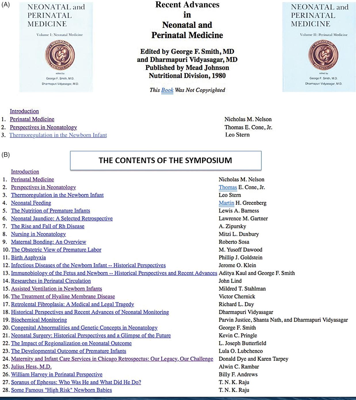
The author of this article is fortunate to have personally seen the evolution of improved neonatal intensive care and neonatal ventilation in the United States over last half century [1]. He along with Dr. George F. Smith, a geneticist and Head of the Department of Pediatrics at the Illinois Masonic Hospital and Professor at the University of Illinois, Chicago, were interested in medical history and organized
a symposium on “Historical Perspective of Perinatal Medicine in 1980.” Many giants in the field of neonatology participated in this symposium. The proceedings were supported and published by the Mead Johnson, Nutritional Division in two volumes (Fig. 2.1A–B); however, they were not copyrighted [2]. Fortunately, later they were placed on the website “Neonatology on the Web” created by Dr. Ray Duncan of Mount Sinai Hospital, Los Angeles. The two volumes on the Internet are readily available for interested readers at the website [3] (permission to reproduce figures by personal communication).
These books contain valuable historical information that would have been lost but for the ingenious method of placing the proceedings on the web. I am grateful to Dr. Duncan for this innovative method of preserving the historical volumes. The material from these books in part form the basis of the current chapter.
The history of assisted ventilation of a newborn is closely intertwined with evolution of neonatology. Therefore, it would be appropriated first to review the evolution of the specialty of neonatology then delve into the evolution of neonatal ventilation.
The story of development of neonatology and respiratory care of a newborn, particularly of the premature babies, has been told by several authors in the past [1,4–10]
The chapter is written from the perspective of both a witness and participant of these developments over the past 50 years. Following narration is based on the above referenced material. The material related to the development of neonatal ventilation is based on several reports [4–8] and three major symposia: Ross symposium in 1968, Paris symposium in 1969, and the Chicago symposium in 1980 [2]
Fig. 2.1 (A) Images of the cover pages of two volumes of symposium; Historical Perspectives and Recent Advances in Neonatal and Perinatal Medicine held in Chicago 1980, published by Mead Johnson Nutritional Division. Columbus OHIO. (B) The list of contents and presenters of two volumes. Note the list of illustrious personalities who participated in the symposium. Neonatology on the web. Available from: http://www.neonatology.org/classics/mj1980/ [3]
The development of neonatology
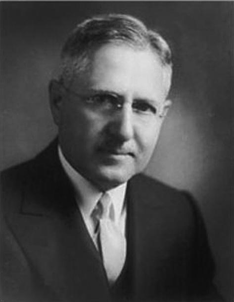
Until the mid-20th century, the primary care of newly born infants was provided by the obstetricians. In the mid-20th century, pediatricians began to take care of the newborn. The premature babies were viewed as a medical curiosity and exhibited for public view at various exhibitions [11]. However, the excellent scientific work of many investigators, both in the United States and Europe, led to better understanding of physiology and pathology of the mature term newborn and premature babies. These studies showed that a premature newborn required special thermal and nutritional care. These understandings lead to the development of premature care centers. Dr. Julius Hess in Chicago [12] was the leading authority on premature care in those days [13]. In 1914, he opened the first 24-bed premature care center at the Sarah Morris Hospital of Michael Reese Hospital (now defunct). Dr. Hess (Fig. 2.2) was the head of Department of Pediatrics at University of Illinois, Chicago and the head of pediatrics at Michael Reese hospital. He along with the help of his nurse Evelyn Lundeen provided the state of
from: http://www.
the art care of its time for premature babies. With their expert care, they showed increased survival of premature babies.
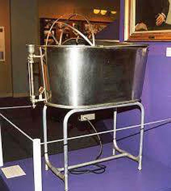
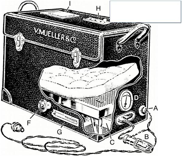
The Chicago Board of Health had established several centers in the city for the care of premature babies. Premature babies born in the community hospitals were mandated to be transported to one of these centers, if they survived the first 24 h after birth. Dr. Hess developed an incubator with the help of an engineer, “the Hess incubator” (Fig. 2.3) [14]. He also developed a transport incubator (Fig. 2.4), which could be plugged into taxis of Chicago for electric power for transportation of the babies to premature care centers within Chicago.
Both Dr. Hess and nurse Lundeen wrote several papers and books [13] on the care of premature babies, mainly on care of the newborn, particularly the premature babies and their feedings. With these advances, the care of the premature infants in Chicago improved greatly. Indeed, the premature care center at the Sarah Morris Hospital gained national and international fame. It became the center of academic learning in premature baby care for doctors and nurses from around the world.
http://www.neonatology.org/classics/mj1980/ [3]
Fig. 2.2 Photograph of Dr. Julius Hess (1876–1953) Who was In-Charge of the Premature Care Center at Sarah Morris Hospital/Michael Reese Hospital in Chicago. Neonatology on the web. Available
neonatology.org/classics/mj1980/ [3]
Fig. 2.3 The Hess Incubator Designed for Care of Premature Babies, Developed by Dr. Hess With the Help of an Engineer. Neonatology on the web. Available from:
Fig. 2.4 The Hess Transport Incubator. Dr. Hess also developed a portable incubator for transporting babies from community hospitals to designated Premature Care Centers in Chicago. Note: the incubator had an adopter to be connected to taxis of Chicago for power during transport. Neonatology on the web. Available from: http://www.neonatology.org/ classics/mj1980/ [3]
The birth of modern neonatal intensive care unit (NICU)
In October 1960, Dr. Loius Gluck established the first known neonatal intensive care unit (NICU) at Yale-New Haven Hospital, United States. Prior to this time, premature infants were often isolated in small cubicles and had little direct contact with doctors and parents because of fear of infections. With focus on hand washing, Dr. Gluck’s design for NICU took shape with the help of US$ 3 million from a benefactor whose premature grandson he had saved [15]. It was set up as a one big open room, filled with newborns in their incubators. This development had a profound influence on the subsequent direction of care of the sick newborn, including premature babies in the United States and rest of the world.
The birth of a new specialty: neonatology—the newborn medicine
The scientific basis of newborn care improved significantly with the work of several physiologists: the work of Joseph Barcroft brought new understanding of the fetus [16,17];
Dr. Geoffrey Dawes in Oxford, England [18–20] and investigators in the laboratories of Julius Comroe, United States studied neonatal physiology extensively. Dr. Clement Smith, Professor of Pediatrics at Harvard Medical School, published his book on the physiology of a newborn infant [21]. These seminal developments in understanding of the fetal and neonatal physiology laid the foundation for the clinicians to develop an evidence-based neonatal care in coming decades.
Dr. Alexander Schaffer [22] was the first one to coin the term Neonatology as the science of newborn medicine and Neonatologist as one practicing neonatal medicine in the preface to his book Diseases of the Newborn (published by Saunders in 1960). It is interesting to note that in a short span of 15 years of coining the term Neonatology, it became an established Board Certifiable Pediatric subspecialty. The first Neonatal–Perinatal Medicine specialty board examination was conducted in 1975. The author was one of the 355 candidates certified at the first board examination.
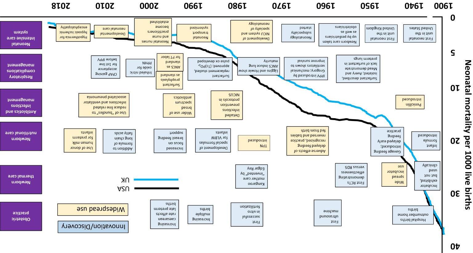
The growth of neonatology continued by leaps and bounds from 1970 onward (Fig. 2.5). The scientific exploration of neonatal illnesses and developing evidence-based therapeutic interventions also grew exponentially leading to steady decrease in neonatal mortality rates (NMR) as shown in Fig. 2.5.
Fig. 2.5 highlights the advances made in different areas of neonatology during the 20th century and also shows the impact of these developments on steady decline of NMR in the United States and the United Kingdom. It shows development in six major areas of neonatology: (1) improved thermal care, (2) improved nutrition, (3) improved nursing care and opening of premature care centers and NICUs, (4) prevention of infections, (5) improved care of infants in respiratory distress and finally, (6) improved perinatal care and resuscitation in the delivery room and ventilation. In the past century, these improvements have resulted in increased neonatal survival.
The evolution of ventilator care of the newborn
As prematurity was the major contributing factor to high NMR and the respiratory problems particularly hyaline membrane disease (HMD) was the major cause of NMR, they received greatest attention in basic and clinical research. These investigative efforts were further boosted with the tragic death of prematurely born son of the then President Kennedy.
Fig. 2.5 The graph shows advances in neonatal medicine in several fields over 100 years and its impact on neonatal mortality rate (NMR), which decreased steadily in the United Kingdom and the United States (from: Born Too Soon published by March of Dimes/WHO 2014). CPAP, Continuous positive airway pressure; NICU, neonatal intensive care unit; TPN, total parental nutrition.
On August 7, 1963, Jacqueline Kennedy, wife of President Kennedy, gave birth to a premature baby (34-week GA, birth weight of 2.1 kg) [2] (Fig. 2.6 , Boston Globe News item) in Boston who developed breathing difficulties, now what is known as the HMD. Usher’s regime [3] , infusion of 10% dextrose water with NaHCO 3 was the only known treatment for HMD. Neonatal ventilator care was not available even for the President’s baby in the United States in 1963. Moreover, sending the President’s baby to neighboring Canada where neonatal ventilation was available was not an option. The baby died after 2 days on August 9, 1963. The death of President Kennedy’s baby was a day for national mourning. As the story of demise of baby Kennedy unfolded HMD, a disease of premature babies, became known to all in America. It was estimated that in 1960s about 25,000 babies died of HMD annually in the United States. With the death of baby Kennedy, the interest in research on disease HMD accelerated. The interest in newborn care increased.
Oxygen therapy
Prior to use of any form of assisted ventilation, administration of oxygen was the only available therapy for infants requiring delivery room resuscitation and infants
in respiratory distress. The use of oxygen in the treatment of neonates with respiratory distress has been reported for more than a century. In 1907, Budin recommended oxygen “supplied through a funnel, the large opening of which is placed beside the infant’s face,” for the treatment of cyanotic episodes in newborns [23]. In the 1930s, Hess developed an incubator capable of delivering approximately 40% oxygen for extended periods of time [12,23]. By the 1940s, a commercially available incubator capable of providing a high concentration of oxygen facilitated the liberal use of oxygen for the treatment of cyanosis, apnea, and periodic breathing of newborns.
Throughout this time, oxygen administration was guided by the clinical observations of skin color, as well as the respiratory rate, regularity, and work of breathing. It wasn’t until the 1960s and 1970s that the technology of microsampling of blood gases was available [24]. In 1980s, noninvasive methods of transcutaneous oxygen [25] and CO2 monitoring became available. Pulse oximetry [26] became available in 1980s for more precise monitoring of oxygen saturation in the blood. It remains the standard method of monitoring blood oxygenation in a newborn.
The overall goal of oxygen therapy was to achieve adequate oxygenation using the lowest concentration of inspired oxygen. However, achieving this goal is complicated due to a number of factors. Routine administration of oxygen to all premature infants led to the catastrophic results of the development of retinopathy of prematurity (ROP) and related blindness [24,27]. However, a study to curtail oxygen therapy was associated with increased cerebral palsy [28–32]
Despite over 75 years of routine oxygen administration to newborn infants, administering optimal level of oxygenation and monitoring—one that avoids the detrimental effects of hypoxia on the one hand, and those caused by hyperoxia on the other hand—has been very difficult [33]. Current recommendations for oxygen saturation targets are different between the United States and Europe. The European recommendations are to keep the target oxygen saturations between 90% and 94% for premature infants requiring supplemental oxygen [34]. The American Academy of Pediatrics states that the ideal target oxygen saturation is not known and in some preterm infants, 91%–95% target may be safer than 85%–89% [33]
In order to achieve the goals of neonatal oxygen therapy, we need to develop and evaluate appropriate devices of oxygen delivery systems. The clinicians today need to have an adequate knowledge of the use of oxygen delivery equipment, and have the training on the concepts of neonatal oxygenation and equipment used to monitor the effects of oxygen therapy.
Fig. 2.6 The News of Death of Prematurely Born Baby Kennedy Printed in Boston Globe.
Usher regime
Prior to the introduction of neonatal ventilation in late 1950s and early 1960s, Dr. Robert Usher of Montreal, Canada after extensive studies in premature infants with HMD showed that they suffer from metabolic acidosis and hyperkalemia [35]. To counteract these changes he proposed a treatment regime of administering NaHCO3 in 10% dextrose to infants in respiratory distress [35]. This therapy became known as Usher regime resulted in significant (50%) reduction of mortality in infants with HMD. The Usher regime was one of the major milestones in the treatment of HMD prior to initiation of assisted ventilation. At this point, a retrospective view of experience with neonatal ventilation and neonatal ventilators is in order.
History of neonatal ventilation
Several reviewers have stated that it is difficult to time exactly when ventilation of the newborn was initiated and probably occurred in the late 1950s and 60s [10,36,37]. Downes in an editorial [38] refers to the work of Smythe and Bull from South Africa to have used successful long-term neonatal ventilation in infants afflicted with tetanus. These infants were treated with d-tubocurarine, tracheostomy, and ventilation; and the mortality was reduced from nearly 100% to 20%. However, these infants had normal lungs [38]. Initial reports of mechanical ventilation of newborns with pulmonary insufficiency were reported by Benson et al. and Donald et al. in 1958 [39,40]. The first highly successful use of mechanical ventilation in premature infants with HMD was reported by Maria Delivoria-Papadopoulos in 1965 [41–43]. In this series, out of 20 infants with severe HMD, 7 survived (35%) and 6 of them were neurologically intact. Since then, several other investigators reported use of assisted ventilation in HMD with increasing success. Some used positive pressure ventilation, including Strang and Reynolds in London [44], Thomas et al. at Stanford [45], and de Heese et al. in Cape Town [46]. Historically, negative pressure ventilation was designed earlier in 1889 by Alexander Graham Bell [47,48]. He presented a paper to the American Association for the advancement of science in Montreal on the use of ventilator for newborn babies and was “met with little enthusiasm.” The design and device are preserved at The Alexander Graham Bell museum in Nova Scotia, Canada [49]. Later in 1960s, Dr. Stahlman in Nashville, Tennessee [50], and Stern in Montreal, Canada [51] used negative pressure ventilation (Fig. 2.7) to treat babies with RDS. Chernick and Vidyasagar in Winnipeg, Canada [52,53] modified negative pressure ventilator to create
Note the respirator has two arts: the closed chamber wherein the baby’s body is placed with the head lays outside open to atmospheric pressure. An adjustable sleeve around the neck seals the body chamber. The incubator is fitted with a vacuum creator underneath the body. Turning the knobs in front allow to create desired negative pressure and adjust the respiratory cycle (operated by vacuum creating machine and a solenoid valve underneath the body).
constant negative distend pressure (similar to continuous positive airway pressure [CPAP]) without an endotracheal tube with success in the management of respiratory failure in newborn.
Readers should note that negative pressure ventilation is no more in use as we have developed several simpler noninvasive methods of ventilation (see chapter on noninvasive ventilation in this book). However, the use of negative pressure respirator to support babies in respiratory distress remains an important phase in the history of neonatal ventilation.
Ross symposium on neonatal intensive care
In 1968, a conference was organized on neonatal intensive care in Vermont by Ross laboratories. Several aspects of neonatal intensive care including design of these units and ventilation techniques were discussed at the conference [54]. Several leading neonatologists of the time from the United States and Canada participated in this conference. The conference was intended to share the experiences of different units and learn the problems of neonatal intensive care units of the day. Presentations by various speakers showed the impact of intensive care on improved survival, complications, and long-term intact survival of babies cared in their respective units.
Fig. 2.7 Photograph of Air-Shield Negative Pressure Respirator.
Survival with assisted ventilation was highest among infants with tetanus neonatorum who had no lung disease. It reversed the 80% mortality in tetanus prior to assisted ventilation to 80% survival with assisted ventilation. Survival rate in RDS although improved was still at 28%. The results of assisted ventilation in other respiratory conditions were not so encouraging. At the end Dr. Lucey, the chairman of the conference, summarized the conference as follows: “ Now that you have read the proceedings of this conference some will be frustrated and discouraged. Others will be encouraged to try to improve the care in their own nurseries. Hopefully this conference will have supplied with early but firm data to encourage you in these efforts and warn you of the problems involved” and cautioned “whereas intensive care is effective we still do not have a clear idea about the key elements of success. The construction of a new nursery or the purchase of a blood gas machine and respirator do not an intensive care nursery make!”. The key elements are intelligent personnel or as one participant put it “people who care intensely” (Dr. Nick Nelson of Harvard)
This is one of the earliest reports on the impact of modern neonatal intensive care including the results of neonatal ventilation.
In 1969, another conference solely on assisted ventilation was organized in Paris by Professor Alex Minkowski. In this symposium, clinician researchers from different countries shared their experiences with neonatal ventilation. Representatives from France, Belgium, England, South Africa, Finland, Canada, and the United States participated in the symposium. A review of published proceedings in Biology of the Neonate (current name of this journal is “Neonatology”) shows the struggles faced by the clinician researchers of the day in finding the right ventilator for the user in newborn, the optimal time for initiating ventilation, monitoring babies on ventilation, and improving outcomes at this time [55–58]
In writing the summary of the symposium, Dr. Paul Swyer from Toronto, Canada who conducted the meeting raised the big question: whether neonatologists should continue to provide assisted ventilation! (Perhaps, more aggressively) or whether the possible complications outweighed neonatal ventilation
However, the efforts to improve the clinical practice of providing assisted ventilation to the sick newborn continued.
The introduction of CPAP in managing infants with HMD by Gregory et al. in 1967 was a major breakthrough in neonatal respiratory management [59]. Using this approach he showed a significant improvement in survival of infants with HMD (16 of the 20 infants survived— including 10 less than 1500 g) [59]
Introduction of surfactant therapy in HMD/RDS
The invention of CPAP and the discovery [60–64] and production of surfactant further reduced mechanical complications of ventilation in preterm newborns [65,66]
In 1959, Avery and Mead [63] reported that the low surface tension in the lining of the lung permits stability of the alveoli at end expiration. Lacking such material, immature infants and infants dying with HMD, surface tension was higher than expected. They speculated that deficiency of surface-active material might be significant in the pathogenesis of HMD. This article was cited 376 times in the period 1961–77.
In 1980, Fujiwara et al. from Japan reported successful use of an artificial surfactant in 10 preterm infants severely ill with HMD [67,68]. Following instillation of artificial surfactant, alveolar–arterial gradients decreased, the levels of inspired oxygen and peak inspiratory pressures decreased, and radiological abnormalities resolved. All survived. Raju et al. [69] reported that replacement therapy with surfactant in a randomized trial significantly improved oxygenation, reduced complications of neonatal ventilation such as airleak syndromes (pneumothorax and pulmonary interstitial emphysema), and improved survival without BPD.
Soon after multiple randomized clinical trials of surfactant replacement therapy substantiating improvement in survival of infants with RDS treated with surfactant [70]. These studies led to FDA approval of a surfactant “Survanta” for clinical use in 1993. Introduction of surfactant in the treatment of RDS remains a major milestone in the history of neonatology
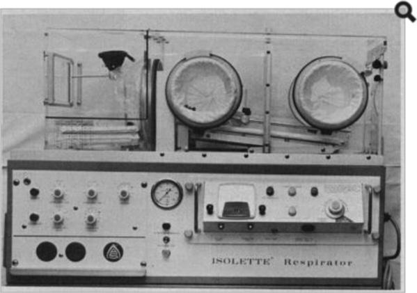
The modern neonatal ventilators
Prior to 1970s, neonatologists had to use modified adult ventilators providing intermittent positive pressure ventilation. However, these ventilators could not match the physiologic pattern of breathing at higher rates. Ventilators designed specifically for the newborn appeared during mid-1970s–90s. Dr. Sola in this book discusses currently available ventilators incorporated with various functional modalities for use by the clinician. Goldsmith et al. [71] describe the milestones of technological developments in designing ventilators specifically for the newborns and premature infants—starting from the modified adult ventilators to current highly sophisticated incorporation of space age technology into ventilators used currently in the NICUs.
The rhetoric question raised at the Paris symposium in 1969 regarding the value of assisted ventilation has been answered by the continued efforts to improve technological, perinatal, and neonatal therapeutic advances to treat babies with HMD. Undoubtedly, assisted ventilation has improved overall survival of tiniest babies with HMD (24% mortality among extremely preterm infants <29 weeks of gestation and 2.9% mortality among moderately preterm infants, 29–33 weeks of gestation) [72], but the persistence of associated complications of ventilator-induced lung injury continue to vex the clinician and stimulate further research.
Summary
In summary technological innovations—the likes of Hess incubator—of early 20th century were the precursors of modern incubators. The premature care centers of early 20th century were the beginning of modern neonatal intensive care units. The evolution of incubator care of the premature infants, combined with the extensive basic and clinical research on fetal and neonatal physiology in the last century, gave birth to the subspecialty of neonatology. Modern ventilatory care evolved from earlier efforts to save babies dying of neonatal tetanus. Similar earlier efforts to ventilate premature babies with lung disease (HMD) were discouragingly poor. Persistent and continued efforts
to develop ventilators to match neonatal physiology combined with modern space age microprocessors resulted in the modern neonatal ventilators. It was also realized that machines alone do not make an intensive care. It is the people (skilled nurses/doctors/respiratory therapists, support staff) who care “intensively” and make the intensive care unit.
With these principles of care the outcome results of modern ventilation in the premature newborn, supplemented with surfactant therapy, nutritional support and nursing care are exceedingly high even in the extreme premature babies (over 92% survival at 28 weeks) [73].
Today, it is estimated that in the United States alone 65,000 babies are ventilated annually [74]. Further, with global efforts of technology transfer to the developing countries, the practice of neonatal ventilator support particularly of the premature baby is widely used even in low- and middle-income countries [75]. While these developments are a very nice welcome, the novice should be cautioned in venturing into this territory without the expertise or the total organizational support needed for the operation of neonatal ventilation.
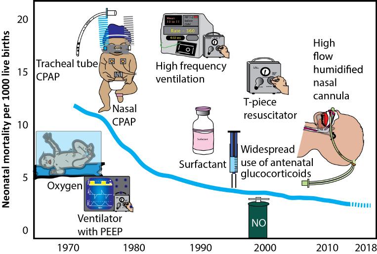
However, long-term ventilation is associated with pulmonary morbidity (CLD/BPD). Experts in the field have discussed various preventive aspects of ventilator-induced lung injury in this book. Prevention of ventilation-induced lung injury (VILI) remains the major challenge for the future generation of neonatologists. A summary of evolution of neonatal ventilation is shown in Fig. 2.8.
Fig. 2.8 Graph Showing Neonatal Mortality Rate and Various Innovations in Neonatal Respiratory Care. CPAP, Continuous positive airway pressure. Copyright: Satyan Lakshminrusimha.