University of Alabama at Birmingham, Birmingham, AL, United States
Rangasamy Ramanathan, MD, FAAP
Professor of Pediatrics at the Keck School of Medicine and Division Chief, Director of Neonatal Respiratory therapy program, Program director, Neonatal Perinatal Fellowship Program, University of Southern California, Los Angeles, CA, United States
P. Syamasundar Rao, MD, FAAP, FACC, FSCAI
Professor of Pediatrics, Division of Pediatric Cardiology, UT Health McGovern Medical School, Houston, TX, United States
Maura Helena Ferrari Resende, MD
Clinical Fellow, The Hospital for Sick Children, Toronto, ON, Canada
Rakesh Sahni, MD
Professor of Pediatrics, Columbia University College of Physicians and Surgeons, New York-Presbyterian
Morgan Stanley Children’s Hospital, Columbia University Medical Center, New York, NY, United States
Mitali Sahni, MD
Fellow in Neonatal-Perinatal Medicine, St. Christopher’s Hospital for Children, Drexel University College of Medicine, Philadelphia, PA, United States
Marwa al Sayyed, MBBS, MSc
Neonatal Registrar, NMC speciality Hospital, Dubai, United Arab Emirates
Bernard Schoonakker, MRCPCH
Nottingham University Hospitals NHS Trust, Nottingham, United Kingdom
Craig Smith, MRCPCH
Nottingham University Hospitals NHS Trust, Nottingham, United Kingdom
Augusto Sola, MD
Director Medico Ejecutivo, SIBEN, Por los recién nacidos
Howard Stein, MD, FAAP
Promedica Toledo Children’s Hospital, University of Toledo Health Science Campus, Toledo, OH, United States
RoseMary S. Stocks, MD, PharmD
Department of Otolaryngology, Head and Neck Surgery, University of Tennessee Health Sciences Center, Memphis, TN, United States
Section Head and Service Chief, Neonatology, Professor of Pediatrics, Texas Children’s Hospital, Baylor College of Medicine, Houston, TX, United States
Ru-Jeng Teng, MD
Children’s Research Institute, Medical College of Wisconsin, Children’s Hospital of Wisconsin, Milwaukee, WI, United States
Vikrum A. Thimmappa, MD Department of Otolaryngology, Head and Neck Surgery, University of Tennessee Health Sciences Center, Memphis, TN, United States
Jerome W. Thompson, MD, MBA Department of Otolaryngology, Head and Neck Surgery, University of Tennessee Health Sciences Center, Memphis, TN, United States
David A. Todd, FIMLS, MSc, PhD, MBBS
Centenary Hospital, Canberra, ACT, Australia
Kirtikumar Upadhyay, MD, FAAP
University of Tennessee Health Science Center, Memphis, TN, United States
Karunakar Vadlamudi, MD
Stollery Children’s Hospital, University of Alberta, Edmonton, AB, Canada
Payam Vali, MD
University of California, Davis; UC Davis Children’s Hospital, Sacramento, CA, United States
Máximo Vento, MD, PhD
University and Polytechnic Hospital La Fe Valencia, València, Spain
Sudeep Verma, MD, FNB
KIMS-Institute of cardiac Sciences, Secunderabad-Hyderabad, Telangana, India
Dharmapuri Vidyasagar, MD, FAAP, FCCM, PhD(Hon)
Professor Emeritus Pediatrics, Division of Neonatology, University of Illinois at Chicago, Chicago, IL, United States
Koert de Waal, PhD, FRACP
John Hunter Children’s Hospital, Newcastle, NSW, Australia
Mark F. Weems, MD, FAAP
University of Tennessee Health Science Center, Memphis, TN, United States
Jen-Tien Wung, MD
Professor of Pediatrics, Columbia
University College of Physicians and Surgeons, Columbia University Medical Center, New York, NY, United States
Hakam Yaseen, MD, CES (Paed), DUN (Neonat) [France], CCST (UK), FRCPCH [UK]
University of Sharjah, Medical Director (CMO), University Hospital Sharjah (UHS), Sharjah, United Arab Emirates
Page left intentionally blank
Page left intentionally blank
Acknowledgments
Rajiv gratefully acknowledges the didactic teaching of his fellow teacher Dr. Elizabeth John, whose extreme sensitivities to the adjustment of CPAP up or down still ring a bell in his ears. This singular caution to optimize continuous positive airway pressure or positive end-expiratory pressure laid the foundation of his strategy in any critical lung disease. This was the fulcrum of his success in neonatal ventilation in the last 30 years.
Rajiv acknowledges the heartfelt help of his teachers Dr. Vidyasagar, Dr. Georg Simbrunner, Dr. Ramanathan, Dr. Martin Keszler, and Dr. Dhanireddy for teaching him and for being authors of many chapters and reviewing many more of them. Dr. Vidyasagar was the first to agree to the concept of this book many years ago and has been the guiding light in the evolution of this book. Rajiv’s close associates Dr. Prakash, Dr. Arun, and Dr. David Todd gave him exceptional chapters at a short notice. His junior associates Dr. Nalinikant and Dr. Srinivas provided very unique, well-researched chapters. He is indebted to coeditor Satyan who joined the team in 2016 for his immortal illustrations and editorial stewardship. His illustrations offer an additional tool for neonatal providers to educate students and parents.
Rajiv also thanks his team members Iftekar, Jason, and Sherly for uncompromising secretarial and artwork. He further thanks Dr. Karunakar, his associate, for responding to the perennial demands of perfection of the chapters, without any hesitation.
This book is a unique joint effort of a highly talented provider-publisher team. Last but not the least, Rajiv thanks his wife Bindoo for silently bearing with him all the timeless lapses at home while he was playing Archimedes for the development of this book.
Dr. Vidyasagar gratefully acknowledges his mentors, Dr. Thomas Boggs, Dr. Jack Downes, and Dr. Victor Chernick who introduced him to neonatal ventilation. He thanks his wife Dr. Nagamani Beligere for her support all through his career. His children Sahana, Sadhana, and Sanjay and grandchildren Kavi, Anika, and Maaya have been the source of his energy.
Satyan thanks his children (Ananya, Aniruddha, and Arun for posing as models during their neonatal period for his illustrations) and his wife Veena Manja, MD, MSc, for her unrelenting support. He expresses gratitude to his parents, sisters, parents-in-law, teachers, and mentors for supporting and guiding him throughout his career.
All the editors sincerely appreciate the exceptional support by Mr. Ayan Dhar and Ms. Sheenam Aggarwal at Elsevier India. Above all, the editors are thankful to all the babies and their parents who contributed to our understanding of neonatal physiology and the functioning of assisted ventilation.
9B. The Importance of Heating and Humidifying the Inspired Gases During Mechanical Ventilation: Identifying the Ideal Settings and Circuit Configuration During Ventilation ...................................................113
David A. Todd, K.Y. Ashok Murthy, P.K. Rajiv 10. Ventilator Graphics.....................................124
Manoj Biniwale, Rangasamy Ramanathan, Mark C. Mammel
11A. Initiation of Mechanical Ventilation 143 Dushyant Batra, Craig Smith, Bernard Schoonakker
11B. Deterioration on the Ventilator ................149
Vikrum A. Thimmappa, Ramasubbareddy Dhanireddy, RoseMary S. Stocks, Jerome W. Thompson
Neonatal Airway Pathology
35. Ventilator-Associated Pneumonia and Infection Control.................................765
Manoj N. Malviya, Prakash Manikoth, Hakam Yaseen
Page left intentionally blank
Introduction
Chapter | 1 |
This book was conceived several years ago, when there appeared to be a distinct lacuna of comprehensive bedside ventilatory management guides in neonatal care history. Currently, there are excellent textbooks to refer to and obtain broad concepts on the approach to providing respiratory assistance to a baby in distress, but a detailed evidence-based book on bedside management is missing. In this book we attempted to provide the readers an evidence-based practice bedside guidelines. In doing so, we sought the contributions from the most experienced leaders in the field. This book is an honest attempt to get the world’s best pioneers in each area to contribute their signature chapters of their research to give the neonatal intensivist, detailed bedside ventilation navigation in critical situations. We earnestly hope readers will find these guidelines useful in managing critically ill neonates.
This book is divided into eight sections. Here are some of the highlights of these sections. Section I reviews the history of neonatal ventilation. Section II deals with basic chapters covering embryology and physiology of pulmonary disease, with the time frame from extreme prematurity at the limits of viability to dysmorphology in the full-term infant. The delivery process and golden first hour are addressed in detail, due to its long-term impact on respiratory and neurological morbidity.
Section III deals with the basics of neonatal ventilation and evolves through the genesis of lung injury to lung mechanics. The chapters on ventilator give deep insight to the reader on the limitations and benefits of its application. The chapters progress to the provision of mechanical ventilation and its attendant complications, which are again discussed in detail.
Section IV is an in-depth analysis in real time of the various respiratory care devices currently available for the neonate. These chapters give an operating framework and the bedside navigation in critically ill babies with trouble shooting algorithms by authentic authors.
Section V is the heart of this book with comprehensive bedside management guidelines of the common respiratory conditions faced in neonatal intensive care. They offer detailed flowcharts, algorithms, and case scenarios in complicated respiratory care management. There is a separate chapter on the management of the 23–25 weeks’ gestation babies: “micropremies”—a challenge for any intensivist.
Section VI deals in-depth for all the common cardiac conditions complicating respiratory care. Management of shock and cyanotic heart disease, PDA, and arrhythmias are discussed. Functional echo is comprehensively discussed as it is evolving as the new standard of care.
No ventilator support will be successful without strong ancillary support. Section VII details all critical aspects of ancillary care of the ventilated neonate, including monitoring, infection control, nutrition, and procedures.
It is heartening to note that there is emergence of an increasing number of neonatal intensive care units (NICUs) to improve survival among low- and middle-income countries (LMCs). Ventilatory support is an essential part of the neonatal intensive care. Proper ventilator care requires a combination of skilled personnel, appropriate equipment, and ancillary support which are the prerequisites for optimal outcome but are difficult to fulfill in some LMCs. Several chapters in the book offer guidelines to assist pioneers in LMCs in establishing ventilatory support in their prospective units and teach physicians, trainees, and nurses.
Besides the rich evidence-based content of the book, it has several unique features to help the practitioner better manage infants requiring ventilatory care. This book is digitally enhanced with illustrations and videos linked to their respective chapters and lecture PowerPoint to most chapters of this book to give the intensivist a 360-degree comprehension of neonatal ventilation.
This book is intended for neonatologists, intensivists, postgraduates (residents and fellows), respiratory therapists, and neonatal nurses as a ready bedside reckoner for urgent consult.
Chapter | 2 |
Evolution of Neonatal Ventilation a Retrospective View
Dharmapuri Vidyasagar, PhD (Hon)
Introduction
The author of this article is fortunate to have personally seen the evolution of improved neonatal intensive care and neonatal ventilation in the United States over last half century [1]. He along with Dr. George F. Smith, a geneticist and Head of the Department of Pediatrics at the Illinois Masonic Hospital and Professor at the University of Illinois, Chicago, were interested in medical history and organized
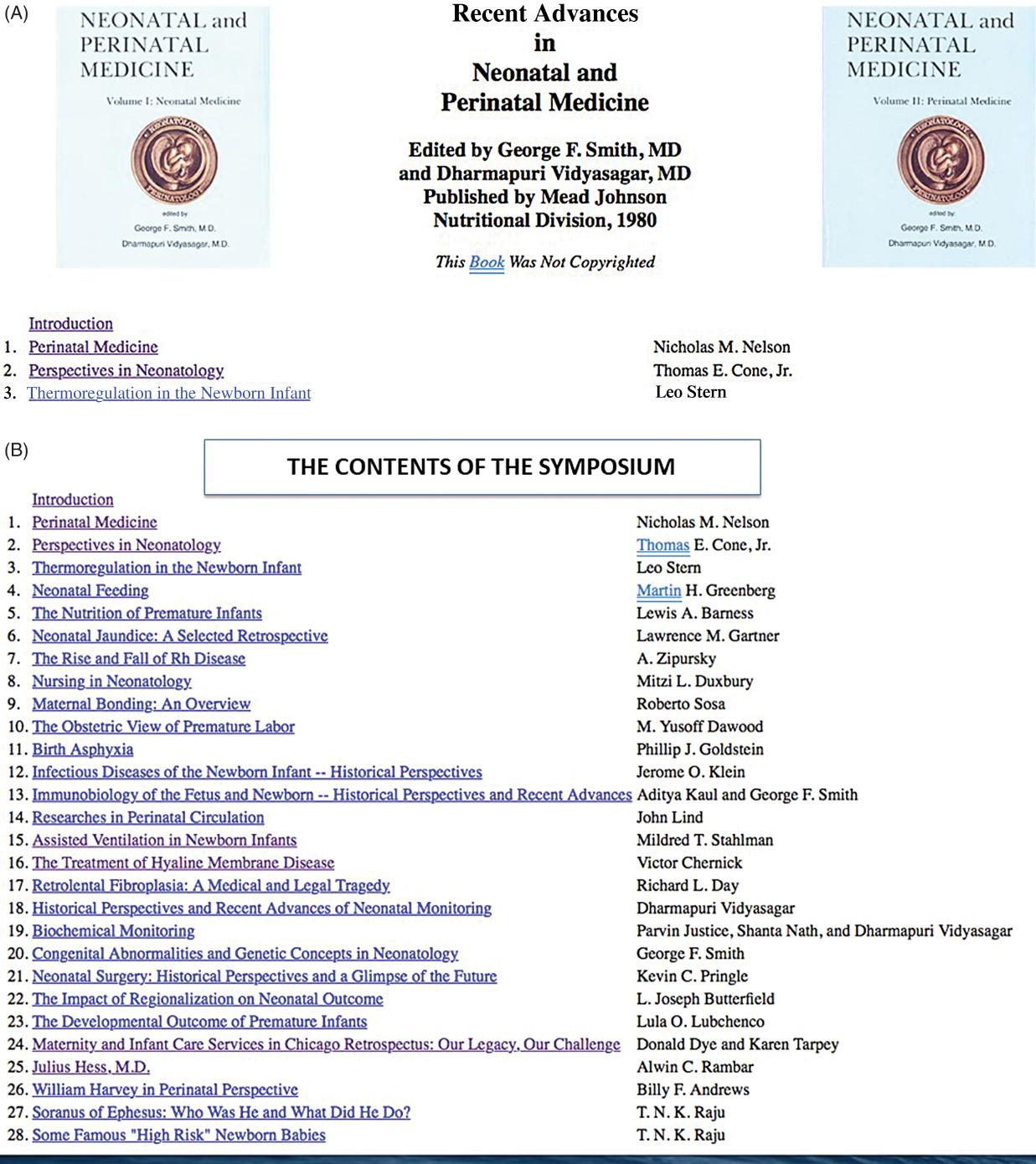
a symposium on “Historical Perspective of Perinatal Medicine in 1980.” Many giants in the field of neonatology participated in this symposium. The proceedings were supported and published by the Mead Johnson, Nutritional Division in two volumes (Fig. 2.1A–B); however, they were not copyrighted [2]. Fortunately, later they were placed on the website “Neonatology on the Web” created by Dr. Ray Duncan of Mount Sinai Hospital, Los Angeles. The two volumes on the Internet are readily available for interested readers at the website [3] (permission to reproduce figures by personal communication).
These books contain valuable historical information that would have been lost but for the ingenious method of placing the proceedings on the web. I am grateful to Dr. Duncan for this innovative method of preserving the historical volumes. The material from these books in part form the basis of the current chapter.
The history of assisted ventilation of a newborn is closely intertwined with evolution of neonatology. Therefore, it would be appropriated first to review the evolution of the specialty of neonatology then delve into the evolution of neonatal ventilation.
The story of development of neonatology and respiratory care of a newborn, particularly of the premature babies, has been told by several authors in the past [1,4–10]
The chapter is written from the perspective of both a witness and participant of these developments over the past 50 years. Following narration is based on the above referenced material. The material related to the development of neonatal ventilation is based on several reports [4–8] and three major symposia: Ross symposium in 1968, Paris symposium in 1969, and the Chicago symposium in 1980 [2]
Fig. 2.1 (A) Images of the cover pages of two volumes of symposium; Historical Perspectives and Recent Advances in Neonatal and Perinatal Medicine held in Chicago 1980, published by Mead Johnson Nutritional Division. Columbus OHIO. (B) The list of contents and presenters of two volumes. Note the list of illustrious personalities who participated in the symposium. Neonatology on the web. Available from: http://www.neonatology.org/classics/mj1980/ [3]
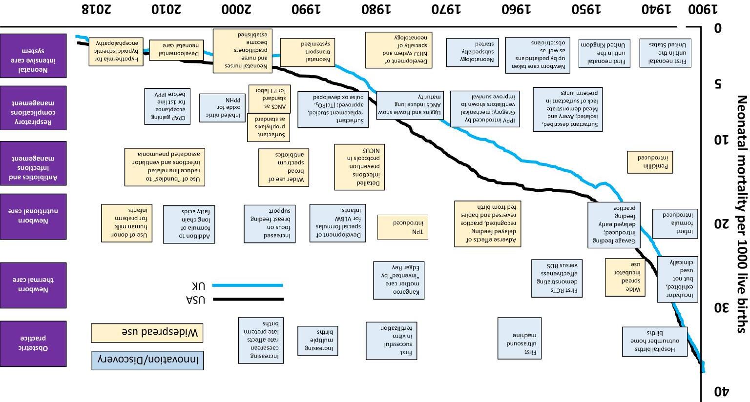
Fig. 2.5 The graph shows advances in neonatal medicine in several fields over 100 years and its impact on neonatal mortality rate (NMR), which decreased steadily in the United Kingdom and the United States (from: Born Too Soon published by March of Dimes/WHO 2014). CPAP, Continuous positive airway pressure; NICU, neonatal intensive care unit; TPN, total parental nutrition.
On August 7, 1963, Jacqueline Kennedy, wife of President Kennedy, gave birth to a premature baby (34-week GA, birth weight of 2.1 kg) [2] (Fig. 2.6 , Boston Globe News item) in Boston who developed breathing difficulties, now what is known as the HMD. Usher’s regime [3] , infusion of 10% dextrose water with NaHCO 3 was the only known treatment for HMD. Neonatal ventilator care was not available even for the President’s baby in the United States in 1963. Moreover, sending the President’s baby to neighboring Canada where neonatal ventilation was available was not an option. The baby died after 2 days on August 9, 1963. The death of President Kennedy’s baby was a day for national mourning. As the story of demise of baby Kennedy unfolded HMD, a disease of premature babies, became known to all in America. It was estimated that in 1960s about 25,000 babies died of HMD annually in the United States. With the death of baby Kennedy, the interest in research on disease HMD accelerated. The interest in newborn care increased.
Oxygen therapy
Prior to use of any form of assisted ventilation, administration of oxygen was the only available therapy for infants requiring delivery room resuscitation and infants
in respiratory distress. The use of oxygen in the treatment of neonates with respiratory distress has been reported for more than a century. In 1907, Budin recommended oxygen “supplied through a funnel, the large opening of which is placed beside the infant’s face,” for the treatment of cyanotic episodes in newborns [23]. In the 1930s, Hess developed an incubator capable of delivering approximately 40% oxygen for extended periods of time [12,23]. By the 1940s, a commercially available incubator capable of providing a high concentration of oxygen facilitated the liberal use of oxygen for the treatment of cyanosis, apnea, and periodic breathing of newborns.
Throughout this time, oxygen administration was guided by the clinical observations of skin color, as well as the respiratory rate, regularity, and work of breathing. It wasn’t until the 1960s and 1970s that the technology of microsampling of blood gases was available [24]. In 1980s, noninvasive methods of transcutaneous oxygen [25] and CO2 monitoring became available. Pulse oximetry [26] became available in 1980s for more precise monitoring of oxygen saturation in the blood. It remains the standard method of monitoring blood oxygenation in a newborn.
The overall goal of oxygen therapy was to achieve adequate oxygenation using the lowest concentration of inspired oxygen. However, achieving this goal is complicated due to a number of factors. Routine administration of oxygen to all premature infants led to the catastrophic results of the development of retinopathy of prematurity (ROP) and related blindness [24,27]. However, a study to curtail oxygen therapy was associated with increased cerebral palsy [28–32]
Despite over 75 years of routine oxygen administration to newborn infants, administering optimal level of oxygenation and monitoring—one that avoids the detrimental effects of hypoxia on the one hand, and those caused by hyperoxia on the other hand—has been very difficult [33]. Current recommendations for oxygen saturation targets are different between the United States and Europe. The European recommendations are to keep the target oxygen saturations between 90% and 94% for premature infants requiring supplemental oxygen [34]. The American Academy of Pediatrics states that the ideal target oxygen saturation is not known and in some preterm infants, 91%–95% target may be safer than 85%–89% [33]
In order to achieve the goals of neonatal oxygen therapy, we need to develop and evaluate appropriate devices of oxygen delivery systems. The clinicians today need to have an adequate knowledge of the use of oxygen delivery equipment, and have the training on the concepts of neonatal oxygenation and equipment used to monitor the effects of oxygen therapy.
Fig. 2.6 The News of Death of Prematurely Born Baby Kennedy Printed in Boston Globe.
The rhetoric question raised at the Paris symposium in 1969 regarding the value of assisted ventilation has been answered by the continued efforts to improve technological, perinatal, and neonatal therapeutic advances to treat babies with HMD. Undoubtedly, assisted ventilation has improved overall survival of tiniest babies with HMD (24% mortality among extremely preterm infants <29 weeks of gestation and 2.9% mortality among moderately preterm infants, 29–33 weeks of gestation) [72], but the persistence of associated complications of ventilator-induced lung injury continue to vex the clinician and stimulate further research.
Summary
In summary technological innovations—the likes of Hess incubator—of early 20th century were the precursors of modern incubators. The premature care centers of early 20th century were the beginning of modern neonatal intensive care units. The evolution of incubator care of the premature infants, combined with the extensive basic and clinical research on fetal and neonatal physiology in the last century, gave birth to the subspecialty of neonatology. Modern ventilatory care evolved from earlier efforts to save babies dying of neonatal tetanus. Similar earlier efforts to ventilate premature babies with lung disease (HMD) were discouragingly poor. Persistent and continued efforts
to develop ventilators to match neonatal physiology combined with modern space age microprocessors resulted in the modern neonatal ventilators. It was also realized that machines alone do not make an intensive care. It is the people (skilled nurses/doctors/respiratory therapists, support staff) who care “intensively” and make the intensive care unit.
With these principles of care the outcome results of modern ventilation in the premature newborn, supplemented with surfactant therapy, nutritional support and nursing care are exceedingly high even in the extreme premature babies (over 92% survival at 28 weeks) [73].
Today, it is estimated that in the United States alone 65,000 babies are ventilated annually [74]. Further, with global efforts of technology transfer to the developing countries, the practice of neonatal ventilator support particularly of the premature baby is widely used even in low- and middle-income countries [75]. While these developments are a very nice welcome, the novice should be cautioned in venturing into this territory without the expertise or the total organizational support needed for the operation of neonatal ventilation.
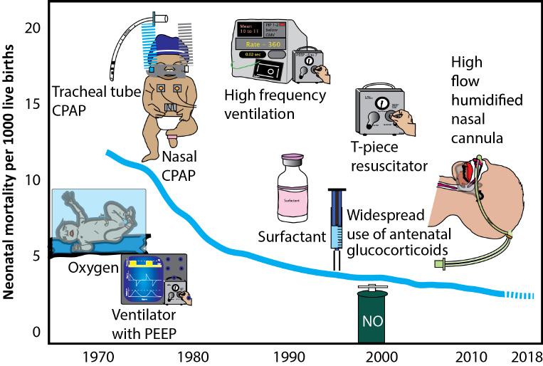
However, long-term ventilation is associated with pulmonary morbidity (CLD/BPD). Experts in the field have discussed various preventive aspects of ventilator-induced lung injury in this book. Prevention of ventilation-induced lung injury (VILI) remains the major challenge for the future generation of neonatologists. A summary of evolution of neonatal ventilation is shown in Fig. 2.8.
Fig. 2.8 Graph Showing Neonatal Mortality Rate and Various Innovations in Neonatal Respiratory Care. CPAP, Continuous positive airway pressure. Copyright: Satyan Lakshminrusimha.