CONTRIBUTORS
Several highly qualified scientists have demonstrated their willingness to contribute to the book project.
Keith J. Betteridge BVSc MVSc PhD FRCVS
University Professor Emeritus
Department of Biomedical Sciences
Ontario Veterinary College University of Guelph, Ontario Canada
Gry Boe-Hansen DVM Phd Lecturer
School of Veterinary Science University of Queensland, Australia
Henrik Callesen DVM PhD DVSc
Research Professor
Department of Genetics and Biotechnology Faculty of Agricultural Sciences, Aarhus University, Denmark
Ernst-Martin Füchtbauer PhD Dr.habil
Associate Professor
Department of Molecular Biology Aarhus University
Denmark
Vanessa Hall PhD Post Doc
Department of Basic Animal and Veterinary Sciences
Faculty of Life Sciences, University of Copenhagen
Denmark
Poul Hyttel DVM Phd DVSc
Professor
Department of Basic Animal and Veterinary Sciences
Faculty of Life Sciences, University of Copenhagen
Denmark
Palle Serup Phd
Director of Research
Department of Developmental Biology
Hagedorn Research Institute
Denmark
Fred Sinowatz Dr.med vet. Dr.med Dr.habil
Professor
Institute of Veterinary Anatomy, Histology and Embryology
LMU Munich, Germany
Gábor Vajta MD PhD DVSc
Scientific Director
Cairns Fertility Centre
Australia
Adjunct Professor, University of Copenhagen, Denmark
Adjunct Professor, James Cook University, Australia
Morten Vejlsted DVM Phd
Assistant Professor
Department of Large Animal Sciences
Faculty of Life Sciences, University of Copenhagen, Denmark
Over the last 20 years, modern life science research efforts have rapidly advanced our knowledge of the normal and abnormal processes of domestic animal development. As our depth of understanding of the cellular and molecular mechanisms has grown, so too has the recognition of the potential for, and successful application of, this knowledge to enhance animal-based food and fiber production. It is during embryo and fetal development, from the formation of competent gametes to parturition, that powerful advancements in molecular genetic manipulation and assisted reproductive technologies are employed, and these efforts have had profound impact on animal production worldwide. As a result of these advances, there remains an unmet need for a contemporaneous text of domestic animal development to support education and training of today’s veterinarians, animal scientists and developmental biologists. Essentials of Domestic Animal Embryology fulfills this need by providing the student, the instructor and the veterinary practitioner with an in depth presentation of the elaborate, chronological processes that culminate in formation of functional embryonic structures from the development of gametes through the peri-partum period. As our understanding of the precisely orchestrated processes of animal development advances, and animal genomes are further unveiled and analyzed, the importance of animal development becomes central to understanding and enhancing animal growth, sustaining health and determining the underlying causes of disease.
Although there continues to be available a number of quality texts of human embryology (for example Langman’s Medical Embryology), focus on a single species remains a serious limitation for veterinary and animal science audiences and prevents a thorough view of the wide and distinct variations that exist among domestic animal species, with particular reference to processes of blastogenesis, implantation and placentation. Certainly, Bradley
Patton’s 1927 benchmark publication in English, Embryology of the Pig, provided a wonderfully illustrated, descriptive account of this often utilized example of mammalian embryonic development. A concise descriptive publication of development in the pig, The Embryonic Pig: A Chronological Account, was later published by A.W. Marrable in 1971. In 1984, Drew Noden and Alexander de Lahunta, similarly recognizing the lack of an adequate text on domestic species embryology that would be useful for veterinary students, published The Embryology of Domestic Animals: Developmental Mechanisms and Malformations. An important contribution to veterinary curricula for many years to follow, this book presented traditional system-bysystem descriptive material on the developmental anatomy of domestic species including birds, and the authors also included many relevant experimental and clinical case references throughout the book. Unfortunately, a revised edition was not forthcoming, and is not currently available. More recently, the finely detailed German text, Lehrbuch der Embryologie der Haustiere (1991), was published by Imogen Russe and Fred Sinowatz (current coauthor). Excellent illustrations and micrographs characterize this comprehensive embryology reference text of domestic species. Printed only in German, broad international adoption has been limited. In 2006, McGaedy and colleagues published Veterinary Embryology, a text targeting the particular needs of the veterinary student. Accordingly, the publication of Essentials of Domestic Animal Embryology is particularly timely as it fills a resource void for those students keen to study and understand the fundamental processes of animal development, be they students, instructors, research scientists or veterinary practitioners.
On reflection, this book project was conceived following from a discussion Poul and I had during a scientific meeting in 2000. As we shared and compared our experiences teaching animal embryology
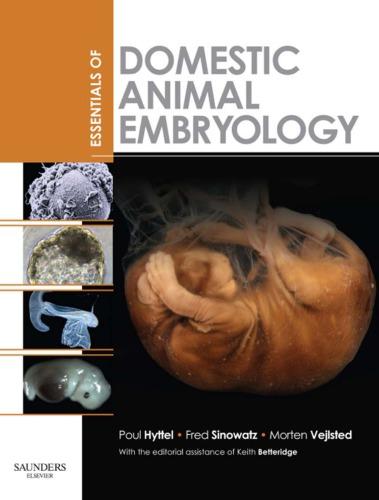
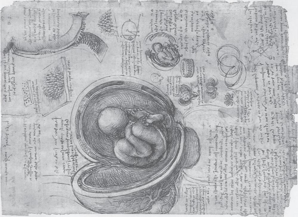
A
B Fig. 1-2: Drawings of Leonardo da Vinci. A: Top: A pregnant bicornuate uterus of the cow. Bottom: Fetus and fetal membranes showing the cotyledons of the placenta (see Chapter 9 ). B: Opened simplex uterus of human showing the fetus with the umbilical cord. Note that the placenta is of the ruminant multiplex type with several placentomes drawn on the cut wall of the uterus and, at the right top, a single cotyledon drawn at a higher magnification displaying its villous surface.
on comparative embryology was De Formato Foetu in 1600 by the Italian anatomist Hieronymus Fabricius of Acquapendente (1533–1619). Fabricius described and illustrated the gross anatomy of embryos and their membranes in that book, but was not actually the first to do so; another Italian anatomist, Bartolomeo Eustachius (1514–1574), had previously published illustrations of dog and sheep embryos in 1552. We now recognize the names of Fabricius, in the term bursa Fabricii (the immunologically competent portion of the bird gut), and of Eustachius in the Eustachian tube.
The work of Eustachius, Fabricius and others gave insight into how organs develop from their immature to mature forms, but left unanswered the basic enigma of how and where the mammalian embryo originates. However, the development of the microscope by Zacharias Janssen, a Dutch eyeglass maker, in 1590 ushered in a new era of embryological science to tackle that 2000yearold question. The Dutch dominance in the optical field at that time may not be just a coincidence; the naval ambitions of their new empire required excellent telescopes and lens systems. Janssen’s microscope in its original form, however, was not really appropriate for cell and tissue research; it was approximately 2 metres long, achieved only 10 to 20 times magnification, and its principal use was to attract an audience at country fairs! In 1672, the Italian medical doctor Marcello Malpighi (1628–1694) published the first microscopic account of chick development, identifying the neural groove, the somites, and circulation of blood in the arteries and veins to and from the yolk. Malpighi also observed that even the unincubated chick egg is considerably structured, leading him to think that a preformed version of the chicken resided in the egg. Later (in 1722), the French ophthalmologist Antoine Maître-Jan (1650–1730) pointed out that although the egg examined by Malpighi was technically ‘unincubated’, it had been left sitting in the Bolognese sun in August and so was certainly not ‘unheated’. Nevertheless, Malpighi’s notion of a preformed chicken initiated one of the great debates in embryology that was to last throughout the 17th and 18th centuries. The question was: are the organs of the embryo formed de novo
(epigenesis), or they are already present in a miniature form in the egg (or the sperm when this cell was discovered), a concept referred to as preformation. We will return to this debate in a moment.
The new microscopic techniques also prompted a vigorous search for the mammalian gametes. The chicken egg and its initial transformation into a chick were obvious, as Aristotle had described, but what mediated the formation of the embryo in mammals? Where was the mammalian egg to be found?
One of the earliest and most influential names in the fascinating story of the discovery of the mammalian egg was that of William Harvey (1578–1657), personal physician to the English kings James I and Charles I, and famous for his description of the circulation of the blood. In 1651, Harvey published De Generatione Animalium (Disputations touching the Generation of Animals) with a famous frontispiece showing Zeus freeing all creation from an egg bearing the inscription Ex ovo omnia (All things come from the egg). However, it should be realized that, far from advancing 17th century knowledge of reproduction and embryology, Harvey’s observations in some ways impeded progress. From having studied with Fabricius, Harvey was imbued with Aristotle’s view that the semen provided a force that interacted with the menstrual blood to materialize as an embryo. Harvey set out to understand this process by looking for the earliest products of conception in female deer killed during the breeding season in the course of King Charles I’s hunts in his Royal forests and parks over a 12year period. In the red and fallow deer that he studied, the male’s rut begins in midSeptember and so Harvey dissected uteri throughout the months of September to December. Believing, wrongly, that copulation coincides with the onset of the rut, Harvey was mystified to find nothing that he recognized as an embryo until midNovember, some two months later. This forced him to the erroneous, but entirely logical conclusion that ‘nothing after coition is to be found in [the] uterus for many days together’. When he did find a conceptus, that, for Harvey, was the egg: ‘Aristotle’s definition of an egg applies to it, namely, an egg is that out of a part of which an animal is
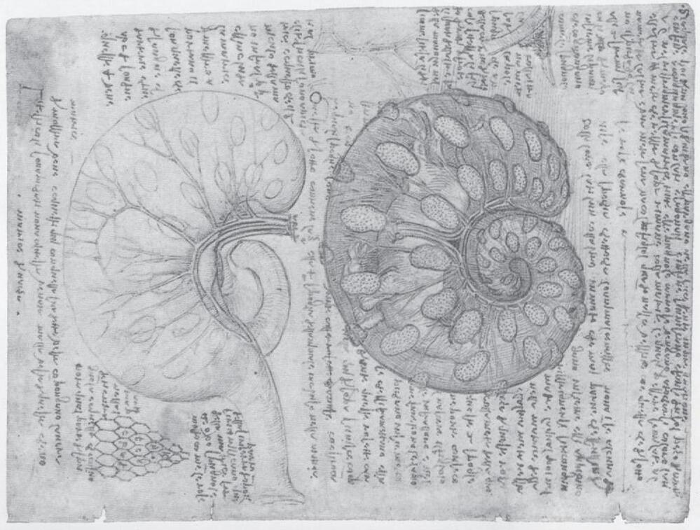
a single germ layer. A remarkable feature of Pander’s book from 1817 is the quality of the illustrations drawn by the German anatomist and artist Eduard Joseph d’Alton (1772–1840); they beautifully depict details that had not yet been defined (Fig. 13). This classical work underlines the necessity for precise observational skills in embryology.
Rathke studied comparative embryology in frogs, salamanders, fish, birds, and mammals and pointed out the similarities in development among all these vertebrate groups. He described for the first time the
pharyngeal arches common to the development of these animals. ‘Rathke’s pouch’ – the ectodermal contribution to the pituitary gland – commemorates him.
In addition to identifying the mammalian ovum, von Baer extended Pander’s observations on chick embryos and described the notochord for the first time. Moreover, von Baer again appreciated the common principles that direct initial embryological development regardless of species; in 1828 he wrote ‘I have two small embryos preserved in alcohol that I forgot to label. At present I am unable to determine the genus to which they belong. They may be lizards, small birds, or even mammals’.
Staining and microscopy techniques continued to improve during the 19th century and allowed for more detailed observations on the initial cleavage stages by the German biologist Theodor Ludwig Wilhelm von Bischoff (1807–1882) in the rabbit, and by the Swiss anatomist and physiologist Rudolph Albert von Kölliker (1817–1905) in man and various domestic animals. Kölliker also published the first textbook on embryology in man and higher animals in 1861.
Thanks to the contributions of Pander, von Baer and Rathke, the preformation school in its radical form ceased in the 1820s. However, the concept survived for another 80 years in the sense that a certain group of scientists regarded the cells of the early cleavage stage embryo to represent right and left halves of the body as it took form. This implied that the information for building the body is segregated regionally in the egg. In 1893, August Weismann (1834–1914) proposed his germ cell plasm theory as an extension of this idea. Based on the sparse knowledge of fertilization available at that time, he was farsighted enough to propose that the egg and the sperm provided equal chromosomal contributions, both quantitatively and qualitatively, to the new organism. Moreover, he postulated that chromosomes carried the inherited potentials of this new organism, which was remarkable at that time considering that the chromosomes had not yet been identified as the carriers of inherited matter. However, Weismann thought that not all information on the chromosomes passed into every cell of
Fig. 1-3: Drawing of a Day 2 chick embryo by Eduard Joseph d’Alton displayed in Pander (1817)
the embryo. Rather, different parts of information went to different cells, explaining their differentiation. Weismann clearly understood the principle of how traits are inherited through fertilization, but he was wrong about the mechanisms of differentiation. Weismann’s differentiation theory was put to the test practically by the German embryologist Wilhelm Roux (1850–1924) who had already, in 1888, published the results of experiments in which individual cells of 2 and 4cell frog embryos were destroyed by a hot needle. As predicted by Weismann’s theory, Roux observed the formation of embryos in which only one side developed normally. These results inspired another German embryologist, Hans Adolf Eduard Driesch (1867–1941) to perform experiments using cell separation instead of Roux’s cell destruction technique. To his enormous surprise, Driesch obtained results that were quite different from those of Roux. Using separated cells from early cleavage stage sea urchin embryos he demonstrated that each of the cells was able to develop into a small but complete embryo and larva (Driesch, 1892). He repeated the same experiment with 4cell embryos and obtained similar results; the larvae were smaller but otherwise looked completely normal.
The final evidence against the RouxWeismann theory was provided by the elegant experiments published by yet another German embryologist, Hans Spemann (1869–1941). Originally, just like Driesch, he had set out to support the theory with his experiments on salamanders. However, by separating the cells of early cleavage stage embryos with a ligature (a hair taken from his newborn son’s head), he soon found that the separated cells were each able to form a small embryo – they were totipotent. In 1928, Spemann conducted the first nuclear transfer experiment, transferring the nucleus of a salamander embryo cell into a cell without a nucleus. Using a hair as a noose, as he had done in his 1902 splitting of the salamander embryo, Spemann tightened the noose around a newly fertilized egg cell, forcing the nucleus to one side and cytoplasm to the other. Next, he waited as the side with the nucleus divided and grew into a 16cell embryo. Then he loosened the noose, and allowed
the nucleus from one of the embryo cells to slip over into the egg cytoplasm on the other side. Spemann then promptly tightened the noose completely, physically breaking the ball of cytoplasm and its new nucleus away from the remains of the 16cell embryo. From this single cell grew a normal salamander embryo, proving that the nucleus from an early embryonic cell was able to direct the complete growth of a salamander. Spemann had created the first clone by nuclear transfer. Spemann published his results in his 1938 book ‘Embryonic Development and Induction’ in which he called for the ‘fantastical experiment’ of cloning from differentiated or adult cells and theoretically paved the way for the cloning by somatic cell nuclear transfer that we know today. Unfortunately, Spemann saw no practical way of realizing such an experiment at that time. Spemann was awarded the Nobel Prize for Physiology or Medicine in 1935 for his discovery of the effect now known as embryonic induction – the influence exercised by various parts of the embryo that directs the development of groups of cells into particular tissues and organs. The works of Driesch and Spemann finally put an end to the concept that inherited information is divided among the cells of the developing embryo.
The whereabouts of inherited materials had still not been determined in the late 19th and early 20th century when a group of American embryologists set out to discover whether inheritance resided in the cytoplasm or the nucleus of the fertilized egg. Edmund Beecher Wilson (1856–1939) was of the opinion that the nucleus is the carrier while Thomas Hunt Morgan (1866–1945) thought the cytoplasm to be responsible. Wilson allied himself with the German biologist Theodor Heinrich Boveri (1862–1915) working at the Naples Zoological Station. Boveri had produced major support for the chromosomal hypothesis of inheritance by fertilizing sea urchin eggs with two spermatozoa. At first cleavage, such eggs produced four mitotic poles and divided into four cells instead of two. Subsequently, Boveri separated the cells and demonstrated that they developed abnormally, each in its own particular way, due to the fact that they carried different chromosomes. Hence, Boveri claimed that each chromo
some is distinct and controls different vital processes. Wilson and Nettie Maria Stevens (1861–1912), one of the first American women to be recognized for her contribution to science, extended the work of Boveri. They demonstrated the relationship between chromosomes and sex: XO or XY embryos developed into males and XX embryos into females (Wilson, 1905; Stevens, 1905a,b). For the first time a particular phenotypical characteristic was clearly correlated with a property of the nucleus. Eventually, Morgan found mutations that correlated with sex and with the X chromosome. This persuaded him that his earlier view that inheritance was through the cytoplasm was wrong and that genes are physically linked to one another on the chromosomes. Consequently, a group of embryologists had laid a cornerstone to the discovery that the chromosomes in the cell nucleus are responsible for the development of inherited characteristics.
In the early 20th century, embryology and genetics were not considered separate sciences. They diverged in the 1920s when Morgan redefined genetics as the science studying the transmission of inherited traits, distinguishing it from embryology, the science studying the expression of those traits. This division did not occur without hostility; geneticists considered the embryologists oldfashioned while embryologists looked upon geneticists as being uninformed about how organisms actually develop! Fortunately, we nowadays see a rapprochement of genetics and embryology in a very fruitful symbiosis. Two of the scientists who advocated synergism between embryology and genetics in the early days were Salome Gluecksohn-Schoenheimer (now GluecksohnWaelsch; 1907–2007) and Conrad Hal Waddington (1905–1975). GluecksohnSchoenheimer received her doctorate in Spemann’s laboratory, but fled Hitler’s Germany for the United States. Her farsighted research demonstrated that mutations in the Brachyury gene of the mouse caused aberrant development of the posterior portion of the embryo, and she localized the defect to the notochord (GluecksohnSchoenheimer, 1938, 1940), providing another example of the close link between embryology and genetics. Interestingly, it took 50 years for her results to be confirmed by DNA hybridization after cloning
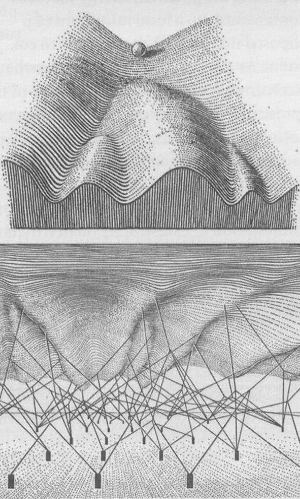
of the Brachyury gene (Wilkinson et al., 1990). Waddington, on the other hand, addressed the causal link between embryology and genetics by isolating several genes that caused wing malformations in fruit flies. Moreover, his interpretation and visual conception of ‘the epigenetic landscape’ affecting initial cell differentiation in the embryo still surfaces during contemporary presentations on embryonic stem cells and their differentiation (Waddington, 1957, Fig. 14).
Fig. 1-4: Top: CH Waddington’s depiction of the epigenetic landscape with the ball representing a cell and the valleys representing different avenues of differentiation. Bottom: A less commonly depicted view behind the epigenetic landscape illustrating how the tension of different genes control the fate of the ball. From Waddington (1957)
The question of totipotency, initially addressed by Driesch and Spemann, was later revisited at a finer level. Thus, whereas Driesch and Spemann had proved the totipotency of the cells of the early cleavage stage embryo, experiments in the 1950s by Robert Briggs (1911–1983) and Thomas King (1921–2000) tested the totipotency of the nucleus or rather the genome. Their nuclear transfer model was an exact realization of the ‘fantastic experiment’ proposed by Hans Spemann, although they had never heard about his suggestion. To accomplish their objective they had to develop methods by which they could remove the genome of an egg without destroying it (enucleation), pick up a donor nucleus of another cell, and transfer that nucleus to the enucleated egg. As their approach was extremely unorthodox, their first grant application to the National Cancer Institute was refused as a ‘harebrained’ idea. However, they eventually obtained some support, and after months of experimentation they produced the first blastocyst from nuclear transfer. Their initial success was shortlived. In their enthusiasm, they gathered the complete staff of the institute to show them the blastocyst. After numerous looks into the microscope, followed by applause and congratulations, they rechecked the dish and found only a completely destroyed embryo. Fortunately, although the first embryo died, the nuclear transfer system worked; in 1952, Briggs and King successfully demonstrated that donor nuclei from frog blastula stages could direct the development of complete tadpoles when transferred into enucleated eggs. This research further paved the way for the somatic cell nuclear transfer that is nowadays used for cloning of mammals. Briggs and King also discovered that when cells from later stages (tailbudstage tadpoles for example), were used as nuclear donors, normal development did not occur unless the nuclei came from the germ cells. Thus, somatic cells appeared to lose their ability to direct development as their degree of differentiation increased. This point was later pursued by John B. Gurdon who worked with another frog species, Xenopus, rather than Briggs and King’s Rana. Gurdon et al. (1975) found that when nuclei of cultured skin cells from adult frogs were transferred into enucleated eggs, development of the
clones never progressed beyond the formation of the neural tube. However, when serial nuclear transfers were made from the cloned embryos to other enucleated eggs, it was possible to generate numerous tadpoles; the genomic totipotency of somatic cells had been proven. It should be noted though that the frog experiments never managed to close the developmental circle by producing an adult organism by transferring a somatic cell nucleus from another adult organism.
Closing the circle did not happen until the nuclear transfer technique was transposed to mammals by the Danish veterinarian Steen Malte Willadsen working in Cambridge during the 1980s. Willadsen (1986) succeeded in transferring not just the nucleus, but the entire cell from sheep morulastage embryos to enucleated eggs by electrical cell fusion. His work resulted in the first mammal to be born after cloning by nuclear transfer. In 1996, this technology was taken one step further by Keith H Campbell, working at the Roslin Institute in Scotland in a research group headed by Ian Wilmut Campbell et al. (1996) succeeded in producing lambs following transfer of nuclei of cultured cells, harvested from the inner cell mass, to enucleated eggs. Key to this success was Campbell’s meticulous cell cycle experimentation that demonstrated the need for a certain degree of synchrony of cell cycle between the donor nucleus and the recipient cytoplasm. The ability to clone mammals from cultured cells represented a major breakthrough in biomedical science, facilitating genetic manipulation of the cells prior to nuclear transfer and opening an avenue for production of transgenic animals (animals in which a foreign gene, the transgene, has been added). Consequently, Angelika Schnieke, working in the same group of researchers, was able to announce the birth of the transgenic sheep Polly, a lamb cloned from cultured fetal sheep fibroblasts into which the gene for human clotting factor IX had been inserted with a promoter that would allow for expression of the transgene in the mammary gland (Schnieke et al., 1997). It was from that event that the concept of ‘biopharming’ (production of valuable proteins in transgenic animals) emerged. The report on Polly, however, was preceded by another from the Roslin group; a
publication that stunned not only the scientific community, but all layers of society, worldwide. That was the report of the birth of the cloned lamb Dolly (Wilmut et al. 1997). Dolly was created by Wilmut and his group by transferring cultured mammary gland cells from a 6yearold ewe into enucleated eggs. Again, the success depended on control of the cell cycle; the mammary gland nuclear donor cells were kept under culture conditions that suppressed their mitotic activity, provoked a state of cellular senescence, and locked them at the G1 state of the cell cycle, or G0. Research since then has resulted in the cloning of many animals of many species (including cattle, mice, goats, pigs, cats, rabbits, horses, dogs, and rats) and has also demonstrated that bringing the nuclear donor cells into quiescence is not a necessity.
In its combination with genetics, embryology is an exponentially developing science; how it will continue to develop, only time can tell. As a subject, embryology made its way into the curriculum of veterinary medicine in the mid 19th century when it became incorporated into the teaching of anatomy. In 1924, the first textbook on veterinary embryology was published by Zeitzschmann and others (though not many) have appeared since. Embryology of the Pig by Bradley M Patten deserves special mention as it has been an admirable resource and inspiration for the authors of this book. Likewise, the ‘bible’ of comparative embryology Developmental Biology by Scott F. Gilbert we have found admirable for its breadth of coverage and for its inimitable style. Because embryology was originally based upon the anatomical descriptive tradition and also entered the curricula of medicine and veterinary medicine as part of anatomy, its nomenclature is Latin and Greekbased. For ease of reading, however, we have anglicized some terms rather than use their strict Latin or Greek forms.
FURTHER READING
Aristotle (ca. 350 BC): The generation of animals. A.L. Peck (trans.). eBooks@Adelaide, 2004 (see http://etext.library. adelaide.edu.au/a/aristotle/generation/).
History of embryology
Baer, K.E.V. (1827): De ovi mammalium et hominis genesi. Voss, Leipzig.
Bonnet, C. (1764): Contemplation de la Nature. MarcMichel Ray, Amsterdam.
Briggs, R. and King, T.J. (1952): Transplantation of living nuclei from blastula cells into enucleated frogs’ eggs. Proc. Natl. Acad. Sci. USA 38:455–464.
Campbell, K.H., McWhir, J., Ritchie, W.A. and Wilmut I. (1996): Sheep cloned by nuclear transfer from a cultured cell line. Nature 380:64–66.
Darwin, C. (1859): On the Origin of Species by Means of Natural Selection, or the Preservation of Favoured Races in the Struggle for Life. John Murray, London.
Fabricius, H. of Aquapendente (1600): De formato foetu. Pasquala, Padova.
Gilbert, S.F. (2003): Developmental biology. 7th edn. Sinauer Associates, Sunderland, Massachusetts.
GluecksohnSchoenheimer, S. (1938): The development of two tailless mutants in the house mouse. Genetics 23:573–584.
GluecksohnSchoenheimer, S. (1940): The effect of an early lethal (t0) in the house mouse. Genetics 25:391–400.
Gurdon, J.B., Laskey, R.A. and Reeves, O.R. (1975): The developmental capacity of nuclei transplanted from keratinized cells of adult frogs. J. Embryol. Exp. Morphol. 34:93–112.
Harvey, W. (1651): Excitationes de generatione animalium. Elzevier, Amsterdam.
Kölliker, A. (1881): Entwiklungsgeschichte des Menschen und der höhere Thiere. Engelmann, Leipzig.
MaîtreJan, A. (1722): Observations sur la formation du poulet. L. d’Houdry, Paris.
Malpighi, M. (1672): De formatione pulli in ovo (London). Reprinted in HB Adelmann ‘Marcello Malpighi and the evolution of embryology’. Cornell University Press, Ithaca, NY, 1966.
Pander, H.C. (1817/18): Beitrage zur Entwiklungsgeschichte des Hühnchens in Eye. Brönner, Wüzburg.
Patten, B.M. (1948): Embryology of the pig. 3rd edn. Blakiston Company, New York, Toronto.
Roux, W. (1888): Contributions to the developmental mechanisms of the embryo. On the artificial production of half embryos by destruction of one of the first two blastomeres and the later development (postgeneration) of the missing half of the body. In B.H. Willier and J.M. Oppenheimer (eds.) 1974 ‘Foundations of experimental embryology’, Hafner, New York, pp. 237.
Schnieke, A., Schnieke, A.E., Kind, A.J., Ritchie, W.A., Mycock, K., Scott, A.R., Ritchie, M., Wilmut, I., Colman, A. and Campbell, K.H. (1997): Human factor IX transgenic sheep produced by transfer of nuclei from transfected fetal fibroblasts. Science 278:2130–2134.
Schwann, T. and Schleyden, M.J. (1847): Microscopical researches into the accordance in the structure and growth of animals and plants. London: Printed for the Sydenham Society.