Eric Whaites, MSc, BDS(Hons), FDSRCS(Edin), FDSRCS(Eng), FRCR, DDRRCR
Senior Lecturer and Honorary Consultant in Dental and Maxillofacial Radiology, King’s College London Faculty of Dentistry, Oral & Craniofacial Sciences, at Guy’s and St THomas’ NHS Foundation Trust, London, UK
Nicholas Drage, BDS (Hons), FDSRCS (Eng), FDSRCPS (Glas), DDRRCR
Consultant and Honorary Senior Lecturer in Dental and Maxillofacial Radiology, University Dental Hospital, Cardiff and Vale University Health Board, Cardiff, UK
Table of Contents
Cover image
Title page
Dedication
Copyright
Preface
Acknowledgements
Part I. Introduction
1. The Radiographic Image
Introduction
Nature of the Traditional Two-Dimensional Radiographic Image
Quality of the Traditional Two-Dimensional Radiographic Image
Perception of the Radiographic Image
Types of Traditional Dental Radiographs
Part II. Radiation Physics, Equipment and Radiation Protection
2. The Production, Properties and Interactions of X-rays
Introduction
Atomic Structure
X-Ray Production
Interaction of X-Rays with Matter
3. Dental X-ray Generating Equipment
Introduction
Ideal Requirements
Main Components of the Tubehead
Other X-Ray Generating Apparatus
4. Image Receptors
Introduction
Digital Receptors
Radiographic Film
Characteristics of Radiographic Film
Background Fog Density
Intensifying Screens
Cassettes
Important Practical Points to Note
5. Image Processing
Computer Digital Processing
Chemical Processing
6. Radiation Dose, Dosimetry and Dose Limitation
Dose Units
Dose Limits
Dose Rate
Dose Limitation
7. The Biological Effects Associated with X-rays, Risk and Practical Radiation Protection
Radiation-Induced Tissue Damage
Classification of the Biological Effects
Practical Radiation Protection
Footnote
Part III. Radiography
8. Dental Radiography – General Patient Considerations including Control of Infection
General Guidelines on Patient Care
Specific Requirements When X-Raying Children and Patients with Disabilities
Control of Infection
Micro-Organisms That May Be Encountered
Standard and Transmission-Based Precautions
Infection Control Measures
Footnote
9. Periapical Radiography
Main Indications
Ideal Positioning Requirements
Radiographic Techniques
Anatomy
Bisected Angle Technique
Comparison of The Paralleling and Bisected Angle Techniques
Conclusion
Positioning Difficulties Often Encountered in Periapical Radiography
10. Bitewing Radiography
Main Indications
Ideal Technique Requirements
Positioning Techniques
Resultant Radiographs
Assessment of Image Quality
11. Occlusal Radiography
Introduction
Terminology and Classification
Upper Standard (or Anterior) Occlusal
Upper Oblique Occlusal
Lower 90° Occlusal
Lower 45° (or Anterior) Occlusal
Lower Oblique Occlusal
12. Oblique Lateral Radiography
Introduction
Terminology
Main Indications
Basic Technique Principles
Positioning Examples For Various Oblique Lateral Radiographs
Bimolar Technique
13. Skull and Maxillofacial Radiography
Equipment, Patient Positioning and Projections
14. Cephalometric Radiography
Introduction
Main Indications
Equipment
Main Radiographic Projections
Cephalometric Posteroanterior of the Jaws (PA Jaws)
15. Tomography and Panoramic Radiography
Introduction
Tomographic Theory
Panoramic Tomography
Selection Criteria
Equipment
Technique and Positioning
Normal Anatomy
Advantages and Disadvantages
Assessment of Image Quality
Assessment of Not Acceptable Images and Determination of Errors
Footnote
16. Cone Beam Computed Tomography
Main Indications
Equipment and Theory
Technique and Positioning
Normal Anatomy
Radiation Dose
Advantages and Disadvantages
Assessment of Image Quality
Quality Assurance
Footnote
17. The Quality of Radiographic Images and Quality Assurance
Introduction
Quality Assurance (QA)
Digital Image Quality
Quality Control Procedures for Digital Receptors, Computer Processing and Monitors
Film-Based Image Quality
Quality Control Procedures for Film, Chemical Processing and Light Boxes
Patient Preparation and Positioning Technique Errors in Both Digital and Film-Based Imaging
Patient Dose and X-Ray Generating Equipment
Working Procedures
Staff Training and Updating
Audits
Footnote
18. Alternative and Specialized Imaging Modalities
Introduction
Contrast Studies
Radioisotope Imaging
Computed Tomography (CT)
Ultrasound Examinations
Magnetic Resonance Imaging (MRI)
Part IV. Radiology
19. Introduction to Radiological Interpretation
Essential Requirements for Interpretation
Conclusion
20. Dental Caries and the Assessment of Restorations
Introduction
Classification of Caries
Diagnosis and Detection of Caries
Other Important Radiographic Appearances
Limitations Of Radiographic Detection of Caries
Radiographic Assessment of Restorations
Limitations of the Radiographic Image
Suggested Guidelines for Interpreting Bitewing Images
Cone Beam Computed Tomography
21. The Periapical Tissues
Introduction
Normal Radiographic Appearances
The Effects of Normal Superimposed Shadows
Radiographic Appearances of Periapical Inflammatory Changes
Treatment and Imaging in Endodontics
Other Important Causes of Periapical Radiolucency
Suggested Guidelines for Interpreting Periapical Images
22. The Periodontal Tissues and Periodontal Disease
Introduction
Selection Criteria
Radiographic Features of Healthy Periodontium
Classification of Periodontal Diseases
Radiographic Features of Periodontal Disease and the Assessment of Bone Loss and Furcation Involvement
Evaluation of Treatment Measures
Limitations of Radiographic Diagnosis
23. Implant Assessment
Introduction
Main Indications
Main Contraindications
Treatment Planning Considerations
Radiographic Examination
Peri-operative Imaging
Post-operative Evaluation and Follow-Up
Footnote
24. Developmental Abnormalities
Introduction
Classification of Developmental Abnormalities
Typical Radiographic Appearances of The More Common and Important Developmental Abnormalities
Radiographic Assessment of Mandibular Third Molars
Radiographic Assessment of Unerupted Maxillary Canines
25. Radiological Differential Diagnosis – Describing a Lesion
Introduction
Detailed Description of a Lesion
Footnote
26. Differential Diagnosis of Radiolucent Lesions of the Jaws
Introduction
Step-By-Step Guide
Typical Radiographic Features of Radiolucent Cysts and Tumours
Typical Radiographic Features of Tumours
Typical Radiographic Features of Allied Lesions
Footnote
27. Differential Diagnosis of Lesions of Variable Radiopacity in the Jaws
STEP I
STEP II
STEP III
STEP IV
STEP V
Typical Radiographic Features of Abnormalities of the Teeth
Typical Radiographic Features of Conditions of Variable Opacity Affecting Bone
Summary
Typical Radiographic Features of Soft Tissue Calcifications
Typical Radiographic Features of Foreign Bodies
28. Bone Diseases of Radiological Importance
Introduction
Developmental or Genetic Disorders
Infective or Inflammatory Conditions
Hormone-Related Diseases
Blood Dyscrasias
Diseases of Unknown Cause
29. Trauma to the Teeth and Facial Skeleton
Introduction
Injuries to the Teeth and Their Supporting Structures
Skeletal Fractures
Fractures of the Mandible
Fractures of the Middle Third of the Facial Skeleton
Other Fractures and Injuries
30. The Temporomandibular Joint
Introduction
Normal Anatomy
Investigations
Main Pathological Conditions Affecting the TMJ
Footnote
31. The Maxillary Antra
Introduction
Normal Anatomy
Normal Appearance of the Antra on Conventional Radiographs
Antral Disease
Investigation and Appearance of Disease Within the Antra
The right of Eric Whaites and Nicholas Drage to be identified as authors of this work has been asserted by them in accordance with the Copyright, Designs and Patents Act 1988.
No part of this publication may be reproduced or transmitted in any form or by any means, electronic or mechanical, including photocopying, recording, or any information storage and retrieval system, without permission in writing from the publisher. Details on how to seek permission, further information about the Publisher’s permissions policies and our arrangements with organizations such as the Copyright Clearance Center and the Copyright Licensing Agency, can be found at our website: www.elsevier.com/permissions .
This book and the individual contributions contained in it are protected under copyright by the Publisher (other than as may be noted herein).
Notices
Practitioners and researchers must always rely on their own experience and knowledge in evaluating and using any information,
methods, compounds or experiments described herein. Because of rapid advances in the medical sciences, in particular, independent verification of diagnoses and drug dosages should be made. To the fullest extent of the law, no responsibility is assumed by Elsevier, authors, editors or contributors for any injury and/or damage to persons or property as a matter of products liability, negligence or otherwise, or from any use or operation of any methods, products, instructions, or ideas contained in the material herein.
It is now nearly 30 years since the first edition of Essentials was published and nearly 6 years since the first co-authored fifth edition. This sixth edition is again co-authored with my friend and colleague Nicholas Drage and marks the last edition that I will be directly involved with, given that I am in the twilight of my career. It is reassuring to know that Essentials will be in Nicholas’ safe hands.
I am delighted that we have been given this final opportunity to update and refresh Essentials together and to be able to introduce colour images throughout – we hope this has given the book a completely new and modern feel. In addition to the new photographic material, we have revised and updated nearly every chapter. We have tried to align the content and terminology more closely with the new international classifications in relation to cysts, tumours, bone lesions and diseases, caries and periodontal diseases. Many new radiographs have been added and we have placed greater emphasis on examples showing normal radiographic anatomy, working on the principle that the main reason for radiographic imaging is to be able to distinguish the difference in appearance between normal and abnormal, to determine the presence or absence of (hard tissue) disease. Another major change has been to reorder the contents of several chapters so that digital imaging is described first, recognising the switch from film-based imaging to digital imaging.
The website created with, and linked to, the previous edition proved popular with students, particularly the online self-assessment questions relating to each chapter, so these have also been reviewed and new questions added as appropriate.
Despite the various updates and revisions, the aims and objectives of this book remain the same, namely to provide a basic, but comprehensive, practical account of what we consider to be the essential subject matter of both dental radiography and radiology as required by undergraduate and postgraduate dental students. As in previous editions, some things have had to be omitted, or sometimes, over-simplified. However, the book remains first and foremost a teaching manual (albeit more colourful) rather than a comprehensive reference. We believe that the content remains sufficiently broad, detailed and up-to-date to satisfy the requirements of most undergraduate and postgraduate examinations and the needs of most general dental practitioners. Students are encouraged to build on the information included here by using the excellent and more comprehensive textbooks available.
We hope that once again the result is a clear, logical and easily understandable textbook that continues to make a positive contribution to the challenging tasks of teaching and learning dental radiology.
EW
Acknowledgements
As with previous editions, this edition has only been possible thanks to the enormous amount of help and encouragement that we have received from our various friends and colleagues in both London and Cardiff.
In particular, we would like to thank Nigel Pearson, Head of the Medical Photography Department at Guy’s and St Thomas’, and Sam Evans from the Dental Photography Unit, Cardiff University, who together are responsible for all the new photographic images. Special thanks to Beatrice Cicchetti for volunteering to be our adult patient model – her patience and ever-present smile made hours of photography bearable for all of us! Our thanks also to Clive and Charlene Bathurst for allowing their daughter Hayley to be our child patient model. Our thanks also to Richard Beal (Clarke Dental Limited), Paul Keeley (Henry Schein Dental) and Jay McKay (SironaDentsply) for supplying some of the equipment for photography purposes.
We are very grateful to several colleagues for their help and advice with specific chapters. They include Melanie Wilson, Senior Lecturer in Oral Microbiology in Cardiff for Chapter 8 ; Dirk Bister, Consultant in Orthodontics at Guy’s for Chapter 14 ; Simon Harvey, Consultant in Dental & Maxillofacial Radiology at Guy’s for Chapter 18 ; Avi Banerjee, Professor of Cariology at King’s College London for Chapter 21 ; Francis Hughes, Professor of Periodontology at King’s College London for Chapter 22 , George Paolinelis, Consultant in Oral Surgery at Guy’s for Chapter 23 and Eddy Odell, Professor of Oral Pathology, King’s College London for Chapters 26 and 27 . In addition, Dental Radiology Departmental colleagues at Guy’s willingly offered their
help and advice – in particular we would like to thank Jackie Brown, Bethan Thomas, John Rout, Niall O’Neill, Amanda Loughlin, Beatrice Cicchetti and Stephen Goss. We are also very grateful to Andrew Gulson, from Public Health England for his permission to allow us to reproduce parts of the 2020 Guidance Notes. Special thanks to Allisson Summerfield for co-ordinating the IT and imagery, the hours she spent photocopying and electronically correcting the proofs, and for always being on hand to ensure the job got done! Many thanks also to Elsevier’s Alison Taylor, our now retired Content Strategist, Fiona Conn for masterminding the manuscript to get it ready for production, and Karthikeyan Murthy and his team for their huge effort in getting the book to final publication.
Last, but by no means least, we would once again like to thank our wives Catriona and Anji and our children Stuart, Felicity and Claudia, and Karisma and Jaimini for their love, understanding and encouragement throughout the production of this edition and for accepting the sacrifice of family time that it has involved.
PART I Introduction OUTLINE
1. The Radiographic Image
1
The Radiographic Image
Introduction
The use of X-rays is an integral part of clinical dentistry, with some form of radiographic examination necessary on the majority of patients. Radiographs are primarily taken to determine the presence or absence of underlying hard tissue disease affecting the teeth and/or bones. As a result, radiographs are often referred to as the clinician’s main diagnostic aid . Traditional radiography enables the creation of conventional two-dimensional radiographic images, but technological advances in computer generated images – so-called cone beam computed tomography (CBCT) - has resulted in imaging in three dimensions and improved diagnostic capability (see Figs. 1.1 and 1.2 ).
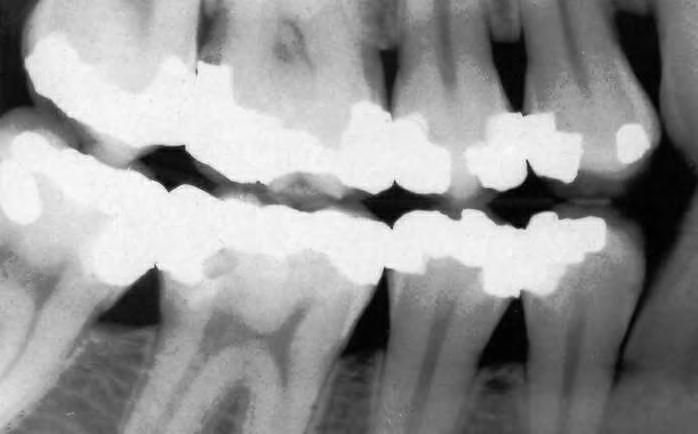
FIG. 1.1 A traditional two-dimensional dental radiograph showing the hard tissues of the teeth, containing metallic amalgam restorations, and the supporting alveolar bone.
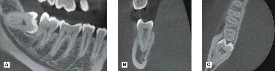
FIG. 1.2 Modern cone beam computed tomography (CBCT) radiographic images in three dimensions of the lower right molar teeth in the (A) sagittal, (B) coronal and (C) axial planes.
The range of knowledge of dental radiography and radiology thus required can be divided conveniently into four main sections:
• Basic physics, X-ray equipment and image receptors – how X-rays