Essential Microbiology for Dentistry
Fifth Edition
Lakshman Samaranayake
DSc(hc) DDS(Glas) FRCPath FDSRCS(Ed) FDS RCPS(Glas) FRACDS FHKCPath FCDSHK
Vice-Dean, College of Dental Medicine, University of Sharjah, UAE
Professor Emeritus & Immediate-Past Dean of Dentistry, University of Hong Kong, Hong Kong
Honorary Professor & Immediate-Past Head, School of Dentistry, University of Queensland, Australia
King James IV Professor, Royal College of Surgeons of Edinburgh, UK (2013)
© 2018 Elsevier Ltd. All rights reserved.
No part of this publication may be reproduced or transmitted in any form or by any means, electronic or mechanical, including photocopying, recording, or any information storage and retrieval system, without permission in writing from the publisher. Details on how to seek permission, further information about the publisher’s permissions policies and our arrangements with organizations such as the Copyright Clearance Center and the Copyright Licensing Agency, can be found at our website: www.elsevier. com/permissions.
This book and the individual contributions contained in it are protected under copyright by the publisher (other than as may be noted herein).
First edition 1996
Second edition 2002
Third edition 2006
Fourth edition 2012
Fifth edition 2018
ISBN 9780702074356
Notice
Practitioners and researchers must always rely on their own experience and knowledge in evaluating and using any information, methods, compounds or experiments described herein. Because of rapid advances in the medical sciences, in particular, independent verification of diagnoses and drug dosages should be made. To the fullest extent of the law, no responsibility is assumed by Elsevier, authors, editors or contributors for any injury and/or damage to persons or property as a matter of products liability, negligence or otherwise, or from any use or operation of any methods, products, instructions, or ideas contained in the material herein.
Printed in Poland Last digit is the print number: 9 8 7 6 5 4 3 2 1
The publisher’s policy is to use paper manufactured from sustainable forests
Welcome to the fifth edition of Essential Microbiology for Dentistry!
It is now 22 years since the first edition of this tome was published in 1996, and since then, the science of microbiomics and infectious diseases has advanced in leaps and bounds. The two major reasons for these transformational changes have been the exploding new technology that delivers novel tools for the identification and reclassification of organisms, and the emergence of new organisms, especially the viruses that change the landscape of dental and medical practice. For instance, next generation sequencing (NGS) technology has revolutionized the field of microbial taxonomy and identification of, in particular, the uncultivable organisms, leading to a radical rethink on the quantity and quality of the flora that inhabit our body, including the oral cavity. In this, the fifth edition of this book, I have attempted to incorporate the new data as much as possible while maintaining its popular concise, yet comprehensive outlook.
The fact that you are now reading the fifth edition of the book is testimony to its popularity, with more than 40,000 copies sold in all five continents; Chinese, Polish and Korean translations as well as Middle East Editions (Al-Farabi Version) of the book are now in print, although the e-print of the book appears to be outstripping the hard copy sales. For this, I am deeply grateful to the microbiology teachers in dental schools/ colleges, as well as the undergraduates and the postgraduates who are avid fans worldwide.
In compiling this completely revised fifth edition, I have retained the popular features of the last few editions. One major feature of this edition is an expanded section on infection control (Part 6), which I co-edited with Dr Caroline Pankhurst, of University of London, UK. Other novel additional features are sections on NGS technology; the oral microbiome and the microbiota; endodontic infections; implant-related infections; plaque biofilms and the systemic disease axis and the current guidelines on antimicrobial prophylaxis.
Of course, a tome of this nature cannot be produced without the help of many friends and colleagues. The legacy authors of the Immunology Section (Part 2) were Dr Brian Jones and Professor Liwei Lu, from the University of Hong Kong, while Professor Glen C Ulett of Griffith University, Australia expanded and embellished these chapters as well as other sections of the tome. To them, my extreme gratitude.
Once again, I am indebted to the following colleagues worldwide, who graciously permitted the reproduction of their work: Professor H Jenkinson, University of Bristol, UK (Fig. 3.9); Dr Bernard Low, Malaysia (Fig. 5.1); Professor Willie van Heerden, University of Pretoria, South Africa (Figs 18.4 and 19.1); Dr Maribasappa Karched of Kuwait University (Fig. 31.2); Dr Leanor Haley, CDC, Atlanta, USA (Fig. 22.5); Dr Annette Motte, Free University of Berlin, Germany (Fig. 31.8); and Professor Saso Ivanowski, Griffith University, Australia (Fig. 33.8). Figures 38.1 and 38.5 are reproduced from UK Health Technical Memorandum No. 01-05, 2009, with permission from Crown Copyright.
As always, the publishing team at Elsevier led by Martin Mellor, Alison Taylor and Helen Leng has pushed me to beat the deadlines despite my myriad duties. Their professionalism and patience has my admiration and gratitude. Last but not least, Hemamali, Dilani and Asanka have lost some quality family time due to this tome, and I am eternally grateful to them for their tolerance and understanding.
Above all, YOU, the reader, are my most important friend and critic! The many features of this edition are due to your feedback over two decades, and I truly hope that the current edition is the finest product thus far. Nevertheless, no book is perfect—so please keep on sending your comments, either good or bad, to me at lakshman@hku.hk
Lakshman Samaranayake
Hong Kong August 2017
Vancomycin-resistant S.aureus
Cyclosporiasis
Multidrug-resistant tuberculosis
Drug-resistant malaria
The reasons for their emergence are manifold and include:
■ societal events: economic impoverishment (especially in the developing world), war and civil conflicts, as well as mass population migration.
■ health care: new medical devices, organ/tissue transplantation, immunosuppression, antibiotic abuse and contaminated blood and blood products.
■ human behaviour: increasing number of sexual partners, injectable drug abuse.
■ environmental changes: deforestation, drought, floods and global warming.
■ microbial adaptation: emergence of new species from the wild (e.g., HIV), changes in virulence and toxin production and development of drug resistance.
About this book
This text is divided into six parts in order to highlight the different features of microbiology related to dentistry, but it should be noted that such division is artificial and is merely an attempt to simplify the learning process.
The first few chapters in Part 1 essentially describe general microbiological features of bacteria and viruses and how they cause human infections (i.e., pathogenesis). Diagnostic microbiology, by which clinical microbiologists ascertain the nature of agents causing various infections, is described in Chapter 6 The laboratory aspect of this fascinating subject is analogous to the work of a crime detection bureau! When a specimen (e.g., pus, urine) from a patient with an infectious disease is sent
O157:H7 H5N1 influenza
Vancomycinresistant S.aureus
to the laboratory for identification of the offending agent, the clinical microbiologist utilizes many methods and techniques, as well as a fair amount of thought and contemplation, to identify the pathogen/s lurking in the clinical sample. In many situations the pathogen may be dead, in which case other, indirect clues via molecular techniques need to be pursued to incriminate the suspect pathogen. Once an offending pathogen is identified, antimicrobial chemotherapy is the mainstay of treatment; a description of chemotherapeutic agents and how they are chosen in the laboratory is given in Chapter 7.
The host responds to infection by mounting an immune response. A highly abbreviated account of basic immunology is given in Part 2; supplemental reading is essential to augment this material, and the reader is referred to the lists of recommended texts for this purpose. Immunological nomenclature is complex and often difficult: a glossary of terms and abbreviations is therefore provided as an appendix.
Although there are thousands of offending pathogens, only some are of direct relevance to dental practice and to the comprehension of the mechanisms of disease; these are described in Part 3. Arguably this section may appear to be the most daunting part of the book because of the complex nomenclature of microbes; hence only the salient bacterial genera—some of which are more closely related to dental practice (e.g., streptococci) than others (e.g., legionellae)—are outlined. Similarly, the chapters on viruses and fungi are relatively brief, with thumbnail sketches of only the most relevant organisms.
The major infections of each organ system are discussed in Part 4, with emphasis on those that are most relevant to dentistry. The student is strongly advised to cross-refer to
Cryptosporidiosis HIV
Ebola haemorrhagic fever
Marburg haemorrhagic fever
Hantavirus pulmonary syndrome
Whitewater Arroyo virus
E. coli
O157:H7
vCJD
Hepatitis C
SARS E. coli
Nipah virus
Hendra virus
Enterovirus 71
Lassa fever
Anthrax bioterrorism
Diphtheria
Typhoid fever
Rift Valley fever
Cholera Yellow fever
Dengue
Human monkeypox
West Nile virus
Lyme disease
Plague
Human monkeypox
Fig. 1.1 Global prevalence of some emerging and re-emerging diseases. E. coli, Escherichia coli; HIV, human immunodeficiency virus; SARS, severe acute respiratory syndrome; S. aureus, Staphylococcus aureus; vCJD, variant Creutzfeldt–Jakob disease.
General microbiology 1
The aim of this section is to present (1) the structural features of microbes and how they cause disease, and (2) a perspective of diagnostic laboratory methods to explain the relationship between the scientific basis of microbiology and its practical application in patient care. Finally, a comprehensive overview of antimicrobial chemotherapy in dentistry is provided, which should be supplemented with additional reading due to its critical relevance to dental care.
■ Bacterial structure and taxonomy
■ Bacterial physiology and genetics
■ Viruses and prions
■ Pathogenesis of microbial disease
■ Diagnostic microbiology and laboratory methods
■ Antimicrobial chemotherapy
Table 2.1 Differential characteristics of major groups of organisms
Visible with light microscope
Capable of free growth
Both DNA and RNA present
Muramic acid in cell wall
Rigid cell wall
Susceptible to penicillin Variable
Susceptible to tetracycline
Reproduce essentially by binary fission
aPrions (agents responsible for Creutzfeldt–Jakob disease) are not included as their status is unclear.
Table 2.2 Major differences among the three domains of life
Organization of the genetic material and replication
DNA free in the cytoplasm
Only one chromosome
DNA associated with histone-like proteins
May contain extrachromosomal elements called plasmids
Introns not found in mRNA
Cell division by binary fission: asexual replication only
Transfer of genetic information occurs by conjugation, transduction and transformation (see Chapter 3)
Cellular organization
Cytoplasmic membrane contains hopanoids
DNA free in the cytoplasm
Only one chromosome
DNA associated with histone-like proteins
Plasmids may be found
Introns not found in most genes
Reproduce asexually and spores are not found
Processes similar to bacterial conjugation enable exchange of genetic material
DNA is contained with a membrane-bound nucleus. A nucleolus is also present
More than one chromosome. Two copies of each chromosome may be present (diploid)
DNA complexed with histone proteins
Plasmids only found in yeast
Introns found in all genes
Cells divide by mitosis
Exchange of genetic information occurs during sexual reproduction. Meiosis leads to the production of haploid cells (gametes), which can fuse
Membranes contain isoprenes
Lipopolysaccharides and teichoic acids found No lipopolysaccharides or teichoic acids found
Energy metabolism associated with the cytoplasmic membrane
Photosynthesis associated with membrane systems and vesicles in cytoplasm
Flagella consist of one protein, flagellin
Ribosomes: 70S
Peptidoglycan cell walls
Contains flagella that derive energy from proton pumps
Ribosomes behave more like eucarya when exposed to inhibitors
Cell walls lack peptidoglycan
Cytoplasmic membrane contains sterols
Mitochondria present in most cases
Chloroplasts present in algal and plant cells
Internal membranes, endoplasmic reticulum and Golgi apparatus present associated with protein synthesis and targeting
Membrane vesicles such as lysosomes and peroxisomes present
Cytoskeleton of microtubules present
Flagella have a complex structure with 9 + 2 microtubular arrangement
Ribosomes: 80S (mitochondrial and chloroplast ribosomes are 70S)
Polysaccharide cell walls, where present, are generally either cellulose or chitin
Gram-positive
Flagella
Pilus
Capsule (variable)
Peptidoglycan Cytoplasm
Cytoplasmic membrane
Outer membrane
Lipoprotein
Periplasmic space
Gram-negative
phospholipid bilayer similar in appearance to that of eukaryotic cells. However, eukaryotic membranes contain sterols, whereas prokaryotes generally do not (the only exception being mycoplasmas). The membrane has the following major functions:
■ active transport and selective diffusion of molecules and solutes in and out of the cell
■ electron transport and oxidative phosphorylation, in aerobic species
■ synthesis of cell wall precursors
■ secretion of enzymes and toxins
■ supporting the receptors and other proteins of the chemotactic and sensory transduction systems.
Mesosome
This is a convoluted invagination of the cytoplasmic membrane that functions as the origin of the transverse septum that divides the cell in half during cell division. It is also the binding site of the DNA that will become the genetic material of each daughter cell.
Cytoplasm
The cytoplasm comprises an inner, nucleoid region (composed of DNA), which is surrounded by an amorphous matrix that contains ribosomes, nutrient granules, metabolites and various ions.
Nuclear material or nucleoid
Bacterial DNA comprises a single, supercoiled, circular chromosome that contains about 2 000 genes, approximately 1 mm long in the unfolded state. (It is analogous to a single, haploid chromosome.) During cell division, it undergoes semiconservative replication bidirectionally from a fixed point.
ribosomes
Ribosomes are the sites of protein synthesis. Bacterial ribosomes differ from those of eukaryotic cells in both size and chemical
composition. They are organized in units of 70S, compared with eukaryotic ribosomes of 80S. These differences are the basis of the selective action of some antibiotics that inhibit bacterial, but not human, protein β-synthesis.
Cytoplasmic inclusions
The cytoplasm contains different types of inclusions, which serve as sources of stored energy; examples include polymetaphosphate, polysaccharide and β-hydroxybutyrate.
Bacterial spores
Spores are formed in response to adverse conditions by the medically important bacteria that belong to the genus Bacillus (which includes the agent of anthrax) and the genus Clostridium (which includes the agents of tetanus and botulism). These bacteria sporulate (form spores) when nutrients, such as sources of carbon and nitrogen, are scarce (Fig. 2.6). The spore develops at the expense of the vegetative cell and contains bacterial DNA, a small amount of cytoplasm, cell membrane, peptidoglycan, very little water and, most importantly, a thick, keratin-like coat. This coat, which contains a high concentration of calcium dipicolinate, is remarkably resistant to heat, dehydration, radiation and chemicals. Once formed, the spore is metabolically inert and can remain dormant for many years. Spores are called either terminal or subterminal, depending on their position in relation to the cell wall of the bacillus from which they developed.
When appropriate conditions supervene (i.e., water, nutrients), there is enzymatic degradation of the coat, and the spore transforms itself into a metabolizing, reproducing bacterial cell once again (Fig. 2.6).
Clinical significance of bacterial spores
The clinical importance of spores lies in their extraordinary resistance to heat and chemicals. Hence they can survive in a dormant state for many years in adverse habitats such as soil, and cause infections once they are implanted into an
Fig. 2.5 Structural features of Gram-positive and Gram-negative cell walls.
Nasal Human microbiome sample
Oral Skin
Gastrointestinal Urogenital Major ecological areas within the microbiome
Which organisms are present?
Amplify 16s RNA gene by PCR and sequence
Bin similar sequences into OTUs
Compare OTUs to databases
OTU
Extract DNA
Identify OTUs in sample and relative frequencies
NCBI RDP Silva
Compare sequences to reference genomes
What are the functions of the community?
Sequence
Compare sequences to databases
NCBI BLAST
MG-RAST img/hmp
img/hmp m
Phylogenetic view of community composition
GATTACA GATTACA GATTTCA GATTTCA
Identify microbial sequences, variants and polymorphisms in sample
Abundance Functions
Identify genes, pathways and relative frequencies in sample
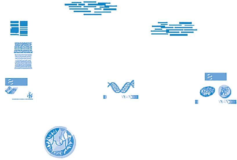
Fig. 2.9 a schematic overview of the uses of bioinformatics for functional metagenome analysis. Microbial community contains numerous bacterial and other species. Once the total DNA has been extracted, the composition of the community is determined by amplifying and sequencing 16S ribosomal RNA (rRNA) gene. Highly similar sequences are then grouped as operational taxonomic units (OTUs), which are then recognized by comparisons with databases of already recognized organisms. OTUs are then analyzed to determine the biomolecular and metabolic functions of the community. (Adapted with permission from Elsevier from Morgan, X.C., Segata N., Huttenhower C. (2013). Biodiversity and functional genomics in the human microbiome. Trends in Genetics, 29(1), 51–58.)
Key facts
Note: clinically relevant facts and practice points are italicized; key words are in bold
• The word ‘microorganism’ (microbe) is used to describe an organism that cannot be seen without the use of a microscope.
• The main groups of microbes are algae, protozoa, fungi, bacteria and viruses, with progressively decreasing size.
• All living cells are either prokaryotic (Archaea and Bacteria) or eukaryotic
• Prokaryotes such as bacteria are simple cells with no internal membranes or organelles.
• Eukaryotes have a nucleus, organelles such as mitochondria and complex internal membranes (e.g., fungi, human cells).
• Bacteria are divided into two major classes according to staining characteristics: Gram-positive (purple) and Gramnegative (pink).
• Structures external to the cell wall of bacteria are flagella (whip-like filaments), fimbriae or pili (fine, short, hair-like filaments), glycocalyx (slime layer) and capsule
• Flagella are used for movement, the fimbriae and pili for adhesion and the glycocalyx for adhesion, protection and biofilm formation.
• Cell wall peptidoglycan is common to both Gram-positive and Gram-negative bacteria but thicker in the former; it gives rigidity and shape to the organism.
• Peptidoglycan comprises long chains of N-acetylmuramic acid and N-acetylglucosamine cross-linked by peptide side chains and cross-bridges.
• Lipopolysaccharides (LPS) are integral components of the outer membranes of Gram-negative (but not Gram-positive) bacteria; LPS is the endotoxin and therefore Gram-positive bacteria cannot produce endotoxin.