No part of this publication may be reproduced or transmi�ed in any form or by any means, electronic or mechanical, including photocopying, recording, or any information storage and retrieval system, without permission in writing from the publisher. Details on how to seek permission, further information about the Publisher’s permissions policies and our arrangements with organizations such as the Copyright Clearance Center and the Copyright Licensing Agency, can be found at our website: www.elsevier.com/permissions.
This book and the individual contributions contained in it are protected under copyright by the Publisher (other than as may be noted herein).
Notices
Knowledge and best practice in this field are constantly changing. As new research and experience broaden our understanding, changes in research methods, professional practices, or medical treatment may become necessary.
Practitioners and researchers must always rely on their own experience and knowledge in evaluating and using any information, methods, compounds, or experiments described herein. In using such information or methods they should be mindful of their own safety and the safety of others, including parties for whom they have a professional responsibility.
To the fullest extent of the law, neither the Publisher nor the authors, contributors, or editors, assume any liability for any injury and/or damage to persons or property as a ma�er of products liability, negligence or otherwise, or from any use or operation of any methods, products, instructions, or ideas contained in the material herein.
Library of Congress Cataloging-in-Publication Data
A catalog record for this book is available from the Library of Congress
British Library Cataloguing-in-Publication Data
A catalogue record for this book is available from the British Library ISBN 978-0-323-99413-2
For information on all Academic Press publications visit our website at h�ps://www.elsevier.com/books-and-journals
Publisher: Andre Gerhard Wolff
Acquisitions Editor: Linda Versteeg-buschman
Editorial Project Manager: Timothy Benne�
Production Project Manager: Swapna Srinivasan
Cover Designer: Kennedy Bonjour (front) and Miles Hitchen (back)
Typeset by STRAIVE, India
About the authors
Rossana
C. N. Melo, MS, PhD
Professor of Cell Biology
Federal University of Juiz de Fora
Juiz de Fora, Minas Gerais, Brazil
Visiting Scientist from 2002 to 2019
Beth Israel Deaconess Medical Center
Harvard University
Boston, Massachuse�s, United States
https://orcid.org/0000-0003-1736-0806
Ann M. Dvorak, MD Professor Emerita of Pathology
Beth
Israel Deaconess Medical Center
Harvard Medical School
Boston, Massachuse�s, United States
Peter F. Weller, MD
William B. Castle Professor of Medicine
Harvard Medical School
Beth Israel Deaconess Medical Center
Professor of Immunology and Infectious Diseases
Harvard Medical School
Harvard T. H. Chan School of Public Health
Boston, Massachuse�s, United States
https://orcid.org/0000-0001-9580-560X
The authors have decades of experience focused on studies of eosinophils in health and disease. The collaborative interactions of the authors include a pioneering pathologist Dr. Ann Dvorak who has singularly advanced our structural knowledge through her elegant career-long electron microscopy of eosinophils, and a physician investigator—Dr. Peter Weller—who has studied eosinophil functioning and the roles of eosinophils in diseases. A distinguished cell biologist Dr. Rossana Melo joining with the other authors, has applied novel electron microscopic techniques that have advanced our insights into the structural bases of eosinophil biology. Drawing on their studies together, the authors provide unique expertise and experience related to human eosinophils, and the atlas of eosinophil ultrastructure is a product of their longstanding studies.
Preface
Rossana C.N. Melo
Ann M. Dvorak
Peter
F. Weller
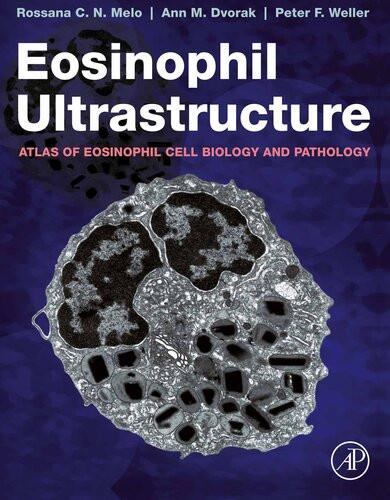
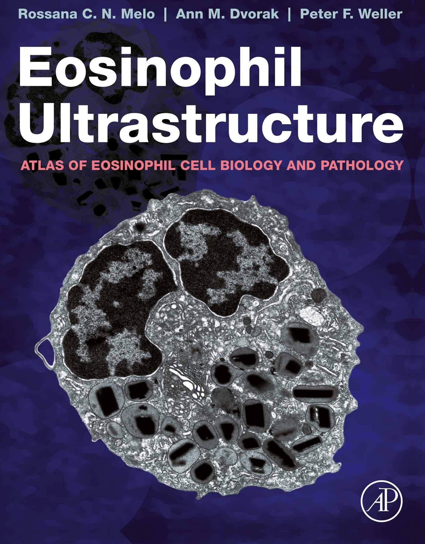
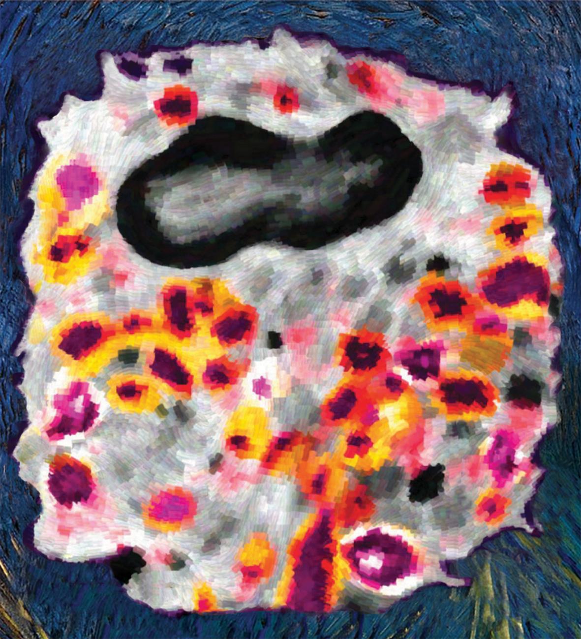
A human eosinophil seen under the transmission electron microscope and pseudocolored in Van Gogh’s style.
Eosin dye was one of the most brilliant hues of Vincent van Gogh’s pale�e applied to create a variety of nuances in his astonishing paintings more than 150 years ago. Around the same time, the eosinophil, a cell of the immune system, was brought to the a�ention when Paul Ehrlich captured the bright pink/orange/red nuances of its granules on eosin staining under the light microscope. Since then, this eosin lover has been considered one of the most beautiful and
enigmatic cells. As important elements of disease processes and maintenance of homeostasis, eosinophils have a vast history of research with remarkable scientific advances, but there is still much to learn regarding their life.
Under the electron microscope, eosinophils were revealed with an additional striking visual their granules bear an electron-dense crystalline core placidly enmeshed in an electron-lucent matrix which is not found in any other cells, thus giving eosinophils an elegant ultrastructural signature and making electron microscopy (EM) a powerful tool to investigate them.Eosinophil granules are complex and contain a multitude of proteins. Release of the granule contents is a critical event of the eosinophil function that only EM can uncover in detail. In this atlas, we explore the ultrastructure of eosinophils, illustrating the moods and the contrasts of their life and death. What emerges is the awesome eosinophil subcellular world that only EM can provide.
The atlas is divided into three main sections. The first section The Cell Biology of Human Eosinophils includes the general ultrastructure of eosinophils found in the bone marrow, peripheral blood, and tissues. We provide a core of essential knowledge to identify both mature and immature eosinophils and to understand eosinophil biological processes, such as activation and degranulation, observed mostly in experimental conditions. Eosinophils are shown under a blend of conventional and advanced electron microscopic techniques such as immunonanogold EM to uncover specific subcellular localization of cytokines and other proteins and electron tomography to show tridimensional aspects of eosinophil organelles.
The second section Eosinophil in Human Diseases explores the ultrastructure of eosinophils seen in body fluids and biopsy specimens from patients with eosinophilic disorders. The cellular features and structural events associated with eosinophil functioning, as well as its microenvironment, are depicted in varied human diseases, thus providing a link between basic science and clinical aspects.
The third section The Cell Biology of Mouse Eosinophils is focused on the ultrastructure of mature and immature eosinophils of mouse
p models, which are extensively used in studies aiming to understand the mechanisms underlying eosinophil-associated diseases. We highlight the ultrastructural differences between human and mouse eosinophils and the responses of mouse eosinophils to activation conditions and in the context of diseases.
This atlas is a unique and comprehensive reference; it is a guide for scientists who use EM as a tool to study eosinophil structural biology, cellular immunology, innate and adaptive immunity, immune responses to pathogens, inflammation, and pathology. By uncovering the eosinophil universe, the atlas also serves as a source to cell biologists pursuing to understand the remarkable structurefunction relationships during a cell journey in a dynamic environment.
As a portrait, every image captured by EM reflects at high resolution a moment of the eosinophil lifetime experience and invites the viewer to engage with and to learn from this multifaceted cell. In this atlas, the eosinophil is under the spotlight in time moments and scenes of its enigmatic life, which have been motivating our scientific careers and fascinating us for decades.
We hope you will enjoy the fantastic eosinophil world!
Acknowledgments
In our compilation of this Eosinophil Ultrastructure Atlas, we have many to thank and recognize for their efforts, encouragement, and support.
For the decades-long studies of eosinophils and their structure and function, we acknowledge with gratitude:
Family support: RCNM: Sarah M. Salles, my daughter, who has made all the difference to me. AMD: Harold Dvorak, MD, Emeritus Professor of Pathology at Harvard Medical School and former Chair of the Department of Pathology at Beth Israel Deaconess Medical Center. PFW: Anne Nicholson-Weller, MD, Professor of Medicine at Harvard Medical School.
Technical support: We thank all the technicians at the Electron Microscopy Unit, Department of Pathology, BIDMC, for the outstanding technical assistance, especially Rita Monahan-Earley, Ellen Morgan, Tracey Sciu�o, and Kit Pyne. The time and dedication that you have put into our studies during decades were just amazing. We are also grateful to the staff at Harvard Medical School EM Facility (in particular to Maria Ericsson), Centro de Microscopia Eletrônica (UFMG, Brazil), and CAPI (ICB, UFMG, Brazil) for kind assistance in more recent years.
We extend our sincere thanks to the members of our research groups at BIDMC/Boston (USA) and Laboratory of Cellular Biology (UFJF, Brazil), past and present, for their collective efforts and enthusiasm. In particular, we thank Thiago P. Silva and Vitor H. Neves for the tremendous assistance with the figures of this atlas and Kennedy Bonjour for the beautiful cover design.
Funding support: We thank the research agencies NIH (USA) and FAPEMIG and CNPq (Brazil). AMD and PFW have been supported
by many NIH grants, including AI05645, AI022571, and AI020241. For decades, AI 020241, first as R01s and then as an R37, along with a Brazilian collaborative funding supplement, supported research on eosinophil form and function. RCNM has been supported by CNPq and FAPEMIG grants, including a fellowship of research productivity granted by CNPq.
We also acknowledge the contributions of those with eosinophilic disorders who provided valuable samples and generated support through the MWV Leukocyte Research Fund to advance our studies.
PFW acknowledges with much appreciation the efforts of his colleague, Rossana Melo. Not only does the compilation and editing of this atlas constitute an enormous effort on Rossana’s part, but also her technical expertise as it has been applied to the study of eosinophils has progressively advanced our insights. Electron microscopy has technically advanced from the early studies of eosinophils. With refined methodological approaches, including be�er preservation of intracellular membranous structures and applications of pre-embedding immunonanogold immunolabeling, Rossana’s contributions have immensely advanced our understandings of the cellular functioning of eosinophils.
RCNM: My deepest gratitude to PFW and AMD. From my initial meeting with Peter in Brazil in 2001 and invitation to join his lab at Harvard, my great appreciation is for continuously inspiring me as an exemplary scientist and human being. And to Ann, a brilliant scientist with a passion for electron microscopy, I could not have wished for a be�er colleague.
Finally, we express our gratitude to the very capable Elsevier team who helped with this project in many ways. Special thanks to the Senior Acquisitions Editor, Linda Versteeg-Buschman, the Editorial Project Manager, Timothy Benne�, and the Production Project Manager Swapna Srinivasan for their kind coordination and a great deal of support.
We dedicate this volume to the late Michael W. Verronneau, who, with his family and friends, was an active advocate for continued research on eosinophils and their associated diseases.
Abbreviations
AA arachidonic acid
[3H]-AA tritiated arachidonic acid
ABPA allergic bronchopulmonary aspergillosis
ADRP (also known as adipophilin or PLIN-2) adipose
differentiation-related protein
ANCA antineutrophil cytoplasmic antibody
APO-1 apoptosis antigen 1
Bcl-2 B-cell lymphoma 2 protein
BFA brefeldin-A
CASP caspase
CCL5 [also known as RANTES (regulated on activation, normal T cell expressed and secreted)] chemokine C-C motif ligand 5
CCL11 (also known as eotaxin-1) chemokine C-C motif ligand 11
CCR3 C-C chemokine receptor type 3
CD9 cluster of differentiation 9
CD63 cluster of differentiation 63
CD34 cluster of differentiation 34
CD95 cluster of differentiation 95
CysLTR cysteinyl leukotriene receptor
CLC Charcot-Leyden crystal
CLC-P Charcot-Leyden crystal protein
COX cyclooxygenase
cPLA2 cytosolic phospholipase A2
DKO double knockout
3D three-dimension
EADs eosinophil-associated diseases
EARs eosinophil-associated RNases
ECM extracellular matrix
ECP (also known as RNase 3) eosinophil cationic protein
ECRS eosinophilic chronic rhinosinusitis
EDN (also known as RNase2) eosinophil-derived neurotoxin
EGIDs eosinophilic gastrointestinal disorders
EGPA eosinophilic granulomatosis with polyangiitis