List of Contributors
The editors would like to acknowledge and offer grateful thanks for the input of all previous editions’ contributors, without whom this new edition would not have been possible.
Hamid Abedi, DDS MS MBA
Clinical endodontist, Irvine, CA, USA
Kenneth Abramovitch, DDS, MS
Professor and Chair, Department of Oral Pathology, Radiology and Medicine, University of Missouri – Kansas City, School of Dentistry, Kansas City, MO, USA
Anita Aminoshariae, DDS, MS
Associate Professor, Endodontics, CWRU School of Dental Medicine, Cleveland, OH, USA
Ana Arias, DDS, MS, PhD
Professor of Conservative Dentistry, School of Dentistry, Complutense University, Madrid, Spain
Adham A. Azim, BDS
Division Head and Director of the Post-Graduate Program, Periodontics and Endodontics, University at Buffalo, Buffalo, New York, NY, USA
Brooke Blicher, DMD, Certificate in Endodontics
Clinical Endodontist, Upper Valley Endodontics, White River Junction, Vermont;
Clinical Instructor, Department of Restorative Dentistry and Biomaterials Science, Harvard School of Dental Medicine, Boston, MA, USA;
Assistant Clinical Professor, Department of Endodontics, Tufts University School of Dental Medicine, Boston, MA, USA; Dental Staff, Department of Oral Surgery, Dartmouth Hitchcock Medical Center, Lebanon, VT, USA
Tatiana Botero, DDS, MS
Clinical Associate Professor, Cariology Restorative Sciences and Endodontics, Ann Arbor, MI, USA
Sami Chogle, BDS, DMD, MSD
Chair and Program Director, Graduate Endodontics, Boston University, Boston, MA, USA;
Adjunct Associate Professor, Graduate Endodontics, Case Western Reserve University, Cleveland, OH, USA
William Herbert Christie, DMD, MS, FRCD(C)
Retired Professor, Senior Scholar, Department of Restorative Dentistry, University of Manitoba, Winnipeg, Manitoba, Canada
Blaine Cleghorn, DMD, MS
Professor and Assistant Dean, Faculty of Dental Clinical Sciences, Dalhousie University, Halifax, Nova Scotia, Canada
Anibal Diogenes, DDS, MS, PhD
Professor and Vice Chair, Department of Endodontics, University of Texas Health Science Center at San Antonio, San Antonio, TX, USA
Melissa Drum, DDS, MS
Professor, Advanced Endodontics Director, Division of Endodontics, College of Dentistry, The Ohio State University, Columbus, OH, USA
Brad Eli, DMD, MS
Staff, Department of Dentistry, Scripps Memorial Hospital, La Jolla, CA, USA
Mohamed I. Fayad, DDS, MS, PhD
Clinical Associate Professor, Director of Research, Department of Endodontics, University of Illinois, College of Dentistry, Chicago, IL, USA
Natasha M. Flake, DDS, PhD, MSD
Associate Professor, Department of Endodontics, University of Washington, Seattle, WA, USA
Ashraf F. Fouad, DDS, MS
Freedland Distinguished Professor, Vice-chair, Division of Comprehensive Oral Health, Adams School of Dentistry, University of North Carolina, Chapel Hill, NC, USA
Brian Goodacre, DDS, MSD
Assistant Professor, Department of Prosthodontics, School of Dentistry, Loma Linda University, Loma Linda, CA, USA
Charles J. Goodacre, DDS, MSD
Distinguished Professor, Department of Restorative Dentistry, School of Dentistry, Loma Linda University, Loma Linda, CA, USA
Robert Handysides, DDS
Dean, Department of Endodontics, School of Dentistry, Loma Linda University, Loma Linda, CA, USA
Brad Johnson, DDS, MHPE
Professor and Head, Director of Postdoctoral Endodontics, Department of Endodontics, University of Illinois at Chicago, Chicago, IL, USA
James David Johnson, DDS, MS
Clinical Professor and Chair, Department of Endodontics, University of Washington School of Dentistry, Seattle, WA, USA
Mo Kang, DDS, PhD
Professor and Chairman, Department of Endodontics, UCLA School of Dentistry, Los Angeles, CA, USA
Philip Michaelson, MS, DMD
Private Practice, Endodontics, Professional Endodontics, Inc., Chagrin Falls, OH, USA
W. Craig Noblett, DDS, MS
Volunteer Faculty, University of California, San Francisco, CA; Private practice, Fresno, CA
Ali Nosrat, DDS, MS, MDS
Clinical Assistant Professor, Department of Endodontics, School of Dentistry, University of Maryland, Baltimore, MD, USA; Private Practice, Centreville, VA, USA
John M. Nusstein, DDS, MS
Professor and Chair, Division of Endodontics, College of Dentistry, The Ohio State University, Columbus, OH, USA
Avina Paranjpe, BDS, MS, MSD, PhD
Associate Professor, Department of Endodontics, University of Washington, Seattle, WA, USA
Masoud Parirokh, DMD, MSc
Distinguished Professor, Department of Endodontics, School of Dentistry, Kerman University of Medical Sciences, Kerman, Islamic Republic of Iran;
Distinguished Professor, Endodontology Research Center, Kerman University of Medical Sciences, Kerman, Islamic Republic of Iran
Ove A. Peters, DMD, MS, PhD
Professor and Discipline Lead, Endodontics, School of Dentistry, The University of Queensland, Brisbane, Qld, Australia
Al Reader, DDS, MS
Professor, Division of Endodontics, College of Dentistry, The Ohio State University, Columbus, OH, USA
Ilan Rotstein, DDS
Professor and Chair, Endodontics, Orthodontics and General Practice Residency, Herman Ostrow School of Dentistry of USC, University of Southern California, Los Angeles, CA, USA; Associate Dean, Continuing Education, Herman Ostrow School of Dentistry of USC, University of Southern California, Los Angeles, CA, USA
Richard A. Rubinstein, DDS, MS, FACD
Adjunct Clinical Associate Professor, Cariology, Restorative Sciences and Endodontics, University of Michigan, Ann Arbor, MI, USA
Nikita B. Ruparel, MS, DDS, PhD
Associate Professor, Department of Endodontics, UTHSCSA, San Antonio, TX, USA
Mohammed Sabeti, DDS, MA
Clinical Professor, PRDS, University of California at San Francisco, San Francisco, CA, USA
Nasser Said-Al-Naief, DDS, MS
Professor and Chair, OMFP Laboratory Director, Pathology and Radiology, OHSU, Portland, OR, USA; Hospital Staff, OMFS, School of Medicine, OHSU, Portland, OR, USA;
Professor, Anatomic Pathology, School of Medicine, OHSU, Portland, OR, USA
Christine Sedgley, MDS, MDSc, FRACDS, MRACDS(ENDO), PhD
Professor and Chair, Department of Endodontology, Oregon Health and Sciences University, Portland, OR, USA
Frank Setzer, DMD, PHD, MS
Assistant Professor, Endodontic Clinic Director, and Predoctoral Endodontic Program Director, Department of Endodontics, University of Pennsylvania, Philadelphia, PA, USA
Shahrokh Shabahang, DDS, MS, PhD
Associate Professor, Department of Endodontics, School of Dentistry, Loma Linda University, Loma Linda, CA, USA
Renato Silva, DDS, MS, PhD
Associate Professor, Department of Endodontics, University of Texas Health Science Center at Houston, Houston, TX, USA
Tory Silvestrin, DDS, MSD, MSHPE
Chairman and Graduate Program Director, Department of Endodontics, School of Dentistry, Loma Linda University, Loma Linda, CA, USA
Fabricio B. Teixeira, DDS, MS, PhD
Professor and Chair, Department of Endodontics, University of Iowa, Iowa City, IA, USA
Yoshitsugu Terauchi, DDS, PhD
President, Endodontics, CT and MicroEndodontic Center, Yamato-shi, Kanagawa, Yamato City, Japan
Mahmoud Torabinejad, DMD, MSD, PhD
Affiliate Professor of Endodontics, Department of Endodontics, University of Washington Seattle, WA, USA;
Adjunct Professor, Department of Endodontics, School of Dentistry, Loma Linda University, Loma Linda, CA, USA; Adjunct Professor, Department of Endodontics, University of Pacific Arthur A. Dugoni School of Dentistry; Adjunct Professor, Department of Preventive and Restorative Dentistry, Section of Endodontics, University of California in San Francisco, School of Dentistry, San Francisco, CA, USA
Marco Aurelio Versiani, Lt. Col., DDS, MSc, PhD
Associate Researcher, Department of Restorative Dentistry, Dental School of Ribeirão Preto, University of São Paulo, Ribeirão Preto, São Paulo, Brazil
Richard Walton, DMD, MS
Professor Emeritus, Department of Endodontics, University of Iowa, Iowa City, IA, USA
Shane N White, BDentSc, MS, MA, PhD
Professor, UCLA School of Dentistry, Los Angeles, CA, USA
Anne E. Williamson, DDS, MS
Associate Professor, Endodontics, University of Iowa, Iowa City, IA, USA
1 Pathogenesis of Pulp and Periapical Diseases
CHRISTINE SEDGLEY, RENATO SILVA, AND ASHRAF F. FOUAD
CHAPTER OUTLINE
Histology and Physiology of Normal Dental Pulp, 1
Etiology of Pulpal and Periapical Diseases, 2
Microbiology of Root Canal Infections, 5
Endodontic Infections Are Biofilm Infections, 5
The Microbiome of Endodontic Infections, 6
Pulpal Diseases, 8
LEARNING OBJECTIVES
After reading this chapter, the student should be able to:
1. Describe the histology and physiology of the normal dental pulp.
2. Identify etiologic factors causing pulp inflammation.
3. Describe the routes of entry of microorganisms to the pulp and periapical tissues.
4. Classify pulpal diseases and their clinical features.
5. Describe the clinical consequences of the spread of pulpal inflammation into periapical tissues.
Histology and Physiology of Normal Dental Pulp
The dental pulp is a unique connective tissue with vascular, lymphatic, and nervous elements that originates from neural crest cells. It resides inside the tooth in a chamber with rigid walls.
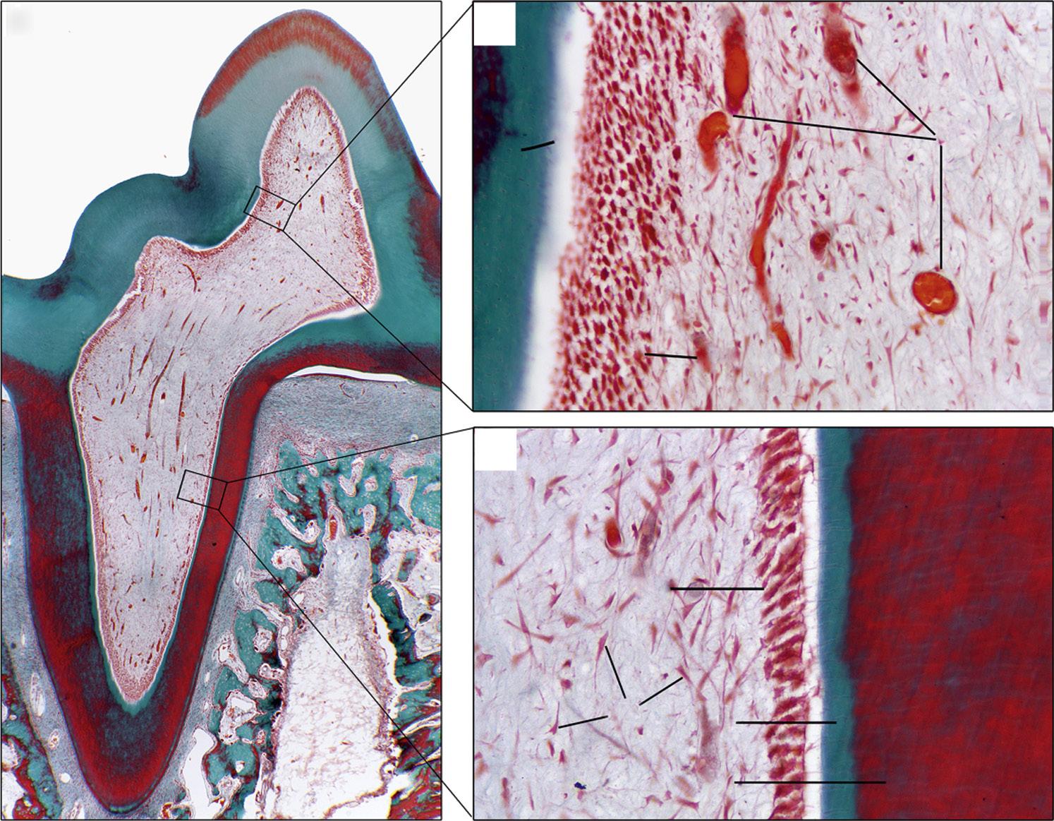
The pulp contains odontoblasts, highly specialized cells with a secretory function, which not only form dentin, but also interact with dental epithelium early in tooth development to initiate the formation of enamel. The pulp also contains fibroblasts, undifferentiated mesenchymal cells, collagen type I and II, proteoglycans, glycoproteins, and water1 (Fig. 1.1).
The histologic structure of the pulp is important, because it reflects a unique architecture suited for the formation of dentin and defense against invading pathogens. Odontoblasts form a
Normal Pulp, 11
Reversible Pulpitis, 11
Irreversible Pulpitis, 11
Pulp Necrosis, 12
Clinical Classification of Periapical (Apical) Conditions, 13
Nonendodontic Pathosis, 15
6. Describe the histopathological diagnoses of periapical lesions of pulpal origin.
7. Identify clinical signs and symptoms of acute apical periodontitis, chronic apical periodontitis, acute and chronic apical abscesses, and condensing osteitis.
8. Discuss the role of residual microorganisms and host response in the outcome of endodontic treatment.
9. Describe the steps involved in repair of periapical pathosis after successful root canal treatment.
palisading layer that lines the walls of the pulp space, and their tubules extend about two thirds of the length of the dentinal tubules. The tubules are larger at a young age and eventually become more sclerotic as the peritubular dentin becomes thicker. The odontoblasts are primarily involved in production of mineralized dentin. They are connected by gap junctions that allow them to form a semipermeable membrane. In addition, odontoblasts play an important role in defense as they express Toll-like receptors (see later), cytokines, and defensins, among other immunologic mediators.
Two main types of sensory fibers innervate the pulp: Aδ-fibers in the periphery and C-fibers in the central pulp. The Aδ-fibers are responsible for the sharp response to thermal changes. They extend between the odontoblasts, lose their myelin sheath, and extend to a distance of 100 to 200 μm into the dentinal tubules. The C-fibers are unmyelinated and are responsible for the dull
• Fig. 1.1 Histologic section of (A) rat molar tooth showing coronal (B) and (C) radicular dental pulp in higher magnification. Masson-Goldner trichrome staining. BV, Blood vessels; D, dentin; FB, fibroblasts; OD, odontoblasts in odontoblastic layer; PD, predentin. An artifact (*) separating the predentin from the odontoblastic layer. (Courtesy Dr. Claudia Biguetti.)
ache that affects patients with symptomatic irreversible pulpitis. The pulp may also have Aβ-fibers and sympathetic fibers in the walls of arterioles.
The pulp vasculature plays a critical role in its response to irritation. When the tooth first erupts into the oral cavity, the root apex is immature, and there is ample blood supply to the pulp. Eventually, the apex matures, and the ability of the pulp to withstand external irritation, such as from trauma or caries, diminishes. However, the pulp of the mature tooth has mechanisms to cope with increased blood flow during inflammation, such as arteriovenous anastomoses and loops that can circulate and increase volume of blood when the need arises. The pulp also contains an elaborate network of arterioles and capillaries around the odontoblasts, which are high-metabolic-rate cells, commonly known as the terminal capillary network.
Etiology of Pulpal and Periapical Diseases
Injury or irritation of pulpal or periapical tissues can result in inflammation. The reactions of the dental pulp to irritants are largely dictated by the type and duration of a stimulus. These irritants can be broadly classified as nonliving (mechanical, thermal, or chemical) or living (microbial) (Video 1.1).
Mechanical Irritants
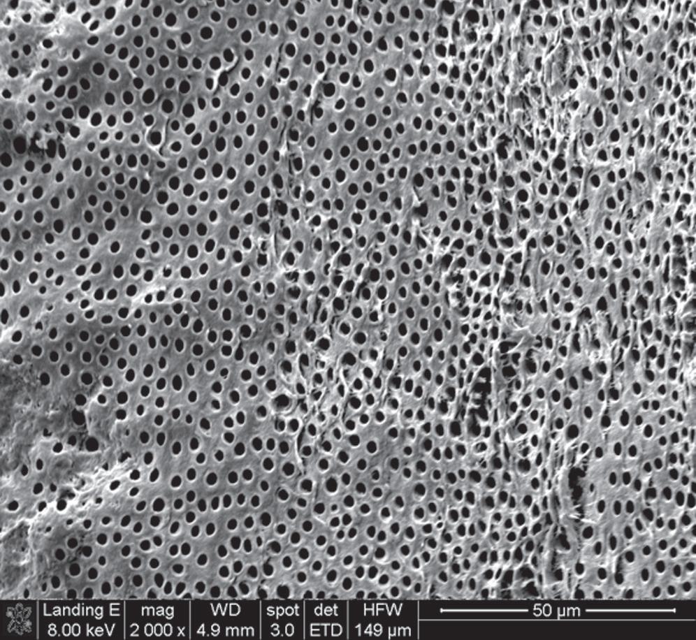
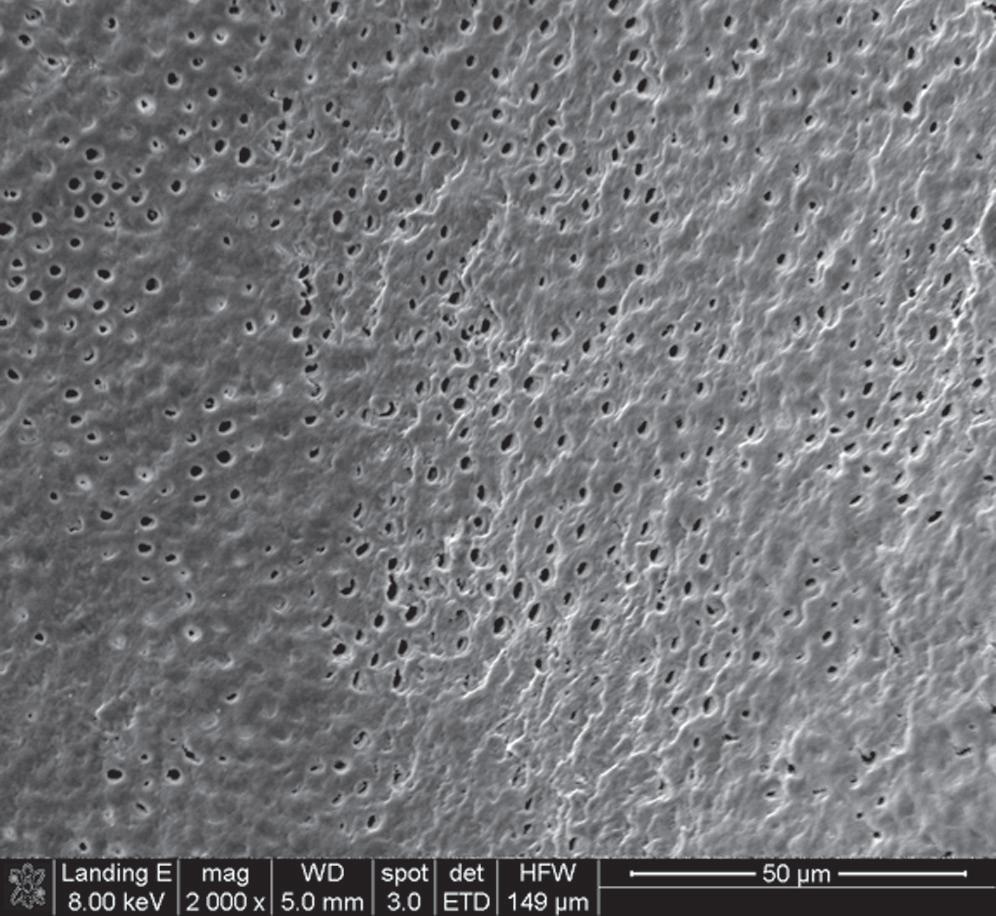
The potential for pulp irritation increases as more dentin is removed during deep cavity preparations because dentinal permeability is greater closer to the pulp2 (Fig. 1.2). The removal of tooth structure without proper cooling may also cause pulp inflammation. Deep scaling and curettage may injure apical vessels and nerves, resulting in pulpal damage.3
Pulpal damage can occur because of impact injuries. Teeth undergoing mild to moderate trauma and those with immature apices have a better chance of pulpal survival in comparison with those suffering severe injury or those with closed apices. Intrusion injuries are more likely to lead to pulp necrosis than are lateral or extrusion injuries4 (Fig.1.3).
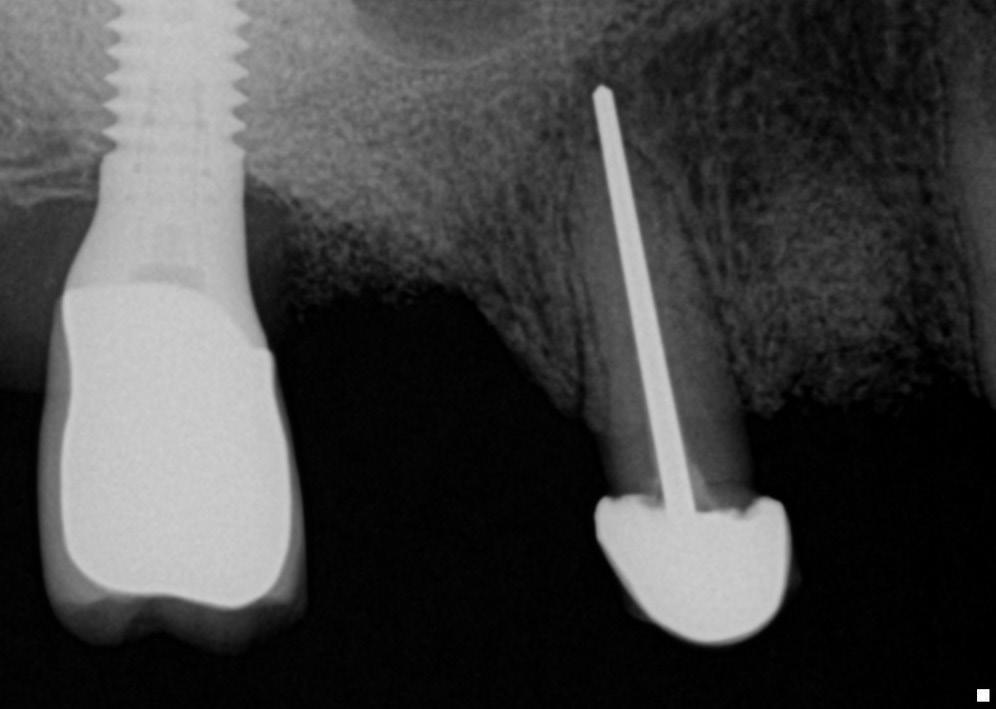
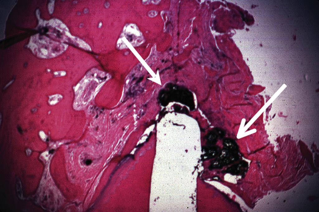
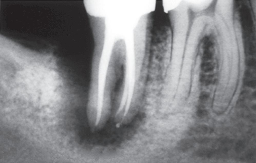
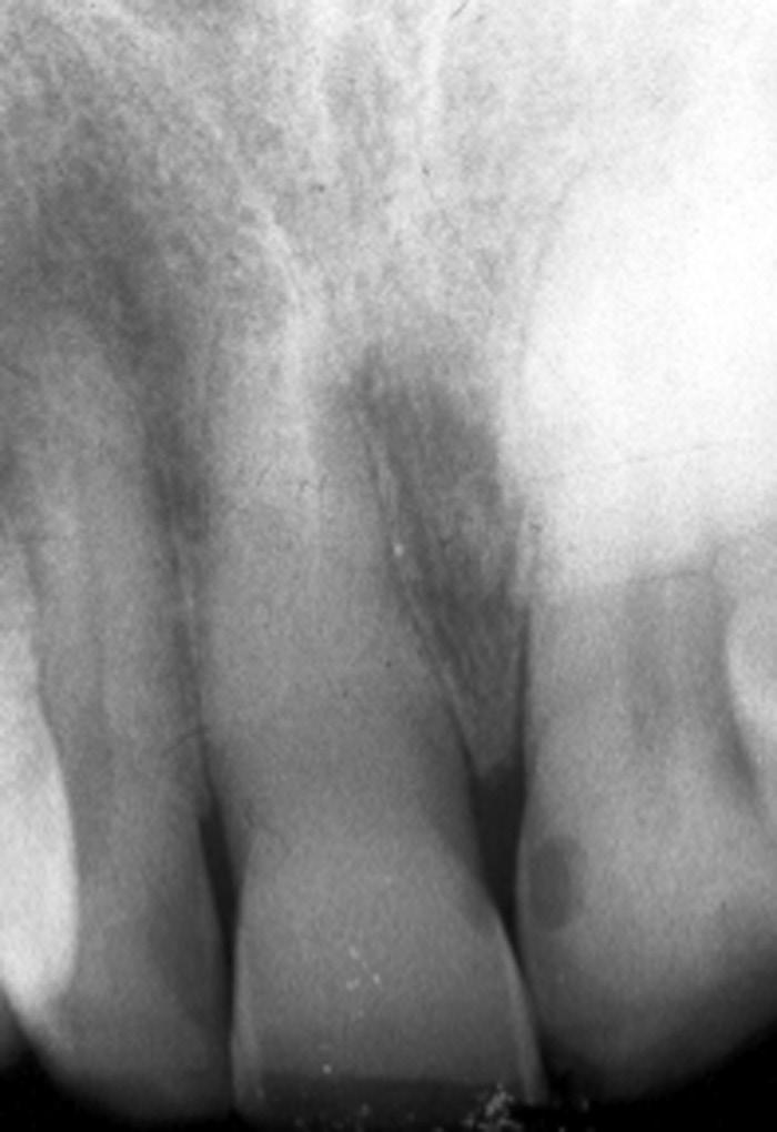
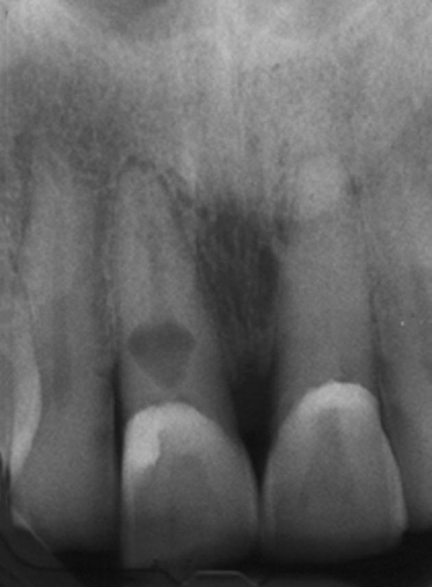
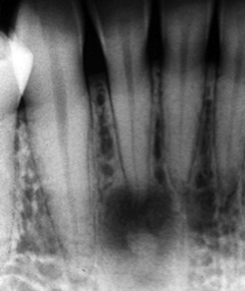
Periapical tissues can be mechanically irritated and inflamed by impact trauma, hyperocclusion, overinstrumentation of root canals, perforation of the root, and overextension of root canal filling materials (Fig. 1.4). Inaccurate determination of root canal length is usually the cause of overinstrumentation and subsequent inflammation. In addition, lack of an adequate apical resistance form created during cleaning and shaping can cause overextension of filling materials into the periapical tissues, causing physical and chemical damage (Fig. 1.5).
Application of forces beyond the physiologic tolerance of the periodontal ligament (PDL) during orthodontic tooth movement results in disturbance of the blood and nerve supply of the pulp tissue.5,6 In addition, orthodontic movement may initiate resorption of the apex, usually without a change in vitality.
Chemical Irritants
Antibacterial agents, such as silver nitrate, phenol with and without camphor, and eugenol, have been used to “sterilize” dentin after cavity preparations. The effectiveness of many of these products is questionable,7 and their cytotoxicity can cause inflammatory changes in the underlying dental pulp.8 Other irritating agents include cavity cleansers, such as alcohol, chloroform, hydrogen peroxide, and various acids; chemicals present in desensitizers, cavity liners and bases; and temporary and permanent restorative materials.
• Fig. 1.2 Scanning Electron Microscopy of Human Dentin. Dentinal permeability is greater closer to the pulp (A) than near the dentinoenamel junction (B) or the cementodentinal junction due to the higher number of tubules per unit and bigger tubule diameter. Therefore the potential for pulp irritation increases as more dentin is removed.
• Fig. 1.3 Graphic representation of pulpal circulation subsequent to various types of luxation injuries to teeth. Pulp circulation is measured in perfusion units over a 36-week observation period.
Antibacterial irrigants used during cleaning and shaping of root canals, intracanal medications, and some compounds present in obturating materials are examples of potential chemical irritants to periapical tissues.9,10 When testing the effects of antimicrobial medications on dental pulp cells, researchers showed that calcium hydroxide and lower concentrations of antibiotic pastes are conducive to cell survival and proliferation, but more concentrated forms of antibiotic pastes have detrimental effects.11
• Fig. 1.4 Periapical radiograph showing overextension of root canal filling material.
• Fig. 1.5 Improper instrumentation and extrusion of filling materials into the periapical tissues causes periradicular inflammation (arrows).
Microbial Irritants
Although mechanical and chemical irritations are predominantly transient in nature, the most significant cause of inflammation is microbial. Studies have shown that even superficial carious lesions in enamel are capable of attracting inflammatory cells in the pulp.12,13 The initial reaction of the pulp to these irritants is mediated through the innate immune response. This early response to caries results in focal accumulation of chronic inflammatory cells, such as macrophages, lymphocytes, and plasma cells.14 As caries progresses toward the pulp, the intensity and character of the infiltrate change. Pulpal tissue may remain inflamed for long periods and may undergo eventual or rapid necrosis. This change depends on several factors: (1) the virulence of the microorganisms; (2) the ability to circulate inflammatory fluids to avoid a marked increase in intrapulpal pressure; (3) host resistance, including genetic variations; (4) the amount of circulation and (5) an important factor, lymphatic drainage. Subsequently, microorganisms or their byproducts and other irritants from the necrotic pulp diffuse from the canal to the periapical region, resulting in the development of an inflammatory lesion (Fig. 1.6).
Pulpal and periapical pathoses do not develop without the presence of bacterial contamination.15,16 Kakehashi and collaborators created pulp exposures in conventional and germ-free rats.15 In the germ-free rats, minimal inflammation only occurred throughout the 72-day observation period. Further, pulpal tissue in these animals was not devitalized but rather showed calcific bridge formation by day 14, with normal tissue apical to the dentin bridge (Fig. 1.7, A). In contrast, infection, pulpal necrosis, and abscess formation occurred by the eighth day in conventional rats (Fig. 1.7, B). The bacteriological investigation by Sundqvist examining the flora of human necrotic pulps supports the findings of Kakehashi and collaborators15 and Möller and coworkers.16 Sundqvist examined previously traumatized intact teeth with necrotic pulps, with and without apical pathosis. The root canals of teeth without apical lesions were aseptic, whereas those with periapical pathosis had positive bacterial cultures.17
Several mechanisms have been proposed for identification of microorganisms as irritants by the immune system. Detection of these pathogens can occur via interaction between pathogen-associated molecular patterns (PAMPs) and specific receptors broadly
identified as pattern recognition receptors (PRRs).18 PRRs recognize PAMPs and initiate host defenses. G-protein coupled receptors and Toll-like receptors (TLRs) are part of the innate immune response and activate phagocytic functions to allow microbial ingestion. G-protein coupled receptors bind to chemokines, lipid mediators (e.g., platelet-activating factor, prostaglandin E2, and leukotriene B4) or bacterial proteins, causing extravasation of leukocytes and production of bactericidal substances. TLRs are transmembrane proteins that are expressed by cells of the innate immune system playing a central role in the initiation of cellular innate immune responses.19 These receptors recognize invading microbes and activate signaling pathways that launch immune and inflammatory responses to destroy the invaders. At least 13 TLRs have been discovered to date with different recognition abilities. Table 1.1 presents some of the currently identified TLRs and their specific interactions.
• Fig. 1.6 Egress of irritants (closed arrow) from the root canal into the periapical tissue causes inflammation (open arrow) and replacement of normal periapical structures with a granulomatous tissue.
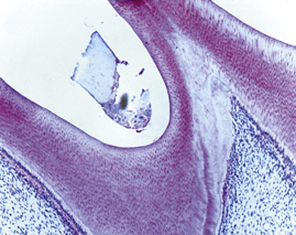
• Fig. 1.7 A, No inflammation is seen in an exposed pulp (P) of a germ-free rat. Food particles and other debris (D) are packed into the chamber. B, Periapical lesion is apparent in a conventional rat after pulp exposure. (Courtesy Dr. H. Stanley.)
TABLE 1.1
Examples of Toll-Like Receptors (TLRS) and Associated Activators
PAMP PRR Pathogen
LPS, Lipid A TLR4 Gram-negative bacteria
Flagellin TLR5 Bacteria, flagellum
dsRNA TLR3 Virus
RNA TLR7,8 Virus
CpG DNA TLR9 Bacteria, DNA
PAMP PRR Pathogen
PAMP, Pathogen-associated molecular patterns; PRR, pattern recognition receptors.
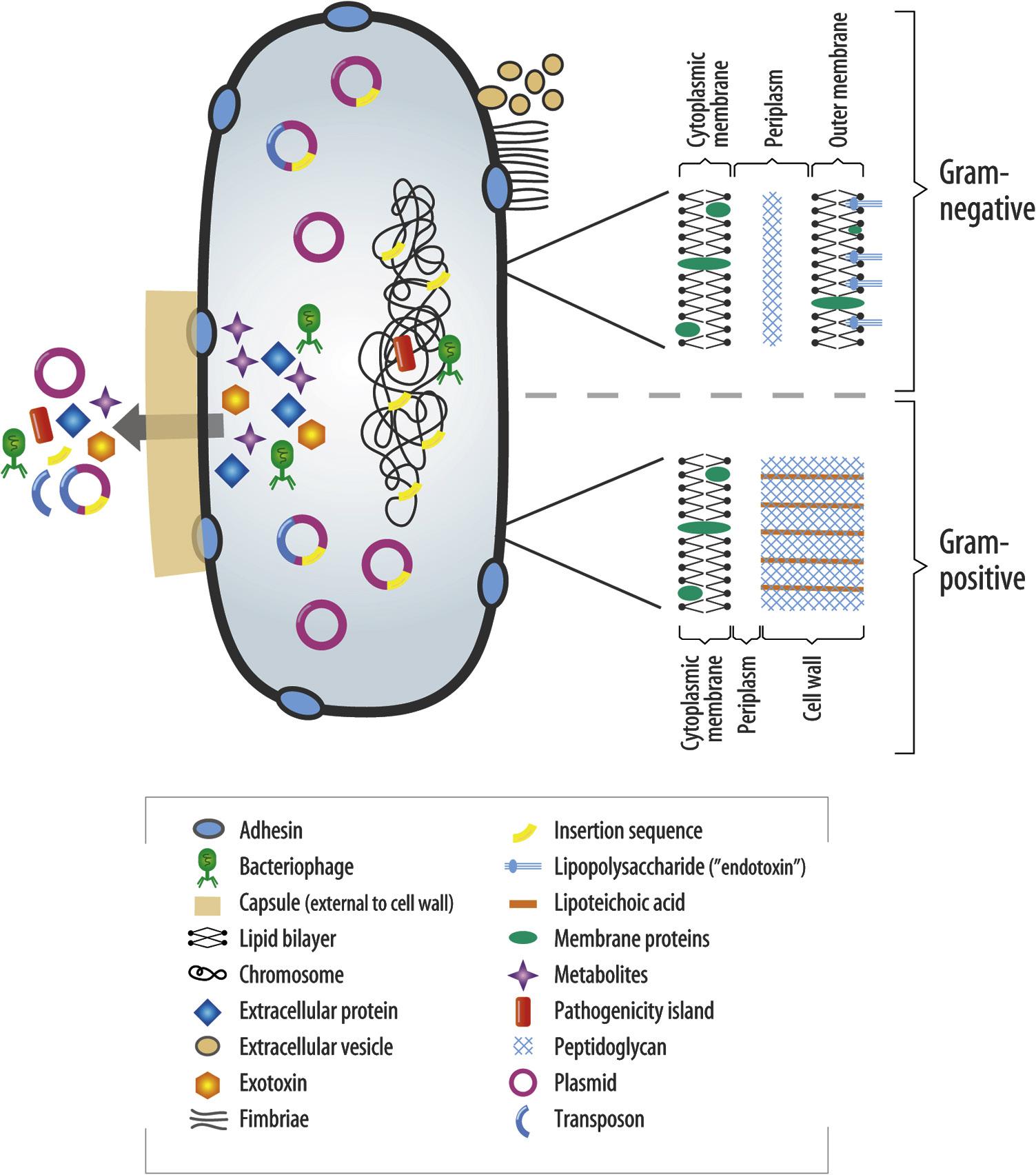
Microbiology of Root Canal Infections
Routes of Root Canal Infection
Under normal conditions, the dental pulp and dentin are isolated from oral microorganisms by overlying enamel and cementum. When the integrity of these protective layers is breached (e.g., as a result of caries, trauma-induced fractures and cracks, restorative procedures, congenital anomalies of teeth, scaling and root planing, attrition, or abrasion) or naturally absent (e.g., because of gaps in the cementoenamel junction at the cervical root surface), the dentin-pulp complex becomes exposed to the oral environment. The pulp then becomes at risk of infection by oral microorganisms present in caries, saliva, and dental plaque. The risk increases with the depth of lesions due to the diameter of dentinal tubules increasing as they approach the pulp (see Fig. 1.2).
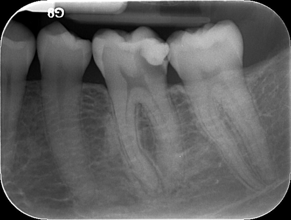
Caries are the most common cause of pulpal exposure (Fig. 1.8). However, microorganisms may also reach the pulp via direct pulpal exposure as a result of iatrogenic restorative procedures, as a result of trauma, and through a periodontal pocket extending to the apical foramen or lateral canal. After pulp necrosis, microorganisms can invade the entire root canal system uninhibited by host defense mechanisms. As a consequence of the interaction between microorganisms and the host defenses, inflammatory changes take place in the periapical tissues and give rise to the development of apical periodontitis.
Endodontic infections can be classified according to their anatomic location as intraradicular or extraradicular. Microorganisms that initially invade and colonize the necrotic pulp tissue cause primary intraradicular infections. Microorganisms that were not present in the primary infection but were introduced into the root canal system during or after initial treatment cause secondary infections. Secondary infections are suspected when a preoperative infection heals after treatment and then recurs at a later time. Persistent infections are caused by microorganisms from a primary infection that resisted intracanal antimicrobial procedures and remained in the prepared root canal system. Persistent and secondary infections are responsible for several clinical problems, including persistent exudation, continuation of symptoms, interappointment flare-ups, and failure of the endodontic treatment.
The goal of root canal treatment is to remove microorganisms from the root canal system. However, in the absence of a strict aseptic technique, microorganisms from caries and dental plaque can be introduced into the canal system during treatment because of lack of rubber dam usage or leakage of the rubber dam.
Contaminated endodontic files and instruments, including delivery systems for antimicrobial agents, are additional potential sources for the introduction of microorganisms into the root canal system during treatment.
Microorganisms can enter the root canal system between appointments via loss or leakage of temporary restorative materials, fracture of the tooth structure, and teeth left open for drainage. Entry of microorganisms after root canal filling occurs by loss or leakage of temporary or permanent restorative materials, during preparation of posts or other intracanal restorations without the rubber dam, fracture of the tooth structure, and by recurrent caries that expose the root canal filling material. Leakage after completion of root canal treatment is more likely to occur if the placement of the permanent restoration is delayed.
Extraradicular infection is characterized by microbial invasion and proliferation into the inflamed periapical tissues and is almost invariably a sequel to intraradicular infection. Once the intraradicular infection is properly controlled by root canal treatment or tooth extraction and drainage of pus, the extraradicular infection can be addressed by the host defenses and usually subsides (Fig. 1.9).
Endodontic Infections Are Biofilm Infections
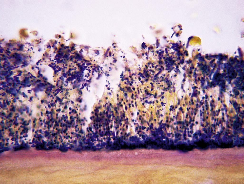
The role of microorganisms as the primary etiologic agents of root canal infections was established in seminal studies published several decades ago.15,16,20 In these and subsequent microbiological analyses of samples recovered from root canal infections, species were isolated and identified by using planktonic (liquid) culture techniques. During the past two decades, it became apparent that microorganisms exist in the root canal system not as planktonic cultures, or as single species, but rather as multispecies biofilm communities composed of microcolonies irreversibly attached to a substratum, to an interface like dentin, and to each other.21,22 All anatomic areas of the infected root canal system can harbor microbial cells organized as highly variable biofilm structures22,23 (Fig. 1.10).
Biofilm formation encompasses attachment of microbial cells to a surface, followed by cell proliferation, adherence to other microorganisms, production of matrix, and microcolony maturation. Dispersal of cells allows the formation of new biofilm microcolonies.24 Microbial cells occupy only a small proportion of the biofilm. Quorum sensing is the expression of specific microbial proteins after the bacterial cells reach a threshold number. It
• Fig. 1.8 Radiograph showing pulp exposure as a result of caries.
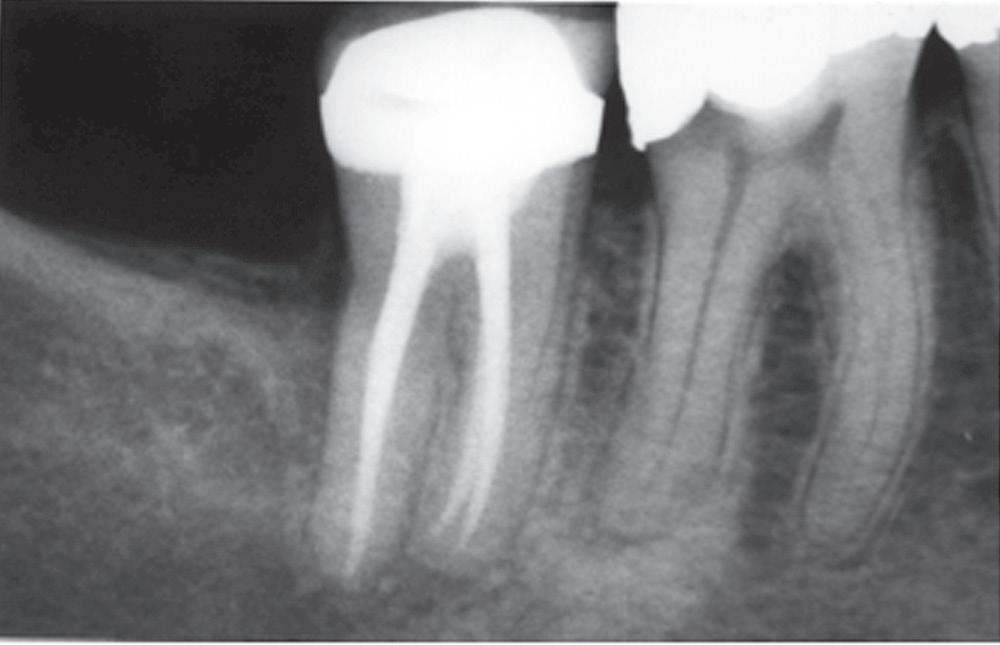
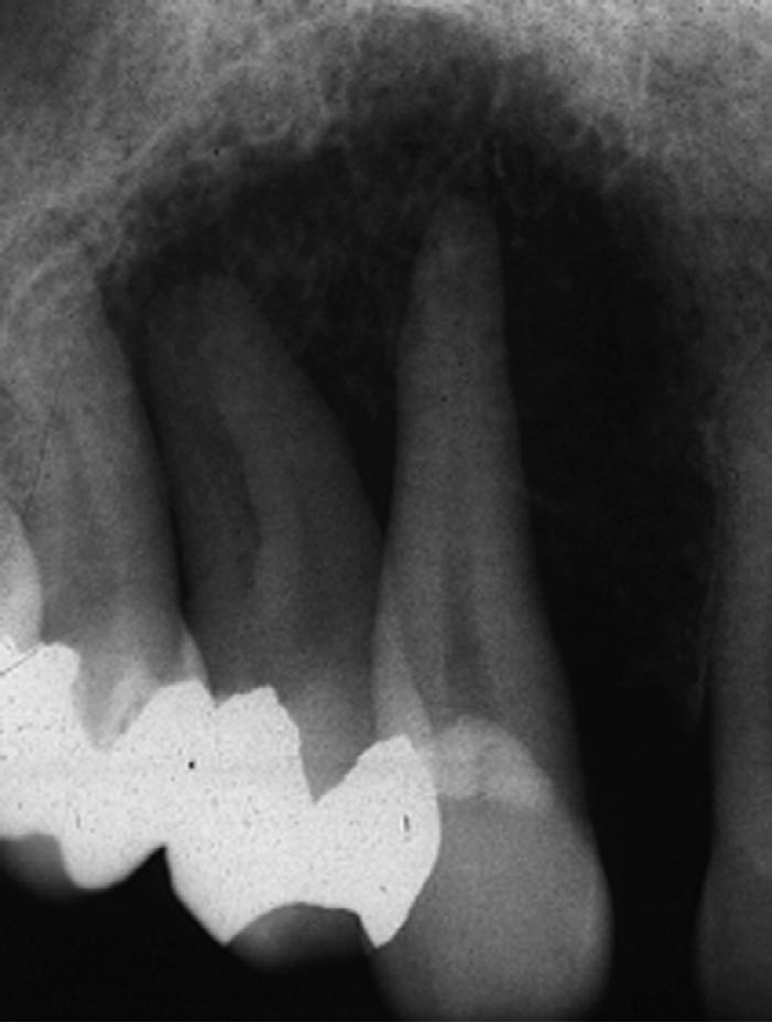
• Fig. 1.9 A, Preoperative radiograph of a second molar with pulpal necrosis and evidence of chronic apical periodontitis. B, Postoperative radiograph of the tooth. C, Postoperative radiograph 2 years after root canal therapy shows complete resolution of the periradicular pathosis.
• Fig. 1.10 Intracanal Biofilms With Predominance of Cocci. Note the high concentration of cells at the bottom of the biofilm and in direct contact with the root canal wall. (From Ricucci D, Siqueira JF Jr: Biofilms and apical periodontitis: study of prevalence and association with clinical and histopathologic findings, J Endod 36(8):1277–1288, 2010.)
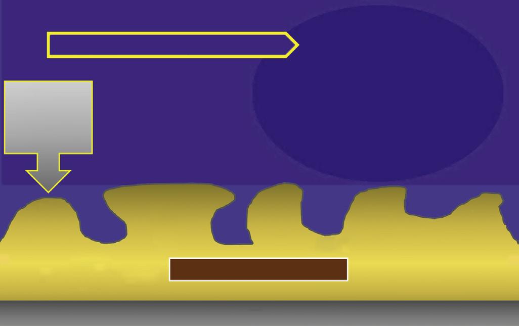
allows the coordinated regulation of expression of these proteins by microorganisms in biofilms to regulate population density and possibly virulence.25 The majority of the biofilm structure is a highly heterogeneous matrix composed of extracellular polymeric substances (EPSs) produced by cells within the biofilm. The EPS matrix provides multiple functions (Fig. 1.11). From a clinical
• Fig. 1.11 Properties of biofilms emerging from life in the extracellular polymeric substance (EPS) matrix. (From Flemming HC: EPS-then and now, Microorganisms 4[4]:pii: E41, 2016 Nov 18.)
perspective, the EPS can act as a physical barrier to antimicrobial agents such as antibiotics and disinfectants.26 Microbial organization into multispecies biofilm communities results in increased pathogenic effects on the host.27
The Microbiome of Endodontic Infections
The identity of specific microorganisms in root canal infections has been a major focus of interest for more than a century.28 Studies using culture-dependent approaches have recovered several species
Bacterial Genera Represented in Endodontic Infections
GRAM-NEGATIVE BACTERIA
Anaerobes
Dialister
Treponema TABLE 1.2
Facultatives
Anaerobes
Rods Rods
Porphyromonas
Tannerella
Prevotella
Fusobacterium
Campylobacter
Pyramidobacter
Catonella
Selenomonas
Centipeda
Capnocytophaga
Eikenella
Haemophilus
Veillonella
Megasphaera
Neisseria
Actinomyces
Pseudoramibacter
Filifactor
Eubacterium
Mogibacterium
Propionibacterium
Eggerthella
Olsenella
Bifidobacterium
Slackia
Atopobium
Solobacterium
Lactobacillus
Parvimonas
Peptostreptococcus
Finegoldia
Peptoniphilus
Anaerococcus
Streptococcus
Gemella
Spirilla
that have been identified as putative endodontic pathogens. The microbiome of carious dentin causing pulpitis and subsequent endodontic infection includes significant numbers of lactobacilli,29 gram-negative bacteria,30 and species from the Firmicutes, Actinobacteria, and Proteobacteria phyla.31 Primary root canal infections harbor a multispecies population of facultative and strict anaerobic gram-positive and gram-negative bacteria, spirochetes, yeasts, Archaea, and other unidentified species.32-36 In addition, EpsteinBarr virus may be associated with irreversible pulpitis and apical periodontitis,37 and papilloma virus and human herpes virus have been found in exudates from acute apical abscesses.38
Microorganisms have been traditionally characterized in terms of their morphology (rods, cocci, spirilla), cell wall characteristics (gram-positive and gram-negative), and oxygen tolerance (anaerobic and facultatively anaerobic). Genera cultured from symptomatic and asymptomatic root canal infections include Prevotella, Porphyromonas, Fusobacterium, Peptostreptococcus, Streptococcus, Lactobacillus, Enterococcus, Actinomyces, Propionibacterium, and Candida.20,39 (Table 1.2)
More recently, the microbiome of endodontic infections has been redefined using culture-independent molecular biology techniques. These studies have both confirmed the findings from culture studies and greatly expanded knowledge. Many species that had already been considered putative pathogens because of their frequency of detection, as reported by culture-dependent methods, have been found in a similar or even higher prevalence by using molecular approaches, strengthening associations with causation of apical periodontitis. Molecular technology has enabled the recognition of many new putative pathogens that had
GRAM-POSITIVE BACTERIA
Facultatives
Actinomyces
Corynebacterium
Lactobacillus
Streptococcus
Enterococcus
Granulicatella
not previously been found in samples from endodontic infections.40,41 A review of 12 studies that used next-generation DNA sequencing (pyrosequencing) methods to evaluate the microbiome of endodontic infections has corroborated previous multiple reports of microbial diversity. The most abundant phyla were Firmicutes, Actinobacteria, Bacteroidetes, Proteobacteria, and Fusobacteria. The most frequently detected genera were Prevotella, Fusobacterium, Porphyromonas, Parvimonas, and Streptococcus.42 (Fig. 1.12)
Many microorganisms isolated from endodontic infections have also been identified as commensals in the oral cavity. The transition from oral “commensal” to root canal “pathogen” likely reflects an innate ability to switch on genes that enable survival and propagation in a different environment and encode a range of virulence factors (Fig. 1.13). The first reported virulence factor associated with endodontic infections was lipopolysaccharide (“endotoxin”), a virulence factor produced by gram-negative bacteria.43
It has been suggested that symptoms increase when certain microbial species are part of the infective endodontic microbiome. Nevertheless, the same species can be found in asymptomatic cases with prevalence comparable to that of symptomatic cases; this apparent discrepancy could be explained in part by the variations in expression of virulence factors by different strains of the same species. Protein analyses (the metaproteome) of endodontic infections, along with the host response, are future steps toward better understanding of interactions between the microbiome of endodontic infections and the host throughout the infection and healing process.44,45
Cocci
Cocci
Firmicutes
Actinobacteria
Bacteroidetes
Proteobacteria
Fusobacteria
Synergistetes
General (n=7) Apical (n=5)
• Fig. 1.12 A heat map of root canal community profiles based on bacterial prevalence in 15 groups of root canal microbiomes gathered from 12 studies using next-generation DNA sequencing. Microbiome profiles are divided into general (entire root canal) and apical samples. Heat map scale ranges from 0 to 1 by taxa where “1” indicates that all samples had that specific taxa as their top 5 phyla or genera. A “0” score indicates that taxa did not appear as a top 5 phyla or genera. (From Shin JM, Luo T, Lee KH, et al: Deciphering endodontic microbial communities by next-generation sequencing, J Endod 44[7]:1080–87, 2018.)
Pulpal Diseases
Host Response in the Dental Pulp
The response of the dental pulp to microbial and other physical and chemical irritants is similar to the response in other connective tissues. An inflammatory process starts in the pulp and corresponds to the location where the irritation reaches it. For example, in an incipient carious lesion at the depth of an occlusal fissure, the pulp at the end of the affected dentinal tubules is seen to have a small inflammatory process histologically. This inflammation progresses throughout the coronal pulp as the carious lesion penetrates deeper in dentin, until the microbial irritants eventually invade the pulp in large numbers and cause severe inflammation (Fig. 1.14).
However, unlike other connective tissues, the dental pulp lacks collateral circulation and is confined within rigid dentinal walls. Therefore at a specific point in the disease process, the inflammation changes from reversible (one that would respond favorably to conservative methods of treatment and eventually heal) to irreversible pulpitis. The transition of reversible to irreversible pulpitis is important to identify clinically because it determines the optimal procedure that should be employed to treat it.
Studies have shown that the inflammatory response in the pulp is associated with several cellular and molecular changes (Video 1.2). The degree of irritation appears to trigger a corresponding level of inflammation. This titration of the
In recent years there has also been considerable interest in molecular mediators of pulpal inflammation. Some inflammatory mediators have been shown to correlate directly with increased pulpal pain and the clinical diagnosis of symptomatic irreversible pulpitis. These mediators include prostaglandins, neuropeptides, bradykinin, cytokines, chemokines, and matrix metalloproteinases.49,50 The dental pulp is heavily innervated by General
inflammatory response to the level of irritation is orchestrated by a balance of proinflammatory and antiinflammatory factors in the pulp. The condition of the pulp deteriorates or improves based on the reaction of pulpal factors to the external environment. Cellular responses include the increase in inflammatory cells, most notably neutrophils, lymphocytes, macrophages, plasma cells, mast cells, and dendritic cells (Figs. 1.15 to 1.17). The inflammatory cell response correlates in its intensity with the depth of the carious lesion.46 Noninflammatory cells, such as odontoblasts and fibroblasts, do contribute to the inflammatory response. Odontoblasts have been shown to express TLRs, cytokines, chemokines, and defensins.47 However, the degree to which noninflammatory cells contribute to the inflammation is much less than inflammatory cells like neutrophils and macrophages. A recent study showed reasonably good agreement between clinical signs and symptoms, and the histologic condition of the pulp in cases with caries.48 In this study, microbial ingress into the pulp, which is indicative of severe pathosis, seemed to be confined to cases diagnosed clinically as irreversible pulpitis.
(n=9)
(n=6)
• Fig. 1.13 Potential Virulence Factors Associated With a Bacterial Cell. (From Sedgley CM. Virulence of endodontic bacterial pathogens. In Fouad AF, editor: Endodontic microbiology, Ames, IA, 2009, WileyBlackwell Publishing, pp. 130–151.)
sensory fibers. During the inflammatory process, there is sprouting of pulp nociceptors, with an increase in secretion of neuropeptides like substance P and CGRP. These mediators lower the pain threshold and increase the permeability of blood vessels to inflammatory cells.
The compromised ability of the pulp to survive severe forms of inflammation, in comparison with other connective tissues, is illustrated by the differences in response to trauma between teeth with immature apex and those with mature apex. After luxation injuries, such as extrusion, intrusion, or even total avulsion and replantation, the tooth with immature apex is much more likely to retain (or revascularize) a vital pulp than a tooth with mature apex. This difference is primarily related to the increased collateral circulation in the immature teeth that can take advantage of the wide apical foramen.
Occasionally, radiographic analysis of the dental pulp reveals an increase in mineralization and deposition of hard dentinal tissues. Increased mineralization occurs gradually due to deposition of secondary dentin throughout life. However, as
a result of traumatic injuries, a substantial increase in mineralization may occur in the tooth or teeth that were traumatized. Increased areas of mineralization, commonly known as pulp stones , are also associated with caries or deep restorations. Excessive pulp stones have also been associated with cardiovascular diseases, 51 and with intake of statin medications. 52 Increased mineralization of the pulp, even when obliterating the entire canal space ( Fig. 1.18 ), in the absence of symptoms or apical pathosis, is not considered pathologic. However, if pulpal disease arises in these conditions, root canal treatment may become very challenging.
Less frequently, the pulp may undergo internal resorptive defects (Fig. 1.19). The cells that typically initiate the resorption, the osteoclasts or odontoclasts, are not normal inhabitants of the healthy pulp. These cells, which arise from monocytes, are normal mediators of bone turnover and are present in the PDL and alveolar bone. When resorptive lesions are detected clinically (internally or externally), they have usually attained a large size and are considered pathologic, even in the absence of symptoms.
• BOX 1.1 Review Study Questions
1. Which of the following is a condition that can be described as irreversible pathosis of the pulp:
a. Dentin hypersensitivity
b. Pulp canal obliteration (total calcification)
c. Internal resorption
d. Sharp, brief pain with thermal changes
2. The following inflammatory mediator(s) is/are not elevated in the pulp, in cases with symptomatic irreversible pulpitis:
a. Prostaglandins
b. Leukotrienes
c. Bradykinin
d. Neuropeptides
e. Matrix metalloproteinases
3. The following cytokine acts to control the expansion of periapical lesions:
a. IL-1
b. IL-8
c. IL-10
d. TNF-alpha
e. IL-17
4. Root canal infections are best described as:
a. Single-species biofilms, predominantly microorganisms
b. Single-species biofilms, predominantly extracellular matrix
c. Multispecies biofilms, within an extracellular matrix, predominantly microorganisms
d. Multispecies biofilms, within an extracellular matrix, predominantly matrix
5. Compared with bacteria in suspension (planktonic), microorganisms in biofilms:
a. Are less resistant to antimicrobial agents
b. Can resist alkaline stress better
c. Have a decreased potential for cell-to-cell interaction
d. A and C
A localized inflammatory reaction containing mainly polymorphonuclear leukocytes (PMNs) at the site of a carious pulpal exposure. The remainder of the coronal pulp is almost free of inflammatory cells. (Courtesy Dr. J.H. Simon.)
• Fig. 1.14
• Fig. 1.15 Mast cells are readily visible as dark-stained cells in this inflamed human dental pulp.
• Fig. 1.16 Some plasma cells stain positively for IgM in inflamed human dental pulp, indicating immunologic activity.
• Fig. 1.17 Many dendritic cells (arrows) are present in an inflamed dental pulp. (Courtesy Dr. M. Jontell.)
Clinical Classification of Pulpal Conditions
The dental pulp is encapsulated by hard tissues and therefore is not amenable to direct visual or tactile examination, or sampling for biopsy. The clinician must rely on signs and symptoms of disease, the results of clinical testing, and radiographic imaging to reach a pulpal diagnosis (see Chapter 4). A diagnosis must be obtained before any endodontic procedure is contemplated. Here the general classification and features of different pulpal conditions will be described to introduce the reader to the disease progression in
the dental pulp. Histological examination of the pulp in various stages of disease, except for total necrosis, reveals that different areas of the pulp may have different stages of normalcy, inflammation, or necrosis. Indeed, the progression from reversible pulpitis to irreversible pulpitis and then to necrosis is gradual and occurs in small compartments of the pulp at any one time, thereby complicating the clinical diagnosis.
Normal Pulp
As noted previously, normal pulp is histologically characterized by the presence of an intact odontoblastic layer, a cell-free zone, a cell-rich zone, and the absence of inflammation or necrosis. The odontoblasts may be interspersed with dendritic cells or nerve endings. Occasionally, inflammatory cells such as lymphocytes, neutrophils, or macrophages may be seen, but they are sporadic and located in the central pulp. However, the pulp tissue is primarily composed of fibroblasts, vascular elements, stem cells, and myelinated and unmyelinated nerve fibers.
Reversible Pulpitis
Pulpal inflammation begins with the onset of a carious lesion, or other irritation, such as a crack, cervical abrasion, crown attrition or fracture, or in response to a dental procedure on an intact tooth. These external stimuli lead to biological or physical irritation of the pulp and result in an increased area of inflammatory response in close proximity to the area of irritation. An example of this response may be dentin hypersensitivity in relation to cervical abrasion or attachment loss treated with scaling and root planing. Clinically, the patient with reversible pulpitis usually has no symptoms, or may have mild hyperalgesia of the pulp. The latter is expressed as increased sharp pain (with thermal changes) that is short in duration. Correction of the clinical problem that caused reversible pulpitis, such as with a restoration of a carious lesion or desensitization of hypersensitive dentin, results in healing and resolution of symptoms.
Irreversible Pulpitis
The transition from reversible to irreversible pulpitis is gradual and may or may not be associated with changes in symptoms. Irreversible pulpitis is characterized histologically by the presence of areas of severe inflammation and partial necrosis in the pulp, commonly in proximity to an area of carious exposure. There is typically microbial invasion of a portion of the pulp, with intense inflammatory response that is attempting to wall off the microbial infection. The rest of the pulp tissue typically has mild inflammation or no inflammation and therefore, is responsive to clinical vitality testing. Irreversible pulpitis may be associated with severe spontaneous and lingering pain, or with milder symptoms or no pain. Because the association of pain with irreversible pulpitis is not consistent, the diagnosis of irreversible pulpitis may be challenging (see Chapter 4).
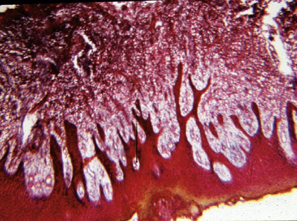
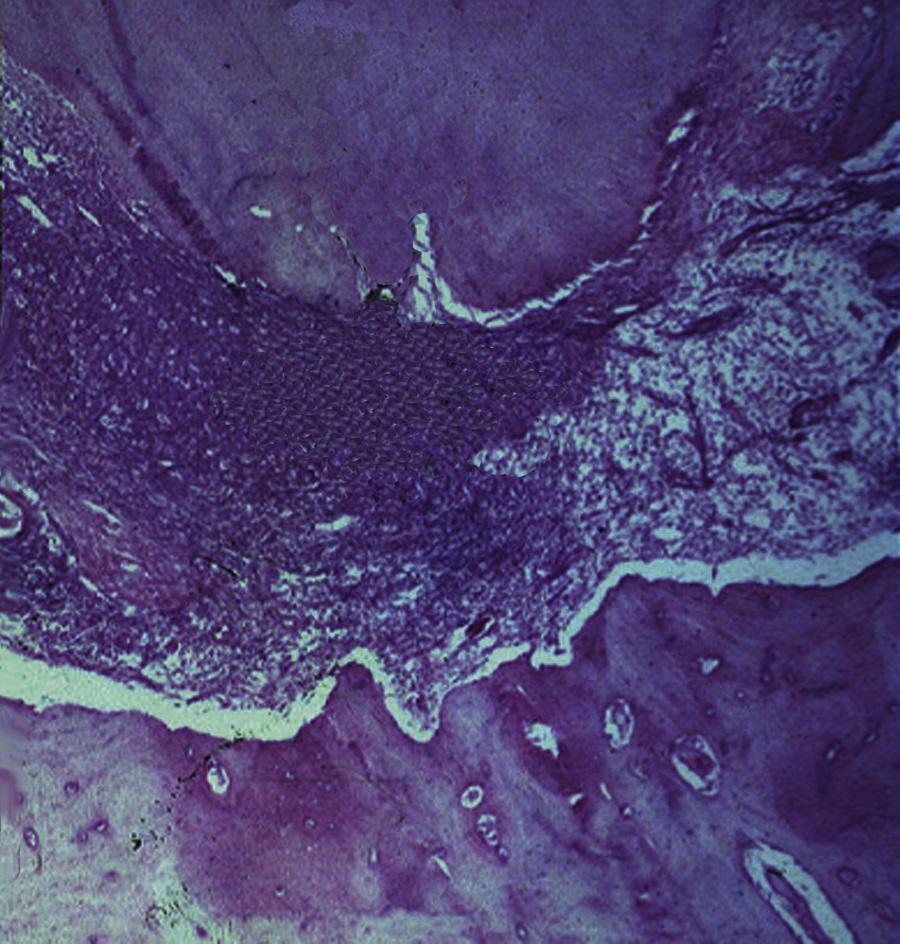
Two additional conditions are usually diagnosed as irreversible pulpitis: cases with internal resorption, with a vital (responsive) pulp (Fig. 1.19) and cases with hyperplastic pulpitis (pulp polyp) (Fig. 1.20). The latter represents a proliferative response that is occasionally seen in the pulp of children, that has been exposed to the oral cavity, and in which the pulp tissue may be covered by desquamated epithelium from oral mucosa.
• Fig. 1.18 Calcific metamorphosis does not represent pathosis per se and may occur with aging or low-grade irritation. It also may occur subsequent to a traumatic injury to the tooth.
• Fig. 1.19 Hard tissue resorption that causes disappearance of normal radiographic evidence of the root canal usually indicates an internal resorption defect.
Pulp Necrosis
In about 40% of cases, irreversible pulpitis develops into pulp necrosis without symptoms.53 In this condition, the dental pulp degenerates completely and is replaced by serous or purulent fluid or by dry necrotic tissue. The root canal environment contains microbial biofilms that vary in their composition, location, and thickness, depending on nutritive factors, pH, oxygen tension, and continued access to the oral cavity. In addition, the biofilm invades dentinal tubules, which are wide enough to accommodate microbial cells, especially in young patients with larger dentinal tubules.54
Pulp necrosis is asymptomatic, because the nerve endings supplying the pulp are no longer present. However, clinically, there are instances when the patient can feel endodontic instruments that are exploring the necrotic pulp space. This sensation may be due to remaining pulpal nerve fibers or fluid pressures that stimulate apical nociceptors. In addition, pulp necrosis may be associated with several apical conditions that are symptomatic (see later section).
Periapical Diseases
As the inflammation progresses throughout the radicular pulp, it gradually starts to affect the periapical tissues. The progression of the pulp disease into the periapical tissues presents a major
biological challenge to those tissues. The PDL is a very thin, highly fibrous tissue that is confined within the bony crypt. As such, these tissues are not capable of mounting an immune response that can stop the advancing microbial ingress. Therefore the hallmark of apical periodontitis is bone resorption to accommodate the formation of a soft tissue lesion, in which the immunologic reactions that can stop the advancing infection can be concentrated.
Animal studies have shown that apical bone resorption and inflammatory response starts soon after the initiation of pulpal inflammation. In one study, researchers found that the pathogenesis of pulpitis and apical periodontitis in the initial phases, and in the absence of severe virulent bacteria, is primarily coordinated by innate rather than adaptive immune responses.55 In another study, adaptive responses were found to be critical in preventing disseminated infections if a significant microbial load of virulent species is introduced.56 As noted before, a variety of microbial factors are important in development of periapical lesions, such as lipopolysaccharides (LPSs), lipoteichoic acid, bacterial enzymes, adhesion factors, toxins, etc. (Fig. 1.13). Although LPS is a well-characterized irritant that can induce pulpal and periapical lesions even in the absence of live bacteria,57,58 periapical lesions have been induced in animal models that do not respond to LPS in a similar manner to normal animals.59 Periapical lesions typically take 5 to 8 weeks in large animal models to be visible radiographically.60
The inflammatory response in the periapical lesion is similar to that in the pulp, except that bone resorption is now a critical component of the host response. Numerous studies have documented the tightly regulated process of periapical bone loss that correlates with the degree of the advancing microbial irritation and the balance of proinflammatory and antiinflammatory factors. Important cytokines in bone resorption include IL-1, IL-6, IL-11, IL-17, and TNF-alpha. Cytokines that limit bone resorption include IL-4 and IL-10. Another key protein that plays a major role in bone resorption is nuclear factor-κB ligand (RANKL). RANKL binds to its receptor (RANK), resulting in osteoclast differentiation. This interaction is inhibited by osteoprotegerin (OPG), a decoy protein that binds to the receptor. Levels of RANKL and the ratio of RANKL to OPG both peak at 2 to 3 weeks, concurrent with the progression of periapical bone destruction. RANKL production tapers off between weeks 4 and 8, whereas production of OPG increases during this time, creating a negative feedback loop that limits the amount of bone destruction caused by bacterial infection.61 The RANKL-RANK interaction is involved in both physiologic and pathologic bone resorption. In periapical tissues, a significant increase in RANKL levels has been found in granulomatous lesions compared with healthy controls.62,63 Finally, T-reg lymphocytes have been shown to control the size of periapical lesions in a significant manner.64
Histologically, periapical lesions have traditionally been classified into granulomas, cysts, or apical abscesses. A granuloma has a collection of inflammatory cells, such as lymphocytes, plasma cells, macrophages, and mast cells, that are arranged in a granular manner, thus the name granuloma. Occasionally, there are multinucleated giant cells and foam cells that may represent osteoclasts or engorged macrophages, respectively. A periapical cyst arises in a part of a granuloma and thus has a granuloma as part of its wall. A cyst represents a proliferation of the epithelial rests of Malassez, which are vestigial embryonic cells that remain after the disintegration of the epithelial root sheath of Hertwig after root development. Cysts may be formed by one of several mechanisms. Either the epithelial strands proliferate to surround an area of the granuloma, restrict the blood supply, and cause degeneration of the central tissue, or the epithelial mass widens to the extent that
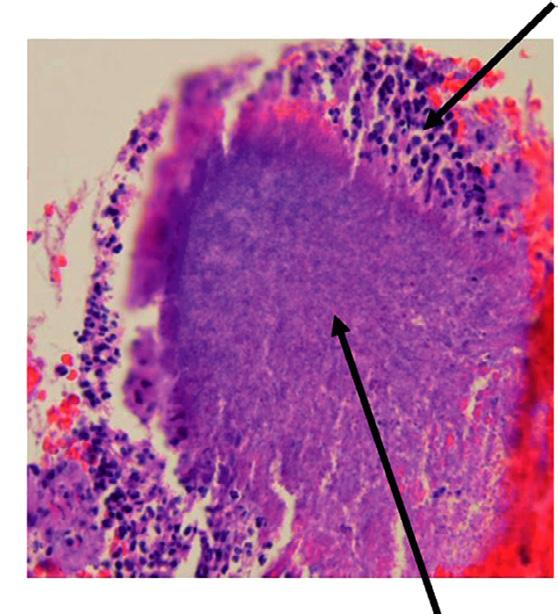
• Fig. 1.20 A, Pulp polyp, also known as hyperplastic pulpitis. The involved tooth is usually carious with extensive loss of tooth structure; the pulp remains vital and proliferates from the exposure site. B, Histologic examination of hyperplastic pulpitis shows surface epithelium and underlying inflamed connective tissue.
central cells degenerate due to their distance from the sources of nutrition. Cysts contain clear cyst fluid that may be filled with eosinophilic material and cholesterol crystals (Fig. 1.21).
The literature shows that the incidence of granulomas and cysts varies considerably. Most of these studies are from pathology services, in which only a select number of cases are submitted for examination, typically when the practitioner suspects nonendodontic pathosis. The sources may be extracted teeth that may or may not have had endodontic treatment, lesions recovered during apical surgery, or lesions in edentulous areas, suspected to have been associated with previously extracted teeth. The tissue may or may not be the complete lesion, and the pathologist may or may not perform serial sectioning to diagnose the entire lesion. In addition, many pathologists would diagnose a cyst upon identifying any epithelial tissue in the lesion, and some pathologists need to see evidence of cyst lining and lumen before making the same diagnosis. Finally, some of the larger studies have documented a high percentage of nonendodontic lesions (27% in one study65) that are submitted for examination, thus raising the possibility that some of these cases may not adhere to other characteristics of apical pathosis. With this diversity, the incidence of cysts has been reported to be from 7% to 54% of apical lesions.
Two types of apical radicular cysts have been described in the literature: true cysts and pocket (or bay) cysts. True cysts are those that do not communicate directly with the apical foramen of the offending tooth but are separated from it by an area of the lesion. Pocket or bay cysts are those in which the root apex and apical foramen open directly into the lumen of the cyst. This distinction between the types of cysts was proposed as a rationale for an argument that true cysts would not respond to nonsurgical treatment and would require surgical enucleation.66 However, there has never been direct evidence to support this hypothesis.67,68
A large body of literature has examined the differences between cysts and granulomas in inflammatory cells or molecular mediators and whether they can be distinguished clinically without obtaining a biopsy. However, it is now thought that cysts arise from granulomas of long standing and in conditions that are conducive to the proliferation of the epithelial rests. It is also generally accepted that the differentiation of cysts and granulomas is not critical, because both can heal equally well after surgical or nonsurgical treatment, 68 provided that bacterial irritants have been controlled. 67 The clinical importance
of apical radicular cysts (or large granulomas) may be in the differential diagnosis from other nonodontogenic lesions (see later section). Cysts may also push roots, causing malalignment of teeth. 69
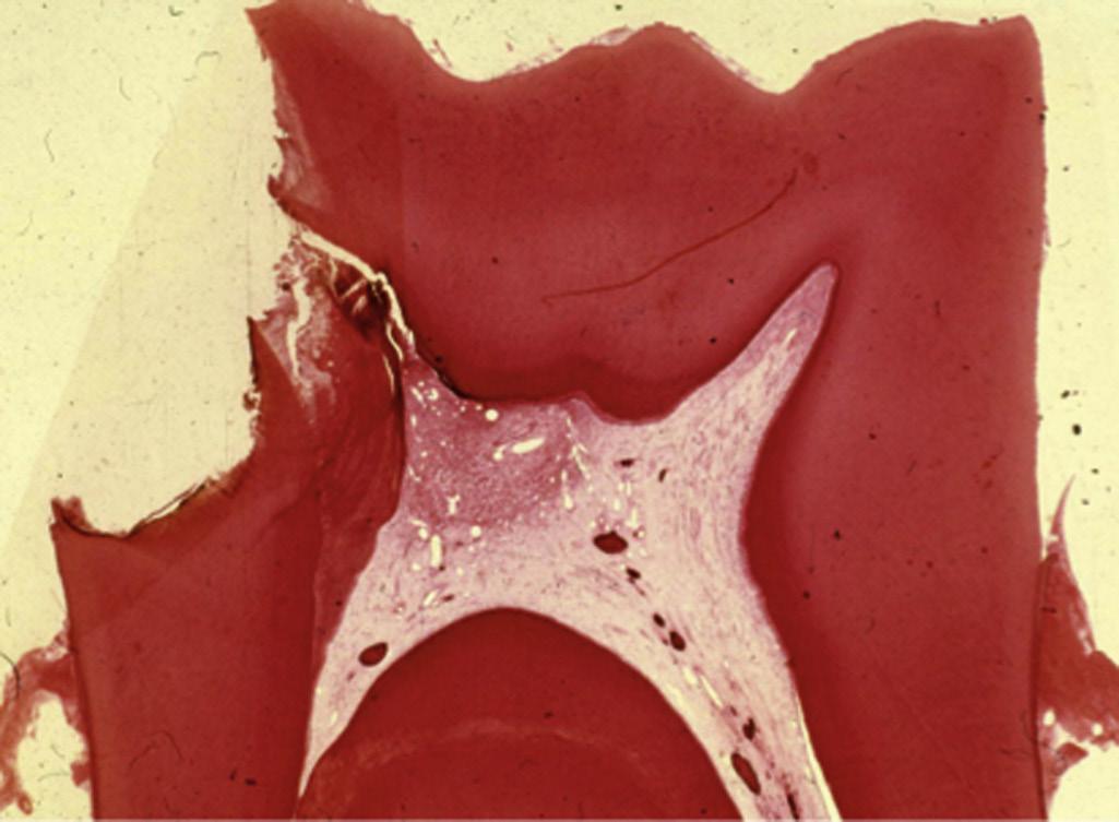
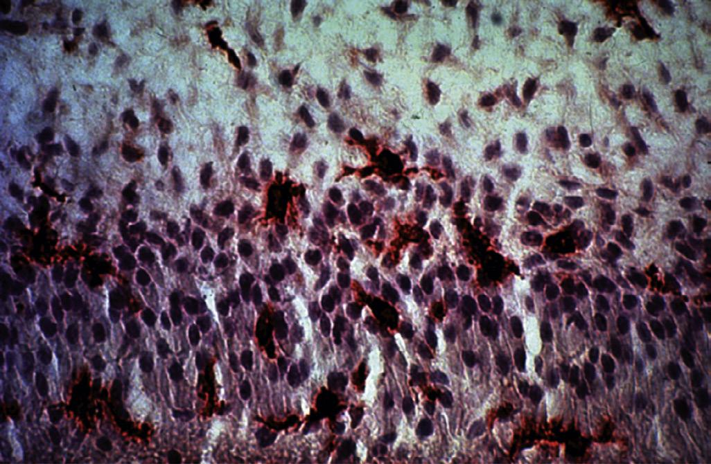
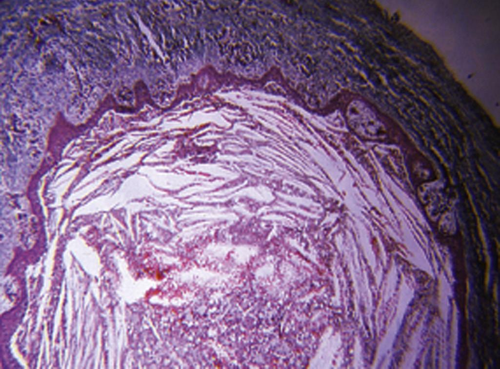
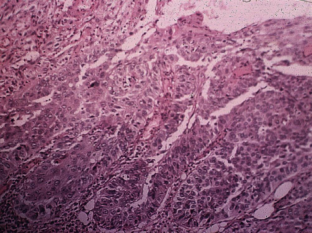
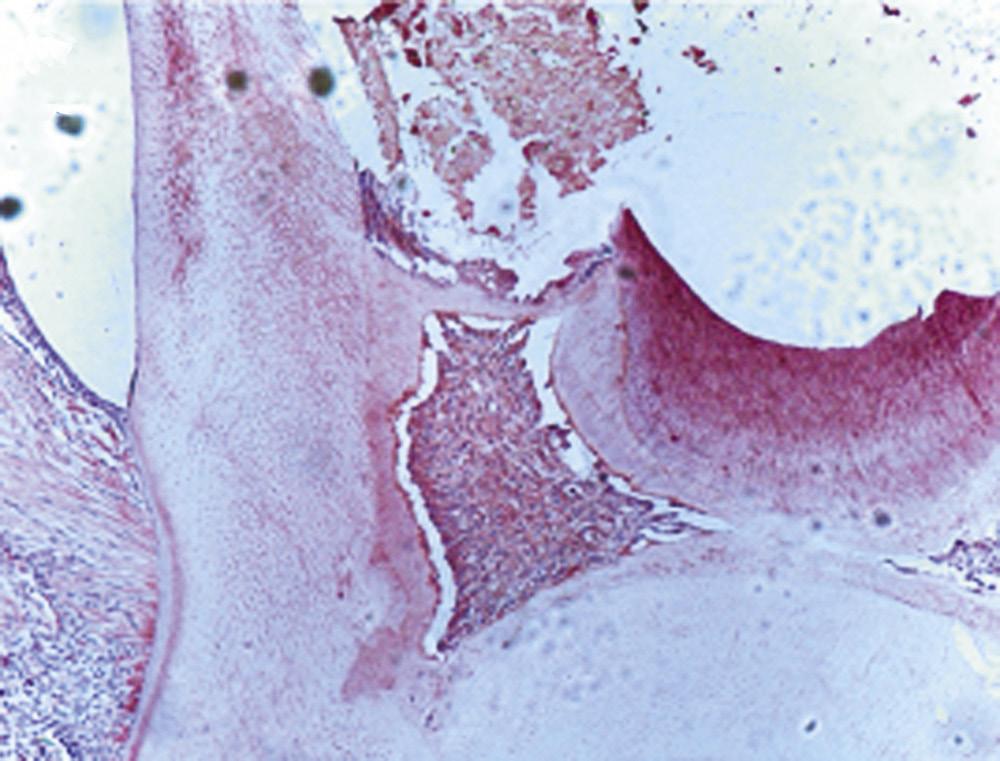
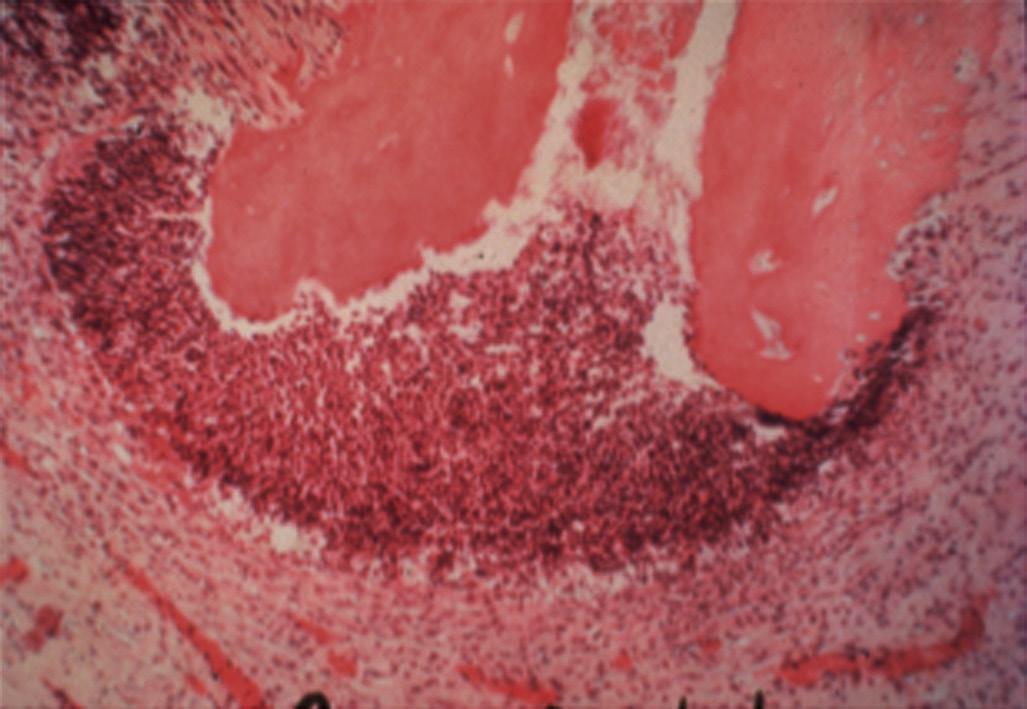
An apical abscess is characterized histologically by the presence of an intense inflammatory response surrounding areas of tissue necrosis. If bacterial stains are used, then bacteria may be physically present in these areas of necrosis. If bacterial invasion is extensive and prolonged, particularly in patients who are immunocompromised, osteomyelitis may develop. Histologically, areas of necrotic bone with empty lacunae are seen surrounded by intense inflammatory response. Microbial presence in asymptomatic apical lesions is relatively infrequent.70,71 However, studies have shown that about 50% of cases with persistent disease after endodontic treatment may have bacterial presence in the lesion.72 Occasionally the predominant bacterial colony is from the Actinomyces spp.73 This organism is filamentous in nature, and the histologic picture is characteristically described as actinic rays (Fig. 1.22)
Clinical Classification of Periapical (Apical) Conditions
Normal Periapex
In this clinical condition, there is no evidence of apical pathosis clinically or radiographically, and the tooth is asymptomatic to percussion and apical palpation. In these cases, it is thought that the inflammation of the pulp has not yet reached the apical tissues but may do so if the pulpal condition is not addressed.
Symptomatic Apical Periodontitis
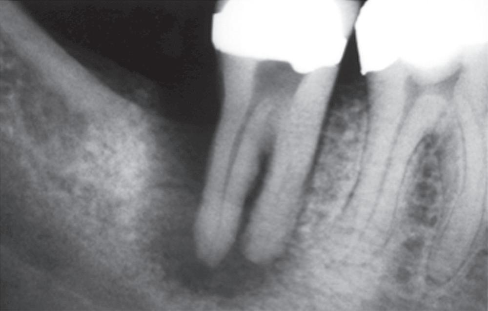
In this condition, the tooth is tender to percussion and/or apical palpation. This condition is also referred to as mechanical allodynia of the involved tooth. The inflammation has progressed in this case to the apical tissues, and therefore the pulpal pathosis is considered irreversible in nature. It is noteworthy, however, that after restorative procedures on a vital normally responsive pulp, if the occlusion on the tooth is inadvertently left high, the tooth is rendered tender to percussion. In these cases, the occlusion needs to be adjusted, and the pulpal condition is considered reversible. Cases with symptomatic apical periodontitis may have widened PDL space on the periapical radiograph (Fig. 1.23), or a small radiolucency on the cone beam computed tomography (CBCT). However, there are also many cases with asymptomatic apical periodontitis, in which an apical lesion is evident radiographically, that eventually develop clinical symptoms and become symptomatic. It is not clear why asymptomatic lesions occasionally exacerbate and become symptomatic; however, it is thought that this change may be due to changes in the microbial composition of the infected root canal causing an increase in virulent strains, an increase in microbial load, or a disruption of the host response that favors a more pathogenic infection.
Asymptomatic Apical Periodontitis
In these conditions, an apical radiolucent lesion is evident radiographically and associated with a necrotic pulp (Fig. 1.24). The lesion is typically associated with one or more roots of the tooth and occasionally extends to the furcation area as well. In fact, the lesion is associated with any area of the root in which there is the
• Fig. 1.21 A region of human apical cyst consists of a central cavity filled with eosinophilic material (EM) and a wall lined with epithelium.
Inflammatory cells
foramen of the main, lateral, or accessory canals. Occasionally, the presence of the lesion indicates the location of a foramen of a large lateral canal that cannot be seen radiographically and may not be treatable directly due to its location and size. Asymptomatic apical periodontitis may remain asymptomatic for years and may reach very large sizes. In these cases, the lesion is detected if the dentist develops a suspicion to image for apical pathosis or as an incidental finding on an intraoral or extraoral radiograph taken for other purposes. These lesions may be granulomas or cysts, but this distinction cannot be determined clinically.
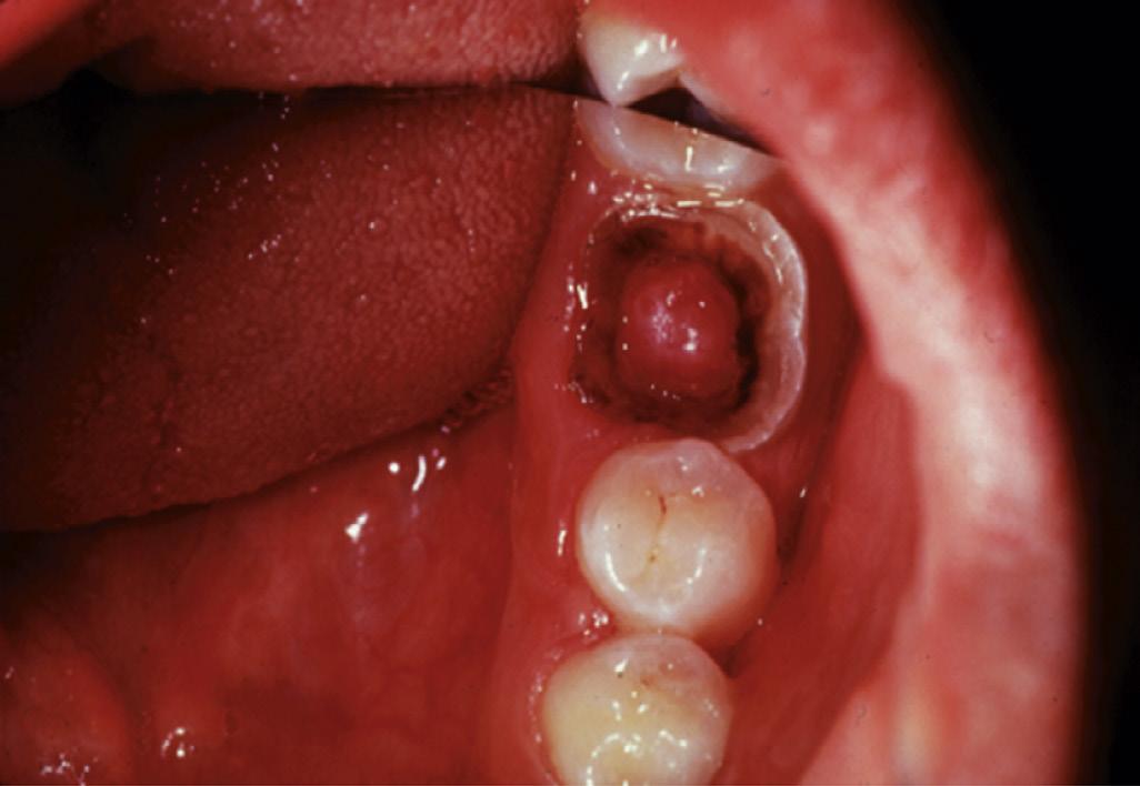
Acute Apical Abscess
This condition is the most serious clinical diagnosis in endodontics. It indicates that virulent bacterial infection has invaded the periapical tissues in sufficient numbers to cause pain and swelling related to the offending tooth (Fig. 1.25). An acute apical abscess ranges in severity from a small localized swelling in the attached gingiva that is not associated with constitutional symptoms, to a large swelling that invades fascial spaces and causes severe morbidity or even mortality of the patient. Microorganisms from the root canal environment are capable of raising systemic immunoglobulins, acute phase proteins, and systemic cytokines such as IL-1.74-77
Chronic Apical Abscess
This condition is less severe than an acute apical abscess, although it is biologically very similar to it. The pulp necrosis in this case is the source of virulent bacteria that invade the apical tissues and cause purulent drainage through a sinus tract, typically in the attached gingiva surrounding the involved tooth. The drainage may also occur through an isolated periodontal pocket that communicates with the apical lesion. The path of the sinus tract is determined by the path of least resistance. In rare instances, the sinus tract travels beyond the insertion of facial muscles and opens on the skin of the patient’s face or chin area. A sinus tract, regardless of location and duration, spontaneously heals once the source of the microorganisms, namely the infected root canal environment, is effectively treated through root canal treatment or tooth extraction.
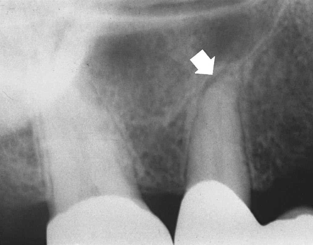
• Fig. 1.23 After cementation of a three-unit bridge, the premolar developed clinical signs and symptoms of acute apical periodontitis. Radiograph shows a widened periodontal ligament space (arrow).
• Fig. 1.24 Some plasma cells stain positively for immunoglobulin M (IgM) in inflamed human dental pulp, indicating immunologic activity.
Bacterial colonies
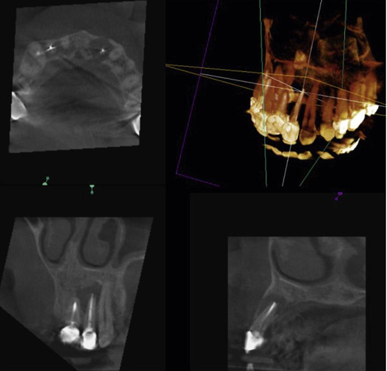
• Fig. 1.22 Apical Actinomycosis. Cone beam computed tomography (CBCT) in (A) is of a patient with persistent apical periodontitis related to previously treated tooth #10. After root end resection and filling, the biopsy result was apical actinomycosis. Photomicrograph of the lesion is shown in (B)
Condensing Osteitis
This condition represents a radiographic feature of the periapex of some teeth with pulpal pathosis. In these cases, the bone surrounding the apical region is more sclerotic than that surrounding neighboring teeth. The pulpal condition may have any of the pathologic conditions described earlier in this chapter. It is thought that low-grade irritation of the bone may result in sclerosis rather than in resorption. In the absence of irreversible pathosis of the pulp, there is no need to address condensing osteitis. Condensing osteitis is often confused with enostosis (sclerotic bone), a nonpathologic entity that may be associated with teeth with normal pulp.
Nonendodontic Pathosis
Benign lesions with radiographic appearances like periapical lesions include (but are not limited to) the initial stages of periapical cemental dysplasia (Fig. 1.26), early stages of fibrous dysplasia, ossifying fibroma, odontogenic keratocyst, lateral periodontal cyst, dentigerous cyst, median maxillary or mandibular cyst, traumatic bone cyst, central giant cell granuloma, central hemangioma, hyperparathyroidism, myxoma, and ameloblastoma. Usually (but not always), radiographically the lamina dura around the root apices is intact, and responses to pulp tests are normal. The final diagnosis of these lesions is often based on surgical biopsy and histopathologic examination.
• Fig. 1.26 A periapical radiolucency in the early stages of cementoma can simulate a periapical lesion of pulpal origin. However, the pulps’ responses are within normal limits.
Malignant lesions that may simulate periapical lesions of pulpal origin and are often metastatic include lymphoma (Fig. 1.27), squamous cell carcinoma, osteogenic sarcoma, chondrosarcoma, and multiple myeloma. Unlike endodontic lesions, these lesions are usually associated with rapid and extensive hard tissue (bone and tooth) destruction. Ordinarily, the teeth in the affected region remain responsive to vitality tests, although occasionally the pulps or sensory nerves are disrupted and nonresponsive. The patient may also experience numbness rather than pain in these situations.
C B
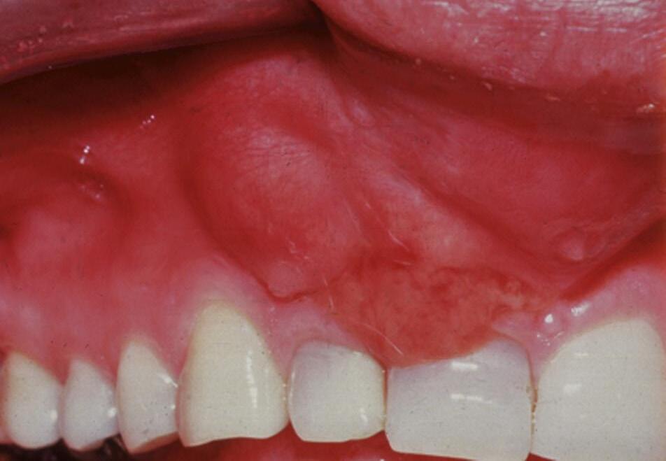
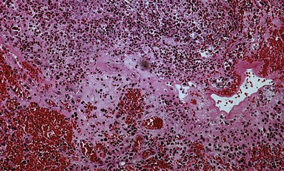
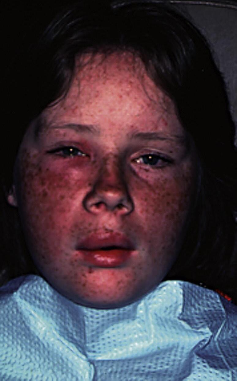
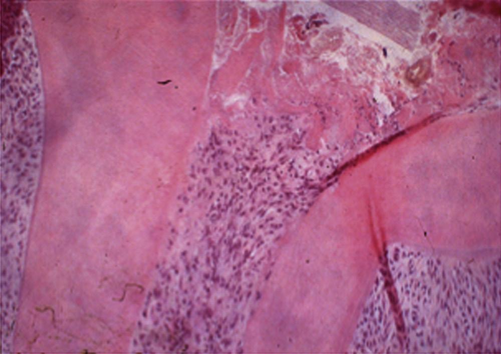
• Fig. 1.25 A, Localized vestibular swelling resulting from the necrotic pulp in the right lateral incisor. B, An acute apical abscess (AAA) has created diffuse facial swelling. C, Histologic examination of the AAA shows edematous tissue heavily infiltrated by degenerating polymorphonuclear leukocytes (PMNs).
• Fig. 1.27 A, Periapical radiolucent lesion of nonpulpal origin. B, Positive results of vitality tests and histologic examination of the tissue confirmed a diagnosis of carcinoma.
The percentages of different histopathologic diagnoses have been presented in one of the largest studies to document endodontic and nonendodontic apical pathosis submitted for biopsy65 (Fig. 1.28).
Role of Residual Microorganisms in the Outcome of Endodontic Treatment
Root canal treatment aims to eliminate necrotic pulp tissue, biofilm, and debris from the root canal system. However, several obstacles to optimal root canal disinfection can exist, resulting in persistent infections, or infections arising from members of the original microbiome that have survived root canal treatment.78 These obstacles include the anatomic complexities of the root canal system, the buffering effects of dentin, and the physical barrier to antimicrobials provided by the EPS matrix of the biofilm. As a consequence, some species survive endodontic treatment procedures, and persistent infection can ensue.79,80 Microbial products from persistent infections diffusing into the apical tissues through root canal ramifications and exposed dentinal tubules can induce an inflammatory response with consequent release of cytokines that activate resorption mechanisms (Fig. 1.29).
The primary cause of endodontic treatment failure has been attributed to intraradicular infections, usually in the form of biofilms.81 Microorganisms present in the apical portion of the root canal, apical deltas, and lateral canals have the potential to sustain long-standing infections.22 Those microorganisms that survive need to be able to resist periods of starvation and withstand possible disruption of the EPS matrix. If communication with the apical tissues is present, then the biofilm is likely to have access to a source of nutrients that facilitates growth and a subsequent host response that sustains or exacerbates periapical inflammation and avoids healing.
Extraradicular infections have been associated with cases of long-standing lesions and persistent root canal infections.82 It is generally accepted that Actinomyces spp. and Propionibacterium propionicum are involved in periapical lesions refractory to endodontic
treatment because of their ability to evade the host response, survive in the apical tissues, and prevent apical healing (Fig. 1.22). However, there is no clear evidence that apical actinomycosis is actually independent of the intraradicular infection.73 In these cases surgical management that includes root end resection and removal of the infected tissue typically provides a successful outcome.
Healing of Pulp and Periapical Tissues
Regeneration is a process by which altered tissues are completely replaced by tissues native to their original architecture and function. Repair is a process by which altered tissues are not completely restored to their original structures. Inflammation and healing are not two separate entities; in fact, they constitute part of one process in response to tissue injury. On the molecular and cellular levels, they are inseparable. Inflammation dominates the early events after tissue injury, shifting toward healing after the early responses have subsided. The level of healing is proportional to the degree and extent of tissue injury and the nature of tissue destruction.
Process of Pulp Healing
In the absence of irritants, a healthy pulp has tremendous capacity to heal. Odontoblasts are the first cells to encounter the invading microorganisms and their components, as well as to detect dentin matrix constituents released during demineralization.83 Together with pulp fibroblasts,84 they are capable of orchestrating an inflammatory response, contributing to the recruitment of immune system cells and releasing cytokines and chemokines.85,86
Human dental pulp stem cells (hDPSCs) are ectodermalderived stem cells, originating from migrating neural crest cells, and possess mesenchymal stem cell (MSC) properties. It has been reported in the literature that stem/progenitor cells from inflamed human dental pulp retain tissue generation potential.87 Pulp capping materials may also contribute to pulpal stem cells’ differentiation and repair.88,89 Andelin and associates used immunostaining with dentin sialoprotein (DSP) to determine the type of tissue
A B
Keratocystic odontogenic tumor Nasopalatine duct cyst Residual Cyst Lateral periodontal cyst
Neurofibroma Central giant cell lesion Traumatic bone cyst Odontogenic fibroma Ameloblastoma Cemento-osseous dysplasia Mucoepidermoid carcinoma Ossifying fibroma Keratinizing odontogenic cys t
Calcifying odontogenic cyst Malignant Lymphoma Odontogenic myxoma Metastatic Schwannoma
Adenomatoid odontogenic tumor Osteoporotic bone marrow defect Langerhans cell disease Plasmacytoma Osteogenic sarcoma Cherubis m Globulomaxillary cyst Squamous odontogenic tumor Chondrogenic sarcoma Stafne bone cyst
• Fig. 1.28 Lesions associated with crowns of unerupted teeth (dentigerous cysts, hyperplastic follicle) which would not be confused with ones from endodontic origin, have been excluded from this list (n = 9.723). Apical cysts and granulomas were the most common, comprising 73% of the considered jaw lesions. Ameloblastomas were reported 144 times (1.2%). Squamous odontogenic tumors and chondrogenic sarcomas were reported twice (0.02%) and a Stafne bone cyst was reported once (0.01%). (From Koivisto T, Bowles WR, Rohrer M: Frequency and distribution of radiolucent jaw lesions: a retrospective analysis of 9,723 cases, J Endod 38[6]:729–32, 2012.)
AB
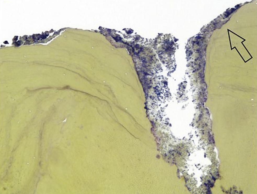
• Fig. 1.29 Apical Foramen and External Surface of Tooth With Persistent Root Canal Infection. A, Longitudinal section cut through the foramen, which appears clogged with a biofilm, extending on the outer apical surface (Taylor-modified Brown-Brenn, original magnification 50). B, High-power view of the external apical surface indicated by the arrow in (A) showing different concentrations of bacteria within the biofilm, which appears firmly attached to the resorbed cementum (original magnification × 400). (From Ricucci D, Loghin S, Gonçalves LS, et al: Histobacteriologic conditions of the apical root canal system and periapical tissues in teeth associated with sinus tracts, J Endod 44[3]:405–413, 2018.)
that forms after pulp capping procedures.88 DSP is present in both bone and dentin but is expressed at an approximately 400-fold higher level in dentin than in bone.90 Depending on the capping material used, dentin healing may occur through regeneration of dentin (identified through heavy DSP staining; Fig. 1.30, A) or through repair with an amorphous mineralized scar tissue (light DSP staining; Fig. 1.30, B).
Mild injury to the pulp and endothelial cells stimulates stemcell recruitment, and proliferation of dental stem cells can be stimulated by platelet-derived growth factor BB (PDGF-BB), vascular endothelial growth factor (VEGF), insulin-like growth factor 1 (IGF-1), and transforming growth factor β1 (TGF-β1).91 On the other hand, prolonged injury (e.g., as seen in microbial infections) results in stem-cell apoptosis and impairment of these cells’ function and ability to repair the pulp.92
Process of Periapical Healing
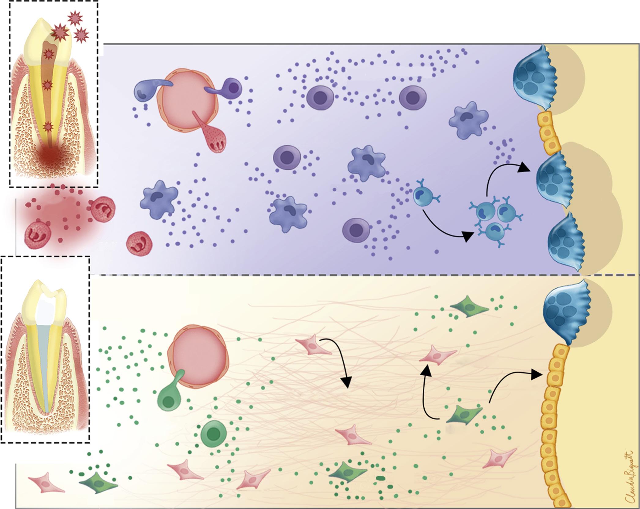
The sequence of events leading to resolution of periapical lesions has not been studied extensively. Based on the processes involved in the repair of extraction sites,93 after removal of irritants, inflammatory responses decrease, and tissue-forming cells (fibroblasts and endothelial cells) increase; finally, tissue organization and maturation ensue. Bone that has resorbed is replaced by new bone; resorbed cementum and dentin are repaired by cellular cementum. The PDL, which is the first tissue affected, is the last to be restored to normal architecture (Fig. 1.31). Histologic examination of healing of periapical lesions shows evidence of healing in the form of cementum deposition, increased vascularity, and increased fibroblastic and osteoblastic activities.94 Studies have shown that some cytokines, MMPs and TIMP1, HSP27, and Serpine1 play an important role during healing of periapical lesions.95-98 MSCs’ mobilization also results in the attenuation of apical periodontitis progression associated with immunosuppressive and prohealing mechanisms.99
Some lesions do not completely regain all the original structures. Variations are seen in different fiber or bone patterns. These variations may be obvious radiographically with a widened lamina dura or altered bony configuration. Certain factors, such as the size of the defect or the extent of injury to the underlying stroma, may affect complete regeneration of the original tissue architecture.
Factors Influencing Healing
Other factors that may affect the healing of periapical lesions include inherent host factors (e.g., leukopenia, impairment of blood supply, inadequate nutrition), corticosteroids, and other systemic diseases. For instance, patients with diabetes mellitus have a significantly lower healing rate after root canal therapy of teeth with apical lesions than do nondiabetic patients.100 Uncontrolled hyperglycemia may also affect pulp healing. In a pulp capping model using streptozotocin-induced diabetes in rats, Garber and coworkers found significant impairment of dentin bridge formation compared with that of normal rats.101 Impairment of dentin bridge formation was directly correlated with the amount of pulp inflammation.
It has been suggested that genetic predisposition can contribute to an individual’s susceptibility to pulpal inflammation and apical periodontitis and that a complex signaling network operates in the determination of the nature and extent of their progression, as well as the associated bone-destructive process. In recent years, polymorphisms in a few genes (IL1B, MMP2, MMP3, HSPA1L,
and HSPA6) mostly belonging to immune response–related pathways, have been shown to be associated with pulpal inflammation and apical periodontitis.102-105 Differential methylation profiles of immune response–related genes, such as FOXP3106 and microRNAs,107 may also have an influence on individual susceptibility to pulpal and apical inflammation and patient treatment outcomes through their potential contributions to altered expression of disease-relevant genes.
• BOX 1.2 Study Questions
6. Biofilms have been observed on the apical root surface of teeth with persistent apical periodontitis:
a. True
b. False
7. Direct pulp exposure to microorganisms is not a prerequisite for pulpal response and inflammation
a. True
b. False
8. The process by which altered or damaged pulp tissues are completely replaced by tissues native to their original architecture and function is described as
a. Repair
b. Revascularization
c. Regeneration
d. Calcification
9. Which of these sentences is considered wrong in regard to the process of pulp healing?
a. In the absence of irritants, a healthy pulp has tremendous capacity to heal.
b. DSP is present in both bone and dentin but is expressed at an approximately 400-fold higher level in bone than in dentin.
c. Mild injury to the pulp and endothelial cells stimulates stem cell recruitment and proliferation of dental stem cells.
d. Prolonged injury results in stem cell apoptosis and impairment of these cells’ function and ability to repair the pulp.
10. Genetic predisposition to pulpal and periapical inflammation has been reported in the literature as an important factor that can influence healing. Which of the genes listed is considered a good candidate gene for the investigation of individual’s susceptibility to develop pulpal and periapical inflammation?
a. TGFB
b. IL1B
c. OPG
d. IL10
ANSWERS
Answer
1 c. Internal resorption
2 b. Leukotrienes
3 c. IL-10
4 d. Multispecies biofilms, within an extracellular matrix, predominantly matrix
5 b. Can resist alkaline stress better
6 a. True
7 a. True
8 c. Regeneration
9 b. DSP is present in both bone and dentin but is expressed at an approximately 400-fold higher level in bone than in dentin
10 b. IL1B
B A
• Fig. 1.30 A, Sclerotic dentin, which appears as an irregularly organized, mineralized tissue and stains lighter than normal tubular dentin with antibody against dentin sialoprotein (DSP). B, Dentin bridge; also, secondary dentin, deposited along canal wall after a pulp capping procedure, which shows a tubular structure and stains similarly to the primary dentin with antibody against DSP.
Vessels
Inflammmator y tissue destruction
Th1/th17
Pro-inflammatory cytokines
macrophage
macrophage
growth factors (e.g. TGFb, VEGF) anti-inflammatory cytokines (e.g. IL10)
IL10, TGFb
osteoclasts
pre osteclasts bone resorption bone formation
osteoblasts
growth factors
• Fig. 1.31 Schematic Illustration of Wound Healing of Periapical Tissues After Endodontic Therapy. Apical periodontitis is an inflammatory lesion caused by microorganisms and their byproducts derived from an infected root canal system. It also represents an important host defense mechanism with the participation of inflammatory cells such as polymorphonuclear leukocytes (PMNs), macrophages, and lymphocytes (Th1/Th17). These cells express several proinflammatory cytokines that will activate osteoclasts contributing to bone resorption. After successful endodontic therapy, T regulatory cells (Tregs), mesenchymal stem cells (MSC), connective tissue fibroblasts, and osteoblasts will initiate the healing/repair process, releasing growth factors and antiinflammatory cytokines, resulting in matrix deposition and new bone formation. IL10, Interleukin 10; LPS, lipopolysaccharides; TGFb, transforming growth factor beta; VEGF, vascular endothelial growth factor. (Courtesy Dr. Claudia Biguetti.)
Fibroblasts
References
1. Yu C, Abbott PV: An overview of the dental pulp: its functions and responses to injury, Aust Dent J 52(Suppl 1):S4–16, 2007.
2. Garberoglio R, Brännström M: Scanning electron microscopic investigation of human dentinal tubules, Arch Oral Biol 21(6):355–362, 1976.
3. Bergenholtz G, Lindhe J: Effect of experimentally induced marginal periodontitis and periodontal scaling on the dental pulp, J Clin Periodontol 5(1):59–73, 1978.
4. Strobl H, Haas M, Norer B, et al.: Evaluation of pulpal blood flow after tooth splinting of luxated permanent maxillary incisors, Dent Traumatol 20(1):36–41, 2004.
5. Kayhan F, Küçükkeleş N, Demirel D: A histologic and histomorphometric evaluation of pulpal reactions following rapid palatal expansion, Am J Orthod Dentofacial Orthop 117(4):465–473, 2000.
6. Taşpinar F, Akgül N, Simşek G, et al.: The histopathological investigation of pulpal tissue following heavy orthopaedic forces produced by rapid maxillary expansion, J Int Med Res 31(3):197–201, 2003.
7. Messer HH, Chen RS: The duration of effectiveness of root canal medicaments, J Endod 10(6):240–245, 1984.
8. Langeland K: Management of the inflamed pulp associated with deep carious lesion, J Endod 7(4):169–181, 1981.
9. Masillamoni CR, Kettering JD, Torabinejad M: The biocompatibility of some root canal medicaments and irrigants, Int Endod J 14(2):115–120, 1981.
10. Karapinar-Kazandağ M, Bayrak OF, Yalvaç ME, et al.: Cytotoxicity of 5 endodontic sealers on L929 cell line and human dental pulp cells, Int Endod J 44(7):626–634, 2011.
11. Ruparel NB, Teixeira FB, Ferraz CC, Diogenes A: Direct effect of intracanal medicaments on survival of stem cells of the apical papilla, J Endod 38(10):1372–1375, 2012.
12. Brännström M, Lind PO: Pulpal response to early dental caries, J Dent Res 44(5):1045–1050, 1965.
13. Baume LJ: Dental pulp conditions in relation to carious lesions, Int Dent J 20(2):309–337, 1970.
14. Jontell M, Okiji T, Dahlgren U, Bergenholtz G: Immune defense mechanisms of the dental pulp, Crit Rev Oral Biol Med 9(2):179–200, 1998.
15. Kakehashi S, Stanley HR, Fitzgerald RJ: The effects of surgical exposures of dental pulps in germ-free and conventional laboratory rats, Oral Surg Oral Med Oral Pathol 20:340–349, 1965.
16. Möller AJ, Fabricius L, Dahlén G, et al.: Influence on periapical tissues of indigenous oral bacteria and necrotic pulp tissue in monkeys, Scand J Dent Res 89(6):475–484, 1981.
17. Sundqvist G: Bacteriological studies of necrotic dental pulps, Umeå, Sweden, 1976, University of Umeå.
18. Janeway Jr CA, Medzhitov R: Innate immune recognition, Annu Rev Immunol 20:197–216, 2002.
19. Vasselon T, Detmers PA: Toll receptors: a central element in innate immune responses, Infect Immun 70(3):1033–1041, 2002.
20. Sundqvist G: Bacteriological studies of necrotic dental pulps [Dissertation], Umeå, Sweden, 1976, University of Umeå.
21. Svensäter G, Bergenholtz G: Biofilms in endodontic infections, Endod Topics 9:27–36, 2004.
22. Ricucci D, Siqueira Jr JF: Biofilms and apical periodontitis: study of prevalence and association with clinical and histopathologic findings, J Endod 36(8):1277–1288, 2010.
23. Nair PN, Henry S, Cano V, Vera J: Microbial status of apical root canal system of human mandibular first molars with primary apical periodontitis after “one-visit” endodontic treatment, Oral Surg Oral Med Oral Pathol Oral Radiol Endod 99(2):231–252, 2005.
24. McDougald D, Rice SA, Barraud N, et al.: Should we stay or should we go: mechanisms and ecological consequences for biofilm dispersal, Nat Rev Microbiol 10(1):39–50, 2012.
25. Parsek MR, Greenberg EP: Sociomicrobiology: the connections between quorum sensing and biofilms, Trends Microbiol 13(1):27–33, 2005.
26. Flemming HC, Wingender J: The biofilm matrix, Nat Rev Microbiol 8(9):623–633, 2010.
27. Siqueira Jr JF: Rôças IN: Community as the unit of pathogenicity: an emerging concept as to the microbial pathogenesis of apical periodontitis, Oral Surg Oral Med Oral Pathol Oral Radiol Endod 107(6):870–878, 2009.
28. Miller WD: An introduction to the study of the bacterio-pathology of the dental pulp, Dental Cosmos 36(7):505–528, 1894.
29. Chhour KL, Nadkarni MA, Byun R, et al.: Molecular analysis of microbial diversity in advanced caries, J Clin Microbiol 43(2):843–849, 2005.
30. Martin FE, Nadkarni MA, Jacques NA, Hunter N: Quantitative microbiological study of human carious dentine by culture and realtime PCR: association of anaerobes with histopathological changes in chronic pulpitis, J Clin Microbiol 40(5):1698–1704, 2002.
31. Roças IN, Alves FRF, Rachid CTCC, et al.: Microbiome of deep dentinal caries lesions in teeth with symptomatic irreversible pulpitis, PLoS One 11(5):e0154653, 2016, https://doi.org/10.1371/ journal.pone.0154653
32. Waltimo TM, Sen BH, Meurman JH, et al.: Yeasts in apical periodontitis, Crit Rev Oral Biol Med 14(2):128–137, 2003.
33. Vickerman MM, Brossard KA, Funk DB, et al.: Phylogenetic analysis of bacterial and archaeal species in symptomatic and asymptomatic endodontic infections, J Med Microbiol 56(Pt 1):110–118, 2007.
34. Li L, Hsiao WW, Nandakumar R, et al.: Analyzing endodontic infections by deep coverage pyrosequencing, J Dent Res 89(9):980–984, 2010.
35. Siqueira Jr JF, Alves FRF, Roças IN: Pyrosequencing analysis of the apical root canal microbiota, J Endod 37(11):1499–1503, 2011.
36. Ozok AR, Persoon IF, Huse SM, et al.: Ecology of the microbiome of the infected root canal system: a comparison between apical and coronal root segments, Int Endod J 45(6):530–541, 2012.
37. Li H, Chen V, Chen Y, et al.: Herpesviruses in endodontic pathoses: association of Epstein-Barr virus with irreversible pulpitis and apical periodontitis, J Endod 35(1):23–29, 2009.
38. Ferreira DC, Paiva SS, Carmo FL, et al.: Identification of herpesviruses types 1 to 8 and human papillomavirus in acute apical abscesses, J Endod 37(1):10–16, 2011.
39. Baumgartner JC, Falkler Jr WA: Bacteria in the apical 5 mm of infected root canals, J Endod 17(8):380–383, 1991.
40. Rolph HJ, Lennon A, Riggio MP, et al.: Molecular identification of microorganisms from endodontic infections, J Clin Microbiol 39(9):3282–3289, 2001.
41. Siqueira Jr JF, Roças IN: Exploiting molecular methods to explore endodontic infections: part 2—redefining the endodontic microbiota, J Endod 31(7):488–498, 2005.
42. Shin JM, Luo T, Lee KH, et al.: Deciphering endodontic microbial communities by next-generation sequencing, J Endod 44(7):1080–1087, 2018.
43. Schein B, Schilder H: Endotoxin content in endodontically involved teeth, J Endod 1(1):19–21, 1975.
44. Provenzano JC, Siqueira Jr JF: Roças IN, et al: Metaproteome analysis of endodontic infections in association with different clinical conditions, PLoS One 8(10):e76108, 2013.
45. Alfenas CF, Mendes TAO, Ramos HJO, et al.: Human exoproteome in acute apical abscesses, J Endod 43(9):1479–1485, 2017.
46. Izumi T, Kobayashi I, Okamura K, Sakai H: Immunohistochemical study on the immunocompetent cells of the pulp in human non-carious and carious teeth, Arch Oral Biol 40(7):609–614, 1995.
47. Horst OV, Horst JA, Samudrala R, Dale BA: Caries induced cytokine network in the odontoblast layer of human teeth, BMC Immunol 12:9, 2011.