No part of this publication may be reproduced or transmitted in any form or by any means, electronic or mechanical, including photocopying, recording, or any information storage and retrieval system, without permission in writing from the publisher. Details on how to seek permission, further information about the Publisher’s permissions policies and our arrangements with organizations such as the Copyright Clearance Center and the Copyright Licensing Agency, can be found at our website: www.elsevier.com/permissions.
This book and the individual contributions contained in it are protected under copyright by the Publisher (other than as may be noted herein).
Notices
Knowledge and best practice in this field are constantly changing. As new research and experience broaden our understanding, changes in research methods, professional practices, or medical treatment may become necessary.
Practitioners and researchers must always rely on their own experience and knowledge in evaluating and using any information, methods, compounds, or experiments described herein. In using such information or methods they should be mindful of their own safety and the safety of others, including parties for whom they have a professional responsibility.
With respect to any drug or pharmaceutical products identified, readers are advised to check the most current information provided (i) on procedures featured or (ii) by the manufacturer of each product to be administered, to verify the recommended dose or formula, the method and duration of administration, and contraindications. It is the responsibility of practitioners, relying on their own experience and knowledge of their patients, to make diagnoses, to determine dosages and the best treatment for each individual patient, and to take all appropriate safety precautions.
To the fullest extent of the law, neither the Publisher nor the authors, contributors, or editors, assume any liability for any injury and/or damage to persons or property as a matter of products liability, negligence or otherwise, or from any use or operation of any methods, products, instructions, or ideas contained in the material herein.
Library of Congress Cataloging-in-Publication Data
Soto, Jorge A., editor.
Lucey, Brian C., editor.
Emergency radiology / [edited by] Jorge A. Soto, Brian C. Lucey. Emergency radiology (Soto) | Requisites in radiology. | Requisites series. Second edition. | Philadelphia, PA : Elsevier, [2017] | Requisites |Requisites radiology series
Includes bibliographical references and index.
LCCN 2015037285
ISBN 9780323376402 (hardcover : alk. paper)
MESH: Diagnostic Imaging. | Emergency Medical Services.
LCC RC78 | NLM WN 180 | DDC 616.07/572--dc23 LC record available at http://lccn.loc.gov/2015037285
Executive Content Strategist: Robin Carter
Content Development Specialist: Amy Meros
Publishing Services Manager: Patricia Tannian
Project Manager: Stephanie Turza
Senior Book Designer: Amy Buxton
Contributors
Carlos A. Anaya, MD
Medical Director
Cardiovascular Interventional Institute Department of Radiology Manati Medical Center Manati, Puerto Rico
Stephan W. Anderson, MD
Associate Professor of Radiology Boston University Medical Center Boston, Massachusetts
Laura L. Avery, MD
Assistant Professor Massachusetts General Hospital Harvard Medical School Boston, Massachusetts
Glenn D. Barest, MD
Assistant Professor of Radiology Boston Medical Center Boston, Massachusetts
Sarah D. Bixby, MD
Assistant Professor of Radiology Harvard Medical School; Pediatric Radiologist Department of Radiology Boston Children’s Hospital Boston, Massachusetts
Anna K. Chacko, MD
Adjunct Professor of Radiology Boston University Boston, Massachusetts; Professor of Telemedicine
John A. Burns School of Medicine University of Hawaii Honolulu, Hawaii
Margaret N. Chapman, MD
Chief of Neuroradiology
Boston VA Healthcare System; Assistant Professor of Radiology Boston Medical Center
Boston University School of Medicine Boston, Massachusetts
Luis E. Diaz, MD
Associate Chief of Radiology VA Boston Health Care System; Associate Professor of Radiology Boston University Boston, Massachusetts
Alejandra Duran-Mendicuti, MD
Department of Radiology
Brigham and Women’s Hospital Boston, Massachusetts
Ana Maria Gomez, MD
Department of Radiology Manati Medical Center Manati, Puerto Rico
Rathachai Kaewlai, MD
Instructor
Division of Emergency Radiology Department of Diagnostic and Therapeutic Radiology
Ramathibodi Hospital Faculty of Medicine
Mahidol University Bangkok, Thailand
Russ Kuker, MD
Department of Radiology University of Miami Hospital Miami, Florida
Christina A. LeBedis, MD
Assistant Professor Boston University Medical Center Boston, Massachusetts
Brian C. Lucey, MD
Associate Professor Department of Radiology
Boston University School of Medicine Boston, Massachusetts; Clinical Director Department of Radiology The Galway Clinic
Doughiska, County Galway, Ireland
Asim Z. Mian, MD
Assistant Professor of Radiology
Boston Medical Center
Boston University Boston, Massachusetts
Sarah S. Milla, MD
Associate Professor
Department of Radiology and Imaging Sciences Emory University; Attending Pediatric Radiologist and Neuroradiologist Children’s Healthcare of Atlanta Egleston Hospital Atlanta, Georgia
Felipe Munera, MD Department of Radiology University of Miami Hospital Miami, Florida
Rohini N. Nadgir, MD
Assistant Professor of Radiology and Radiological Science
Johns Hopkins Medical Institutions Baltimore, Maryland
Osamu Sakai, MD, PhD
Chief of Neuroradiology
Professor of Radiology, Otolaryngology–Head and Neck Surgery and Radiation Oncology
Boston Medical Center
Boston University School of Medicine Boston, Massachusetts
Rashmikant B. Shah, MD
Diagnostic Radiology
St. James Healthcare Butte, Montana
Ajay Singh, MD Department of Radiology
Massachusetts General Hospital Boston, Massachusetts
Aaron D. Sodickson, MD, PhD Department of Radiology
Brigham and Women’s Hospital Boston, Massachusetts
Jorge A. Soto, MD
Professor of Radiology Department of Radiology
Boston University School of Medicine; Vice Chairman Department of Radiology Boston Medical Center Boston, Massachusetts
Michael Stella, MD Department of Radiology
Brigham and Women’s Hospital Boston, Massachusetts
Joshua W. Stuhlfaut, MD
Beth Israel Deaconess Hospital Plymouth, Massachusetts
Jennifer C. Talmadge, MD Department of Radiology
Children’s Hospital Boston Boston, Massachusetts
Salvatore G. Viscomi, MD
Clinical Instructor
Harvard Medical School; Attending Radiologist Department of Radiology
Brigham and Women’s Hospital Boston, Massachusetts; Chairman Department of Radiology
Cape Cod Hospital Hyannis, Massachusetts
Scott White, MD Department of Radiology
Brigham and Women’s Hospital Boston, Massachusetts
Ryan T. Whitesell, MD St. Paul Radiology Regions Hospital St. Paul, Minnesota
Preface
Emergency Radiology is a unique title in THE REQUISITES series. Although both the organ system–based and modality-based divisions of radiology have existed for some time, this REQUISITES title is the first to embrace a multimodality, multisystem approach to radiology. There is an ongoing paradigm shift in medical management over the past 25 years or so, away from inpatient-oriented health care toward an increasingly outpatient-based system. Nowhere is this more apparent than in emergency departments across the United States and around the world. The reliance on imaging for diagnosis and guiding management decisions throughout medicine has been increasing, and this is exemplified in the emergency setting. All imaging modalities are available to the emergency physician. More than in any other modality, the massive increase in the use of computed tomograpy (CT ) has led to the development and growth of the specialty of emergency radiology. The value of CT in the setting of trauma, investigation of severe headache, abdominal pain, and the evaluation of patients with suspected pulmonary embolus forms the bedrock of emergency imaging, although there is an increasing role for MR and ultrasound imaging in the emergency setting, particularly for the rapid evaluation of musculoskeletal injury and emergent neurologic evaluation. The book is an attempt to collate all the radiology information required in today’s emergency department setting into one succinct, practical, and current text that can be used by both residents in training and general radiologists in practice, as well as emergency department physicians and trauma surgeons.
The goal of this revision is to provide updates to address the rapid changes in emergency imaging requirements,
including CT angiography in the emergency department for coronary, aorta, brain/neck, visceral, and extremity arteries, updated CT protocols in trauma and nontraumatic emergencies, and new and better quality images obtained with the latest imaging technology. Stepping away from the organ- and modality-based divisions, we acknowledge that there is potential for overlap among this text and others in THE REQUISITES series. However, to avoid this, we have endeavored to confine the text to medical and surgical conditions that commonly present through the emergency department rather than including every imaging possibility that may present. We apologize in advance if any overlap is identified—it was included for completeness—or for any deficiencies; some rare entities may have been omitted for the sake of brevity.The fundamental division of the book is in two parts, one dealing with acute trauma and the other with nontraumatic acute processes, and the division of the chapters reflects this. This makes it possible to easily select those chapters relevant to an individual radiology practice. Some departments, especially large academic departments with residency programs, will have trauma units, whereas some community practices may run an emergency department without dealing with acute trauma.
We are pleased with how this revision has developed from an abstract concept into reality and built upon the first edition. It has taken substantial effort, and we fully appreciate the contributions from the authors, all of whom have considerable experience in emergency imaging. We hope that the revision will be as well received as the first edition and will act as an integral resource for all radiology departments and training programs.
Contents
Chapter 1
Traumatic and Nontraumatic Emergencies of the Brain, Head, and Neck 1
Glenn D. Barest, Asim Z. Mian, Rohini N. Nadgir, and Osamu Sakai
Chapter 2
Chest Trauma 61
Ryan T. Whitesell and Laura L. Avery
Chapter 3
Abdomen Trauma 81
Joshua W. Stuhlfaut, Christina A. LeBedis, and Jorge A. Soto
Chapter 4
Extremity Trauma 115
Rathachai Kaewlai and Ajay Singh
Chapter 5
Extremities: Nontrauma 165
Luis E. Diaz
Chapter 6
Imaging Evaluation of Common Pediatric Emergencies 186
Jennifer C. Talmadge, Sarah S. Milla, and Sarah D. Bixby
Chapter 7
Traumatic and Nontraumatic Spine Emergencies 221
Glenn D. Barest and Margaret N. Chapman
Chapter 8
Nontraumatic Emergency Radiology of the Thorax 243
Alejandra Duran-Mendicuti, Scott White, Salvatore G. Viscomi, Michael Stella, and Aaron D. Sodickson
Chapter 9
Nontrauma Abdomen 281
Stephan W. Anderson, Brian C. Lucey, and Jorge A. Soto
Chapter 10
Pelvic Emergencies 316
Brian C. Lucey
Chapter 11
Vascular Emergencies 327
Russ Kuker, Carlos A. Anaya, Ana Maria Gomez, and Felipe Munera
Chapter 12
Emergency Nuclear Radiology 369
Anna K. Chacko and Rashmikant B. Shah
Index 395
Chapter 1
Traumatic and Nontraumatic Emergencies of the Brain, Head, and Neck
Glenn D. Barest, Asim Z. Mian, Rohini N. Nadgir, and Osamu Sakai
Imagine you are asked to create a list of the disorders of the brain, head, and neck that one might commonly expect to encounter at an emergency department (ED) and describe the typical imaging features. At first, this challenge seems straightforward enough. However, upon beginning the task, it soon becomes clear that almost every disorder within the realm of neuroradiology/head and neck radiology might at one time or another present as an acute emergency. Inclusion of certain diagnoses such as stroke, fractures, and epiglottitis is a must. Other diagnoses, such as oligodendroglioma or perhaps a slowly growing lesion, might seem less clear-cut. Ultimately, it is important to realize that a wide variety of processes will result in an alteration in mental status leading to an ED visit, with imaging playing a key role in diagnosis and appropriate management.
Upon admission, inpatient workups now occur on a 24/7 basis, with many complex examinations completed during the night shift. On-call radiologists (often residents or fellows) are expected to provide “wet readings” or complete interpretations for complex cases covering the full spectrum of medicine, pediatrics, surgery, and related subspecialties. It was not that many years ago that the radiologist was faced with a seemingly never-ending stack of plain films from the ED, inpatient wards, and intensive care units requiring rapid interpretations. This work was interrupted by an occasional computed tomography (CT) scan. In this new millennium, during a typical shift the radiologist must maintain a rapid pace to review thousands of cross-sectional CT and magnetic resonance images (MRI) with two-dimensional (2D) and three-dimensional (3D) reformats. For this reason, the majority of the discussion and most of the examples in this chapter are based on these modalities and the latest techniques.
The most daunting part of preparing this chapter was to boil down all of the disorders and details to a set of requisites. Division of this chapter into sections is not quite as neat as one might think. For example, it is not possible to separate the vascular system from discussion of the brain, head and neck, or spine, and the imaging methods applied to the extracranial vessels in the setting of stroke are similar to those used for blunt or penetrating trauma to the neck. One may therefore notice mention of similar techniques and findings in several places with examples appropriate to the context. All readers would do well
to study the other volumes in the Requisites series (especially Neuroradiology, Musculoskeletal Imaging, and Pediatric Radiology), which cover this material in great detail. In this attempt at condensing so much material into one useful volume, important topics inevitably have been neglected. We hope that this volume can serve as a starting point for further study and become a valuable reference to on-call radiologists, emergency department physicians, and residents of both specialties.
INTRACRANIAL HEMORRHAGE AND TRAUMATIC BRAIN INJURY
Whether in the setting of head trauma, spontaneous development of headache, or alteration of mental status, the ability to diagnose intracranial hemorrhage (ICH) is of primary importance for all practitioners. These presentations are some of the most common indications for brain imaging in the emergency setting. Almost invariably, the requisition will read, “Rule out bleed.” An understanding of traumatic and nontraumatic causes of ICH, the usual workup, and recognition of ICH is therefore important and seems like a natural starting point. A discussion of the important types of mass effect resulting from ICH and traumatic brain injury is also included in this section. An understanding of hemorrhage and herniation syndromes is central to the discussion of other topics that follow, such as stroke and neoplasms.
The word hemorrhage has Greek origins: the prefix haima-, meaning “blood,” and the suffix -rrhage, meaning “to gush or burst forth.” Incidence of ICH is approximately 25 to 30 per 100,000 adults in the United States, with a higher incidence in elderly hypertensive patients. ICH is typically more common in the African American and Asian populations. Bleeding may take place within the substance of the brain (intraaxial) or along the surface of the brain (extraaxial). Intraaxial hemorrhage implies parenchymal hemorrhage located in the cerebrum, cerebellum, or brainstem. Extraaxial hemorrhages include epidural, subdural, and subarachnoid hemorrhages, and intraventricular hemorrhage can be considered in this group as well. Hemorrhages can lead to different types of brain herniation, from direct mass effect and associated edema or development of hydrocephalus, causing significant morbidity and mortality.
TABLE
1-1 Usual Magnetic Resonance Signal Characteristics of Hemorrhage
Stage Time Component
Hyperacute (0-12 h)
Acute (12 h-3 days)
Early subacute (3-7 days)
Late subacute (1 wk-1 mo)
Chronic (>1 mo)
General Imaging Characteristics of Hemorrhage
Oxyhemoglobin Isointense Hyperintense
Deoxyhemoglobin Isointense Hypointense
Methemoglobin (intracellular)
Methemoglobin (extracellular)
Hemosiderin
The appearance of ICH on a CT scan can vary depending on the age of the hemorrhage and the hemoglobin level. The attenuation of blood is typically based on the protein content, of which hemoglobin contributes a major portion. Therefore the appearance of hyperacute/acute blood is easily detected on a CT scan in patients with normal hemoglobin levels (approximately 15 g/dL) and typically appears as a hyperattenuating mass. This appearance is typical because, immediately after extravasation, clot formation occurs with a progressive increase in attenuation over 72 hours as a result of increased hemoglobin concentration and separation of low-density serum. On the other hand, in anemic patients with a hemoglobin level less than 10 g/dL, acute hemorrhage can appear isoattenuating to the brain and can make detection difficult. Subsequently, after breakdown and hemolysis, the attenuation of the clot decreases until it becomes nearly isoattenuating to cerebrospinal fluid (CSF) by approximately 2 months. In the emergency setting, one should be aware of the “swirl” sign with an unretracted clot that appears to be hypoattenuating and resembles a whirlpool; this sign may indicate active bleeding and typically occurs in a posttraumatic setting. It is important to recognize this sign, because prompt surgical evacuation may be required. The amount of mass effect on nearby tissues will depend on the size and location of the hemorrhage, as well as the amount of secondary vasogenic edema that develops.
Use of an intravenous contrast agent usually is not necessary for CT detection of ICH. If a contrast agent is used, an intraaxial hemorrhage can demonstrate an enhancing ring that is usually due to reactive changes and formation of a vascularized capsule, which typically occurs 5 to 7 days after the event and can last up to 6 months. Subacute and chronic extraaxial hematomas also can demonstrate peripheral enhancement, usually because of reactive changes and formation of granulation tissue. Unexpected areas of enhancement should raise concern, because active bleeding can appear as contrast pooling. Refer to the section on aneurysms and vascular malformations in this chapter for a discussion of CT angiography in the setting of acute ICH.
MRI has greatly revolutionized the evaluation of ICH. The evolution of hemorrhage from the hyperacute to the chronic stage will have corresponding signal changes on T1-weighted images (T1WIs), T2-weighted images (T2WIs), fluid-attenuated inversion recovery (FLAIR) images, and gradient-echo sequences. These properties can assist in detection and understanding of the time course
Hyperintense Hypointense
Hyperintense Hyperintense
Hypointense Hypointense
of the injury. Although it is beyond the scope of this chapter, a description of the physics of the signal characteristics of blood products on MRI is generally based on the paramagnetic effects of iron and the diamagnetic effects of protein in the hemoglobin molecule. The usual signal characteristics of hemorrhage and the general time course over which hemorrhages evolve are summarized in Table 1-1.
EXTRAAXIAL HEMORRHAGE
Extraaxial hemorrhage occurs within the cranial vault but outside of brain tissue. Hemorrhage can collect in the epidural, subdural, or subarachnoid spaces and may be traumatic or spontaneous. It is important to recognize these entities because of their potential for significant morbidity and mortality. Poor clinical outcomes are usually the result of mass effect from the hemorrhage, which can lead to herniation, increased intracranial pressure, and ischemia. Intraventricular hemorrhage will be considered with these other types of extracerebral hemorrhage.
Epidural Hemorrhage
Epidural hematoma is the term generally applied to a hemorrhage that forms between the inner table of the calvarium and the outer layer of the dura because of its masslike behavior. More than 90% of epidural hematomas are associated with fractures in the temporoparietal, frontal, and parieto-occipital regions. CT is usually the most efficient method for evaluation of this type of hemorrhage.An epidural hematoma typically has a hyperdense, biconvex appearance. It may cross the midline but generally does not cross sutures (because the dura has its attachment at the sutures), although this might not hold true if a fracture disrupts the suture. Epidural hematomas usually have an arterial source, commonly a tear of the middle meningeal artery, and much less commonly (in less than 10% of cases) a tear of the middle meningeal vein, diploic vein, or venous sinus (Figs. 1-1 and 1-2). The classic clinical presentation describes a patient with a “lucid” interval, although the incidence of this finding varies from 5% to 50% in the literature. Prompt identification of an epidural hematoma is critical, because evacuation or early reevaluation may be required. Management is based on clinical status, and therefore alert and oriented patients with small hematomas may be safely observed. The timing of follow-up CT depends on the patient’s condition, but generally the first follow-up CT scan may be obtained after 6 to 8 hours and, if the patient is stable, follow-up may be extended to 24 hours or more afterward.
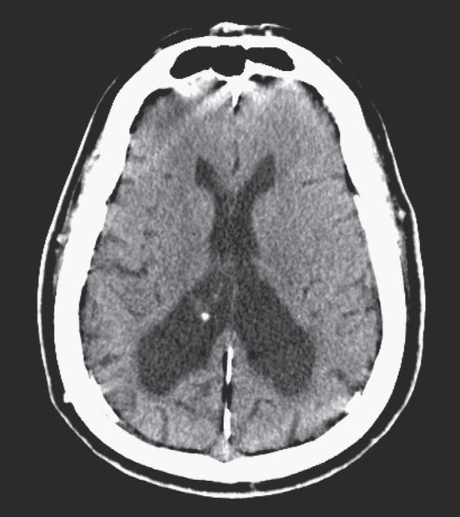
in a patient prone to repeated falls who is brought in because of a change in mental status. On both CT and MRI, these collections typically have a crescentic shape and may demonstrate enhancing septations and membranes surrounding the collection after administration of a contrast agent. Calcification of chronic SDH can occur and may be quite extensive (Fig. 1-5). Areas of hyperdensity within a larger hypodense SDH may indicate an acute component due to recurrent bleeding, termed an “acute on chronic subdural hematoma.” Mixed density collections also may be acute as a result of active bleeding or CSF accumulation as a result of tearing of the arachnoid membrane. A chronic SDH is usually isointense to CSF on both T1WIs and T2WIs, but the appearance can be variable depending on any recurrent bleeding within the collection. The FLAIR sequence is typically very sensitive for detection of chronic SDH as a result of hyperintensity based on protein content. Hemosiderin within the hematoma will cause a signal void because of the susceptibility effect, and “blooming” (i.e., the hematoma appears to be larger than its true size) will be noted on a gradient-echo sequence.
A subdural hygroma is another type of collection that is commonly thought to be synonymous with a chronic subdural hematoma. The actual definition of a hygroma is an accumulation of fluid due to a tear in the arachnoid membrane, usually by some type of trauma or from rapid ventricular decompression with associated accumulation of CSF within the subdural space. Many persons still use this term interchangeably with chronic subdural hematoma. CT demonstrates a fluid collection isodense to CSF in the subdural space. MRI can be useful in differentiating CSF from a chronic hematoma based on the imaging characteristics of the fluid on all sequences. Occasionally hygromas are difficult to differentiate from the prominence of the extraaxial CSF space associated with cerebral atrophy. The position of the cortical veins can be a helpful clue. In the presence of atrophy, the cortical veins are visible traversing the subarachnoid space, whereas
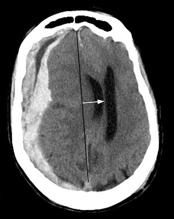
FIGURE 1-3 A subdural hematoma with a mixed density layered pattern due to recurrent hemorrhages. The image (arrow) shows one method of measuring midline shift.
FIGURE 1-4 An isodense subdural hematoma. A, Sulcal effacement and a midline shift to the right are clues to the presence of a left-sided subdural hematoma. B, Reexpansion of the left Sylvian fissure and a reduction in midline shift after evacuation.
with a hygroma, they are displaced inward along with the arachnoid membrane by the fluid in the subdural space.
Subarachnoid Hemorrhage
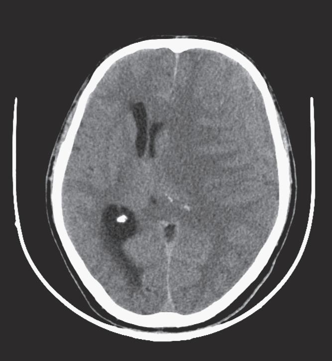
Subarachnoid hemorrhage (SAH) fills the space between the pia and the arachnoid membrane, outlining the sulci and basilar cisterns. SAH can be due to a variety of causes, including trauma, a ruptured aneurysm, hypertension, arteriovenous malformation, occult spinal vascular malformation, and hemorrhagic transformation of an ischemic infarction. SAH is often associated with overlying traumatic SDH. SAHs generally do not cause mass effect or focal regions of edema. However, in patients presenting with ominous signs on clinical grading scales, such as stupor or coma, diffuse cerebral edema may be evident. On CT, hyperdensity is seen within the sulci and/or basilar cisterns (Figs. 1-6 and 1-7).
Although MRI may be as sensitive as CT for the detection of acute parenchymal hemorrhage and SAH, CT generally remains the modality of choice (and the imaging gold standard). The sensitivity of CT for the detection of SAH compared with CSF analysis can vary from up to 98% to 100% within 12 hours to approximately 85% to 90% after 24 hours of symptom onset. Other factors affecting sensitivity are the hemoglobin concentration and the size and location of the hemorrhage. CT is widely available, can be performed rapidly, and is relatively inexpensive. In several small studies, MRI has demonstrated sensitivity equivalent to CT for detection of acute parenchymal hemorrhage and SAH. In some cases of “CT-negative” (subacute) hemorrhage, MRI has shown greater sensitivity. However, results may
be confounded by artifacts from CSF pulsations, an elevated level of protein (meningitis), or oxygen concentration (i.e., a high fraction of inspired oxygen) in CSF on FLAIR images and the presence of blood products from previous microhemorrhages on gradient-echo images.
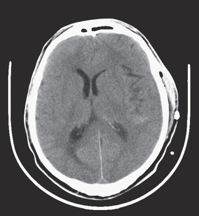
Intraventricular Hemorrhage
In the adult population, intraventricular hemorrhage (IVH) is typically caused by trauma. It can result from extension of a parenchymal hemorrhage into the ventricles or from redistribution of SAH. Primary IVH is uncommon and is usually caused by a ruptured aneurysm, an intraventricular tumor, vascular malformation, or coagulopathy (Fig. 1-8). Large IVHs are quite conspicuous on CT or MRI. They may occupy a majority of the ventricle(s) and may result in hydrocephalus and increased intracranial pressure. Small amounts of IVH may be difficult to detect; one must check carefully for dependent densities within the atria and occipital horns of the lateral ventricles. Normal choroid plexus calcifications in the atria of lateral ventricles, in the fourth ventricle, and extending through the foramina of Luschka should not be mistaken for acute IVH.
Another less common type of extracerebral ICH that may present acutely is a pituitary hemorrhage, which is usually associated with pituitary apoplexy due to pituitary necrosis that may become hemorrhagic. Presenting symptoms may include headache, visual loss, ophthalmoplegia, nausea, and vomiting. Other causes of pituitary hemorrhage include tumors (e.g., macroadenoma and germinoma) and, less commonly, trauma.
B A
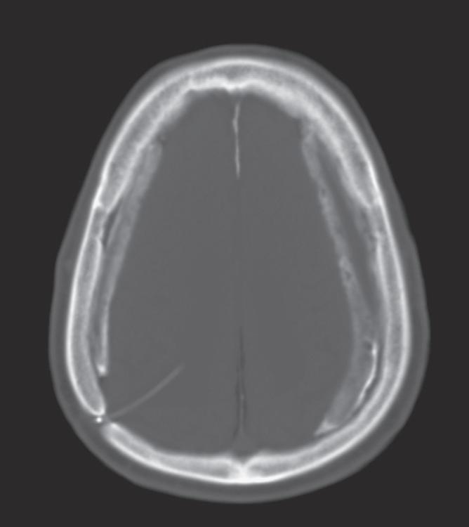
FIGURE 1-5 Calcified subdural hematomas. A, Colpocephaly configuration of the lateral ventricles. B, Bone window/level settings more clearly show the calcified subdurals in this adult patient who, as a child, had a shunt implanted because of congenital hydrocephalus.