No part of this publication may be reproduced or transmitted in any form or by any means, electronic or mechanical, including photocopying, recording, or any information storage and retrieval system, without permission in writing from the publisher. Details on how to seek permission, further information about the Publisher’s permissions policies and our arrangements with organizations such as the Copyright Clearance Center and the Copyright Licensing Agency, can be found at our website: www.elsevier.com/permissions.
This book and the individual contributions contained in it are protected under copyright by the Publisher (other than as may be noted herein).
Notice
Practitioners and researchers must always rely on their own experience and knowledge in evaluating and using any information, methods, compounds or experiments described herein. Because of rapid advances in the medical sciences, in particular, independent verification of diagnoses and drug dosages should be made. To the fullest extent of the law, no responsibility is assumed by Elsevier, authors, editors or contributors for any injury and/or damage to persons or property as a matter of products liability, negligence or otherwise, or from any use or operation of any methods, products, instructions, or ideas contained in the material herein.
Obstetrics and Gynecology; Division of Gynecologic Oncology
The Ohio State University Wexner Medical Center and James Comprehensive Cancer Center, Columbus Ohio
United States
Lindsey B. Beffa, MD
Cleveland Clinic
Division of Gynecologic Oncology, Cleveland Ohio
United States
Caroline C. Billingsley, MD
Associate Professor Obstetrics and Gynecology
Univerisity of Cincinnati, Cincinnati Ohio
United States
Michael J. Birrer, MD, PhD Director
Winthrop P. Rockefeller Cancer Institute UAMS, Little Rock Arkansas
United States
Kristin Bixel, MD
Assistant Professor Obstetrics & Gynecology, Division of Gynecologic Oncology
The Ohio State University Wexner Medical Center and James Comprehensive Cancer Center, Columbus Ohio
United States
Teresa K.L. Boitano, MD
Division of Gynecologic Oncology
Department of Obstetrics and Gynecology
University of Alabama at Birmingham, Birmingham Alabama United States
Wendy R. Brewster, MD, PhD
Professor
Obstetrics and Gynecology
University of North Carolina, Chapel Hill, Chapel Hill
North Carolina
United States Director,
Center for Women’s Health Research University of North Carolina, Chapel Hill
North Carolina
United States
Dana M. Chase, MD
Associate Professor
Creighton University School of Medicine
Phoenix, Arizona
United States
Shaina Bruce, MD
Gynecologic Oncology Fellow Obstetrics and Gynecology
Washington University School of Medicine, St Louis Missouri
United States
Angelena Crown, MD
Breast Surgery, True Family Women’s Cancer Center, Swedish Cancer Institute, Seattle
Washington
United States
Christina S. Chu, MD Professor
Division of Gynecologic Oncology
Fox Chase Cancer Center, Philadelphia
Pennsylvania
United States
Daniel L. Clarke-Pearson, MD
Robert A. Ross Distinguished Professor and Chair (Emeritus)
Department of Obstetrics and Gynecology
University of North Carolina, Chapel Hill
North Carolina
United States
Brian Crosland, MD
University of California, Irvine, Orange California
United States
Joshua G. Cohen, MD
Division of Gynecologic Oncology
University of California Los Angeles Medical Center, Los Angeles California
United States
Robert L. Coleman, MD, FACOG, FACS Professor & Deputy Chair
Department of Gynecologic Oncology & Reproductive Medicine
Chief Scientific Officer, US Oncology Research and Texas Oncology, The Woodlands, United States
Paul A. DiSilvestro, MD Director
Program in Women’s Oncology
Women and Infants Hospital, Department of Pathology & Laboratory Medicine
Providence, Rhode Island
United States
Oliver Dorigo, MD, PhD
Director and Associate Professor
Obstetrics and Gynecology, Division of Gynecologic Oncology, Department of Obstetrics & Gynecology
Stanford University, Palo Alto
United States
Linda R. Duska, MD, MPH Professor
Department of Obstetrics and Gynecology
UVA, Charlottesville
Virginia
United States
Ramez Nassef Eskander, MD
Associate Professor
Obstetrics and Gynecology, Reproductive Sciences
UC San Diego, La Jolla
California
United States
Mary L. Gemignani, MD, MPH
Breast Service
Department of Surgery
Memorial Sloan Kettering Cancer Center, New York
New York
United States
Camille Catherine Gunderson, MD
University of Oklahoma, Mercy Hospital
Section of Gynecologic Oncology
Oklahoma City
Oklahoma
United States
Andrea R. Hagemann, MD, MSCI
Associate Professor
Department of Obstetrics and Gynecology, Division of Gynecologic Oncology,
Washington University School of Medicine, Saint Louis
Missouri
United States
Thomas J. Herzog, MD Professor
Paul & Carolyn Flory Professor of Obstetrics & Gynecology
Deputy Director, University of Cincinnati Cancer Center
Univerisity of Cincinnati, Cincinnati, Ohio
United States
Travis R. Korenaga, MD
University of California, Irvine, Orange California
United States
Warner K. Huh, MD
Division of Gynecologic Oncology
Department of Obstetrics and Gynecology
University of Alabama at Birmingham, Birmingham
Alabama
United States
Lindsay Kuroki, MD, MSCI
Assistant Professor
Obstetrics and Gynecology
Washington University School of Medicine, St. Louis
Missouri
United States
Katherine Kurnit, MD, MPH
Assistant Professor
Department of Obstetrics & Gynecology
Section of Gynecologic Oncology
University of Chicago Medicine
Robert S. Mannel, MD Director,
University of Oklahoma Stephenson Cancer Center
Obstetrics and Gynecology
University of Oklahoma Health Sciences Center, Oklahoma City
Oklahoma
United States
L. Stewart Massad Jr., MD
Departement of Obstetrics and Gynecology
Washington University St. Louis, St. Louis
Missouri
United States
Cara A. Mathews, MD
Associate Professor
Obstetrics and Gynecology, Division of Gynecologic Oncology
Alpert Medical School of Brown University, Providence
Rhode Island
United States
David S. Miller, MD, FACOG, FACS
Division of Gynecologic Oncology in the Department of Obstetrics and Gynecology, UT Southwestern, Dallas, Texas
United States
Bradley J. Monk, MD Professor
Division of Gynecologic Oncology
University of Arizona College of Medicine
Creighton University School of Medicine
Phoenix
United States
Director, Principal Investigator Community Research Development, HonorHealth Research Institute
David G. Mutch, MD
Judith and Ira Gall Professor
Department of Obstetrics and Gynecology, Division of Gynecologic Oncology
Washington University School of Medicine, St Louis
Missouri
United States
Rachita Nikam, CGC
Washington University in St. Louis School of Medicine, Department of Pediatrics, Division of Genetics & Genomic Medicine
United States
JoAnn V. Pinkerton, MD, NCMP Professor
Department of Obstetrics and Gynecology
University of Virginia Health Sciences, Charlottesville
Virginia
United States
Matthew Powell, MD
Department of Obstetrics and Gynecology
Washington University School of Medicine, St Louis
Missouri
United States
Dominique L. Rash, MD
Department of Radiation Medicine and Applied Sciences
Radiation Medicine and Applied Sciences
University of California San Diego Health, La Jolla
California
United States
Lisa M. Landrum, MD, PhD
Indiana University
Department of Obstetrics and Gynecology
Indianapolis, IN
Kari L. Ring, MD
Assistant Professor
Obstetrics and Gynecology
University of Virginia, Charlottesville
Virginia
United States
Malte Renz, MD, PhD
Gynecologic Oncology Division, Department of Obstetrics & Gynecology, Stanford University, Palo Alto
United States
Brandon Roane, MD Gynecologic Oncologist, Texas Oncology–Methodist Charlton Cancer Center and Methodist Dallas Cancer Center Gynecologic Oncology
Texas
United States
Stephen C. Rubin, MD Professor of Gynecologic Oncology, Grotzinger-Raab Chair in Surgical Oncology
Division of Gynecologic Oncology Fox Chase Cancer Center, Philadelphia Pennsylvania
United States
Ritu Salani, MD, MBA Professor
Obstetrics & Gynecology Division of Gynecologic Oncology
University of California Los Angeles, Los Angeles
California
United States
Jane Satero, PharmD, BCOP Pharmacy Manager at Ironwood Cancer and Research Centers Scottsdale
Arizona
United States
Anil K. Sood, MD, FACOG Professor and Vice Chair Department of Gynecologic Oncology & Reproductive Medicine
M.D. Anderson Cancer Center, Houston
Texas
United States
John T. Soper, MD
Distinguished Professor of Gynecologic Oncology
Obstetrics and Gynecology
University of North Carolina, Chapel Hill
North Carolina
United States
Elizabeth Christina Stock, MD Gynecologic Oncology Fellow
Obstetrics and Gynecology
Washington University School of Medicine, St Louis
Missouri
United States
C. James Sung, MD
Women & Infants Hospital
Department of Pathology & Laboratory Medicine, Providence
Rhode Island
United States
Krishnansu Sujata Tewari, MD
Professor & Director of Research
Obstetrics & Gynecology; Division of Gynecologic Oncology
University of California, Irvine Medical Center, Orange
California
United States
Director of Gynecologic Oncology
The Center for Cancer Prevention and Treatment
St. Joseph’s Medical Center, Orange California
United States
Michael D. Toboni, MD, MPH
Obstetrics and Gynecology
Washington University, St. Louis
Missouri
United States
Katherine Tucker, MD
Assistant Professor
Division of Gynecologic Oncology, Department of Obstetrics and Gynecology`
University of North Carolina School of Medicine, Chapel Hill
North Carolina
United States
Joan L. Walker, MD
Professor
Obstetrics and Gynecology
Stephenson Cancer Center, OUHSC, Oklahoma City
Oklahoma
United States
Jaclyn A. Wall, MD
Division of Gynecologic Oncology
Obstetrics and Gynecology
University of Alabama at Birmingham, Birmingham
Alabama
United States
Christina Washington, MD
Gynecologic Oncology Fellow
Department of Obstetrics and Gynecology
University of Oklahoma Health Science Center, Oklahoma City
Oklahoma
United States
Lari B. Wenzel, PhD
Professor
University of California, Irvine
California
United States
Shannon N. Westin, MD, FACOG
Associate Professor
Department of Gynecologic Oncology and Reproductive Medicine
The University of Texas MD Anderson Cancer Center, Houston
Texas
United States
Catheryn M. Yashar, MD, FACR, FABS, FASTRO
Professor
Department of Radiation Medicine and Applied Sciences
University of California San Diego, La Jolla
California
United States
William T. Creasman, MD
Medical University of South Carolina, Charleston
South Carolina
United States
Rosemary E. Zuna, MD
The Univeristy of Oklahoma Department of Pathology, Oklahoma City
Oklahoma
United States
DiSaia and Creasman
CLINICAL GYNECOLOGIC ONCOLOGY
William T. Creasman, MD
Medical University of South Carolina, Charleston, SC, USA
David G. Mutch, MD
Judith and Ira Gall Professor
Obstetrics and Gynecology
Washington University School of Medicine
St Louis MO, USA
Robert S. Mannel, MD
Director, University of Oklahoma Stephenson Cancer Center
Obstetrics and Gynecology
University of Oklahoma Health Sciences Center Oklahoma City OK, USA
Krishnansu Sujata Tewari, MD
Professor & Director of Research
Obstetrics & Gynecology; Division of Gynecologic Oncology
University of California, Irvine Medical Center
Orange CA, USA
Director of Gynecologic Oncology
The Center for Cancer Prevention and Treatment
St Joseph’s Medical Center Orange CA, USA
PREFACE
Over 40 years ago, Phil DiSaia was stimulated by a recognized need for a readable textbook on Gynecologic Oncology and related subjects. Our paths crossed while we were both at the MD Anderson Hospital, and we developed a close relationship both collegially and personally. After leaving Anderson and although being on opposite coasts, we were jointly involved with several clinical research endeavors and, when he asked me if I would be interested in joining his project, I was delighted to accept his kind offer. He identified a publisher and sketched out a table of contents. He was adamant that this would address primarily community physicians, residents, and other students involved with these patients. The clinical presentation and management of these problems were to be heavily employed. In areas where more than one approach to management was present, he felt that the author should inject their own bias. It should be easily readable, in which one could comfortably read a chapter in one sitting. It would not be a reference book, but most major topics would be created in depth and supplemented with ample references to current literature. It would provide a comprehensive resource for study by the resident, fellow, or student of Gynecologic Oncology and serve as a source for review material. A noble project! It was intense, but enjoyable. We would do a chapter and send it via mail (no internet) to each other to critique for corrections and/or additions.
This was continued in this manner for the first six or seven editions at which time we invited three associate editors to join us in a successful project. The subsequent editions have expanded as new information in therapy have been developed, including topics not previously introduced, and expanded areas as new information became available. Not only have we added three associate editors, but also several new authors, taking advantage of their expertise. The first edition contained 16 chapters and was 478 pages including the bibliography and 3 appendices. The
ninth edition contained 16 chapters and was 631 pages long, including 5 appendices with the bibliography being online.
The physician must be prepared to treat the malignancy in light of today’s knowledge and to deal with the patient and her family in a compassionate and honest manner. Patients with gynecological cancer need to feel their physicians are confident and goal oriented. Although, unfortunately, gynecological cancers will cause the demise of some individuals, it is hoped that the information collected in this book will help increase the survival rate of these patients by bringing current practical knowledge to the attention of the primary care and specialized physician.
We were all looking forward to what we consider to be a landmark 10th edition. Before this could be accomplished, unfortunately, our friend and colleague Phil DiSaia, became ill and passed away. It is with renewed energy and endeavor that those of us remaining wanted to make sure this would be not only an excellent edition but truly a fitting attribute to Phil and his multiple contributions to our specialty. The 10th edition is dedicated to everything he has accomplished during his lifetime.
“Our ideas are only intellectual instruments which we use to break into phenomena; we must change them when they have served their purpose, as we change a blunt lancet that we have used long enough”
—Claude Bernard (1813–1878)
“Some patients, though conscious that their condition is perilous, recover their health simply through their contentment with the goodness of the physician”
—Hippocrates (440–370 bc)
William T. Creasman, MD
David G. Mutch, MD
Robert S. Mannel, MD
Krishnansu Sujata Tewari, MD
1. Preinvasive Disease of the Cervix, 1
Jaclyn A. Wall, Teresa K.L. Boitano, L. Stewart Massad Jr. and Warner K. Huh
2. Preinvasive Disease and Dystrophies of the Vagina and Vulva and Related Disorders, 20
Cara A. Mathews and Joan L. Walker
3. Invasive Cervical Cancer, 40
Krishnansu Sujata Tewari and Bradley J. Monk
4. Endometrial Hyperplasia, Estrogen Therapy, and the Prevention of Endometrial Cancer, 104
Kari L. Ring, JoAnn V. Pinkerton, Lisa M. Landrum, Rosemary E. Zuna and Linda R. Duska
5. Adenocarcinoma of the Uterine Corpus and Sarcomas of the Uterus, 125
William T. Creasman, David S. Miller, Ramez Nassef Eskander and Matthew Powell
6. Invasive Cancer of the Vulva, 175
Thomas J. Herzog and Caroline C. Billingsley
7. Gestational Trophoblastic Disease, 204
John T. Soper
8. Adnexal Masses, 229
Christina Washington, Camille Catherine Gunderson and Robert S. Mannel
9. Epithelial Ovarian Cancer, 250
Katherine Kurnit, Shannon N. Westin and Ritu Salani
10. Germ Cell, Stromal, and Other Ovarian Tumors, 282
Lindsey B. Beffa, C. James Sung and Paul A. DiSilvestro
11. Breast Diseases, 311
Angelena Crown and Mary L. Gemignani
12. Cancer in Pregnancy, 345
Travis R. Korenaga, Brian Crosland and Krishnansu Sujata Tewari
13. Complications of Disease and Therapy, 415
Katherine Tucker and Daniel L. Clarke-Pearson
14. Basic Principles of Chemotherapy and Other Systemic Therapies, 443
Christina S. Chu and Stephen C. Rubin
15. Targeted Therapy and Molecular Genetics, 464
Shannon N. Westin, Anil K. Sood and Robert L. Coleman
16. Molecular Hallmarks of Cancer, 489
Michael J. Birrer and Brandon Roane
17. Immunotherapy in Gynecologic Malignancies, 506
Malte Renz and Oliver Dorigo
18. Genes and Cancer: Genetic Counselling and Clinical Management, 521
Andrea R. Hagemann, Rachita Nikam and David G. Mutch
19. Palliative Care and Quality of Life, 560
Dana M. Chase, Jane Satero, Lari B. Wenzel and Bradley J. Monk
20. Role of Minimally Invasive Surgery in Gynecologic Malignancies, 594
Joshua G. Cohen, Kristin Bixel and Floor J. Backes
21. Epidemiology of Commonly Used Statistical Terms, and Analysis of Clinical Studies, 615
Wendy R. Brewster
22. Basic Principles in Gynecologic Radiotherapy, 624
Dominique L. Rash and Catheryn M. Yashar
Appendix A Staging, 646
Michael D. Toboni, David Mutch
Appendix B Modified from Common Terminology Criteria for Adverse Events (CTCAE), 648
Shaina Bruce, David Mutch
Appendix C Blood Component Therapy, 649
Lindsay Kuroki, David Mutch
Appendix D Suggested Recommendations for Routine Cancer Screening, 653
Elizabeth Christina Stock, David Mutch
Appendix E Normal Nutrition, 655
Elizabeth Christina Stock, David Mutch
Index, 660
Preinvasive Disease of the Cervix
Jaclyn A. Wall, MD, Teresa K.L. Boitano, MD, L. Stewart Massad Jr., MD, Warner K. Huh, MD
OUTLINE
Introduction, 1
Human Papillomavirus Natural History, 1 Epidemiology, 3
Human Papillomavirus Vaccination, 4 Screening, 5
KEY POINTS
1. Persistent expression of human papillomavirus (HPV) is required for progression to cancer.
2. HPV vaccination has the potential to eradicate cervical cancer.
3. Screening guidelines have changed dramatically with the use of contesting and increased intervals between screenings.
INTRODUCTION
Worldwide, cervical cancer is a leading cause of cancer-related death and morbidity in women. In the United States, it is the third most common and lethal gynecologic malignancy. Despite the implementation of cytologic screening (Papanicolaou [Pap] testing) in the United States, it is estimated that in 2021 there will be over 14,000 new cases of cervical cancer, and almost 4000 cervical-cancer related deaths. The majority of cervical cancers are caused by persistent infection of the human papillomavirus (HPV). Women’s health providers must understand and comply with primary screening recommendations and management of abnormal screening results, particularly in ways that balance benefits and harms of screening and are cost-conscious.
HUMAN PAPILLOMAVIRUS NATURAL HISTORY
Over 99% of cervical cancers arise from persistent infection with HPV (Fig. 1.1). HPV is an approximately 8000 base pair DNA virus, encoding eight major proteins. Approximately 200 strains of HPV have been identified, with 12 classified as carcinogenic (HPV-16, -18, -31, -33, -35, -39, -45, -51, -52, -56, -58, and -59). Other types have been described as possibly carcinogenic (HPV-26, -53, -66, -67, -68, -70, -73, and -82). New genotypes are based on DNA sequencing and must share less than 90% DNA homology in the L1, E6, and E7 coding domains compared with existing HPV types. HPV-16 is the most oncogenic, and perhaps the most clinically significant, genotype, and historically has accounted for
Management of Abnormal Screening Results, 8 Management of Post-Colposcopic Results, 13 Management of Abnormal Screening Results in Pregnancy, 17 Future Directions, 18
4. The main screening objective is to allow for modified evaluation and treatment based on an individual woman’s risk.
5. Self-collection HPV testing may increase screening by reaching those in traditionally under-screening areas or with other barriers to clinic-based screening.
over 50% of cervical cancers. HPV-18 is found in approximately 10% of cervical cancers, and is more frequently found in adenocarcinoma, which is commonly missed by Papanicolaou (Pap) testing. The other types are less oncogenic but have been reported in large typing studies of cervical cancers. HPV-18 and related HPV-45 are linked to cancers found in younger women.
Interactions of the HPV E6 and E7 gene products with p53 and pRb are critical for HPV-induced carcinogenesis. By inactivating or activating degradation of their targets, E6 and E7 eliminate genetic surveillance and allow unchecked cell cycling, leading to the accumulation of mutations and the potential for invasive cancer. HPV-16 E6 and E7 bind their targets with greater affinity than gene products of other HPV types which may partly explain its greater oncogenicity.
The progression from persistent HPV infection to cancer requires several key steps. First, infection of basal epithelial cells leads to establishment of a ring chromosome from which carcinogenic proteins are amplified while virion production occurs in maturing epithelium. Subsequently, disruption of the ring, often at the HPV E2 regulatory region, allows integration of E6 and E7 sequence into the host genome. The accumulation of mutations leads to nuclear changes visible cytologically as high-grade squamous intraepithelial lesions (HSILs) and histologically as highgrade cervical intraepithelial neoplasia (CIN) (Fig. 1.2). Selection for invasiveness and metastasis through additional mutations and gene methylation ultimately results in the development of cancer. Notably, preexisting HPV infections do not appear to predispose to or protect from infection by unrelated types.
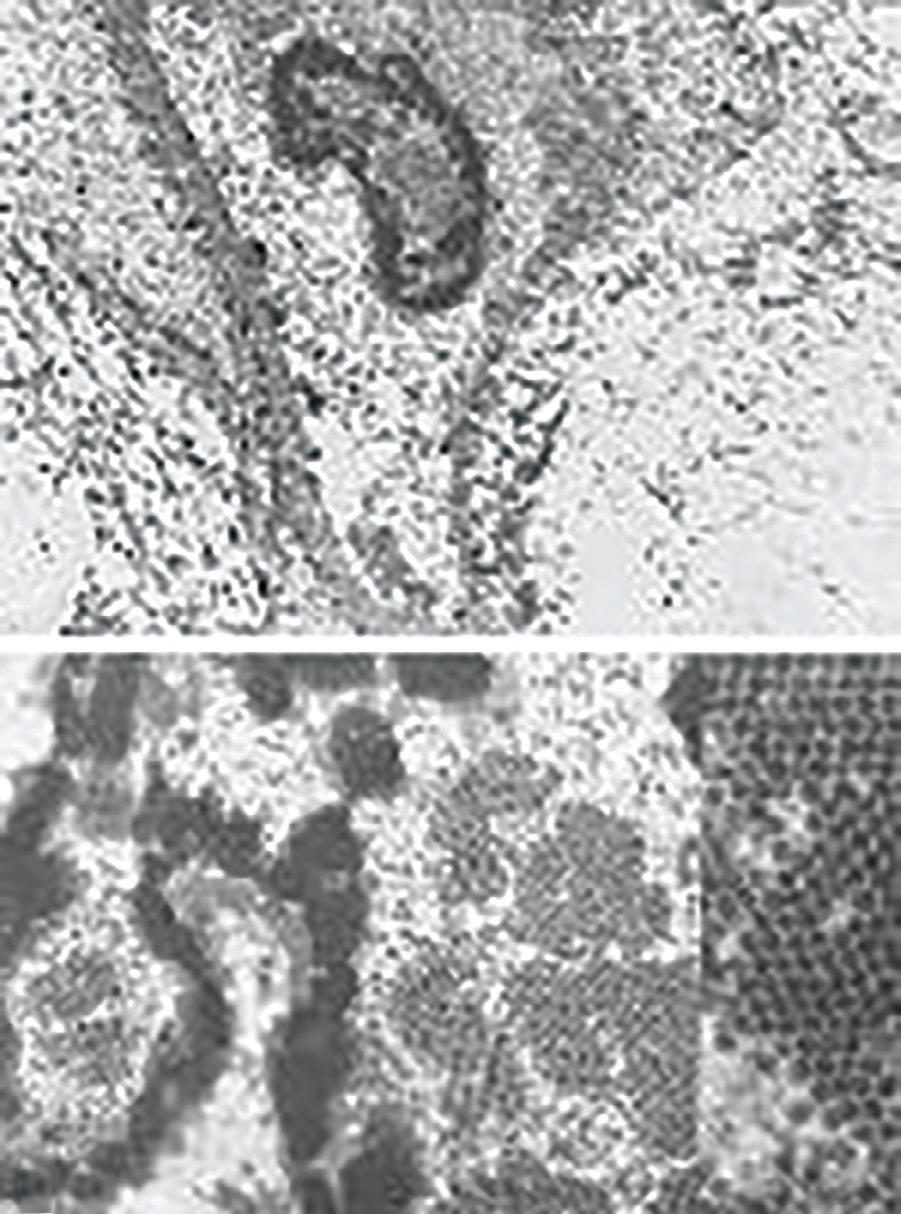
1.1 A, Koilocytotic cells with intranuclear virions (36900). B, Human papillomavirus particles. Note the intranuclear crystalline array (“honeycomb”) arrangement of virions (320,500). See the insert (380,000). (Courtesy of Alex Ferenczy, MD, Montreal, Canada.)
While lifetime abstinence protects against genital HPV infection, nonpenetrative sexual behaviors may still transmit the virus, and additional male exposures increase female risk. For example, spouses of men who engaged in sex with prostitutes were at higher risk for cervical cancer than those of men who did not, and cervical cancer risk is higher among women whose husbands had more sexual partners. Women who report recent sex only with women are also at risk, though their risk may be lower than that of heterosexual women. Condom use is not fully protective against HPV infection because condoms fail to cover wide areas of genital skin, though it speeds clearance of HPV infections. Male circumcision also reduces but does not eliminate HPV and cancer risks. For these reasons, all women with prior sexual experience, including those who have not been sexually active for years, remain at risk for cervical cancer and require screening until they meet criteria for screening exit as discussed later.
While the percentage of women who at some point develop HPV infection is high, most women infected with carcinogenic HPV do not develop cervical cancer. The majority of infections are cleared immunologically, and women who clear the virus are at low risk for progression to cancer or reappearance of CIN21. HPV is an intraepithelial virus, and clearance appears to require recognition of infection by cell-mediated immune cells. Roughly half of new infections are cleared within 6 months, with half of the remainder cleared by the end of the first year after infection. Clearance is associated with greater density of CD81 cells and lower density of T-regulatory cells in underlying stroma. Cervical treatment speeds clearance and reduces risk for posttreatment acquisition of new HPV infections. The type distribution of HPV infection after hysterectomy shows that HPV-16 and HPV-18 have a greater predilection for cervical rather than vaginal epithelium, with HPV types of lesser oncogenicity dominating in the post-hysterectomy vagina. Women infected at an increased age do not typically develop preinvasive disease or cancer of the vagina; however, increased age may result in immune senescence which suggests that some HPV infections in older women derive from reactivation of previously acquired latent infections.
Although determinants of HPV persistence and progression of HPV infection to invasive cancer are poorly understood, several risk factors are known. HPV infection of a cervix undergoing active metaplasia increases risk, as reflected by the epidemiologic observations that early onset of first intercourse is associated with cancer. Smoking is linked to both CIN and cervical cancer. Benzopyrenes have been identified in cervical mucus, and the interaction of tobacco carcinogens with carcinogenic HPV increases risk substantially. Smoking also reduces immune-mediated HPV clearance. Cervical adenocarcinoma and adenocarcinoma in situ (AIS) have been linked to oral contraceptive use. Variants of common HPV types that segregate by ethnicity and polymorphisms in genes related to HPV immune recognition or HPV protein products also modulate HPV persistence and carcinogenic progression. Perhaps most important, lack of screening is a high-risk factor for progression of HPV infection to precancer and cancer. Appropriately screened and managed women with multiple risk factors are at relatively low risk, and women with few risk factors who are not screened are at higher risk.
Figure
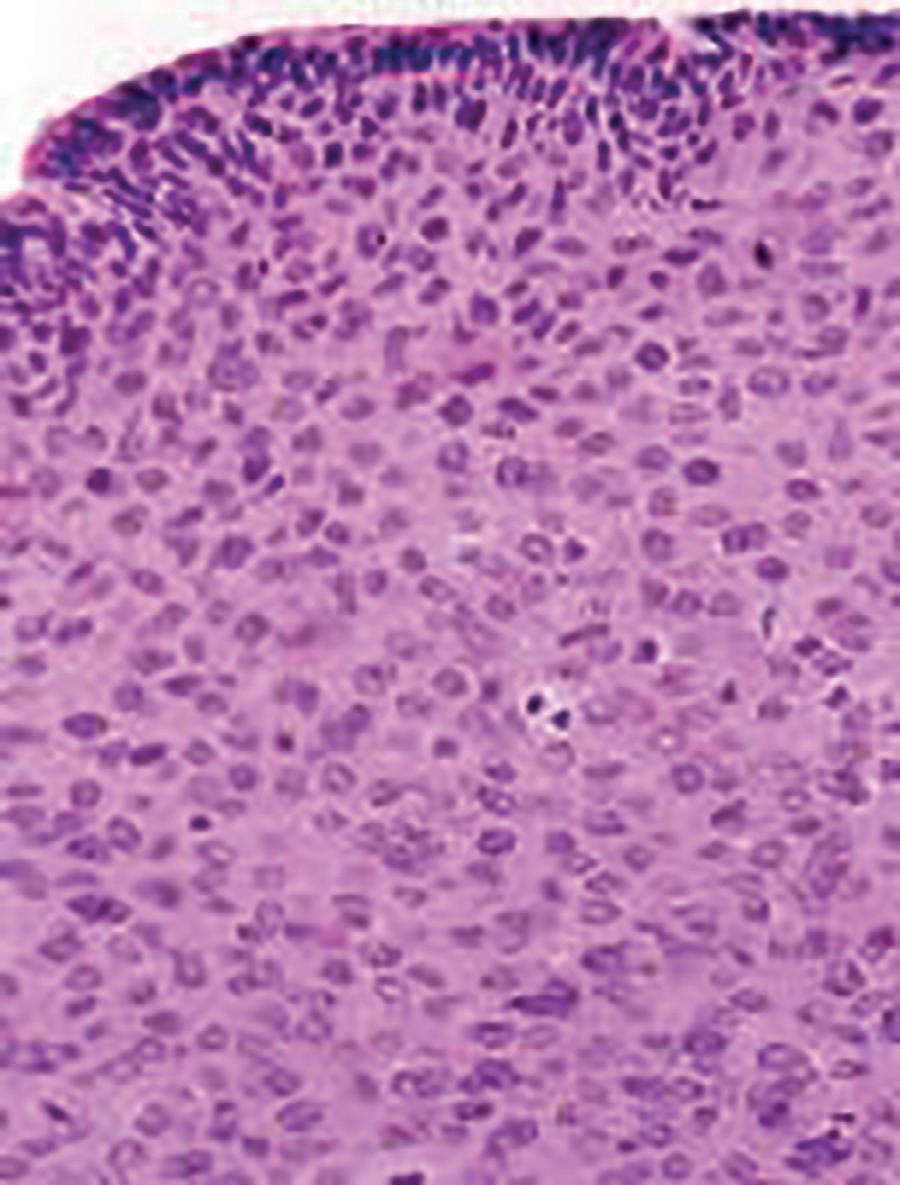
Figure 1.2 A cervical intraepithelial neoplasia lesion with multiple mitotic figures.
Immune factors play a clear role in the clearance or persistence of HPV-related cervical lesions, but the nature of immune defects is poorly understood. Immunosuppression related to coinfection with the human immunodeficiency virus (HIV-1) illustrates the importance of immunity in the typical control of HPV. Women with HIV have much higher rates of HPV infection, including multitype infections. HPV clearance rates are lower, although most women do clear their HPV infections if observed long enough, especially if immune reserve as measured by CD4 lymphocyte count remains above 200 cells/mm3. Although most HPV infections in HIV-seropositive women are cleared to such low levels of viral expression that they become nondetectable even with sensitive assays, reactivation appears to occur. This is apparent in cohort studies as the reappearance of previously cleared infections in women who deny sexual activity, often because of illness. Risks in other immunosuppressed states appear to be similar.
HPV infection predicts risk for subsequent CIN21, even among cytologically normal women. In most cases, persistent HPV infections result first in cytologically detectable abnormalities and then in colposcopically visible lesions that grow before developing into invasive cancers. The 10-year risk of CIN21 after a single detected HPV infection exceeds 10%. As developed by Richart through observational studies of the cervix using cytology and colpomicroscopy, a diagnosis of CIN was based on progressively severe nuclear aneuploidy, abnormal mitotic figures, and loss of epithelial maturation. Initially considered a progressive lesion, CIN was thought to begin as a small lesion with atypia near the basement membrane of the cervical transformation zone, gradually increasing in size and becoming less differentiated with an increasing proportion of the epithelium taken up by atypical cells until a full-thickness carcinoma in situ developed and then became invasive. Given this concept of progression from low-grade to CIN21 disease to cancer, lesions of all grades were treated. When progression does occur, however, it appears to require years. HPV acquisition commonly follows sexual debut, the median age being approximately 17 in the United States, but the peak age of cervical cancer diagnosis lags by some 3 decades. This long transition time allows for even moderately sensitive screening tests to identify persistent lesions for treatment before invasive cancer develops.
Gradually, the regressive nature of most low- and midgrade lesions becomes apparent. Low-grade lesions, which include genital warts and CIN1, are histologic expressions of HPV infection and are thus not considered to be premalignant. Greenberg and associates found that of 163 women with CIN1 after low-grade cytology followed for a median of 36 months, 49% regressed, 43% persisted, and only 8% progressed to CIN3. In the Atypical Squamous Cells of Undetermined Significance/Low Grade Squamous Intraepithelial Lesion Triage Study (ALTS), a large randomized trial of management options for women with borderline cytology results conducted under the auspices of the US National Cancer Institute (NCI), 2-year risk for CIN3 was 10% among women with CIN1. As reported by Castle et al., after controlling for HPV genotype, with HPV-16–associated CIN1 progressing to CIN3 in 19% of cases, biopsy-proven CIN1 was not a risk factor for progression. However, these risk estimates may be substantially higher for women with prior CIN21 cytology.
CIN2 lesions or greater (CIN21) appear to represent clonal lesions arising from single-type HPV infections. Although women may harbor multiple HPV types in the genital tract, most multitype infections are associated with multifocal lesions. Moscicki et al. showed that 63% of adolescents and young women with CIN2 lesions had resolution without treatment within 2 years; subsequent clearance was minimal, rising only to 68% after an additional year. McAllum et al. showed a similar 62% regression after only 8 months of observation for women with CIN2 younger than 25 years of age. No patients in either study progressed to cancer during observation. In both studies, CIN2 likely represented recent HPV infections, not true premalignant lesions. Regression rates are lower in older women, at least in part because lesions detected later may have been persistent for years, and lesions that have evolved mechanisms to evade host immune-mediated clearance are likely to continue to persist. Castle et al. compared CIN2 rates in the immediate colposcopy and cytology surveillance arms of the ALTS. They found that over 2 years, approximately 40% of CIN2 regressed. Trimble and colleagues showed that HPV-16–associated lesions are less likely to resolve. Their finding of associations with human leukocyte antigen (HLA) alleles and regression support a role for HLA-restricted HPV-specific immune responses in determining clearance.
Untreated, CIN3 poses considerable risk of progression to invasive cancer. An observational study of women in New Zealand with CIN3 diagnosed between 1955 and 1976 were observed; among 143 women reported by McCredie et al. managed only by punch or wedge biopsy, 31 progressed to cancer of the cervix or vagina after 30 years. Risk rose to 59% in 92 women with persistent disease after 2 years of observation. These findings show both that treatment of CIN3 should be strongly considered regardless of age or other factors, but also that not all CIN3 lesions will inevitably progress to cancer.
Even treated CIN3 continues to pose a risk of progression to cancer. Women in the New Zealand study whose treatment appeared adequate by current standards faced only 0.7% cancer risk after 30 years. Studies from Scandinavian countries with integrated health systems can link databases on procedures and subsequent cancers and provide accurate long-term results with minimal loss to follow-up. Strander et al. showed that risk for cervical cancer rose significantly in previously treated women after age 50 years, with standardized incidence ratios compared with untreated women ranging from 3 to 5. Vaginal cancer risks were elevated across all ages, although the absolute risk of vaginal cancer was low. In Finland, Kalliala et al. confirmed this long-term increased risk and also found an elevated risk for nongenital smoking-related cancers. Jakobsson et al. found that in addition to cervical cancer, women treated for CIN faced higher mortality rates from cardiovascular disease, alcoholrelated, and traumatic death, consistent with the demographic and behavioral factors linked to CIN.
EPIDEMIOLOGY
Approximately 20 million Americans and 630 million persons worldwide are infected with HPV. In the United States, about
6.2 million people will acquire a new infection annually. More than 80% of sexually active individuals acquire genital HPV infections. Prevalence rates are highest among women in their late teens and early 20s. Risk factors for HPV acquisition include smoking, oral contraceptive use, and new male partners.
Among high-risk HPV types, HPV-53 is most common, detected in 5.8% of US women ages 14 to 59 years screened in the National Health and Nutrition Examination Survey (NHANES) between 2003 and 2006. This was followed by HPV-16 (4.7%), HPV-51 (4.1%), HPV-52 (3.6%), and HPV-66 (3.4%). HPV-18 was present in only 1.8% of screened women. In this study, demographic risk factors for prevalent HPV infection included younger age (peaking at ages 20 to 24 years), non-Hispanic black ethnicity, non-married status, non-college educated, and living below the poverty line. Behavioral risk factors included reporting ever having sex, age of first intercourse before 16 years, increased number of lifetime partners, and number of partners in the past year.
HPV infection determines subsequent risk for precancer. Among women enrolled in a Portland health maintenance organization who had HPV-16, the 10-year risk for CIN3, AIS, or cancer was more than 15% after HPV-16 infection, almost 15% after HPV-18 infection, less than 3% after other oncogenic HPV infections, and less than 1% after a negative HPV test result. A Danish study of over 8000 women found that infection with, and especially persistence of, HPV-16/18/31/33, was associated with high absolute risks of development of CIN31. Additionally, in a study by Elfgren et al., the authors demonstrated persistence of HPV infection at 1- and 3-year intervals following initial screening was predictive of CIN21
It is estimated that there were approximately 14,000 new cases of cervical cancer in the United States in 2020. However, the annual incidence of CIN21 is approximately 6 to 10 times higher than cervical cancer incidence. Preinvasive lesions begin to appear on average 2 years after infection. Cancer risk is quite low soon after infection; despite a high prevalence of HPV among sexually active teens, cervical cancer incidence is only about 1 in 1,000,000 before 20 years of age. Among women who develop CIN21, only 30% to 50% will develop cancer over years of observation.
Clear risks for CIN and cervical cancer have been identified. The international Collaboration of Epidemiological Studies of Cervical Cancer reviewed evidence for various risk factors for cervical cancer and carcinoma in situ (though their studies were not linked to HPV data). They found that oral contraceptive use raised the risk for cervical disease by 1.9-fold for every 5 years of use. First intercourse before 15 years of age was associated with twice the risk of cervical cancer found in women with first intercourse after 23 years of age, and having more than five lifetime sexual partners carried more than double the cervical cancer risk of lifetime monogamy. Lesser but still significant increases in risk were associated with number of pregnancies and earlier age at first term pregnancy. Both squamous cancers and adenocarcinomas share epidemiologic risk factors, except that smoking is linked only to squamous cancers. The most notable risk factor for the development of invasive cervical cancer, however, is infection with and persistence of HPV, particularly HPV-16, which is found in nearly 50% of cases.
The role of family history in determining cervical cancer risk is complex. Dissociating genetic components of familial risk from cultural ones is difficult, as sexual attitudes and behaviors, reproductive patterns, and smoking are often linked to family. Zelmanowicz et al. assessed the role of family history in cohorts of women prospectively studied in Costa Rica and the United States. A family history of cervical cancer in a first-degree relative tripled the risk for CIN3 or squamous cervical cancer. The effect persisted after controlling for HPV exposure. No effect of family history on adenocarcinoma risk was seen. Although several genome-wide association studies (GWASs) have identified a range of genetic variants in candidate pathways that might contribute to cervical oncogenesis, Chen et al. in a large Chinese GWAS found that only HLA and major histocompatibility class I polypeptide-related sequence A genes were identified as candidate risk genes across several populations.
Lower socioeconomic status (SES) and minority ethnicity are also linked to CIN and cervical cancer risk in the United States, although distinguishing cultural contributions to cervical cancer risk, such as a sense of fatalism, distrust of the medical care system providing screening services, and lack of health education about the benefits of screening, are difficult to distinguish from biologic risks related to ethnicity and SES, such as genetic predisposition, toxin exposure, and micronutrient deficiencies.
HUMAN PAPILLOMAVIRUS VACCINATION
HPV vaccination has the potential to eliminate cervical cancer, given HPV causes the vast majority of cases. Intramuscular delivery of synthetic HPV L1 capsid antigens results in humor immunity; current vaccines are created in protein synthesis using cell culture systems. No actual live or killed virions are used, and therefore HPV vaccines cannot cause HPV-related cancer. Despite early concerns that humoral immunity would be insufficient to prevent infection, vaccine efficacy in preventing type-specific HPV persistence approaches 100%. Currently available vaccines are largely prophylactic. However, a recent meta-analysis demonstrated that patients who underwent surgical excision followed by HPV vaccination were less likely to develop recurrent CIN.
Three HPV vaccines have been used in the United States. The quadrivalent HPV vaccine (available 2006–17) protected against HPV-16 and -18, which together account for almost 70% of all cervical cancers, as well as HPV-6 and -11, which are the most common causes of genital warts. The benefit of cervical cancer prevention, which might take decades to become manifest, is augmented by its ability to prevent genital warts, a concern for many young women. The bivalent HPV vaccine protects against only HPV-16 and -18. It is no longer available for use in the United States and may have superior antigenicity and may have some cross-protection against HPV types related to HPV-16 and -18. The nonavalent vaccine (available since 2017) is effective against the same types as the quadrivalent vaccine and also includes coverage against HPV types 31, 33, 45, 52, and 58; enhanced coverage should prevent 90% of all cervical cancers and many other HPV-related cancers at other sites.
Ideally, because HPV vaccines are prophylactic, populationbased vaccination should begin before the initiation of sexual activity. Because 5% of US 13-year-old girls are sexually active, the target age for HPV vaccination is the ages of 11 to 12 years. However, vaccination can be initiated at 9 years of age. The vaccination schedule is dependent on patient age. For patients younger than 15, two doses of the vaccine should be administered, with the second administration between 6 and 12 months following the initial. Because teen sexual activity is unpredictable, delaying vaccination until girls are more mature risks missing the vaccination window for many. Nevertheless, many sexually active young women show no evidence of infection by target HPV types, and “catch-up” vaccination up to 26 years of age should be considered. Vaccination through age 45 years has been approved but appears cost-ineffective with minimal population-level cancer prevention impact. Testing of cervicovaginal secretions and serum antibody testing are both insensitive for detecting prior HPV vaccination and are not recommended before a decision about HPV vaccination. After age 15, three doses of the vaccine should be administered (at 0, 1 to 2, and 6 months). These vaccination strategies are endorsed by the CDC.
Several countries have instituted organized vaccination programs, either mandatory or using a school-based opt-in mechanism with high uptake. Countries that used quadrivalent vaccine have documented a dramatic decrease in genital warts among teens but not older women, and abnormal cytology rates have also fallen in the youngest women.
In the United States, vaccination rates are suboptimal. Data in 2019 demonstrated that in adolescents aged 13 to 17, 71.5% had started the vaccine series and nearly 54.2% had completed it. However, this is still well below the Healthy People 2030 goal of 80%. Regrettably, despite the potential for vaccination to eliminate the disparately high risk of cervical cancer among women of minority ethnicity and lower SES, uptake has been lowest in these groups, potentially widening cancer disparities in future years. Work by the CDC’s HPV-IMPACT group has demonstrated vaccine effectiveness of 1, 2, and 3 doses of the HPV vaccine and falling rates of CIN21 lesions positive for HPV-16 and -18, particularly in the youngest women receiving the vaccine.
Risks of vaccination are low and the most commonly cited adverse effects are tolerable. These include fever, rash, injection site pain, nausea, headache, and dizziness. Anaphylactic and vagal reactions may be fatal, so vaccination should only be administered in sites with ability to manage anaphylaxis and fainting. Despite initial concerns, HPV vaccination status does not enter into young women’s decisions to initiate sexual activity. Vaccination is contraindicated for pregnant women, although no congenital anomalies or adverse pregnancy outcomes have been linked to HPV vaccination, and the series may begin after delivery. Interruption of vaccination does not appear to require re-initiation of the three-shot series. The duration of vaccine effectiveness is unclear, but antibody levels remain elevated for several years after vaccination.
Presently, history of HPV vaccination does not alter screening recommendations for US women, but this is expected to change in the future. This is because many women of screening
age were not vaccinated before initiating intercourse, so vaccine effectiveness is unclear. There is no central US vaccine registry, and identifying vaccinated women by self-report may be inaccurate. No HPV vaccine covers all carcinogenic HPV types, so women vaccinated before first intercourse remain at risk for infection and cancer due to strains not covered by the vaccine.
In October of 2018, the Food and Drug Administration (FDA) extended the approval of the nonavalent HPV vaccine to previously unvaccinated individuals up to age 45 for men and women for the prevention of all HPV-related cervical, vaginal, vulvar, anal, oropharyngeal, and other head and neck cancers. Previous approval extended only through age 26. However, modeled studies have shown that reductions in cases of CIN21 and cervical cancer by extending vaccine approval are extremely modest while significantly increasing costs.
SCREENING
The goal of any cancer prevention program is the reduction of morbidity and mortality through intervention prior to the onset of symptoms. For cervical cancer, the current mechanism to achieve this goal is the identification and destruction of CIN21 lesions that are presumed precancers.
Classically, screening has relied on Papanicolaou cytology testing followed by colposcopic assessment of women with Pap abnormalities, directed biopsies, and treatment of biopsy proven CIN21 lesions. However, Papanicolaou testing is relatively insensitive, and a single Pap test may be negative in almost half of women with CIN21. Simultaneously, progression from HPV infection to cancer usually requires several years, allowing for multiple rounds of screening, with greater sensitivity than single tests.
Cytologic interpretation includes all mutations, methylations, and other genetic modifications that alter the nuclear and cytoplasmic appearance of cells. To be clinically useful, these changes must be categorized in a manner that reflect a common natural history. Papanicolaou developed a five-class grading system, from normal to invasive cancer, with atypia, dysplasia, and carcinoma in situ between. To unify terminology, the NCI convened a consensus meeting that developed the 1988 terminology known as the Bethesda System for cervicovaginal cytologic diagnosis. The Bethesda System has been revised multiple times, most recently in 2014. It identifies cytology as satisfactory or unsatisfactory, includes nonneoplastic changes, and divides epithelial cell abnormalities into squamous and glandular changes of varying degrees of severity ( Table 1.1). Distinguishing squamous from glandular abnormalities is critical because glandular abnormalities carry much higher risk for CIN21, including squamous abnormalities, as well as endometrial cancer and cervical adenocarcinoma and AIS. Squamous changes related to HPV are termed “squamous intraepithelial lesions (SILs)” because some lesser changes do not reflect dysplasia or neoplasia, only cytomorphologic changes of HPV infection. Indeterminate lesions are termed “atypical squamous cells (ASCs),” and these are subdivided into ASC “of undetermined significance (ASC-US),” which carries a low risk of associated CIN21, or “cannot exclude high-grade SIL (ASC-H),”
TABLE 1.1 Bethesda 2001 Classification
1. Negative for intraepithelial lesion or malignancy
which is a more worrisome finding that requires immediate colposcopy. An online atlas allows pathologists to standardize findings and interpretations against national norms (https:// screening.iarc.fr/atlascyto.php).
Most cytologic tests in the United States are conducted using liquid-based assays. In these tests, cells are collected and suspended in preservative solution and then transferred to a slide. Liquid-based cytology was marketed as more sensitive than conventional Pap tests. However, a meta-analysis by Arbyn et al. showed that, although liquid-based cytology yields more abnormalities, including CIN21, it is not superior to conventional smears in cancer prevention. Nevertheless, it remains preferred in the United States primarily because it allows for molecular triage of equivocal results using HPV and other assays. Interpretation is still done visually, although some centers use automated imaging and pattern recognition software to eliminate the least abnormal slides. Cytotechnologists perform initial assessment, with slides containing abnormal findings and a proportion of normal slides read by cytopathologists.
The effectiveness of screening has not been demonstrated in randomized trials, but population studies have shown clear benefits. An NCI study by Erickson et al. assessed cytology screening by vaginal aspiration for 108,000 women in Shelby County, Tennessee. They showed a high yield of unsuspected CIN21 and early cancer at the first screen, with a substantial reduction in invasive lesions in the second screen. Gustafsson et al. reviewed data from 17 cancer registries and showed marked effects, especially in Scandinavian countries. Eddy assessed the impact of screening on cervical cancer incidence and death. Without screening, a 20-year-old average-risk woman faces a 2.5% risk of cancer and a 1.2% risk of cancer death. Triennial screening between ages 20 and 75 years reduces risk to less than
0.4% and 0.1%. Annual screening does improve effectiveness, but by less than 5% and with a notable increase in cost. Initially, screening was opportunistic, and women underwent screening when they presented for care, which was generally at annual visits. This screening strategy remains the norm in the United States, although recommended screening intervals have lengthened; some electronic medical records prompt clinicians when screening is due; and some health care organizations have developed standards, rewards, and reminders. Several countries with centralized medical care systems have developed organized screening, with coordinated identification, invitation, and management of women due for screening (though this is not the case in the United States). Serraino et al. showed that the move from opportunistic to organized screening in Italy resulted in a decline in cervical cancer incidence and a downstaging of incident cervical cancers after an increase in precursor detection. Additionally, Quinn et al. found that a national call/recall system with incentive payments to general practitioners in Britain instituted in 1988 increased screening coverage to 85% of the target population, increased detection of CIN21, and reduced mortality in women younger than 55.
Despite its overall benefit, cytology-based screening has several weaknesses. Most fundamentally, the process of screening, triage, and treatment is cumbersome, and noncompliance, which can be a significant problem, at any point renders it ineffective. Cytology results are reported in ways that can be confusing, and efficient, effective management may require integration of current results with prior abnormalities. Costs of combined HPV testing with cytology are increased compared to cytologic testing alone. Multiple studies have shown that most women who develop cervical cancer in developed countries, especially those presenting at advanced stages, are inadequately screened. Sung et al. studied incident cancers in a US prepaid health plan and found that 53% were nonadherent to screening, 28% had false-negative Pap tests, 4% had inadequate follow-up after an abnormal Pap test result, and the rest either developed cancer despite appropriate investigations or were unclassified. Kinney et al. in the same US health maintenance organization found that 60% of cervical cancer patients were inadequately screened. Deeper exploration of the records of long-standing plan members with inadequate screening showed that 70% had missed opportunities for screening in primary care clinics.
In addition, cytology-based screening performs poorly in younger women. Sasieni et al. showed that cervical screening in women ages 20 to 24 years had little impact on actual cancer risk until those women reached 30 years of age, but screening older women results in an immediate benefit. Because younger women have low rates of cervical cancer incidence and death but high rates of HPV infection, abnormal cytology, and CIN destined to regress, the benefits of early initiation of screening may be difficult to balance against potential harms. Cytology preferentially detects squamous cell carcinomas, and the impact of cytology screening on adenocarcinoma incidence has been muted.
Over 99% of all cervical cancer is caused by HPV. The incorporation of HPV testing into screening has allowed for the
implementation of longer screening intervals. As HPV assays have been developed, protocols have utilized HPV testing as a primary screening test as well as in combination with cytology (co-testing) and as a triage test for women with borderline cytology results. Only high-risk HPV types have a role in screening; because low-risk types essentially play no role in identification of HPV infection in the absence of visible genital warts, testing for low-risk types is contraindicated. HPV genotyping assays for types 16 and 18 identifies women at higher risk. The disadvantage of HPV testing is its lower specificity, with up to 30% of young women testing positive in some studies. Because all commercially available HPV tests have a detection threshold designed to balance sensitivity and specificity, a negative test result does not exclude low-level HPV infection, and prior cytologic or histologic abnormalities may mandate close followup or even treatment despite absence of detectable HPV. HPV tests should not be collected in media whose effects of test performance have not been evaluated by the FDA.
Screening does have potential harms. Identification of HPV infection, abnormal cytology, and cervical cancer precursors is not without consequences, including anxiety, relationship disruption after diagnosis of a sexually transmitted infection, the inconvenience and cost of accelerated follow-up visits, and the pain of repeated examinations. Treatment of precursor lesions also carries risks, including bleeding, infection, and injury to adjacent organs. Some studies have suggested that destructive cervical treatments increase the risk for preterm delivery and pregnancy loss. US studies have failed to replicate these results in women after cervical loop electrosurgical excision procedure (LEEP). Women with CIN21 are at higher risk for pregnancy loss than those who do not, perhaps because of common risk factors, including smoking, nutritional deficiencies, and lower SES, and these confounding factors may account for differences. However, there may be a threshold effect for treatment, and women with deep or repeated excisional procedures may be at higher risk for pregnancy loss. Harms of excessive screening are limited, and include stigmatization, unfounded fear of cancer, and the potential of undergoing interventions without cancer prevention benefit. Sharp et al. showed that depression, distress, and anxiety occurred in 15% to 30% of women in the months after reporting of marginal cytology abnormalities.
After the utility of screening is accepted, societies, women at risk, and clinicians must decide when to initiate screening, which screening tests to use, how often to screen, and when toward the end of life the identification of asymptomatic disease ceases to be beneficial. With all choices, sensitivity and specificity must be balanced. Earlier screening starts with more sensitive tests at shorter intervals until later in life will decrease cancer incidence and mortality, but costs and harms from diagnosing lesions that would never have progressed to cancer will increase. In developed societies, guidelines for screening have been developed by experts assessing evidence for benefit and harm and deciding how these can best be balanced. In 2012, guidelines were released by the US Preventive Services Task Force (USPSTF) and a consensus conference sponsored by the American Cancer Society (ACS), the American Society for Colposcopy and Cervical Pathology (ASCCP), and the American
TABLE 1.2 Comparison of Current US Cervical Cancer Screening Guidelines
When to start? Age 21 years Age 21 yearsa
How often? Pap tests every 3 years
Co-tests every 5 years at ages 30–64
When to stop? Age 65 years if adequate prior screens
Pap tests every 3 years at ages 21–29 years
Co-tests every 5 years at ages 30–64 years preferred Pap tests every 3 years remain an option
Age 65 years if the patient has had three negative Pap tests or two negative co-tests After hysterectomy for benign disease
aIn the most recent update from the ACS, primary recommendation is initiation of screening at age 25 with primary HPV testing; if unavailable, co-testing every 5 years or cytology alone every 3 years are acceptable alternatives
ACS, American Cancer Society; ASCCP, American Society for Colposcopy and Cervical Pathology; ASCP, American Society for Clinical Pathology; HPV, human papillomavirus; USPSTF, US Preventive Services Task Force.
Society for Clinical Pathology (ASCP) ( Table 1.2). Guidelines for screening recommended by the USPSTF were updated in 2018 and those by the ACS were updated in 2020.
The USPSTF recommends the initiation of screening at age 21 with cytology every 3 years until the age of 30; beginning at age 30, patients may continue with cytology every 3 years or have co-testing or HPV testing alone every 5 years. In women with continued normal screening results and no history of CIN21, screening should continue until age 65 or time of hysterectomy, after which no further screening is indicated. The most recent screening guidelines from the ACS recommend commencement of screening at age 25 with primary HPV testing every 5 years. If primary HPV testing is not available, cotesting every 5 years or cytology alone every 3 years are acceptable. Similar to the recommendations put forth by the USPSTF, screening should be discontinued at age 65 or at the time of hysterectomy, provided certain criteria are met regarding their prior screening history.
Again, HPV infections are common in sexually active young women. The rationale for initiating screening at 21 years of age is founded on the low risk of cervical cancer in teens: with only one to two cases per 1,000,000 women, few cancers will be missed by a later screening start, and there will be fewer unnecessary interventions for lesions that will spontaneously regress. Although CIN21 is more common, most HPV infections, abnormal cytology test results, and CIN1 and CIN2 in young women will regress with time. As girls who were vaccinated before their first intercourse reach the age of screening initiation, the population risks for cancer and precancer have declined.
Screening options include cytology alone, primary HPV testing, or co-testing. The frequency of screening depends both on the method used as well as patient age. In general, the screening interval with cytology alone is every 3 years; primary
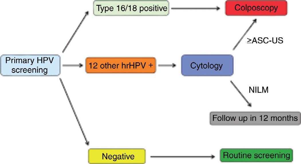
Figure 1.3 Strategies for incorporating primary human papillomavirus screening into practice. ASC-US, Atypical squamous cells undetermined significance; hrHPV, high risk human papillomavirus; NILM, negative for intraepithelial lesion or malignancy.
HPV testing every 5 years; and co-testing every 5 years. These intervals assume no prior abnormal screening history and normal results upon screening. Surveillance describes the followup and management of women with abnormal screening results or treatment history and differs based on the specific abnormality and management strategy. Other factors, such as age and fertility desires, may also play a role. Notably, screening intervals should be based on documented results; Boyce et al. have shown that women’s ability to recall prior screening is flawed. In 2014, the FDA approved HPV testing as a primary screening test for cervical cancer. Approval was granted only for the cobas HPV test (Roche). At an initial consensus meeting sponsored by the Society of Gynecologic Oncologists and ASCCP, Huh and fellow consensus meeting delegates developed interim guidelines to inform clinicians and women at risk on strategies to incorporate primary screening into practice. When used according to an algorithm (Fig. 1.3) that appears to optimize disease detection while minimizing colposcopy, HPV testing is more sensitive than Pap testing when initiated at 25 years of age. Screening intervals for primary HPV testing are controversial. Ultimately, the authors recommended that screening be no more often than every 3 years, language that reflected disagreement about 3-year versus 5-year testing intervals. The pivotal trial for the cobas HPV test did not extend beyond 3 years, so performance data are unknown. However, other HPV tests with similar sensitivity and specificity have been tested, and a 5-year interval seems to provide sensitivity superior to 3-year cytology screening while allowing more time for transient lesions to regress. Performance characteristics of HPV-based screening among women ages 25 to 29 years should improve as HPV risk declines as vaccinated girls age into that range.
The risk for cervical cancer in adequately screened women after 65 years of age becomes negligible. Prior HPV infection that has been cleared without development of CIN21 does not appear to increase risk substantially. New HPV infections may be acquired after 65 years of age despite prior negative screening, but in the absence of active metaplasia, these are unlikely to progress to cancer for years and so are unlikely to result in morbidity or mortality, but evaluation and treatment of atrophic
postmenopausal women is technically difficult and painful. Vaginal cancer risks are low after hysterectomy for benign disease and does not justify continued screening in women with no abnormal screening history. Women with CIN21 remain at risk for cervical and vaginal cancer despite treatment, including hysterectomy, and evaluation until comorbidity suggests a short residual lifespan remains indicated.
Women at low risk for disease during their remaining lifespans should not be screened. This is most apparent for women with illnesses that will be fatal in the medium term, as the discomfort and risks of screening and treatment of identified precursors are not justified when the woman is unlikely to survive long enough to develop symptomatic cervical cancer. Similarly, cervical and vaginal cancer risk in the absence of a cervix approaches zero. After hysterectomy for CIN, screening appears justified based on risk for coexistent vaginal intraepithelial neoplasia and vaginal cancer, and posttreatment surveillance for recurrent cervical cancer treated with hysterectomy is recommended. Women who reach 65 years of age after multiple negative cytology results, including three in the previous decade and two in the previous 5 years, are at low risk for cervical cancer, as are women with two negative HPV–cytology co-test results, including one in the previous 5 years. These women can stop undergoing screening. Women without adequate prior screening should continue undergoing screening until they meet these criteria. Although older women can acquire new HPV infections, they are not undergoing active metaplasia, and transition time to cancer appears to be decades long, as in younger women, so few are likely to survive to develop cancer. For this reason, acquisition of new sexual partners by women who have otherwise met criteria for stopping should not be a consideration for continuing screening.
There are few predictors of CIN21 identified that would allow clinicians to focus more intensive screening efforts on high-risk groups. Boardman and colleagues found that smokers faced a higher risk of CIN21 than nonsmokers, but the odds ratio of 1.6 did not provide sufficient discrimination to allow observation of nonsmokers. Fundamentally, cytology and HPV testing are powerful tools for identifying risk, and the low positive predictive value of borderline cytology grades can be refined by triage using HPV testing or genotyping. Women with abnormal cytology or high-risk HPV infections merit further assessment regardless of demographic or behavioral risks.
MANAGEMENT OF ABNORMAL SCREENING RESULTS
In 2007, Castle et al. proposed that management of abnormal cervical cancer screening tests should be based on the associated risk for significant disease. Cancer mortality would be the ideal risk outcome for comparison across tests, but it is too uncommon in screened populations and its frequency is determined by downstream interventions after most tests. Katki and associates proposed instead that risk after various test results and combinations should be benchmarked to CIN3 or worse (CIN31, including CIN3, AIS, and cancer). Because some lesions are present but inapparent initially because women fail to
present for assessment or lesions are clinically too small for detection, the optimal benchmark endorsed by a 2012 consensus conference is 5-year risk for CIN31. Using this benchmark, clinicians can use similar management strategies for women with similar levels of risk, without regard to how risk was determined. Conventionally, risk was defined by cytology and colposcopy findings. More recently, HPV testing allowed recalibration of risk, especially for women with borderline findings such as ASC-US. HPV genotyping for types 16 and 18 further defines risk categories. On the horizon are other molecular tests whose results will modify risk stratification and management, such as spectroscopic analysis and molecular testing for p16ink4a, the Ki-67 proliferation marker, methylation status, and others.
Determining optimal management for each test or combination can be arduous and confusing. In an ideal world, comparative trials would define which triage tests were optimal. Unfortunately, few comparative trials have been undertaken. National health budgets across the developed world are increasingly constrained. Industry lacks incentive to fund trials that might find their products inferior. With the proliferation of screening tests, management options become increasingly complex because algorithms must incorporate all options clinicians might select. Previously management guidelines have been criticized as too complex for even experts to master. Fortunately, the adoption of risk-based management introduced with the new 2019 guidelines has eliminated the need for clinicians to memorize cervical cancer prevention strategies. Electronic medical record platforms can be programmed to generate reminders when women come due for screening. Smartphone apps allow entry of patient information and lead clinicians to relevant algorithms.
Previously, Katki et al. analyzed data from more than 1 million women screened by the Kaiser Permanente of Northern California health care system to define 5-year CIN31 risk after various tests and test combinations. At the base of management options is the 5-year follow-up for women who test negative in Pap/HPV co-testing; these women have a 5-year CIN31 risk of less than 0.01%. Women screened with cytology alone are followed at 3-year intervals when their test results are negative, with a 5-year CIN31 risk of less than 0.1%. One-year retesting is standard for women with a positive test result for HPV but negative cytology, with a risk less than 5%. The consensus threshold for colposcopy in the United States is a low-grade SIL result, with 5-year CIN31 risk of just above 5%; lesser results are triaged by serial testing or triage tests. The threshold for treatment is CIN2, although not all women with CIN2 require treatment, and few studies have assessed use of other test results except CIN21 cytology as a treatment indication. More recently, data was expanded and analyzed to include approximately 1.5 million individuals between 2003 and 2017 to estimate immediate and 5-year rates of CIN31 using various combinations of screening history and current testing results; these data were critical to the updated risk-based (rather than test-based) guidelines developed in 2019.
Traditionally, colposcopy was used as the triage modality to identify women with CIN21 for treatment. Colposcopy is the magnified stereoscopic visualization of the cervix under intense
TABLE 1.3 Abnormal Colposcopic Findings
Atypical transformation zone
Keratosis
Acetowhite epithelium
Punctation
Mosaicism
Atypical vessels
Suspect frank invasive carcinoma
Unsatisfactory colposcopic findings
illumination. Magnification ranges from 33 to 303. A green filter over the light source accentuates vascular patterns and lesion margins. Although colposcopy without staining has been advocated to maximize visualization of cancer, most colposcopic assessments are augmented by the application of vital stains such as 3% to 5% acetic acid and Lugol’s iodine solution.
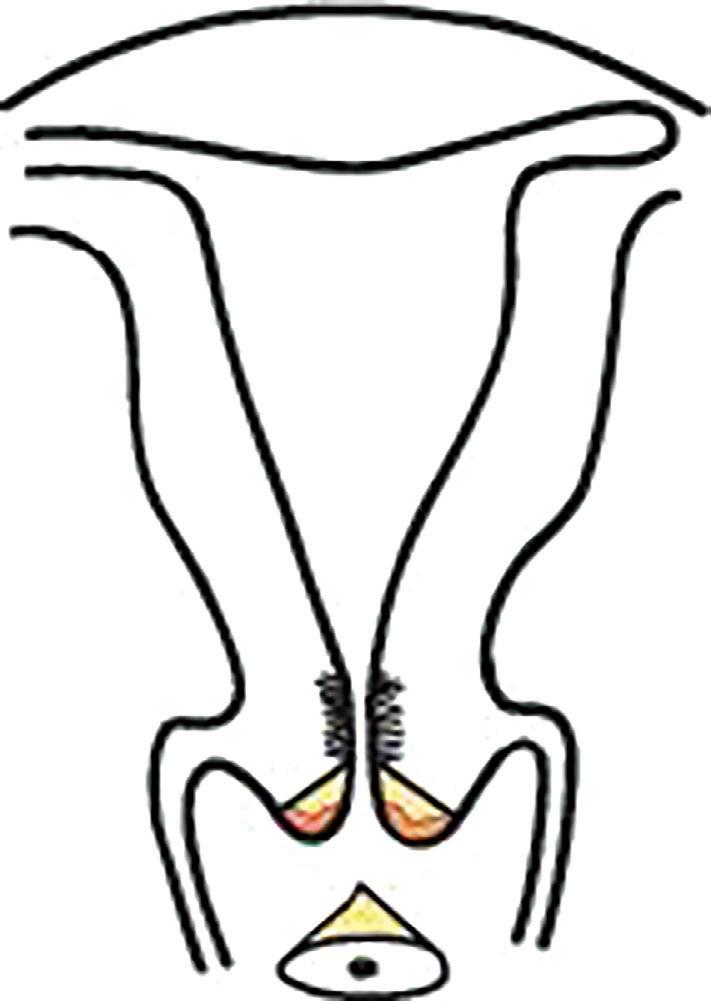
Colposcopy
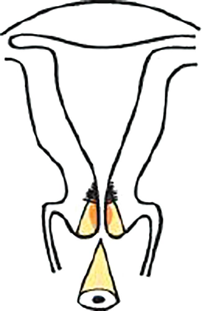
In colposcopy, the cervical transformation zone is assessed. The transformation zone is that area of the cervix and vagina initially covered by columnar epithelium that has undergone metaplasia to squamous epithelium. A range of terms are used to describe colposcopic findings ( Table 1.3 and Figs. 1.4–1.7). The procedure for colposcopy is inspection of the cervix without stains and then cleansing of the cervix with an application of 3% to 5% acetic acid for at least 90 seconds. This removes
Figure 1.4 A, Squamocolumnar junction (transformation zone). B, Large transformation zone.
mucus and debris and accentuates vascular and epithelial patterns. Most cervical lesions stain white with acetic acid (acetowhitening).
In 2017, the ASCCP discussed risk-based colposcopy standards with four main working groups. These included: (1) role
of colposcopy, benefits and harms of terminology, (2) risk-based colposcopy and biopsy, (3) colposcopy procedures and adjuncts, and (4) quality control. One of the main goals of the second working group was to define risk-based colposcopy and to evaluate how results prior to colposcopy and colposcopy results could be combined in order to improve outcomes. Previous studies had shown that only taking a single biopsy may miss up to 40% of precancers and that there was an increase in CIN21 detection when at least two targeted biopsies were taken. The recommendation was made for “adapting colposcopy practice to previous risk and colposcopy impression.” In other words, based on the risk level, colposcopy practice can be modified. For most women, colposcopy with 2 to 4 biopsies is required for optimal sensitivity. However, in low-risk women with no acetowhitening, no HPV-16/18, and cytology less than HSIL, colposcopy can be deferred and women followed with co-testing. In women where the risk of precancer is very high, immediate excisional treatment can be performed in order to decrease costs and repeat visits. In patients who are of intermediate risk, multiple targeted biopsies leads to increased detection of precancerous changes. HPV vaccination promises to reduce cervical cancer risk in coming decades but more immediately will reduce the prevalence of CIN21 lesions. This in turn will reduce the specificity of screening tests and lower the sensitivity of colposcopy. Opportunities for further changes to screening and management strategies will follow, especially longer screening intervals, more HPV-based assessment, more intermediate triage tests before colposcopy, and a move toward immediate treatment without colposcopy for women at highest risk.
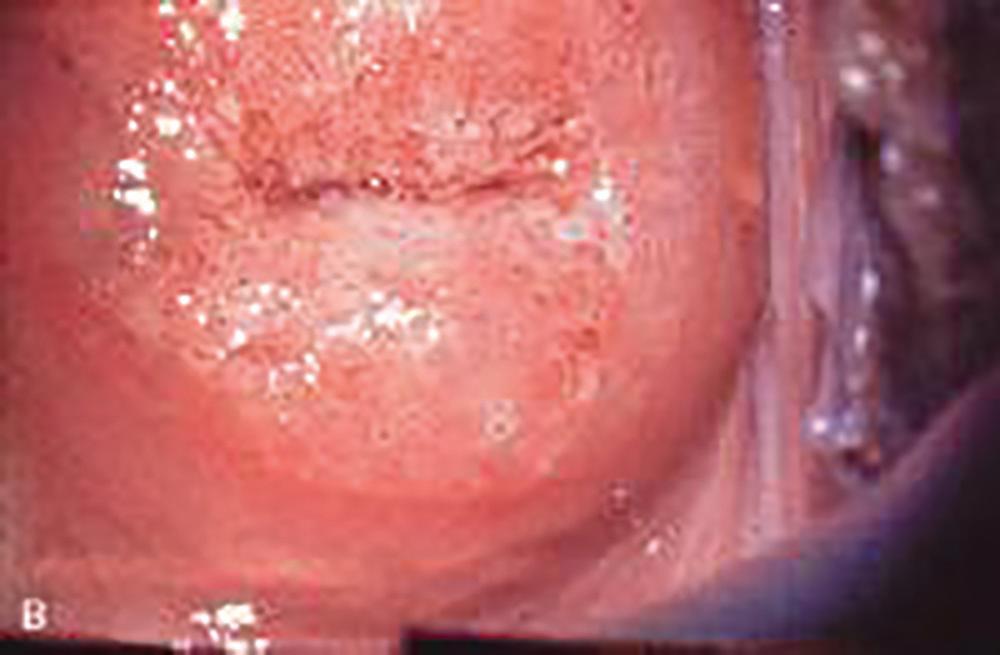
Figure 1.5 White epithelium at the cervical os (a colposcopic view).
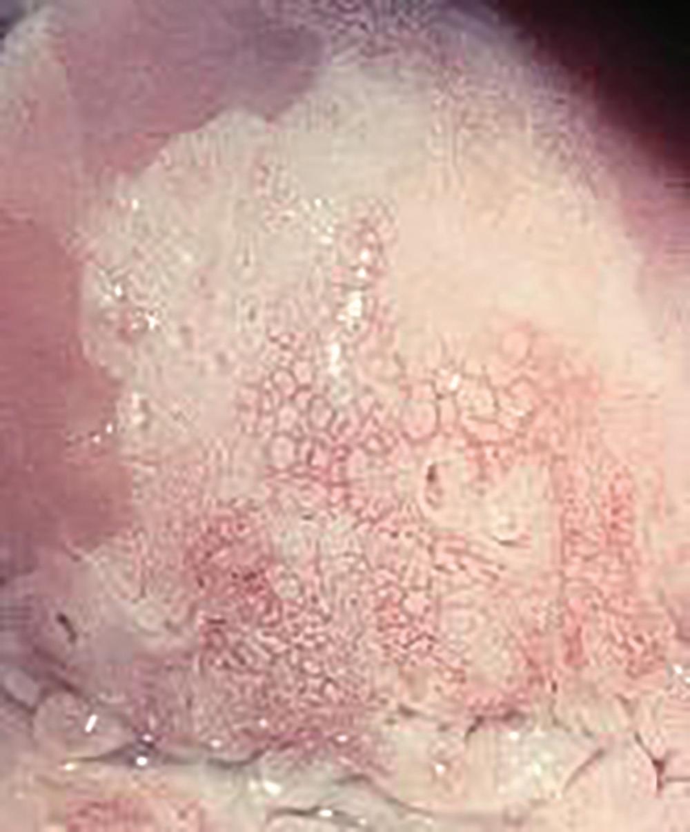
Figure 1.6 A punctation pattern is seen clearly above a mosaic structure (a colposcopic view).
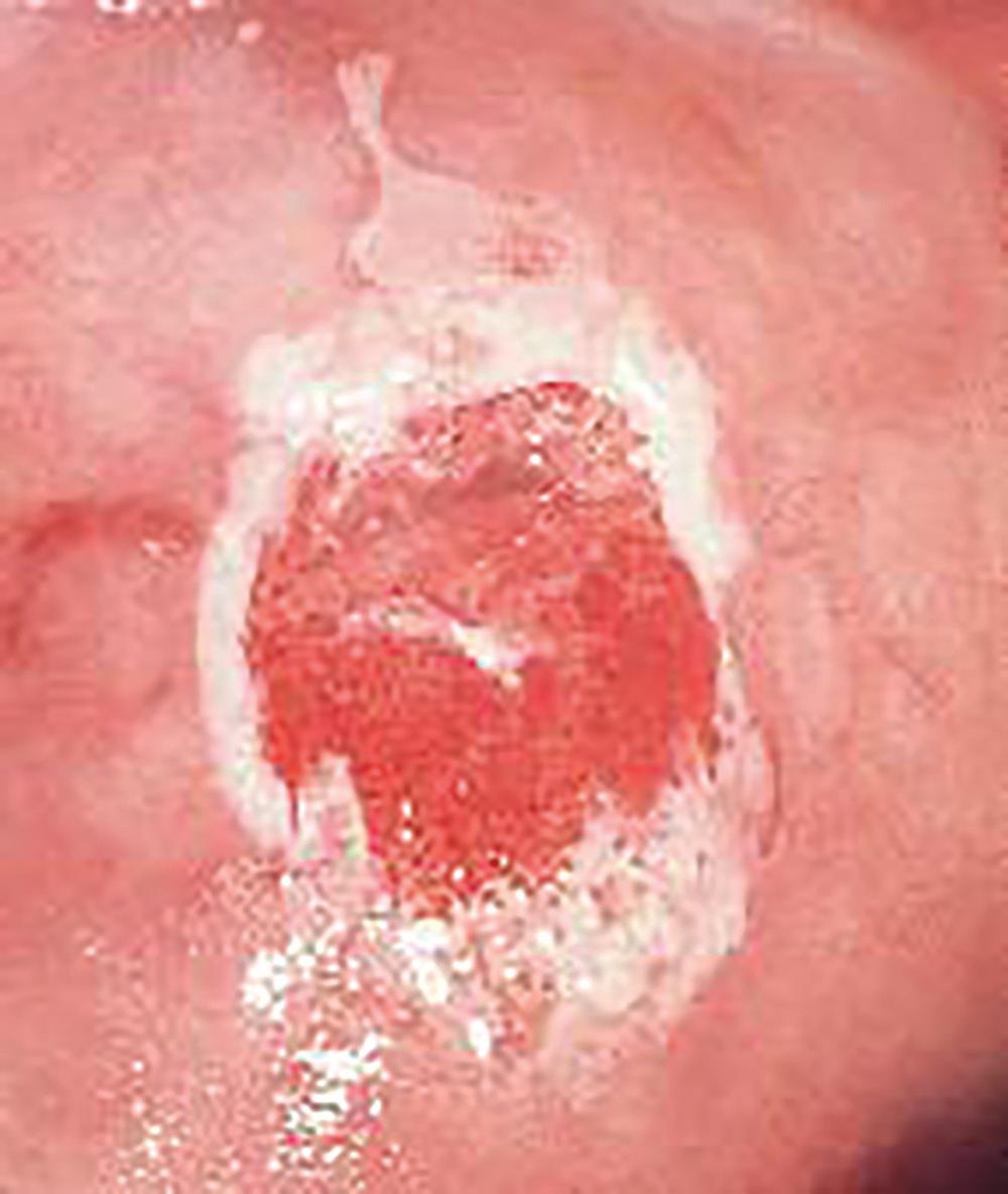
Figure 1.7 A large anterior lip lesion with white epithelium punctation and mosaic patterns.
Screening and Treatment in Immunocompromised Women
The screening for immunocompromised women differs from immunocompetent women and management guidelines are directed by recommendations from the CDC. Immunocompromised patients include (but are not limited to) those with HIV, solid organ transplant, allogenic hematopoietic stem cell transplant (HSCT), systemic lupus erythematous (SLE), or patients requiring immunosuppressive treatments. Women with HIV are screened according to the guidelines for the prevention and treatment of opportunistic infections in HIV-infected adolescents and adults from the CDC. Patients with HIV are more likely to have persistent HPV infections and should start screening within 1 year of onset of sexual activity (if prior to 21 years old) but no later than 21 years of age. One group demonstrated that up to 1 in 4 women do not get annually tested despite regular visits to their primary care physician. These women should be screened twice in their first year following their diagnosis, followed by yearly screening as long as initial results are normal. If the results of three consecutive tests are normal, the screening interval can change to every 3 years. Screening should continue indefinitely and not cease at age 65 as in noninfected women.
Immunocompromised women without HIV infection includes those women with solid organ transplant, HSCT, and autoimmune diseases (i.e., SLE, rheumatoid arthritis [RA], and inflammatory bowel disease [IBS]). The evidence is limited, but expert recommendations were published in 2019. For patients with solid organ transplant less than 30 years old, screen with cytology annually for 3 years and if normal, patients can transition to every 3 years. For patients aged 30 and older, the recommendation is for co-testing every 3 years if normal. If the transplant occurs before the age of 21, then screening should begin within 1 year of sexual debut. Screening should continue throughout the patient’s lifetime (do not stop at 65 years old). Recommendations are similar for patients with HSCT, IBS on immunosuppressant treatments, and SLE and RA on immunosuppressant treatments. For patients less than 30 years old, screen with cytology annually for 3 years and if normal normal, patients can transition to every 3 years. For patients aged 30 and older, the recommendation is for cotesting every 3 years if normal. If a patient is started on an immunosuppressant treatment prior to the age of 21 or undergoes HSCT, they should start cervical cancer screening within 1 year of sexual debut. All screening should be continued throughout the patient’s lifetime. Patients with IBS, SLE, or RA not on immunosuppressant treatments can follow the general population screening guidelines.
Patients exposed to diethylstilbestrol (DES) in utero are recommended to undergo yearly surveillance by both ACOG and the NCI including a Pap test that obtains cells from the cervix and vaginal walls. Similar to immunocompromised women, there is no age cut-off for when Pap tests should stop in DES-exposed women. Vaginal cytology should continue to be performed in DES-exposed women who have undergone a hysterectomy.
Management of Abnormal Screening Results
Since 2001, the ASCCP has led four consensus development conferences (2001, 2006, 2012, 2019) to define standard guidelines for management of US women with abnormal cervical cancer screening test results and CIN or AIS. Risk-based guidelines were initially introduced in 2012. The most recent guidelines, released in 2019, more closely align management recommendations with the growing understanding of the natural history of HPV and cervical carcinogenesis. Healthcare providers can use these guidelines via the http://asccp.org website, the ASCCP phone application, or the tables in Egemen et al. A patient’s risk is assessed through age, current screening results, past screening/biopsy results, and if they have immunosuppression. These guidelines also included adjustments for women with an unknown history of screening. All of these factors are used to decide on surveillance, colposcopy, and treatment for patients. If a patient’s risk is 4% or greater for CIN 31, then immediate management with colposcopy or treatment is recommended. In those with less than 4% risk, the 5-year risk of CIN 31 is evaluated and triages the patients into a 1-, 3-, or 5-year return.
The overarching goal of current ASCCP risk-based guidelines is to allow modified evaluation and treatment in alignment with a woman’s risk. Subsequent management recommendations are based on risks, not merely results, and promulgate the concept of “equal management for equal risk.” Additionally, these guidelines importantly factor in that time matters, and that the longer an HPV infection has been present, the higher the risk for precancer and cancer (CIN3 and higher); this serves as the framework and foundation of the updated guidelines. The four guiding principles of this update are: (1) HPV-based testing is the basis of risk estimation; (2) personalized risk-based management is possible with knowledge of current results and past history; (3) guidelines must allow updates to incorporate new test methods as they are validated and to adjust for decreasing CIN31 risks as more patients who received HPV vaccination reach screening age; and (4) colposcopy practice must follow guidance detailed in the ASCCP Colposcopy Standards.
Using combined clinical data from patients’ prior and current screening results, recommendations for management are made based on predefined clinical action thresholds. These correspond to a patient’s particular risk strata that is calculated within the electronic app based on validated large-cohort perspective data, all based on the immediate risk of CIN31 (Fig. 1.8). As introduced previously, these clinical action thresholds are for 5-, 3-, or 1-year return to clinic follow-up, immediate colposcopy, expedited treatment or colposcopy acceptable, or immediate treatment preferred.
The primary driver of risk stratification into a particular clinical action threshold is the persistence or absence of highrisk HPV. For example, consecutive negative HPV screening is highly predictive that CIN31 will not develop. A 2018 study demonstrated that the 5-year risk of invasive cancer following a negative HPV result decreased from 0.0092% for the first negative result to 0.0015% for a negative result on a third test preceded by two negative screening tests. On the opposite end of the spectrum, long-term persistent HPV is particularly high-risk
Is immediate CIN3+ risk greater than or equal to 4%?
Look at 5–year CIN3+ risk and find the sur veillance inter val.
Return in 1 year 0.55% (5–year CIN3+risk)
Return in 3 years 0.15%–0.54% (5–year CIN3+risk)
Return in 5 years 0.15% (5–year CIN3+risk)
Figure 1.8 Determination of patient risk based on current screening results and screening history.
(with persistent HPV-16 conferring the highest risk); a study of over 8500 women followed for 12 years for the development of CIN31 showed that two consecutive tests positive for HPV-16 conferred a 47% risk of CIN31 over the follow-up period.
In the following sections, we will detail the specific management of select scenarios of abnormal screening. However, given the improved and more dynamic nature of individualizing screening follow-up and management put forth by the new guidelines outlined earlier, we will not fully outline all results and management strategies as previously put forth. The reader is encouraged to utilize the ASCCP app and web-based program for the most individualized results as they pertain to a specific patient and clinical scenario.
Managing Abnormal Results in Young Women
Cervical cancer risk is low among women 21 to 24 years of age, but high-risk HPV infections can be found in more than 20%. Most CIN1 and many CIN2 lesions in this age group will regress without intervention, and CIN3 is unlikely to progress to cancer if missed prior to subsequent screening. Treatment of intermediate-risk lesions may impact future pregnancies. For these reasons, women in this age group are managed less aggressively than older women. For women in this age group, most abnormal cytology results reflect HPV infection; HPV
testing should not be ordered, and if obtained, results should not modify management.
Additionally, because of concerns about impact on subsequent pregnancies, women with biopsies read as CIN2, CIN21, or HSIL can be observed, provided colposcopy is satisfactory. There is no specific age range for observational management of these lesions, and a decision to treat or follow these lesions depends on a discussion between the woman and her clinician, balancing the potential risk to future pregnancies against the risk of progression to cancer during observation; both risks appear to be low. Treatment is recommended for women in this age group for which CIN3 is specified as CIN3 is considered an immediate precursor to cancer.
Managing Unsatisfactory Cytology
When cytology is unsatisfactory for evaluation and HPV status is either unknown, not tested for, or negative, it must be repeated. The ideal timeframe is within 2 to 4 months. For women of age greater than 25 with unsatisfactory cytology and an HPV-positive result, repeat cytology with concurrent colposcopy in 2 to 4 months is acceptable. For women with atrophy, a short course of vaginal estrogen may improve sample cellularity. Women with obscuring inflammation should be assessed for specific infections and treated for any that might be
identified; empiric vaginal antibiotic therapy does not appear beneficial. Samples that are unsatisfactory because of obscuring blood raise concern for cancer. When repeated samples are unsatisfactory, colposcopy should be considered.
Cytology-Negative, Human Papillomavirus-Positive Women
Identification of a carcinogenic HPV type carries substantial risk for CIN31, but the 5-year risk does not reach the 5% threshold for colposcopy. A positive HPV result is most often encountered in HPV–cytology co-testing for women ages 30 to 65 years. Specific management differs based on if HPV genotyping was performed. If not initially performed and not available, women should have repeat co-testing in 1 year, in which time HPV infection may regress. If at 1 year either result is abnormal, colposcopy should be performed.
If HPV-genotyping was performed and is either HPV-16 or HPV-18 positive, colposcopy should be performed as the risk exceeds the 5% threshold.
Management of ASCUS, low-grade squamous intraepithelial lesions (LSILs), and atypical squamous cells, cannot exclude HSIL (ASC-H).
For specific management guidance, we recommend the reader reference the new interactive guidelines via the app or through tables available at asccp.org. General notes only are provided.
ASC-US results are common. Triage using HPV testing on the liquid-based cytology sample appears to be cost effective and minimizes delay and loss to follow-up. If HPV testing is positive, then colposcopy is indicated; immediate risk of CIN31 is approximately 4.4%. A negative HPV result in the context of ASC-US indicates a low risk of CIN31 (less than 1%), though the risk is higher than among women with negative co-testing, and 3-year co-testing is recommended.
HPV genotyping distinguishes between higher-risk women with HPV-16/-18 and lower-risk women negative for HPV-16/18, but for all HPV-positive women, 5-year CIN31 risk approximates or exceeds the threshold for colposcopy. Because management is not changed by results, HPV genotyping is not recommended for triage of ASC-US cytology.
LSIL previously set a paradigmatic threshold for colposcopy in the United States. Most women triage into colposcopy, though variables such as age and HPV status/history will more accurately triage patients into either follow-up or colposcopy. Immediate treatment is unacceptable.
Management of High-Grade Squamous Intraepithelial Lesion
CIN21 is found in some 60% of women with HSIL cytology, and approximately 2% will have cancer at time of colposcopy. Given such a substantial risk, aggressive management is justified. In many settings, especially for women in clinics with high rates of loss to follow-up and for women who have completed childbearing, immediate loop excision is an efficient approach to management. Immediate excision is unacceptable for women up to 24 years of age because of their low near-term risk for cancer and the likelihood that CIN1 and CIN2, but not CIN3,
will regress. HPV results modulate risk, but even women with HPV-HSIL face up to a 30% 5-year risk for CIN31. For this reason, colposcopy is required regardless of HPV result, and triage of HSIL cytology using HPV testing is unacceptable, as is observation with repeat cytology.
Atypical Glandular Cells
Despite its name, atypical glandular cells (AGCs) cytology is more often associated with squamous than glandular lesions. The risk of CIN3, AIS, or cancer (CIN31) after a Pap test read as AGC is almost 10%, and risk for cancer is 3%. AIS has been on the rise in the last 20 years, especially in younger women between the ages of 30 to 40. Squamous and glandular lesions can coexist, and identification of CIN does not rule out adenocarcinoma. Many associated cancers are of endometrial origin and would be HPV negative; endometrial lesions are more common in older women and in women with such risk factors as obesity, unexplained bleeding, and anovulation. Thorough evaluation is needed regardless of HPV test results.
Women with AGC cytology need colposcopy with endocervical sampling and biopsies of any acetowhite cervical lesion. Endometrial sampling is needed if the patient is older than 35 years of age or if endometrial cancer risk factors are present. For women with AGC subcategorized as “atypical endometrial cells,” evaluation can be truncated: initial endometrial biopsy and endocervical curettage are needed, with colposcopy only if no endometrial pathology is found. In contrast to ASC-US, triage of AGC results with HPV testing is not appropriate, nor is cytologic testing. Management of AGC in women 21 to 24 years of age is identical to that of older women. Pregnant women with AGC cytology results need colposcopy, but endocervical curettage and endometrial sampling are contraindicated until following pregnancy.
Management of Endometrial Cells in Older Women
Among postmenopausal women, a finding of benign endometrial cells on cytology is associated with a 5% risk of clinically important pathology, including cancer. However, endometrial cells, endometrial stromal cells, or histiocytes in premenopausal women are not associated with disease; as such, no further work-up is necessary. For postmenopausal women with endometrial cells on cytology, endometrial assessment is indicated using either endometrial sampling or imaging of endometrial thickness.
MANAGEMENT OF POST-COLPOSCOPIC RESULTS
Management of Women With No Lesion or CIN1 at Colposcopy
A colposcopic biopsy result of no lesion or CIN1 does not exclude the presence of a higher-grade lesion in an unsampled area of the cervix. The risk for subsequent CIN31 depends on patient age as well as the prior abnormality. Risk is lower for women with what ASCCP terms “lesser abnormalities” than for women with ASC-H, HSIL, or AGC. Lesser abnormalities
include negative cytology with either HPV-16 or repeated HPV positivity, ASC-US, or LSIL.
As mentioned previously, for women with lesser abnormalities and no lesion or only CIN1 on colposcopy, observation with serial co-testing is indicated. If a first co-test result is negative, repeat testing is indicated 3 years later. If all test results are negative, then routine screening is appropriate. If any test result is abnormal, repeat colposcopy is required. This can become burdensome. Treatment of CIN1 is acceptable once disease has persisted for 2 years, although continuing observation is also acceptable if fertility is a concern. Treatment of persistent HPVpositive tests or persistent ASC-US/LSIL cytology in the absence of a cervical lesion is not indicated because many of these women will have vaginal lesions. This includes topical therapies such as trichloroacetic acid. Hysterectomy is never indicated for CIN1 or abnormal cytology.
For women 21 to 24 years of age with no lesion or CIN1 after ASC-US or LSIL, observation with repeat cytology annually is indicated. Repeat colposcopy is only needed if cytology progresses to ASC-H, HSIL, or AGC, or if cytology remains borderline abnormal for 2 years. After a negative result, routine screening at 3 years is indicated. HPV testing and co-testing are not indicated in this age group. For pregnant women with no lesion or CIN1 after lesser abnormalities during pregnancy, colposcopy should be deferred until postpartum.
Management differs if colposcopy reveals no lesion or CIN1 after ASC-H or HSIL cytology, as the risk for CIN31 is much higher (Fig. 1.9). Histology and colposcopic biopsy results can be reviewed by experts to confirm diagnosis. Observation at
1 and 2 years is also appropriate granted colposcopy was adequate. A diagnostic excisional procedure is recommended if HSIL/ASC-H persists at 1 year in women older than 25 or for 2 years in women younger than 25. For pregnant women with no lesion or only CIN1 after ASC-H or HSIL, colposcopy should be repeated postpartum: the short-term cancer risk is low, and no other intervention is needed during pregnancy. When no lesion or CIN1 is found after AGC cytology, annual co-testing at 1 and 2 years is indicated, with colposcopy for any abnormality; after two negative co-test results, risk declines, and resumption of three regular co-testing intervals is appropriate (Fig. 1.10). Colposcopy is needed if any result is abnormal. Hysterectomy is unacceptable as primary therapy for any grade of CIN, although after prior excision, hysterectomy may be indicated if lesions recur or persist and repeat diagnostic excision is not feasible.
Management of Women With CIN21
A colposcopic biopsy of CIN21 is the threshold for treatment. CIN2 is an intermediate lesion: many represent transient but exuberant HPV infection, and half or more resolve without therapy. However, some will progress to CIN3 and cancer. For women younger than 25 and CIN2 is specified, observation is acceptable; observation is also acceptable in women older than 25 with concerns regarding future fertility (Fig. 1.11). Again, specific situations can be managed to tailor each patient’s individual circumstance utilizing the updated ASCCP guidelines and app. All lesions read as CIN3 should be treated regardless of the patient’s age or desire for future childbearing, because there is
Observation:
Cytology 25 years
HPV-based testing 25years at 1 and 2 years*
Negative cytology or HPV-based testing at 1 year and 2 years
HPV-based testing in 3 years
Abnormal test results Manage per 2019 ASCCP guidelines
OR
Age 25years: cytology HSIL at 1 or 2 years or ASC-H at 2 years
Age 25 yrs: cytology HSIL or ASC-H at 2 years
Review of cytologic, histologic, and colposcopic findings
Diagnostic excisional procedure^
Manage per 2019 ASCCP guideline for revised diagnosis
*Only if colposcopy visualizes the entire squamocolumnar junction and upper limit of lesion(s), and endocervical sampling negative ^Except in pregnant women
@ Copyright, 2020, ASCCP All rights reserved.
Figure 1.10 Subsequent management of atypical glandular cells (AGC). ASC-H, Cannot exclude high-grade SIL; ASCCP, American Society for Colposcopy and Cervical Pathology; HPV, human papillomavirus; HSIL, high-grade squamous intraepithelial lesions.
CIN2 specified
Observation preferred 25 years
Acceptable 25 years if concerns about treatment effects on future pregnancy outweigh concerns about cancer
HSIL, unspecified Observation acceptable 25 years. Treatment preferred 25 years.
CIN3 specified; any histologic HSIL with SCJ or upper limit of lesion(s) not visualized; or ECC with CIN 2+ or ungraded
Observation:
Colposcopy & cytology 25 years
Colposcopy & HPV-based testing 25 years at 6 and 12 months
2 consecutive evaluations both ASC-H cytology and CIN2 histology
HPV-based testing ( 25 years) or cytology ( 25 years) in 1 year
Test results negative
HPV-based testing annually x 3 total years, then test at 3 year intervals for at least 25 years
Colposcopy remains CIN2 or cytology ASC-H2 AGC or HSIL with biopsy CIN2
Any test abnormal
Diagnostic excisional procedure
CIN3 develops at any point or CIN2 histology or ASC-H, HSIL, or AGC cytology persists for 2 years
Continue observation: Colposcopy & cytology 25 years
Colposcopy & HPV-based testing 25 years at 6-month intervals up to a total of 2 years since first CIN2 diagnosis
@ Copyright, 2020, ASCCP All rights reserved.
Figure 1.11 Management of CIN21 in patients whose concerns about the effects of treatment on a future pregnancy outweigh their concerns about cancer. AGC, Atypical glandular cells; ASC-H, cannot exclude highgrade SIL; ECC, endocervical curettage; HPV, human papillomavirus; HSIL, high-grade squamous intraepithelial lesions; SCJ, squamocolumnar junction.
no margin for progression, and some will harbor undiagnosed cancer; ongoing pregnancy is the main exception to this standard. Additionally, treatment is preferred if histologic HSIL cannot be specified (reported as histologic HSIL or HSIL [CIN2,3] (CIN3)).
Observation is an acceptable option for women who, after counseling, consider risk to future pregnancies from treatment to outweigh cancer risk from observation. There is no specific age threshold: a 21-year-old woman who has had a tubal ligation should be treated, but a 43-year-old woman undergoing infertility treatment after a diagnosis of incompetent cervix might prefer to be observed. Observation is only acceptable when the entire lesion and squamocolumnar junction is observed colposcopically. Observation consists of colposcopy and cytology at 6-month intervals until the lesion resolves, as evidenced by regression of colposcopic abnormalities and normalization of cytology (i.e., results are , ASC-H cytology and/or CIN21 histology). After that occurs, women should be followed with a co-test 1 year later and then 3 additional years later before returning to routine screening. If histology remains CIN2 or cytology is ASC-H, HSIL or AGC with histology , CIN2, continued observation at 6-month intervals for up to a total of 2 years since the initial CIN2 diagnosis.
In 2020, Teoh et al. published recommendations regarding the diagnosis and management of cervical AIS. In women with AIS, an excisional procedure is needed to rule out invasive adenocarcinoma before definitive treatment is performed (Fig. 1.12). An intact specimen with a length of 10 mm and negative margins is recommended. If a provider is unable to consistently perform this, a referral to a gynecologic oncologist should be considered. When a patient has completed childbearing, a hysterectomy is preferred given that even in women with negative margins there is 20% risk of persistent AIS and lesser risk of invasive cancer. In patients who desire future fertility, conservative management with close follow-up is acceptable in the setting of negative margins. These patients should undergo co-testing with endocervical sample every 6 months for 3 years and then annually for at least 2 years. Patients who have had negative co-testing results for 5 years may then undergo testing every 3 years. Hysterectomy is still recommended once childbearing is complete.
Treatment of Cervical Disease
Cure rates for CIN21 requires excisional or destructive procedures. In the United States, ablative therapies include cryotherapy and laser ablation. Excision can be accomplished with
*Those desiring fertility-sparing management cotesting with endocervical sampling **For patients with AIS and persistent positive margins for whom additional excisional procedures are not feasible, either simple or modified radical hysterectomy is acceptable
Diagnostic excision procedure to rule out invasive cancer
Figure 1.12 Management of adenocarcinoma in situ (AIS) ECC, Endocervical curettage; HPV, human papillomavirus.
an electrosurgical wire loop or needle or with a scalpel. In the United States, excisional procedures are performed more commonly than ablative procedures.
Cervical ablation results in the elimination of lesions without rendering a specimen for pathologic analysis and while an option, is not the preferred option for management. For this reason, many gynecologists consider outpatient loop excision a preferred approach. However, randomized trials showed similar failure rates for excision and ablation. This suggests that as long as invasive cancer is microinvasive, with little risk for metastasis, the method of destruction is irrelevant. Nevertheless, using ablative therapies for cervical disease requires careful attention to well-defined exclusion criteria. The 2019 ASCCP guidelines have specific criteria for ablation though stress that excision is the preferred treatment. Ablation cannot be undertaken unless the entire squamocolumnar junction, including all tissue with lesions, is visible colposcopically. Ablation of such an unsatisfactory transformation zone risks missing invasive cancer within the endocervix that then progresses undetected. Furthermore, if disease extends into the endocervical canal and lesion covers more than 75% of the surface area of the ectocervix, ablation is inappropriate. The entire squamocolumnar junction must be visualized to perform an ablative procedure. Additional criteria that must be met include: an endocervical curettage (ECC) should not contain CIN21, there should be no history of CIN21 that has been treated, all obtained biopsies/ cytologics should have been adequate to ensure diagnosis, and obviously, invasive cancer should not be highly suspected.
Cryotherapy is performed using nitrous oxide, a delivery gun, and various sizes of metal probes designed to cover the transformation zone of the cervix. Figge and Creasman described use of a freeze–thaw–freeze technique with good success rates, but regardless of the use of double- or single-freeze approaches, identifying the development of at least a 5-mm “ice ball” or zone of freezing lateral to the cryotherapy probe is essential to achieving deep thermal destruction. Advantages of cryotherapy include relatively low cost and low risk for injury. Disadvantages include a copious discharge from cervical tissues suffering from sublethal thermal injury and lack of a surgical specimen.
Cervical laser ablation involves the use of carbon dioxide laser energy to destroy abnormal cervical tissue. When optimally used, the laser energy is delivered at a power density of 750 to 1250 W/cm2. This results in flash boiling of impacted cells, and energy is dissipated from the operating field through the smoke plume without causing deeper tissue injury. Lower power density may create the illusion of greater control of tissue destruction, but it requires longer beam application, with greater hidden, delayed coagulation injury to underlying stroma. As with all therapies, laser should be used to ablate the entire at-risk transformation zone. CIN can involve the metaplastic epithelium of cervical glands, which extend some 5 mm into the cervical stroma; for this reason, ablation should be carried to a depth of about 7 mm and should encompass all lesions. If the lesion extends onto the vagina, ablation is carried only to 1 to 2 mm in depth. As described by Stanhope and associates, laser therapy can result in persistent CIN risk of less
than 90%. Advantages include precision in application, allowing extension of therapy to lesions that involve the vagina, and rapid recovery from injury. Disadvantages include the need for an expensive laser generator that requires frequent maintenance. Unskilled use of laser energy can result in both immediate and delayed injury.
Most gynecologists in the United States treat cervical precancers by excision with electrosurgical loops, termed LEEP. The entire transformation zone is excised using loops with diameters of 1.5 to 2.5 cm. Excision should extend to the deepest glands, or 7 to 8 mm. For women with disease extending into the endocervical canal and those with inadequate colposcopy, either use deeper loops or a second endocervical pass, colloquially termed a “top-hat” excision for the appearance of the stacked specimen that results; this endocervical excision must encompass the lateral extent of endocervical glands, so 6 to 8 mm from the endocervical surface. For most women, LEEP can be done in an office setting under local anesthesia. Epinephrine is injected with the local anesthetic to minimize blood loss. The injection should be subepithelial rather than stromal, and a paracervical block does not provide the same hemostatic benefit of intracervical injection. Colposcopic visualization allows optimal tailoring of the excision. Hemostasis is achieved with a combination of fulguration using ball electrodes and application of Monsel’s solution. Delayed bleeding should occur in fewer than 5% of cases but is a known complication of the procedure and women should be counseled as such. Obese women and those with anatomic variations or anxiety that prevents optimal cervical visualization in the office may require general anesthesia. Advantages to LEEP include the availability of a surgical specimen with minimal thermal artifact at margins and the ability to adapt excision to the extent of lesions and metaplasia. Disadvantages include risk of surgical injury.
Lesions such as AIS and microinvasive cancers require intact diagnostic specimens to exclude invasive cancer and assess margins. Traditionally, these lesions have been treated with knife conization (Figs. 1.13 and 1.14) which results in an optimal diagnostic specimen while treating in situ and microinvasive lesions for women who wish to retain fertility. As LEEP has become pervasive in training programs, knife conization skills may be disappearing in general gynecologists. Needle or straight-wire electrosurgical conization has been developed as an alternative, allowing similar adaptation of excision while minimizing blood loss and tissue injury.
MANAGEMENT OF ABNORMAL SCREENING RESULTS IN PREGNANCY
The risk is remote for cancer developing during pregnancy in women who have CIN, including CIN3. ASC-US and LSIL cytology results do not require colposcopy until postpartum, even if associated with HPV infection, although colposcopy is acceptable. Wetta et al. found no cancers and few CIN21 lesions among 625 pregnant women with these borderline cytology results. For women with ASC-H, HSIL, or AGC cytology results, colposcopy is indicated. Because of the vascularity of the pregnant cervix, often only one biopsy can be obtained; mini-Tischler forceps
Figure 1.13 Cone biopsy for endocervical disease. Limits of the lesion were not seen colposcopically.
Figure 1.14 Cone biopsy for cervical intraepithelial neoplasia of the exocervix. Limits of the lesion were identified colposcopically.
should be used and immediate direct pressure applied to the biopsy site. Clinicians without experience with colposcopy during pregnancy should consider referral to a center with expertise because pregnancy will cause colposcopic abnormalities. Biopsy should be done only for lesions that appear high grade because limiting biopsy to malignant-appearing lesions results in missed cancers. Women with CIN21 may be followed with postpartum colposcopy and repeat biopsy or with serial colposcopy during pregnancy, with repeat biopsy only if the colposcopic impression
worsens. Conization is indicated only when cancer is suspected by examination, cytology is read as showing malignant cells, or colposcopy or biopsy shows possible invasion. When performed, complete excision of the transformation zone is not needed because the goal is to determine if a deeply invasive cancer is present, not to definitively treat the cervix. Limited excision using an electrosurgical loop with blended current in a controlled operating room setting that allows for cervical suturing may be considered; Israeli studies have demonstrated the safety of LEEP when performed in the first 15 weeks of pregnancy. Endocervical curettage, endometrial biopsy, and definitive treatment without histologic biopsy are unacceptable in pregnant patients.
FUTURE DIRECTIONS
Among patients diagnosed with cervical cancer, over half have not been screened appropriately. Further, approximately half of patients diagnosed with cervical cancer have never had a screening Pap test; another 10% have not been screened in at least 5 years. This strongly demonstrates the vast importance of screening and calls for action of widespread screening and vaccination programs so cervical cancer can reach its potential as a true preventable disease.
Perhaps most important to decline in cervical cancer is the widespread adoption of HPV vaccination. A Swedish study published in the New England Journal of Medicine demonstrated that women aged 10 to 30 years old who were vaccinated with the quadrivalent HPV vaccination had a substantially reduced risk of invasive cervical cancer at the population level. As HPV-16 and -18 prevalence fall among at-risk women as vaccinated girls age, the prevalence of CIN31 women will decline. This will result in a lower incidence of abnormal Pap test results and more abnormalities that occur will reflect transient HPV infections or benign cytologic changes. As the disease prevalence falls, the positive predictive value of an abnormality should also decline; this should drive a shift away from cytology as a primary screening test, and primary HPV testing should eventually replace cytology as screening. Although cytology may be retained as a triage test for women with HPV types other than 16 or 18, other tests may prove superior, either because of greater predictive accuracy or because they can be automated. Management recommendations will also likely continue to shift as women who were vaccinated as children/ adolescents before HPV exposure enter the screening pool.
Another promising method to increase cervical cancer screening is the use of self-collection HPV testing. This method bypasses some barriers including living in an underscreened area, financial difficulties, work commitments, lack of access, history of sexual trauma, or various cultural beliefs. With self-collection, a woman takes the sampling device, inserts it into her own vagina, and then returns it to the healthcare center. The sample is then processed to detect HPV. HPV-based testing relies on detecting viral nucleic acid rather than morphological cell changes. Viral nucleic acid is shed from the cervix into the vaginal canal, so a woman only needs to be able to swab the inside of the vagina in order to get a sample. It allows for screening in rural and low-resource settings, in women who