No part of this publication may be reproduced or transmitted in any form or by any means, electronic or mechanical, including photocopying, recording, or any information storage and retrieval system, without permission in writing from the publisher. Details on how to seek permission, further information about the Publisher’s permissions policies and our arrangements with organizations such as the Copyright Clearance Center and the Copyright Licensing Agency, can be found at our website: www.elsevier.com/permissions.
This book and the individual contributions contained in it are protected under copyright by the Publisher (other than as may be noted herein).
Notice
Practitioners and researchers must always rely on their own experience and knowledge in evaluating and using any information, methods, compounds or experiments described herein. Because of rapid advances in the medical sciences, in particular, independent verification of diagnoses and drug dosages should be made. To the fullest extent of the law, no responsibility is assumed by Elsevier, authors, editors or contributors for any injury and/or damage to persons or property as a matter of products liability, negligence or otherwise, or from any use or operation of any methods, products, instructions, or ideas contained in the material herein.
Previous editions copyrighted 2008.
Executive Content Strategist: Robin Carter
Content Development Specialist: Sara Watkins
Publishing Services Manager: Deepthi Unni
Senior Project Manager: Haritha Dharmarajan
Book Designer: Ryan Cook
Conf idence is ClinicalKey
Evidence-based answers, continually updated
The latest answers, always at your fingertips
A subscription to ClinicalKey draws content from countless procedural videos, peer-reviewed journals, patient education materials, and books authored by the most respected names in medicine.
Your patients trust you. You can trust ClinicalKey. Equip yourself with trusted, current content that provides you with the clinical knowledge to improve patient outcomes.
Øyvind Senstad Andersen, MD
Cardiology
Oslo University Hospital
Cardiologic Department and Institute of Surgical Research Oslo, Norway
Department of Internal Medicine, Division of Cardiology
University of Texas Southwestern Medical Dallas, Texas
Aldo L. Schenone, MD
Chief Non-invasive Cardiovascular Imaging Fellow
Non-invasive Cardiovascular Imaging Department
Brigham and Women’s Hospital/Harvard Medical School Boston, Massachusetts
Partho Sengupta, MD
Professor of Medicine
J.W. Ruby Memorial Hospital
Chief, Division of Cardiology
Director, Cardiovascular Imaging
J.W. Ruby Memorial Hospital
WVU Heart and Vascular Institute
Morgantown, West Virginia
Otto A. Smiseth, MD, PhD, FESC, FACC, FASE
Professor of Medicine
Division Head
Division of Cardiovascular and Pulmonary Diseases
Oslo University Hospital Oslo, Norway
Randall C. Starling, MD, MPH, FACC, FAHA, FESC, FHFSA
Professor of Medicine
Cleveland Clinic Lerner College of Medicine of Case Western Reserve University Department of Cardiovascular Medicine
Heart, Vascular, and Thoracic Institute
Cleveland Clinic Cleveland, Ohio
Marie Stugaard, MD, PhD, FESC Doctor of Medicine
Department of Health Sciences
Osaka University Graduate School of Medicine
Suita, Osaka, Japan
Madhav Swaminathan, MD, MMCi, FASE, FAHA
Professor Vice chair, Faculty Development Department of Anesthesiology
Duke University Durham, North Carolina
Edlira Tam, DO, MS
Division of Cardiology
Montefiore-Einstein Heart Center Bronx, New York
W.H. Wilson Tang, MD FACC, FAHA, FHFSA
Professor of Medicine
Cleveland Clinic Lerner College of Medicine of Case Western Reserve University
Research Director
Section of Heart Failure and Cardiac Transplantation Medicine
Department of Cardiovascular Medicine
Heart, Vascular, and Thoracic Institute
Cleveland Clinic
Cleveland, Ohio
Harsh V. Thakkar, MBBS
Clinical Lecturer
School of Medicine
University of Tasmania Department of Cardiology
The Royal Hobart Hospital Hobart CBD, Tasmania, Australia
James D. Thomas, MD, FACC, FASE
Professor of Medicine
Feinberg School of Medicine
Northwestern University Director, Center for Heart Valve Disease and Co-director
Center for Artificial Intelligence in Cardiovascular Disease
Division of Cardiology in the Bluhm Cardiovascular Institute
Northwestern Medicine
Chicago, Illinois
Lynne Williams, MBBCh, FRCP, PhD
Consultant Cardiologist
Department of Cardiology
Royal Papworth Hospital NHS Foundation Trust
Cambridge, United Kingdom
Dmitry M. Yaranov, MD
Advanced Heart Failure Failure and Transplant Cardiologist
Advanced Heart Failure, Heart Transplant, Mechanical Circulatory Support
Baptist Memorial Hospital
Memphis, Tennessee
Laura Young, MD
Clinical Instructor of Medicine
Cleveland Clinic Lerner College of Medicine of Case Western Reserve University
Interventional Cardiology Fellow
Department of Cardiovascular Medicine
Heart, Vascular, and Thoracic Institute
Cleveland Clinic
Cleveland, Ohio
Michael R. Zile, MD
Charles Ezra Daniel Professor of Medicine
Distinguished University Professor
Medical University of South Carolina
Chief, Division of Cardiology
RHJ Department of Veterans Affairs Medical Center
Charleston, South Carolina
Diastolic dysfunction and its clinical cousin, heart failure with preserved ejection fraction (HFpEF), are among the most baffling of topics in clinical cardiology. Is it primarily a cardiac condition? Related to comorbidities? A manifestation of poor peripheral muscular oxygen extraction? Maybe a bit of each. And so many cardiac parameters to consider: E wave, A wave, tissue Doppler, LA size, torsion, RV pressure, diastolic strain rate, just to name a few! How to put them together in a unified way is nearly impossible. It is rare to find a resource that addresses all the myriad manifestations of diastolic function: basic principles from cellular physiology to the physics of intracardiac blood flow, multimodality assessment and diagnosis, as well as standard and evolving therapies for this challenging syndrome. In 2008, Drs. Allan Klein and Mario Garcia brought forth their landmark book Diastology, which for the first time brought all aspects of diastolic function, especially the diagnostic aspects, under one cover. It’s hard to believe it’s been 12 years since that initial publication, but the science of diastology has progressed rapidly in that time. Building on the great achievement of the first edition of Diastology, Allan and Mario now bring us their second edition, completely updated with the latest basic and clinical information. In addition to updating all the chapters from the first edition, they bring out four new chapters. These include the diastolic function stress test, the perioperative assessment of diastolic function, and the important entity of pulmonary hypertension in HFpEF. An important new
chapter addresses the ASE/EACVI Diastolic Guidelines (of which there have been two since the first edition), examining the strengths and limitations of these important formulations.
On a personal note, I congratulate dear friends Allan and Mario on this great achievement. I have known Allan for over 30 years and well recall planning the first conference on diastolic function in 1992, where we coined for the first time (I think) the word diastology. Soon thereafter, Mario joined us at the Cleveland Clinic, and together the three of us and our colleagues produced some of the landmark research and education in diastolic function. With this book, Allan and Mario continue their masterful scholarship into the science of diastolic function. It is thus with great enthusiasm that I commend the second edition of Diastology to you.
James D. Thomas, MD, FASE, FACC, FESC Professor of Medicine
Feinberg School of Medicine
Northwestern University Director, Center for Heart Valve Disease and Co-director, Center for Artificial Intelligence in Cardiovascular Disease Division of Cardiology in the Bluhm Cardiovascular Institute Northwestern Medicine Chicago, Illinois
FOREWORD
I am pleased to say a few words about the 2nd Edition of Diastology edited by Allan Klein and Mario Garcia. I have known both editors for more than 20 years and have followed their careers with great pride. The first edition of this book in 2008 was a magnificent achievement. Diastolic heart failure at that time had evolved substantially to a point where we were beginning to better understand how to define it and manage its various phenotypes. New and improved imaging techniques, including magnetic resonance imaging and use of echo to image strain rate were quickly evolving. The book very much captured our attention regarding the diagnosis and management of this very important clinical syndrome as it emerged from a somewhat obscure clinical entity in the 1970s to a major diagnostic entity with multiple etiologies by 2008.
Now it is 2020, and the clinical syndrome of diastolic heart failure is one of the more common diagnoses in cardiology. Heart failure with preserved ejection fraction (HFpEF) is now similar in prevalence to heart failure with reduced ejection fraction (HFrEF) in terms of hospitalization for heart failure in the United States. Hospitalization for HFrEF has steadily declined between 2002 and 2013, while increases in heart failure hospitalizations are now being driven by more cases of HFpEF. Of course, other conditions such as amyloidosis, hypertrophic cardiomyopathy, left ventricular hypertrophy due to long-standing hypertension, chronic renal disease, coronary disease as well as primary
restrictive, infiltrative and storage cardiomyopathies are also now well recognized as etiologies of diastolic dysfunction leading to the development of heart failure. Valvular heart disease and pericardial disorders are still with us and may also lead to diastolic dysfunction and heart failure.
Each of these entities are discussed in great detail by expert contributing authors in the new edition of Diastology. Clinical and laboratory diagnoses are also featured in great detail and treatment options are carefully discussed. Each of the contributing authors brings substantial experience to bear in describing the subtleties of diastolic dysfunction. The book is richly endowed with many tables and figures.
This very comprehensive book is the best we have in the field of Diastology. Doctors Klein, Garcia and their contributing authors are to be congratulated for their magnificent contribution. It will be most useful for clinicians, but others will find it to be the definitive text in this complex but important field of cardiac relaxation and filling. Trainees, internists, cardiologists and sonographers will benefit from this wholly updated and outstanding text.
Gary
S. Francis, M.D. Professor of Medicine University of Minnesota Minneapolis, Minnesota
ACKNOWLEDGMENTS
We would like to especially thank our parents Jean and Sam Klein as well as Marilyn, Jared, Lauren, and Jordan Klein, Anna Kezerashvili and Melinda, and Olivia Garcia for their inspiration and encouragement and unwavering support of our careers. We especially would like to express our thanks to Marie Phillips, who helped and guided us in the journey of putting this book together. Finally, we would like to express our gratitude to the editors of Elsevier including Robin Carter, Sara Watkins and Haritha Dharmarajan for their guidance in making the second edition of this book.
A 56-year-old obese woman with exertional dyspnea was referred for evaluation of suspected heart failure with preserved LV ejection fraction. She had a past medical history of Crohn disease, hypothyroidism, and asthma. She had been medically treated for hypertension for several years. Over the last several years, she was hospitalized several times with pneumonia and exacerbations of asthma. Between these admissions, she was experiencing shortness of breath on moderate activity, which was attributed to the combination of moderate asthma and obesity. She was referred to echocardiography to determine if cardiac dysfunction contributed to her dyspnea.
Echocardiography revealed normal systolic function, with LV EF of 54%. She had moderate LV hypertrophy with septal wall thickness of 1.3 cm. Peak mitral early diastolic velocity (E) was 73 cm/sec, mitral early diastolic velocity/atrial velocity ratio (E/A) was 0.9 with an E deceleration time (DT) of 222 msec. Septal mitral annulus velocity (e′) was 7 and lateral e′ 8 cm/sec. Previously using the ASE/EACVI 2009 guidelines, this patient would not have met diagnostic criteria for elevated LV filling pressure, since mitral E/A was 0.9. However, in the 2016 ASE/EACVI guidelines by following Algorithm B in the setting of myocardial pathology (LVH), she is in the intermediate diastolic function group where three additional criteria—E/e′ (10), left atrial (LA) volume (35 mL/m2), and peak tricuspid regurgitation (TR) velocity (2.9 cm/sec)—need to be considered to assess for grade 2 diastolic dysfunction and elevated filling pressures (see Chapter 19).
Since the first edition of Diastology, published over a decade ago, there have been significant advances in our general understanding of the pathophysiologic mechanisms, epidemiology, and diagnostic approaches to the syndrome of diastolic heart failure, now reclassified as “heart failure with preserved ejection fraction (HFpEF).” Like in the previous edition, this textbook comprehensively discusses the basic molecular and hemodynamic assessment principles, epidemiology, clinical presentation, diagnostic approaches, and treatment of the different cardiovascular diseases that lead to HFpEF. The content of this book is targeted to a broad audience encompassing noninvasive and invasive cardiologists, physiology scientists, cardiology fellows, and cardiac sonographers.
Why is the study of diastology relevant? The answer is that a complete understanding of the pathophysiology of diastolic function and a complete characterization through diagnostic imaging are fundamental to the management of all patients with congestive heart failure syndromes, independent of etiology.
HISTORICAL PERSPECTIVE
As early as the Renaissance, Leonardo da Vinci described how the lower cardiac chambers of the heart filled with blood by drawing it from the upper chambers. In the 1940s, Carl J. Wiggers proposed the term inherent elasticity to describe the passive properties of the heart. In the 1970s, cardiac physiologists assessed the properties of active ventricular relaxation and passive filling using invasive quantification of intracavity pressure and volume. During the following decade, clinicians recognized that diastolic heart failure was an important cause of congestive heart failure, and Doppler echocardiography emerged as an important noninvasive method to assess the diastolic filling properties of the heart. The term diastology was coined in the early 1990s; imaging modalities, such as Doppler echocardiography and cardiac magnetic resonance imaging (MRI), advanced our understanding of diastolic
function. Over the past decade, newer methods such as myocardial strain imaging, contrast cardiac MRI, and nuclear scintigraphy have been able to enhance our ability to establish the diagnosis of HFpEF and diagnose infiltrative disorders at earlier stages of the disease. Recent results from large-scale clinical trials have now identified targeted treatment for many HFpEF patients, leading to improved outcomes.
ABOUT THE AUTHORS
In the late 1980s, Allan Klein started his interest in this field as a Canadian Heart Foundation fellow at the Mayo Clinic studying Doppler assessment of LV filling during acute myocardial infarction and after reperfusion. His first impression was that the quick bedside echocardiographic evaluation, including the mitral E/A ratio and deceleration time, was a simple but powerful measure of LV diastolic filling, relaxation, and prognosis. Also, he was struck by how the grades of diastolic filling related to the clinical exam, including the extra heart sounds (S3 and S4). As a student of the field, he also learned that the study of diastolic function was more complex than the simple analysis of the mitral E/A ratio. During his training, Dr. Klein was very fortunate to have excellent mentors, including Liv Hatle, Jamil Tajik, and James Seward. Dr. Mario Garcia developed his interest in the field while at the Cleveland Clinic in the early days of tissue Doppler echocardiography, color M mode Doppler, and strain rate imaging. His clinical observations and hemodynamic validation of early annular velocities (e′) and the slope of the flow propagation (Vp) as well as the relationship of mitral early filling/annular e-wave (E/e′) and mitral early filling/flow propagation slope (E/Vp) as measures of LV filling pressure were important for the advancement of the field. Their work as well as that of the other leaders who have contributed to the new edition of Diastology makes this textbook an essential read for all cardiovascular specialists.
CONTENTS OF THE BOOK
In this second edition, Diastology is organized into five main sections: basic determinants, noninvasive and invasive diagnosis, specific cardiac disease, emerging topics, and treatment. It includes a comprehensive analysis of the major areas of knowledge in the field from the molecular, genetic, and cellular mechanisms to clinical presentation and treatment of HFpEF. The book discusses traditional and newer diagnostic methods, including 2-D and 3-D Doppler echocardiography, LV and LA strain imaging, and nuclear and cardiac MRI techniques. The strengths and weaknesses of the ASE/EACVI 2016 guidelines on diastolic function are analyzed in depth. A review of the prototypical diseases that show diastolic dysfunction, including hypertension, coronary artery disease, hypertrophic cardiomyopathy, restrictive cardiomyopathies, diabetes mellitus, and pericardial disease provide an important clinical perspective. Newer topics, including the effect of pacing, aging, pulmonary hypertension, and perioperative assessment, are also included. General treatment, analyzing the results of clinical trials such as I-PRESERVE, TOPCAT, and PARAGON, and future therapies are reviewed. Of note, the book includes 50 interactive cases and 150 review questions.
Finally, it is important to recognize that the field of HFpEF is a fastmoving target. We have made a concerted effort to keep the content current and to avoid overlap between chapters.
We surely hope that you enjoy our latest version of the book.
VIDEO TABLE OF CONTENTS
1. Video 12.1 Supplementary material for Case Study 2.
2. Video 17.1 Supplementary material for Case Study 2.
3. Video 17.2 Supplementary material for Case Study 2.
4. Video 17.3 Supplementary material for Case Study 2.
5. Video 17.4 Supplementary material for Case Study 2.
6. Video 17.5 Supplementary material for Case Study 2.
7. Video 21.1 Supplementary material for Case Study 1.
8. Video 21.2 Supplementary material for Case Study 1.
9. Video 21.3 Supplementary material for Case Study 2.
10. Video 21.4 Supplementary material for Case Study 2.
11. Video 24.1 Supplementary material for Case Study 1. Apical four-chamber view illustrating lateral wall akinesis (arrow); this akinesis was not present on a prior echo obtained 6 months prior to admission.
12. Video 24.2 Supplementary material for Case Study 1. Apical four-chamber view illustrating lateral wall akinesis (arrow); this akinesis was not present on a prior echo obtained 6 months prior to admission.
13. Video 26.1 Supplementary material for Case Study 1. Neck veins in patient with constrictive pericarditis. Note the marked elevation in central venous pressure with distended jugular veins and the prominent x and y descents, typical of constrictive pericarditis.
14. Video 26.2 Supplementary material for Case Study 1. Respirophasic septal shift in a patient with constrictive pericarditis. Apical four-chamber view shows striking respirophasic septal shift; upon inspiration the ventricular septum shifts to toward the left ventricle (left of the screen) with reciprocal changes seen upon expiration.
15. Video 26.3 Coronary angiography in patient with constrictive pericarditis. Left anterior oblique view shows fixation of the acute marginal branches of the right coronary artery in patients with constrictive pericarditis following aortic valve replacement. Calcification of the diaphragm is also present.
16. Video 26.4 Coronary angiography in patient with constrictive pericarditis. Right anterior oblique caudal view shows fixation of the obtuse marginal branches of the left circumflex coronary artery in patients with constrictive pericarditis following aortic valve replacement.
17. Video 33.1 Supplementary material for Case Study 1. Transthoracic echocardiogram 1.
18. Video 33.2 Supplementary material for Case Study 1. Transthoracic echocardiogram 2.
19. Video 33.3 Supplementary material for Case Study 1. Transthoracic echocardiogram 3.
20. Video 33.4 Supplementary material for Case Study 1. Transthoracic echocardiogram 4.
21. Video 35.1 Supplementary material for Case Study 1. A4C.
22. Video 37.1 2-D parasternal long axis, Case Study 1.
23. Video 37.2 2-D parasternal long axis with color Doppler, Case Study 1.
24. Video 37.3 2-D apical four chamber, Case Study 1.
25. Video 37.4 2-D apical two chamber, Case Study 1.
26. Video 37.5 2-D apical three chamber, Case Study 1.
27. Video 37.6 2-D parasternal long axis, Case Study 2.
28. Video 37.7 2-D parasternal long axis with color Doppler, Case Study 2.
29. Video 37.8 2-D apical four chamber, Case Study 2.
30. Video 37.9 2-D apical two chamber, Case Study 2.
31. Video 37.10 2-D apical three chamber, Case Study 2.
32. Video 37.11 2-D parasternal long axis, Case Study 3.
33. Video 37.12 2-D parasternal long axis with color Doppler, Case Study 3.
34. Video 37.13 2-D apical four chamber, Case Study 3.
35. Video 37.14 2-D apical two chamber, Case Study 3.
36. Video 37.15 2-D apical three chamber, Case Study 3.
37. Video 37.16 2-D parasternal long axis, Case Study 4.
38. Video 37.17 2-D parasternal long axis, Case Study 4.
39. Video 37.18 2-D apical four chamber, Case Study 4.
40. Video 37.19 2-D apical two chamber, Case Study 4.
41. Video 37.20 2-D apical three chamber, Case Study 4.
42. Video 37.21 2-D parasternal long axis, Case Study 5.
43. Video 37.22 2-D parasternal long axis, Case Study 5.
44. Video 37.23 2-D apical four chamber, Case Study 5.
45. Video 37.24 2-D apical two chamber, Case Study 5.
46. Video 37.25 2-D apical three chamber, Case Study 5.
47. Video 37.26 2-D parasternal long axis, Case Study 6.
48. Video 37.27 2-D parasternal long axis, Case Study 6.
49. Video 37.28 2-D apical four chamber, Case Study 6.
50. Video 37.-29 2-D apical two chamber, Case Study 6.
51. Video 37.30 2-D apical three chamber, Case Study 6.
52. Video 37.31 2-D parasternal long axis, Case Study 7.
53. Video 37.32 2-D parasternal long axis, Case Study 7.
54. Video 37.33 2-D apical four chamber, Case Study 7.
55. Video 37.34 2-D apical two chamber, Case Study 7.
56. Video 37.35 2-D apical three chamber, Case Study 7.
57. Video 37.36 2-D short axis view, Case Study 7.
58. Video 37.37 2-D subcostal view, Case Study 7.
59. Video 37.38 2-D parasternal long axis, Case Study 8.
60. Video 37.39 2-D parasternal long axis with color, Case Study 8.
61. Video 37.40 2-D apical four chamber, Case Study 8.
62. Video 37.41 2-D apical two chamber, Case Study 8.
63. Video 37.42 2-D apical three chamber, Case Study 8.
64. Video 37.43 2-D apical five chamber of the LVOT, Case Study 8.
65. Video 37.44 2-D parasternal long axis, Case Study 9.
66. Video 37.45 2-D parasternal long axis, Case Study 9.
67. Video 37.46 2-D apical four chamber, Case Study 9.
68. Video 37.47 2-D apical two chamber, Case Study 9.
69. Video 37.48 2-D apical three chamber, Case Study 9.
70. Video 37.49 2-D parasternal long axis, Case Study 10.
71. Video 37.50 2-D parasternal long axis, Case Study 10.
72. Video 37.51 2-D apical four chamber, Case Study 10.
73. Video 37.52 2-D apical two chamber, Case Study 10.
74. Video 37.53 2-D apical three chamber, Case Study 10.
1
Molecular, Gene, and Cellular Mechanism
Amy D. Bradshaw, Kristine Y. DeLeon-Pennell, and Donald R. Menick
OUTLINE
Myocyte Stiffness, 1
Calcium Dysregulation, 1
Posttranslational Modification of Myofibrillar Proteins, 2
Other Posttranslational Modifications, 3
Transthyretin Amyloidosis, 4
Mitochondrial Dysfunction and Age, 4
Myocardial Collagen, 4
Cardiac Fibrillar Collagen, 4
Collagen Deposition in HFpEF, 5
Transcriptional Regulation of Collagen I, 5
Although heart failure (HF) has been traditionally affiliated with reduced contractile function and dilation of the left ventricle (LV) resulting in reduced ejection fraction (HFrEF), nearly half of HF patients have an ejection fraction that is normal. These patients present with abnormal LV relaxation, diastolic distensibility, or diastolic stiffness.1 The number of patients hospitalized and the mortality risk for patients with heart failure with preserved ejection fraction (HFpEF) is equivalent to patients with HFrEF (∼50% die within 3 years).2–4 Importantly, whereas HFrEF patients treated with angiotensin-converting enzyme (ACE) inhibitors, angiotensin receptor blockers (ARBs), and mineralocorticoid receptor antagonist have improved clinical outcomes,5 no such benefit has been seen in patients with HFpEF.6,7 Therefore determining the signaling pathways and molecular mechanisms that trigger the decline into diastolic HF is one of the major challenges facing cardiovascular medicine. The identification of these pathways will hopefully lead to new therapies for this dramatically growing health problem.
Multiple risk factors are associated with HFpEF, including age (>65), hypertension, renal disease, and diabetes mellitus, and HFpEF presents more often in women than in men. But the significance of each risk factor, in terms of molecular mechanisms, remains uncertain in part because access to live human heart tissue at various times during disease progression is limited, and animal models do not fully recapitulate the integrative complexity of human disease. To point, the majority of animal models in use focus on a single defect such as pressure overload (PO), hypertension, obesity, diabetes, renal insufficiency, or age. For technical reasons and efforts to limit confounding variables, rarely are multiple defects combined in animal models. Nonetheless, this chapter will focus on the leading contributors that have been
Postsynthetic Procollagen Processing and Deposition, 5
ECM Degradation, 5
Tissue Inhibitors of Metalloproteinases, 6
Inflammatory Mediators in HFpEF, 6
Neutrophils, 6
Macrophages, 6
Future Directions, 7 Key Points, 7
Review Questions, 7 References, 7
identified in the research setting, including myocyte stiffness, reactive oxygen species (ROS), age, mitochondrial dysfunction, myocardial interstitial fibrosis, and inflammation.
MYOCYTE STIFFNESS
Myocardial stiffness is a hallmark of diastolic heart disease and an important contributor to HFpEF. The contributors of myocardial stiffness and impaired diastolic filling are naturally divided into those specific to the myocyte itself and those factors affecting the extracellular matrix (ECM). We will first discuss myocyte-specific factors identified as determinants of myocyte stiffness, which include calcium dysregulation, mitochondrial energetics, posttranslational modification of titin and other sarcomeric proteins, and infiltration of amyloids.
Calcium Dysregulation
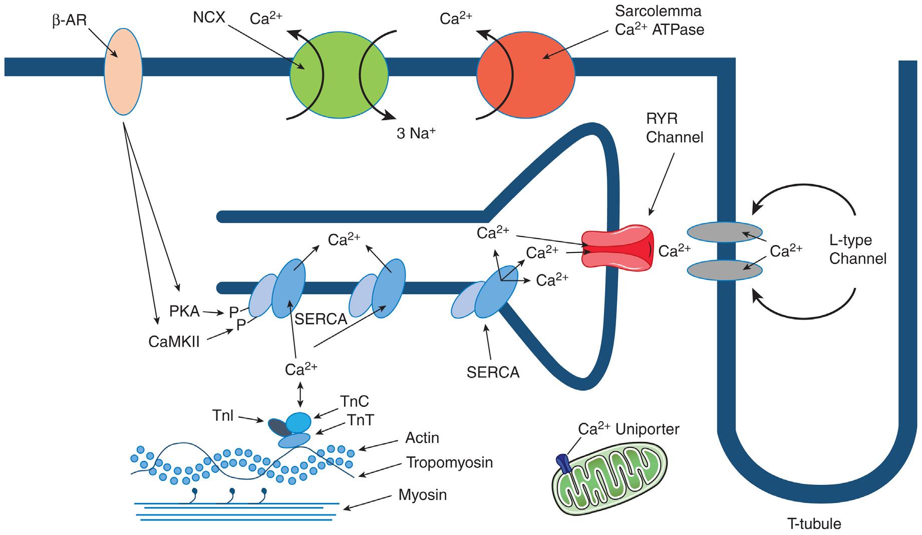
Calcium (Ca2+) plays a central role in the excitation-contraction and repolarization-relaxation of the myocardium.8 Hence factors that regulate Ca2+ flux in myocytes are poised to be critical regulators of diastolic function. Depolarization of the sarcolemma results in Ca2+ influx into the cytosol via the voltage-gated L-type channels. This inward Ca2+ current (ICa) promotes the release of Ca2+ from the sarcoplasmic reticulum (SR) by Ca2+–induced Ca2+–release via the ryanodine receptor 2 (RYR). The ICa and SR Ca2+ release raise intracellular free Ca2+ allowing Ca2+ to bind to the myofilament protein troponin C (TnC). When cytosolic Ca2+ is low, the troponin-tropomyosin complex inhibits the formation of the actinomyosin complex. When Ca2+ binds to TnC, it releases the inhibition and allows cross-bridge cycling and
Fig. 1.1 Diagram of cardiomyocyte Ca2+ flux during excitation-contraction coupling. Ca2+ enters via L-type Ca channels, which triggers Ca2+-induced Ca2+ release from the sarcoplasmic reticulum (SR). The increase in cytosolic Ca2+ results in Ca2+ binding to troponin C (TnC) initiating myofilament activation. For relaxation, cytosolic Ca2+ is transported into the SR via SR Ca2+-ATPase (SERCA) and into the extracellular space via sarcolemmal Na/Ca exchanger. β-AR, β-Adrenergic; NCX, Na/Ca exchanger; PKA, protein kinase A; RYR, ryanodine receptor; TnT, troponin T; TnI, troponin I.
contraction of the sarcomeres. In cardiac relaxation, cytosolic free Ca2+ must decline to result in Ca2+ dissociation from TnC. Ca2+ is transported from the cytosol by four pathways: (1) SR Ca2+-ATPase (SERCA), (2) sarcolemmal Na/Ca exchanger (NCX), (3) sarcolemmal Ca2+-ATPase, and (4) mitochondrial Ca2+ uniporter (Fig. 1.1). For Ca2+ homeostasis at each heart beat, the amount of Ca2+ pumped back into the SR by SERCA must equal the amount released through the RYR2 channel, and levels of Ca2+ extruded from the cell must equal the amount that entered via the L-type channel.
During relaxation Ca2+ is pumped back into the SR lumen by SERCA, which is regulated by phospholamban (PLN). When PLN is dephosphorylated it binds to SERCA and inhibits its Ca2+ affinity. Phosphorylation of PLN relieves SERCA inhibition and enhances Ca2+ sequestration in the SR increasing the rate of relaxation. PLN is phosphorylated by cyclic adenosine monophosphate (cAMP) activated–protein kinase A (PKA) and Ca-CaM–dependent protein kinase (CaMK) as a result of β-adrenergic stimulation. The major substrates for the cAMP-PKA axis include PLN, L-type Ca channels, RYR, troponin I (TnI), and myosin-binding protein C (cMyBP-C). The relaxant effect of PKA is mediated mainly by phosphorylation of PLN and TnI. PLN phosphorylation (at Ser-16) speeds up SR Ca2+ sequestration, while phosphorylation of TnI speeds up dissociation of Ca2+ from the myofilaments. CaMKII phosphorylation of PLN (at Thr-17) also increases SR Ca2+-ATPase activity. Both PKA and CaMKII are likely to be coactivated during normal sympathetic stimulation (β adrenergic), creating synergy between these important regulatory signaling pathways (see Fig. 1.1). SERCA expression and function is decreased in most HF models. Several studies have also shown reduced SERCA/ PLN ratio with age. In addition, there are data that point to reduced phosphorylation state of PLN in HF.9 This would result in reduced Ca2+ sensitivity of SERCA and lower SR Ca2+ uptake at physiologic
cytoplasmic Ca2+ levels [Ca]I. The reduction of SERCA activity is consistent with the characteristic slowed relaxation and [Ca]I seen in diastolic HF. In animal models when SERCA expression is increased or PLN expression is decreased, myocardial relaxation and [Ca]I decline are accelerated resulting in improved diastolic function.9 Consequently, factors that might target SERCA and other mediators of Ca2+ flux are an active area of research for therapeutic opportunities.
Posttranslational Modification of Myofibrillar Proteins
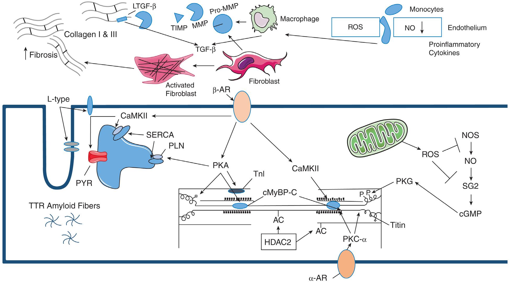
The sarcomeric protein titin is the largest known protein with a length greater than 1 um. It spans half the sarcomere connecting the Z-line to the M-line. It functions as a very large molecular spring contributing to force transmission at the Z-line and resting tension in the I-band region.10 Titin limits the range of sarcomere motion and is a major molecular contributor to myocyte passive stiffness. Titin activity can be modulated both by isoform expression and by phosphorylation. The differences in titin isoforms are correlated with the differences in the mechanical properties of cardiac, skeletal, and smooth muscle and differences of cardiac passive tension across species.11 Importantly, phosphorylation of titin by PKA, PKG, and PKC-α modulates its stiffness.12–15 Consequently, the activity of these kinases in myocytes has a direct influence on diastolic parameters. For example, PKA activated by β-adrenergic stimulation can phosphorylate titin as well as thick and thin filaments of the sarcomere.16 Nitric oxide (NO) and natriuretic peptides initiate signaling pathways activating PKG, which phosphorylates some of the same titin residues in the N2B spring element as PKA. Phosphorylation of the N2B element by either PKA or PKG results in a reduction in passive tension. α-Adrenergic stimulation activates PKC-α in cardiomyocytes, which is known to phosphorylate titin in the proline-valine-glutamate-lysine (PVEK) sequence increasing titinbased passive tension (Fig. 1.2). Therefore phosphorylation of the N2B
Fig. 1.2 Diagram of pathways contributing to diastolic dysfunction. Intersitial response: Cardiac connective tissue, composed primarily of collagen types I and III, is maintained by resident cardiac fibroblasts. In response to pressure overload (PO), the recruitment of monocytes through activated endothelium occurs, triggered by increases in cytokine expression, and results in increases in macrophage populations as well as activation of resident fibroblasts. These cell types express extracellular matrix (ECM) components, matricellular proteins, and matrix metalloproteinases (MMPs), which drive remodeling of the myocardium. Sequestered factors in the ECM such as latent TGF-β (LTGF-β), are released through the action of MMPs to further propagate remodeling events. Tissue inhibitors of MMPs (TIMPs), also act to modulate myocardial remodeling by limiting MMP activity on both sequestered cytokines and structural ECM components. Macrophages also contribute fibrotic deposition of collagen in the PO myocardium through matricellular protein and MMP production. Myocyte response: Increased levels of nitric oxide (NO) promote myocyte relaxation through activation of protein kinase G (PKG) and phosphorylation of titin. Increased production of reactive oxygen species (ROS) can foster diastolic dysfunction by reducing the bioavailability of NO. PKA activated by βadrenergic (β-AR) stimulation can phosphorylate titin decreasing passive tension. α-Adrenergic stimulation activates PKC-α in cardiomyocytes, which is known to phosphorylate titin, increasing titin-based passive tension. β-AR stimulated activation of CaMKII and protein kinase (PKA), also results in the phosphorylation of phospholamban (PLN), the ryanodine receptor (RYR), and L-type calcium channels, which increases diastolic cytosolic Ca2+ content consistent with the characteristic slowed relaxation seen in diastolic HF. α-AR, α-Adrenergic; cGMP, cyclic guanosine monophosphate; cMyBP-C, myosin-binding protein C; HDAC2, histone deacetylase 2; NOS, nitric oxide synthase; SERCA, SR Ca2+-ATPase SG2, soluble guanylate cyclase; TGF-β, transforming growth factor-β; TnI, troponin I.
element in titin by PKA or PKG decreases passive tension, whereas phosphorylation of titin’s PEVK element by PKC-α increases passive tension.11 The increased oxidative pressure or ROS in diastolic dysfunction has been proposed to deplete NO reserve, lowering PKG activity and leading to hypophosphorylation of the N2B element and titin stiffing in HFpEF.14,17 Increased ROS can also result in the oxidation of cysteine residues in the N2B element resulting in disulfide bond formation and increased passive tension in mouse models.18,19
In addition to titin, posttranslational modification of thick and thin filaments of myofibrils can affect cardiomyocyte relaxation. As mentioned, TnI and cMyBP-C are targets for phosphorylation by β-adrenergic stimulation. Phosphorylation of cardiac TnI at serine 23/24 by PKA reduces TnC-TnI interaction strength while reducing calcium sensitivity. The weakened C-I interaction may slow thin filament activation and result in faster relaxation kinetics thus increasing early phase relaxation with β-adrenergic stimulation.20 The cardiac cMyBP-C is a thick filament accessory protein that when
unphosphorylated represses both cross-bridge attachment and detachment. It is phosphorylated by multiple kinases, including PKA, PKC, PKD, CaMKII, glycogen synthase kinase 3β, and ribosomal S6 kinase. Phosphorylation of cMyBP-C results in increased rates of cross-bridge cycling.21 Hypophosphorylation of cMyBP-C is associated with diastolic dysfunction in human patients.22 A recent study examined phosphorylation-deficient and phosphomimetic mutants of PKA-targeted cMyBP-C sites. They found that PKA phosphorylation of cMyBP-C threonine 35 results in accelerated cross-bridge detachment of myosin and actin, thereby enhancing relaxation.23
Other Posttranslational Modifications
Advanced aging is associated with increased posttranslational modifications and has been associated with a systemic proinflammatory state (inflamm-aging) and development of HFpEF. Inflammation of the coronary microvascular endothelial cells leads to increased production of ROS. Oxidative stress can promote diastolic dysfunction
by reducing the bioavailability of NO. Cardiac relaxation is regulated by NO. ROS can affect NO-related signaling at multiple sites. NO is generated by NO synthase (NOS), which requires tetrahydrobiopterin as a cofactor for the reaction. Hypertension and activation of the renin-angiotensin system lead to a depletion of tetrahydrobiopterin. The loss of tetrahydrobiopterin leads to NOS uncoupling, the production of superoxide instead of NO, and diastolic dysfunction.24 Some of the mechanisms of how NO modulates myofilament contractility have been recently revealed. Depletion of tetrahydrobiopterin in hypertension can repress NO synthesis and is associated with S-glutathionylation of MyBP-C, which reduces cross-bridge cycling. S-glutathionylation is an oxidative posttranslation modification of cysteines. Tetrahydrobiopterin supplementation lowers S-glutathionylation of MyBP-C, reduces the changes in actin-myosin cross-bridge cycling, and improves diastolic dysfunction.25 Further, excessive ROS leads to oxidation of guanylate cyclase and affects its responsiveness to NO to synthesize cyclic guanosine monophosphate (cGMP).26 Lower cGMP level decreases PKG activity in cardiomyocytes leading to hypophosphorylation of titin resulting in increased cardiac stiffness.
Many cardiac myofibrillar proteins are posttranslationally modified by acetylation in the healthy heart.27–29 Unfortunately, we know very little about the changes in acetylation with cardiac pathologies. One recent study demonstrated that histone deacetylase (HDAC) inhibitors were efficacious in two murine models of diastolic dysfunction. In addition, the investigators showed that HDAC2 copurified with myofibrils. Although the target(s) were not identified, the study showed that ex vivo deacetylation of isolated myofibrils with recombinant HDAC2 significantly increased the rate of myofibril relaxation, whereas acetylation with recombinant p300 decreased myofibril relaxation duration.30 Hence HDAC inhibitors might be a promising avenue for future research into potential HFpEF therapies.
Transthyretin Amyloidosis
Cardiac amyloid deposition has also been linked with HFpEF. Over 30 different proteins have been shown to form amyloid fibrils and five (immunoglobulin light chain, immunoglobulin heavy chain, transthyretin, serum amyloid A, and apolipoprotein AI) have been found to infiltrate the heart.31 Autopsy from patients who were diagnosed with HFpEF revealed that transthyretin amyloidosis was present in 32% of those greater than 75 years of age.32 Another study using nuclear scintigraphy to detect amyloids has indicated that 13% of hospitalized patients with HFpEF have transthyretin amyloidosis.33 Interestingly, some HF patients have amyloidosis caused by a mutation in transthyretin. Over 80 transthyretin mutations, with autosomal dominant inheritance, have been associated with tissue amyloid deposition, some within the heart.34 Nearly 25% of African Americans with cardiac transthyretin amyloidosis were heterozygous for a transthyretin V122I mutation.34 Although this condition is rare, interstitial deposition of wild-type and mutant transthyretin is an underrecognized trigger of HF in the elderly.35
Mitochondrial Dysfunction and Age
As evidenced in the preceding sections, there are many factors that contribute to cellular changes observed in HFpEF. However, the fact that aging is a critical and overarching factor in the development of HFpEF is clearly noted.36 LV diastolic stiffness increases and LV diastolic filling rate decreases with age.37,38 Kaushik et al. showed that age-related increases in vinculin, a cytoskeletal protein, was linked to cortical stiffening and contractility.39 Vinculin is found localized to integrin-mediated cell–ECM and cadherin-mediated cell–cell adhesions. Vinculin acts as one of several proteins involved in anchoring
F-actin to the membrane. Senescent rats have twofold the ECM content in the myocardium compared to younger rats.40 The senescent myocardium has increased levels of ROS, which can activate transforming growth factor-β (TGF-β), inducing conversion of cardiac fibroblast to myofibroblasts, an activated fibroblast phenotype.41 The aging heart has lower responsiveness to β-adrenergic stimulation. There is lowered PKA and CaMKII phosphorylation of PLN and RYR receptor. Together this results in a lowering of the Ca2+ uptake and relaxation rate.42
The mitochondrial deoxyribonucleic acid (DNA) in aging hearts in both man and mice have up to 16-fold more point mutations and deletions than those of younger animals. Myocardial energetics have been examined as a potential mechanism for reduced systolic reserve in HFpEF with increased age. Patients with HFpEF have been shown to have a reduced phosphocreatine/adenosine triphosphate (ATP) ratio when compared to controls.43 Several studies suggest that abnormal skeletal muscle performance is a contributor to exertional intolerance rather than just limited cardiac reserve.44 One study found that HFpEF patients had reduced type I oxidative muscle fibers, type I/II fiber ratio, and a reduced capillary to fiber ratio in skeletal muscle compared with controls.45
Comorbid diseases, including hypertension and renal failure, are much more common in the elderly. There is increased inflammation with increasing age. Although the cellular mechanisms are not yet clearly defined, myocardial aging is interconnected to molecular events that influence both myocyte contractility and changes in the collagenous ECM (see upcoming discussion) that appear to provide a favorable milieu for the development of HFpEF, particularly when in combination with other comorbitidies such as hypertension and diabetes.
MYOCARDIAL COLLAGEN
Cardiac Fibrillar Collagen
Fibrillar collagens play a vital role in homeostatic function and pathologic dysfunction in the heart. Collagen types I, III, and V are the most highly represented myocardial fibrillar collagens.46 These three types of collagens form composite fibrils that then form the collagen fibers of the heart. Three categories of collagen fibers in and around myocytes have been described based on their morphologic characteristics: coils, struts, and weaves.47 Presumably each category of collagen fiber has a unique role in providing structural support to myocytes. A perimysial fibrillar collagen weave composed of smaller collagen fibers surrounds each myocyte, whereas the larger collagen fibers present as struts and coils to connect adjacent myocytes and blood vessels. Myocytes are organized in aligned bundles within the myocardium. The bundles are further organized into sheets that slide by one another during each heartbeat to generate the torsional motion to expel blood from the ventricles. Collagen fibers likely also participate in the organization of higher order structures such as bundles and sheets, although these types of structures are more difficult to identify in traditional two-dimensional tissue sections. Recent advances in generating and imaging decellularized hearts have afforded opportunities to visualize cardiac collagenous ECM in three dimensions where these structures can be better appreciated. As imaging technologies advance, the ability to visualize collagen structures in living human hearts in three dimensions will provide critical information for diagnosing and evaluating myocardial fibrosis in patients.
Age-related changes in the cardiac ECM have been observed both in man and in animal models.48 In mice, levels of fibrillar collagen were found to be low in neonatal hearts where abundant levels of
fibronectin were detected.49 In adult hearts, fibronectin was significantly decreased, whereas fibrillar collagen levels increased to become the dominant fibrillar ECM component. Interestingly, in aged hearts, levels of fibrillar collagen further increased in comparison to adult ages, and increases were also noted in levels of fibronectin. Increases in fibrillar collagen in aged hearts were associated with increases in tissue stiffness even in the absence of hypertension or PO suggesting a tendency for the development of myocardial fibrosis with age alone.50 Accordingly, age-dependent changes in ECM, coupled with those in myocytes, appear to set the stage for increases in myocardial stiffness that is further exacerbated by comorbid conditions.
Collagen Deposition in HFpEF
In patients diagnosed with HFpEF, increases in fibrillar collagen content are significant. In the study by Zile et al., collagen volume fraction was assessed in patient biopsies recovered from three groups: referent control, hypertensive heart disease only, and hypertensive heart disease with HF.51 Collagen content and myocardial stiffness were significantly increased only in biopsies from patients with hypertensive heart disease with HF. Furthermore, collagen-dependent stiffness was measured and shown to make a significant contribution to overall muscle stiffness when the sarcomeric component was removed.51 These results are consistent with increases in collagenous ECM observed in the myocardium of HFpEF patients making a critical contribution to diastolic stiffness.
Similarly, animal models of PO that recapitulate many aspects of human fibrotic disease also support a key role for fibrillar collagen in diastolic stiffness. Resident cardiac fibroblasts are the primary cardiac cell type that produces fibrillar collagen both in homeostasis and in hearts subjected to PO.52 Activated fibroblasts, often referred to as myofibroblasts, express elevated levels of contractile cytoskeletal elements and ECM components. Although activated fibroblasts are strongly implicated in cardiac remodeling following infarction, the role of activated fibroblasts in PO is less clear.53 Nonetheless, resident cardiac fibroblasts are the primary producers of fibrillar collagen and, with the assistance of inflammatory cells, are a critical mediator of collagen degradation (see Fig. 1.2).
Although imperfect, animal models provide a platform for evaluating cause-and-effect mechanisms of cardiac collagen deposition. Accumulation of ECM derives from (1) increases in transcription, translation, and secretion of ECM proteins; (2) procollagen processing and deposition to insoluble ECM in the extracellular space; and (3) degradation of ECM by myocardial matrix metalloproteinases (MMPs). Evidence that each of these mechanisms might contribute to increases in collagen content in response to PO has been shown in animal models.
Transcriptional Regulation of Collagen I
Transcriptional control of messenger ribonucleic acid (mRNA) encoding procollagen I is one mechanism by which levels of secreted procollagen can increase. Measurements of mRNA encoding the subunits of collagen I have been shown to significantly increase as soon as 3 days following the induction of PO.54 The increased levels of mRNA encoding fibrillar collagen subunits coincide with significant increases in MMP expression indicative of extensive ECM remodeling that accompanies hypertrophic growth of the myocardium.55 The expansion of individual myocytes by addition of sarcomeres requires renewed synthesis of basal lamina surrounding each myocyte as well as ECM in and around blood vessels. Likely the interstitial connective tissue also undergoes remodeling in response to hypertrophic growth. Interestingly, the increase in mRNA encoding fibrillar collagens at day
3 does not immediately result in an increase in collagen deposition in the extracellular space.54 Significant increases in levels of collagen protein incorporated into collagen fibers is not detected until 1 week after induction of PO. Hence, transcriptional control of fibrillar collagen genes is not the sole mechanism that controls levels of myocardial collagen.
Postsynthetic Procollagen Processing and Deposition
Studies from Laurent et al. suggested that deposition of collagen protein into an insoluble ECM is a critical step in controlling the amount of collagen deposited following PO.56 In the heart, ∼50% of newly synthesized collagen is degraded prior to incorporation into the ECM.56 Upon induction of PO, the amount of procollagen degraded decreases accompanied by increases in insoluble collagen deposition. These studies suggest that the efficiency of procollagen processing increases in response to PO and results in increases in collagen content. Procollagen maturation in the extracellular space requires cleavage of both the N-terminal and C-terminal propeptides from the procollagen monomer.57 In addition, modification of amino acids by enzymes that facilitate collagen crosslinking, such as lysyl oxidase, occurs during procollagen processing.58 Other proteins secreted into the extracellular space influence assembly of collagen into insoluble fibrils and include matricellular proteins such as SPARC and periostin as well as other collagen family members and proteoglycans.59 Proteins involved in procollagen processing and assembly are essential factors influencing myocardial collagen content in homeostasis and in response to fibrotic stimuli. For example, the absence of SPARC expression results in reduced amounts of insoluble myocardial collagen deposited in response to PO.60 Likewise, a decrease in insoluble collagen in SPARC-null hearts correlated with a reduction in myocardial stiffness in comparison to wild-type hearts. Hence, like in humans, increases in collagen content in mice is also associated with increases in myocardial stiffness.
ECM Degradation
Matrix Metalloproteinases
Once collagen has been incorporated into an insoluble ECM, the only known biologic mechanism for reducing collagen content is through enzymatic degradation. MMPs are zinc-dependent endopeptidases primarily responsible for ECM degradation. Of note, HFpEF patients have been found to have elevated levels of circulating MMP-1, MMP-2, MMP-8, and MMP-9.61–64 In addition to ECM degradation, MMPs also perform proteolytic cleavage of inflammatory mediators, matricellular proteins, and other MMPs within the myocardium.65,66 Hence increases in MMP activity are associated with active remodeling of the myocardial ECM that does not necessarily lead directly to reductions in ECM. For example, MMP-dependent cleavage of latent TGF- β, a potent profibrotic cytokine sequestered in the ECM, can lead to increases in ECM production.
Of the 25 MMPs described to date, MMP-2 and MMP-9 are the most widely studied (see Refs. 55 and 66). Interestingly, circulating MMP-2 has been shown to be elevated to a higher extent in patients with severe diastolic dysfunction as well as those with diastolic HF.62 Increased MMP-2 activity has also been shown to induce cardiomyocyte hypertrophy and degrade contractile and structural proteins (i.e., TnI, myosin light chain 1, α-actinin, and titin) resulting in decreased cardiac function.67,68 Hence a prominent function of this MMP in cardiac remodeling beyond ECM degradation alone is suggested. In a murine model of LV PO, genetic deletion of MMP-2 actually reduced the degree of myocardial fibrillar collagen accumulation and improved indices of diastolic function.69
PART I Basic Determinants of Diastolic Function
Neutrophils and monocyte-derived macrophages are a major source of MMP-94. In aged mice, MMP-9 deletion was found to attenuate the age-related decline in diastolic function through a reduction in TGF-β signaling. Reductions in the profibrotic matricellular proteins, periostin and CCN2 (connective tissue growth factor [CTGF]), were detected in the absence of MMP-9.70 MMP-9 is thought to regulate inflammation through its proteolytic activity on both ECM components and cytokine substrates. For example, in a mouse model of myocardial infarction (MI) MMP-9 mediated the post-MI degradation of CD36 leading to a decrease in macrophage phagocytosis of apoptotic neutrophils.71 Although a number of MMP-9 substrates have been identified, including collagens (type I, IV, V, VII, X, and XIV), fibronectin, elastin, interleukin (IL)-8, Cxcl4, and IL-1β, the mechanisms whereby MMP-9 modulates LV remodeling and diastolic dysfunction have not been completely elucidated.72–75
Other MMPs implicated in diastolic dysfunction include MMP-1, which has the highest affinity for fibrillar collagens and preferentially degrades collagens type I and III. Increased MMP-1 activity results in excessive collagen deposition and diastolic dysfunction.76 In addition, MMP-8, a collagenase primarily secreted by neutrophils, negatively correlates with development of HFpEF.64,77 Similarly, MMP-12 inhibition exacerbates cardiac dysfunction by prolonging neutrophil-mediated inflammation highlighting the importance of MMPs in regulation of inflammation and the subsequent development of cardiac dysfunction.78 Taken together, MMPs are critical mediators of myocardial ECM content and might represent a viable target for therapeutic intervention in future applications.
Tissue Inhibitors of Metalloproteinases
MMP activity is dependent not only on the concentration of active enzymes but also on the levels of a family of naturally occurring tissue inhibitors of metalloproteinases (TIMPs).79 After chronic stimulation of proinflammatory cytokines, TIMP levels increase leading to decreased MMP/TIMP ratio and increases in fibrillar collagen deposition.80 For example, the interaction between MMP-1 and TIMP-1 is of critical relevance in the maintenance of the integrity of the cardiac collagen network. TIMP-1 colocalizes with MMP-1 in healthy myocardium and is expressed by cardiac fibroblasts and myocytes.81–84 Circulating levels of TIMP-1 closely associate with markers of systemic inflammation, diastolic dysfunction, and HF severity.62,85 In hypertensive subjects with HFpEF, TIMP-1 moderately predicts the presence of HF.86 Using a single MMP to TIMP ratio, however, can be somewhat misleading as there are multiple MMPs and TIMPs present in diseased myocardium.
In addition to TIMP-1, TIMP-2 levels correlate with deposition of cardiac ECM.87 In response to angiotensin II infusion, TIMP-2 deleted mice showed enhanced hypertrophy, reduced collagen crosslinking, and suppressed collagen deposition resulting in impaired active relaxation.88 TIMP-3 has cardioprotective functions, as TIMP-3 deletion leads to spontaneous dilated cardiomyopathy,89 whereas TIMP-4 levels increase with hypertrophy and decrease with the onset of HF in spontaneously hypertensive rats.90
Inflammatory Mediators in HFpEF
A significant role for inflammatory mediators in the heart following MI has been well established in a number of studies where inflammation has been shown to be a major component contributing to healing and scar formation.91–95 Initiation of the inflammatory response following MI is necessary for adequate wound healing, yet too much or too little inflammation can result in increased dilation and poor cardiac function highlighting the importance of a balanced immune response to
injury.96 Relatively less is known about the role of inflammatory mediators in HFpEF although this actively expanding area of research will certainly lead to new discoveries that are likely to influence treatment strategies.
As mentioned, aging is a risk factor for HFpEF and has been linked to increased systemic inflammation termed inflamm-aging. Inflammaging is defined as elevated levels of proinflammatory markers in the blood and other tissues often detected in older individuals. It is hypothesized that this increase in inflammation is the common biologic factor responsible for the decline and the onset of cardiovascular diseases, frailty, multimorbidity, and decline of physical and cognitive function in the elderly. Possible mechanisms potentially underlying inflamm-aging include genomic instability, cell senescence, mitochondria dysfunction, NLRP3 inflammasome activation, and primary dysregulation of immune cells.
Obesity is another risk factor for HFpEF that has been linked to an increase in baseline systemic inflammation.97 More than 80% of patients with HFpEF are overweight or obese. Visceral fat produces proinflammatory and chemotactic compounds and is infiltrated by inflammatory cells such as macrophages and lymphocytes.98 Older, obese HFpEF patients that underwent a 20-week caloric restriction diet had significantly improved peak oxygen consumption and quality of life scores. This improvement was found to correlate with reduced body fat mass, increased percent lean body mass, higher thigh muscle/intermuscular fat ratio, and lower biomarkers of inflammation supporting the hypothesis that systemic inflammation due to obesity contributes to exercise intolerance in HFpEF.99 As mentioned, inflammatory cells produce MMPs, which have a significant impact on cardiac remodeling. Studies that support a function for inflammatory cell types in contributing to fibrotic deposition of cardiac collagen are emerging, particularly for neutrophil and macrophage populations.
Neutrophils
In response to ischemia, neutrophils are the first cells to respond to the site of injury.100,101 Similarly, neutrophils might also be an important early contributor to PO-induced hypertrophy. In a murine model of PO, the recruitment of neutrophils was characterized as an early event as increases in neutrophil numbers noted at day 1, decreased by day 3.102 Early infiltration of neutrophils was also associated with detection of neutrophil extracellular traps (NETs) in the heart. The release of chromatin by neutrophils in the form of NETs is thought to be an antimicrobial defense mechanism. However, NETs are also thought to lead to further recruitment of platelets and other types of inflammatory cells. In the study by Martinod et al., transgenic mice that were unable to form NETs demonstrated significant reductions in myocardial collagen content in comparison to wild-type mice after 4 weeks of PO.102 Whether neutrophil activity and NET deposition might also contribute to human disease is yet to be established.
Macrophages
Similar to neutrophils, increasing evidence that macrophages might also influence fibrotic deposition of collagen after PO is emerging. In a murine model of PO induced by transverse aortic constriction (TAC), cardiac macrophage populations were increased at day 6.103 McDonald et al. also reported increases in macrophage populations at 1 week after TAC.54 Importantly, the time course of collagen accumulation paralleled that of macrophage expansion in the PO myocardium.54 Macrophage expression of the matricellular protein SPARC, a key regulator of myocardial fibrosis, was also found suggesting that
macrophage expression of SPARC might increase procollagen processing and enhance collagen deposition in PO hearts.
Evidence that macrophages might also contribute to fibrosis in humans was supported by an increase in macrophages in biopsies from patients diagnosed with HFpEF.104 Tissue samples from referent controls and from those with hypertension in the absence of HFpEF did not demonstrate significant increases in myocardial macrophages. Hulsmans et al. went on to show, in another murine model of diastolic dysfunction, an association with macrophage recruitment and cardiac fibrosis. In mice with macrophage-specific deletion of IL-10, an improvement in diastolic function was found.104 Hence a growing body of evidence suggests that macrophages are a critical inflammatory cell population influencing the development of myocardial fibrosis and diastolic dysfunction.
KEY POINTS
• Heart failure with preserved ejection fraction (HFpEF) is characterized by increases in myocardial stiffness contributed by both cellular and molecular mechanisms that arise from alterations in myocytes and in extracellular matrix.
• Myocardial energetics are a potential mechanism for reduced systolic reserve in HFpEF with increased age.
• A reduction of SERCA activity is consistent with the characteristic slowed relaxation and [Ca]I seen in diastolic HF. In animal models when SERCA expression is increased or PLN expression is decreased, myocardial relaxation and [Ca]I decline are accelerated resulting in improved diastolic function.
• Phosphorylation of the N2B element of titin by PKA or PKG decreases passive tension, whereas phosphorylation of titin’s PEVK element by PKC-α increases passive tension.
• Cardiac transthyretin amyloid deposition is linked with HFpEF.
REVIEW QUESTIONS
1. How does NO regulate cardiac relaxation?
a. Generation of NO by NO synthase requires a cofactor, tetrahydrobiopterin, which can reduce actin-myosin cross-bridge cycling
b. Through depletion of tetrahydrobiopterin and oxidation of guanylate cyclase
c. Through NO synthase uncoupling and production of superoxide
2. What are the important steps for procollagen I production, processing, and deposition (in sequential order)?
i. Transcription and translation inside the cell
ii. Cleavage by MMPs
iii. Incorporation into extracellular matrix enhanced by matricellular proteins
REFERENCES
1. Sharma K, Kass DA. Heart failure with preserved ejection fraction: mechanisms, clinical features, and therapies. Circ Res. 2014;115:79–96.
2. Owan TE, Hodge DO, Herges RM, Jacobsen SJ, Roger VL, Redfield MM. Trends in prevalence and outcome of heart failure with preserved ejection fraction. N Engl J Med. 2006;355:251–259.
3. Bhatia RS, Tu JV, Lee DS, et al. Outcome of heart failure with preserved ejection fraction in a population-based study. N Engl J Med. 2006;355:260–269.
4. Steinberg BA, Zhao X, Heidenreich PA, et al. Trends in patients hospitalized with heart failure and preserved left ventricular ejection fraction: prevalence, therapies, and outcomes. Circulation. 2012;126:65–75.
FUTURE DIRECTIONS
Cellular mechanisms that influence diastolic function are multivariant in nature. A decrease in cardiomyocyte number, cardiomyocyte hypertrophy, increased collagen deposition, and functional changes at the cellular level can all contribute to LV stiffness and abnormal diastolic function. Determining what factors trigger the decline into HF has never been more important. Clearly, factors that render the aged myocardium more susceptible to diastolic dysfunction include changes in myocyte contractility, age-related ECM remodeling, and inflammation. New strategies that arise from basic research into cellular mechanisms of diastolic dysfunction are highly likely to lead to significant advances for the treatment of patients diagnosed with HFpEF in the coming years.
• Three primary cellular mechanisms contribute to increases in myocardial collagen accumulation:
1. Increased transcription, translation, and secretion of collagen proteins
2. Increased procollagen processing in the extracellular space
3. Decreases in extracellular matrix degradation
• Procollagen processing is influenced by expression of collagenbinding proteins, such as the matricellular protein SPARC, that drives collagen deposition in response to pressure overload.
• ECM degradation is controlled by the balance between matrix metalloproteinases (MMPs) and tissue inhibitor of MMPs (TIMPs).
• Cardiac fibroblasts are the primary cell type producing fibrillar collagen in the heart; however, expression of collagen-binding proteins, MMPs, and TIMPs by resident and infiltrating immune cells can influence levels of myocardial ECM.
• Increases in fibrillar collagen and alterations in ECM assembly, such as differential collagen crosslinking, contribute to fibrotic ECM and myocardial dysfunction in HFpEF.
iv. Cleavage by propeptidases
a. i, ii, iv
b. ii, iv, iii, i
c. i, iv, iii
d. iv, ii, iii
e. i, ii, iii, iv
3. What are NETs?
a. Release of chromatin by neutrophils
b. Antimicrobial defense mechanism
c. Neutrophil extracellular traps
d. All the above
5. McMurray JJ, Packer M, Desai AS, et al. Angiotensin-neprilysin inhibition versus enalapril in heart failure. N Engl J Med. 2014;371: 993–1004.
6. Pitt B, Pfeffer MA, Assmann SF, et al. Spironolactone for heart failure with preserved ejection fraction. N Engl J Med . 2014;370: 1383–1392.
7. Paulus WJ, Tschope C. A novel paradigm for heart failure with preserved ejection fraction: comorbidities drive myocardial dysfunction and remodeling through coronary microvascular endothelial inflammation. J Am Coll Cardiol. 2013;62:263–271.
8. Bers DM. Cardiac sarcoplasmic reticulum calcium leak: basis and roles in cardiac dysfunction. Ann Rev Physiol. 2014;76:107–127.
9. Kranias EG, Hajjar RJ. Modulation of cardiac contractility by the phospholamban/SERCA2a regulatome. Circ Res. 2012;110:1646–1660.
10. Borbély A, van der Velden J, Papp Z, et al. Cardiomyocyte stiffness in diastolic heart failure. Circulation. 2005;111:774–781.
11. Hidalgo C, Granzier H. Tuning the molecular giant titin through phosphorylation: role in health and disease. Trends Cardiov Med. 2013;23: 165–171.
12. Krüger M, Kötter S, Grützner A, et al. Protein kinase G modulates human myocardial passive stiffness by phosphorylation of the titin springs. Circ Res. 2009;104:87–94.
13. Hidalgo C, Hudson B, Bogomolovas J, et al. PKC phosphorylation of titin’s PEVK element: a novel and conserved pathway for modulating myocardial stiffness. Circ Res. 2009;105(7): 631–638 + 17 pp.
14. Hamdani N, Bishu KG, von Frieling-Salewsky M, Redfield MM, Linke WA. Deranged myofilament phosphorylation and function in experimental heart failure with preserved ejection fraction. Cardiov Res. 2013;97:464–471.
15. Hamdani N, Krysiak J, Kreusser MM, et al. Crucial role for Ca2(+)/calmodulin-dependent protein kinase-II in regulating diastolic stress of normal and failing hearts via titin phosphorylation. Circ Res. 2013;112:664–674.
16. Yamasaki R, Wu Y, McNabb M, Greaser M, Labeit S, Granzier H. Protein kinase A phosphorylates titin’s cardiac-specific N2B domain and reduces passive tension in rat cardiac myocytes. Circ Res. 2002;90:1181–1188.
17. van Heerebeek L, Franssen CP, Hamdani N, Verheugt FW, Somsen GA, Paulus WJ. Molecular and cellular basis for diastolic dysfunction. Curr Heart Failure Rep. 2012;9:293–302.
18. Grutzner A, Garcia-Manyes S, Kötter S, Badilla CL, Fernandez JM, Linke WA. Modulation of titin-based stiffness by disulfide bonding in the cardiac titin N2-B unique sequence. Biophys J. 2009;97:825–834.
19. Nedrud J, Labeit S, Gotthardt M, Granzier H. Mechanics on myocardium deficient in the N2B region of titin: the cardiac-unique spring element improves efficiency of the cardiac cycle. Biophys J. 2011;101:1385–1392.
20. Rao V, Cheng Y, Lindert S, et al. PKA phosphorylation of cardiac troponin I modulates activation and relaxation kinetics of ventricular myofibrils. Biophys J. 2014;107:1196–1204.
21. Tong CW, Stelzer JE, Greaser ML, Powers PA, Moss RL. Acceleration of crossbridge kinetics by protein kinase A phosphorylation of cardiac myosin binding protein C modulates cardiac function. Circ Res. 2008;103:974–982.
22. Donaldson C, Palmer BM, Zile M, et al. Myosin cross-bridge dynamics in patients with hypertension and concentric left ventricular remodeling. Circ Heart Failure. 2012;5:803–811.
23. Rosas PC, Liu Y, Abdalla MI, et al. Phosphorylation of cardiac myocinbinding protein-C is a critical mediator of diastolic function. Circ Heart Failure. 2015;8:582–594.
24. Silberman GA, Fan TH, Liu H, et al. Uncoupled cardiac nitric oxide synthase mediates diastolic dysfunction. Circulation . 2010;121: 519–528.
25. Jeong EM, Monasky MM, Gu L, et al. Tetrahydrobiopterin improves diastolic dysfunction by reversing changes in myofilament properties. J Molecul Cell Cardiol. 2013;56:44–54.
26. Tsai EJ, Liu Y, Koitabashi N, et al. Pressure-overload-induced subcellular relocalization/oxidation of soluble guanylyl cyclase in the heart modulates enzyme stimulation. Circ Res. 2012;110:295–303.
27. Choudhary C, Kumar C, Gnad F, et al. Lysine acetylation targets protein complexes and co-regulates major cellular functions. Science (New York, N.Y.). 2009;325:834–840.
28. Foster DB, Liu T, Rucker J, et al. The cardiac acetyl-lysine proteome. PLoS One. 2013;8:e67513.
29. Lundby A, Lage K, Weinert BT, et al. Proteomic analysis of lysine acetylation sites in rat tissues reveals organ specificity and subcellular patterns. Cell Rep. 2012;2:419–431.
30. Jeong MY, Lin YH, Wennersten SA, et al. Histone deacetylase activity governs diastolic dysfunction through a nongenomic mechanism. Sci Transl Med. 2018 Feb 7;10(427):eaao0144.
31. Maurer MS, Elliott P, Comenzo R, Semigran M, Rapezzi C. Addressing common questions encountered in the diagnosis and management of cardiac amyloidosis. Circulation. 2017;135:1357–1377.
32. Mohammed SF, Mirzoyev SA, Edwards WD, et al. Left ventricular amyloid deposition in patients with heart failure and preserved ejection fraction. JACC. Heart Failure. 2014;2:113–122.
33. González-López E, Gallego-Delgado M, Guzzo-Merello G, et al. Wildtype transthyretin amyloidosis as a cause of heart failure with preserved ejection fraction. Eur Heart J. 2015;36:2585–2594.
34. Buxbaum J, Jacobson DR, Tagoe C, et al. Transthyretin V122I in African Americans with congestive heart failure. J Am Coll Cardiol. 2006;47: 1724–1725.
35. Quarta CC, Buxbaum JN, Shah AM, et al. The amyloidogenic V122I transthyretin variant in elderly black Americans. N Engl J Med. 2015;372:21–29.
36. Loffredo FS, Nikolova AP, Pancoast JR, Lee RT. Heart failure with preserved ejection fraction: molecular pathways of the aging myocardium. Circ Res. 2014;115:97–107.
37. Redfield MM, Jacobsen SJ, Borlaug BA, Rodeheffer RJ, Kass DA. Age- and gender-related ventricular-vascular stiffening: a community-based study. Circulation. 2005;112:2254–2262.
38. Bursi F, Weston SA, Redfield MM, et al. Systolic and diastolic heart failure in the community. JAMA. 2006;296:2209–2216.
39. Kaushik G, Spenlehauer A, Sessions AO, et al. Vinculin network-mediated cytoskeletal remodeling regulates contractile function in the aging heart. Sci Trans. Med. 2015 Jun 17;7:(292):292ra99.
40. Biernacka A, Frangogiannis NG. Aging and cardiac fibrosis. Aging Dis. 2011;2:158–173.
41. Cucoranu I, Clempus R, Dikalova A, et al. NAD(P)H oxidase 4 mediates transforming growth factor-beta1-induced differentiation of cardiac fibroblasts into myofibroblasts. Circ Res. 2005;97:900–907.
42. Chu G, Lester JW, Young KB, Luo W, Zhai J, Kranias EG. A single site (Ser16) phosphorylation in phospholamban is sufficient in mediating its maximal cardiac responses to beta-agonists. J Biol Chem. 2000;275:38938–38943.
43. Phan TT, Abozguia K, Nallur Shivu G, et al. Heart failure with preserved ejection fraction is characterized by dynamic impairment of active relaxation and contraction of the left ventricle on exercise and associated with myocardial energy deficiency. J Am Coll Cardiol 2009;54:402–409.
44. Bhella PS, Prasad A, Heinicke K, et al. Abnormal haemodynamic response to exercise in heart failure with preserved ejection fraction. Eur J Heart Failure. 2011;13: 1296–1304.
45. Kitzman DW, Nicklas B, Kraus WE, et al. Skeletal muscle abnormalities and exercise intolerance in older patients with heart failure and preserved ejection fraction. Am J Physiol. Heart Circ Physiol. 2014;306:H1364–H1370.
46. Eghbali M, Weber KT. Collagen and the myocardium: fibrillar structure, biosynthesis and degradation in relation to hypertrophy and its regression. Molecul Cell Biochem. 1990;96:1–14.
47. Caulfield JB, Borg TK. The collagen network of the heart. Lab Invest 1979;40:364–372.
48. Gazoti Debessa CR, Mesiano Maifrino LB, Rodrigues de Souza R. Age related changes of the collagen network of the human heart. Mech Ageing Develop. 2001;122:1049–1058.
49. Trombetta-Esilva J, Eadie EP, Zhang Y, Norris RA, Borg TK, Bradshaw AD. The effects of age and the expression of SPARC on extracellular matrix production by cardiac fibroblasts in 3-D cultures. PLoS One 2013;8:e79715.
50. Bradshaw AD, Baicu CF, Rentz TJ, Van Laer AO, Bonnema DD, Zile MR. Age-dependent alterations in fibrillar collagen content and myocardial diastolic function: role of SPARC in post-synthetic procollagen processing. Am J Physiol. Heart Circ Physiol . 2010;298: H614–H622.
51. Zile MR, Baicu CF, Ikonomidis J, et al. Myocardial stiffness in patients with heart failure and a preserved ejection fraction: contributions of collagen and titin. Circulation. 2015;131:1247–1259.
52. Moore-Morris T, Guimarães-Camboa N, Banerjee I, et al. Resident fibroblast lineages mediate pressure overload-induced cardiac fibrosis. J Clin Invest. 2014;124:2921–2934.
53. Kanisicak O, Khalil H, Ivey MJ, et al. Genetic lineage tracing defines myofibroblast origin and function in the injured heart. Nature Comm 2016;7:12260.
54. McDonald LT, Zile MR, Zhang Y, et al. Increased macrophage-derived SPARC precedes collagen deposition in myocardial fibrosis. Am J Physiol. Heart Circ Physiol. 2018.
56. Bishop JE, Rhodes S, Laurent GJ, Low RB, Stirewalt WS. Increased collagen synthesis and decreased collagen degradation in right ventricular hypertrophy induced by pressure overload. Cardiov Res. 1994;28: 1581–1585.
57. Bradshaw AD. The extracellular matrix. Encycl Cell Biol. 2016;2:694–703.
58. Eyre DR, Paz MA, Gallop PM. Cross-linking in collagen and elastin. Ann Rev Biochem. 1984;53:717–748.
59. Bradshaw AD. The role of SPARC in extracellular matrix assembly. J Cell Comm Signal. 2009;3:239–246.
60. Bradshaw AD, Baicu CF, Rentz TJ, et al. Pressure overload-induced alterations in fibrillar collagen content and myocardial diastolic function: role of secreted protein acidic and rich in cysteine (SPARC) in post-synthetic procollagen processing. Circulation. 2009;119:269–280.
61. Gonzalez A, López B, Querejeta R, Zubillaga E, Echeverria T, Diez J. Filling pressures and collagen metabolism in hypertensive patients with heart failure and normal ejection fraction. Hypertension. 2010;55: 1418–1424.
62. Martos R, Baugh J, Ledwidge M, et al. Diastolic heart failure: evidence of increased myocardial collagen turnover linked to diastolic dysfunction. Circulation. 2007;115:888–895.
63. Martos R, Baugh J, Ledwidge M, et al. Diagnosis of heart failure with preserved ejection fraction: improved accuracy with the use of markers of collagen turnover. Eur J Heart Failure. 2009;11:191–197.
64. Zile MR, Desantis SM, Baicu CF, et al. Plasma biomarkers that reflect determinants of matrix composition identify the presence of left ventricular hypertrophy and diastolic heart failure. Circ Heart Failure. 2011;4: 246–256.
65. Newby AC. Metalloproteinases promote plaque rupture and myocardial infarction: a persuasive concept waiting for clinical translation. Matrix Biol: J Int Soc Matrix Biol. 2015;44–46:157–166.
66. Rohani MG, Parks WC. Matrix remodeling by MMPs during wound repair. Matrix Biol. 2015;44–46:113–121.
67. Wang W, Schulze CJ, Suarez-Pinzon WL, Dyck JR, Sawicki G, Schulz R. Intracellular action of matrix metalloproteinase-2 accounts for acute myocardial ischemia and reperfusion injury. Circulation. 2002;106:1543–1549.
69. Matsusaka H, Ide T, Matsushima S, et al. Targeted deletion of matrix metalloproteinase 2 ameliorates myocardial remodeling in mice with chronic pressure overload. Hypertension. 2006;47:711–717.
70. Chiao YA, Ramirez TA, Zamilpa R, et al. Matrix metalloproteinase-9 deletion attenuates myocardial fibrosis and diastolic dysfunction in ageing mice. Cardiov Res. 2012;96:444–455.
71. DeLeon-Pennell KY, Tian Y, Zhang B, et al. CD36 is a matrix metalloproteinase-9 substrate that stimulates neutrophil apoptosis and removal during cardiac remodeling. Circ Cardiov Gen. 2016;9:14–25.
72. de Castro Brás LE, Ramirez TA, DeLeon-Pennell KY, Chiao YA, et al. Texas 3-step decellularization protocol: looking at the cardiac extracellular matrix. J Proteomics. 2013;86:43–52.
73. DeLeon KY, Yabluchanskiy A, Winniford MD, Lange RA, Chilton RJ, Lindsey ML. Modifying matrix remodeling to prevent heart failure. In: Li R, Weisel RD, eds. Cardiac Regeneration and Repair. Volume I: Pathology and Therapies. Woodhead Cambridge, England; 2013.
74. Lindsey ML, Iyer RP, Zamilpa R, et al. A novel collagen matricryptin reduces left ventricular dilation post-myocardial infarction by promoting scar formation and angiogenesis. J Am Coll Cardiol. 2015;66:1364–1374.
75. Zamilpa R, Lopez EF, Chiao YA, et al. Proteomic analysis identifies in vivo candidate matrix metalloproteinase-9 substrates in the left ventricle postmyocardial infarction. Proteomics. 2010;10:2214–2223.
76. Reed AL, Tanaka A, Sorescu D, et al. Diastolic dysfunction is associated with cardiac fibrosis in the senescence-accelerated mouse. Am J Physiol. Heart Circ Physiol. 2011;301:H824–H831.
77. Erkol A, Pala S, Oduncu V, et al. Relation of plasma matrix metalloproteinase-8 levels late after myocardial infarction with left ventricular volumes and ejection fraction. Turk Kardiyol Dern Ars. 2013;41(7): 617–624.
78. Iyer RP, Patterson NL, Zouein FA, et al. Early matrix metalloproteinase-12 inhibition worsens post-myocardial infarction cardiac dysfunction by delaying inflammation resolution. Int J Cardiol. 2015;185:198–208.
79. Visse R, Nagase H. Matrix metalloproteinases and tissue inhibitors of metalloproteinases: structure, function, and biochemistry. Circ Res 2003;92:827–839.
80. Mann DL. Inflammatory mediators and the failing heart: past, present, and the foreseeable future. Circ Res. 2002;91:988–998.
81. Lindsey ML, Goshorn DK, Squires CE, et al. Age-dependent changes in myocardial matrix metalloproteinase/tissue inhibitor of metalloproteinase profiles and fibroblast function. Cardiov Res. 2005;66:410–419.
82. Tyagi SC, Kumar SG, Banks J, Fortson W. Co-expression of tissue inhibitor and matrix metalloproteinase in myocardium. J Mol Cell Cardiol. 1995;27:2177–2189.
83. Tyagi SC, Kumar S, Glover G. Induction of tissue inhibitor and matrix metalloproteinase by serum in human heart-derived fibroblast and endomyocardial endothelial cells. J Cell Biochem. 1995;58:360–371.
84. Romanic AM, Burns-Kurtis CL, Gout B, Berrebi-Bertrand I, Ohlstein EH. Matrix metalloproteinase expression in cardiac myocytes following myocardial infarction in the rabbit. Life Sci. 2001;68:799–814.
85. Cavusoglu E, Ruwende C, Chopra V, et al. Tissue inhibitor of metalloproteinase-1 (TIMP-1) is an independent predictor of all-cause mortality, cardiac mortality, and myocardial infarction. Am Heart J 2006;151(1101):e1–e8.
86. Janicki JS, Brower GL. The role of myocardial fibrillar collagen in ventricular remodeling and function. J Cardiac Failure. 2002;8:S319–S325.
87. Peterson JT, Li H, Dillon L, Bryant JW. Evolution of matrix metalloprotease and tissue inhibitor expression during heart failure progression in the infarcted rat. Cardiov Res. 2000;46:307–315.
88. Fan D, Takawale A, Basu R, et al. Differential role of TIMP2 and TIMP3 in cardiac hypertrophy, fibrosis, and diastolic dysfunction. Cardiov Res. 2014. 2014;103:268–280.
89. Eckhouse SR, Purcell BP, McGarvey JR, et al. Local hydrogel release of recombinant TIMP-3 attenuates adverse left ventricular remodeling after experimental myocardial infarction. Sci Transl Med . 2014;6:223ra21.
90. Mujumdar VS, Tyagi SC. Temporal regulation of extracellular matrix components in transition from compensatory hypertrophy to decompensatory heart failure. J Hyperten. 1999;17:261–270.
92. DeLeon-Pennell KY, Meschiari CA, Jung M, Lindsey ML. Matrix metalloproteinases in myocardial infarction and heart failure. Prog Mol Biol Transl Sci. 2017;147:75–100.
93. Lindsey ML. Regulating macrophage infiltration to alter wound healing following myocardial infarction. Cardiov Res . 2018;114: 1571–1572.
94. Mouton AJ, Rivera OJ, Lindsey ML. Myocardial infarction remodeling that progresses to heart failure: a signaling misunderstanding. Am J Physiol Heart Circ Physiol. 2018;315:H71–H79.
95. Kain V, Liu F, Kozlovskaya V, et al. Resolution agonist 15-epi-Lipoxin A4 programs early activation of resolving phase in post-myocardial infarction healing. Sci Rep. 2017;7:9999.
96. Grzybowski M, Welch RD, Parsons L, et al. The association between white blood cell count and acute myocardial infarction in-hospital mortality: findings from the National Registry of Myocardial Infarction. Acad Emerg Med. 2004;11:1049–1060.
97. Pandey A, Patel KV, Vaduganathan M, et al. Physical activity, fitness, and obesity in heart failure with preserved ejection fraction. JACC Heart Fail 2018;6:975–982.
10 PART I Basic Determinants of Diastolic Function
98. Surmi BK, Hasty AH. Macrophage infiltration into adipose tissue: initiation, propagation and remodeling. Future Lipidol . 2008;3: 545–556.
99. Kitzman DW, Brubaker P, Morgan T, et al. Effect of caloric restriction or aerobic exercise training on peak oxygen consumption and quality of life in obese older patients with heart failure with preserved ejection fraction: a randomized clinical trial. JAMA. 2016;315:36–46.
100. Borregaard N, Cowland JB. Granules of the human neutrophilic polymorphonuclear leukocyte. Blood. 1997;89:3503–3521.
101. Faurschou M, Borregaard N. Neutrophil granules and secretory vesicles in inflammation. Microbes Infect. 2003;5:1317–1327.
102. Martinod K, Witsch T, Erpenbeck L, et al. Peptidylarginine deiminase 4 promotes age-related organ fibrosis. J Experiment Med. 2017;214:439–458.
103. Weisheit C, Zhang Y, Faron A, et al. Ly6C(low) and not Ly6C(high) macrophages accumulate first in the heart in a model of murine pressure-overload. PLoS One. 2014;9:e112710.
104. Hulsmans M, Sager HB, Roh JD, et al. Cardiac macrophages promote diastolic dysfunction. J Experiment Med. 2018;215:423–440.